Baseline Chromatin Modification Levels May Predict
Interindividual Variability in Ozone-Induced Gene
Expression
Shaun D. McCullough,*
,1Emma C. Bowers,
†Doan M. On,* David S. Morgan,*
Lisa A. Dailey,* Ronald N. Hines,* Robert B. Devlin,* and David Diaz-Sanchez*
*National Health and Environmental Effects Research Laboratory, U.S. Environmental Protection Agency,
Research Triangle Park, North Carolina 27711; and
†Curriculum in Toxicology, University of North Carolina –
Chapel Hill, Chapel Hill, North Carolina 27599
1To whom correspondence should be addressed at Environmental Public Health Division, National Health and Environmental Effects Research Laboratory, U.S. Environmental Protection Agency, Human Studies Facility, 104 Mason Farm Road, Chapel Hill, NC 27599. Fax: (919) 966-6271.E-mail: [email protected].
ABSTRACT
Traditional toxicological paradigms have relied on factors such as age, genotype, and disease status to explain variability in responsiveness to toxicant exposure; however, these are neither sufficient to faithfully identify differentially responsive individuals nor are they modifiable factors that can be leveraged to mitigate the exposure effects. Unlike these factors, the epigenome is dynamic and shaped by an individual’s environment. We sought to determine whether baseline levels of specific chromatin modifications correlated with the interindividual variability in their ozone (O3)-mediated induction in an air–liquid interface model using primary human bronchial epithelial cells from a panel of 11 donors. We characterized the relationship between the baseline abundance of 6 epigenetic markers with established roles as key regulators of gene expression—histone H3 lysine 4 trimethylation (H3K4me3), H3K27 acetylation (H3K27ac), pan-acetyl H4 (H4ac), histone H3K27 di/trimethylation (H3K27me2/3), unmodified H3, and 5-hydroxymethylcytosine (5-hmC)—and the variability in the O3-induced expression of IL-8, IL-6, COX2, and HMOX1. Baseline levels of H3K4me3, H3K27me2/3, and 5-hmC, but not H3K27ac, H4ac, and total H3, correlated with the interindividual variability in O3-mediated induction of HMOX1 and COX2. In contrast, none of the chromatin modifications that we examined correlated with the induction of IL-8 and IL-6. From these findings, we propose an “epigenetic seed and soil” model in which chromatin modification states between individuals differ in the relative abundance of specific modifications (the “soil”) that govern how receptive the gene is to toxicant-mediated cellular signals (the “seed”) and thus regulate the magnitude of exposure-related gene induction.
Key words: chromatin; DNA methylation; histone; ozone; epigenetics
The response to toxicant exposure can vary greatly among indi-viduals, and in the case of inhaled toxicants is thought to result from intrinsic factors (Bastain et al., 2003; Holz et al., 1999; McDonnellet al., 1985). In fact, the variability between healthy individuals is often greater than the difference between healthy individuals and those with airway diseases such as asthma. Although traditionally studied susceptibility factors such as genes, age, and disease status may provide a partial
explanation, these factors are neither sufficient to completely explain interindividual variability in exposure responsiveness nor are they modifiable factors that can be leveraged to mitigate the adverse health effects of toxicant exposures. Unlike these static factors, an individual’s epigenome is dynamic and shaped by interactions with chemical and nonchemical aspects of their environment (Baccarelli andBollati, 2009;Houet al.,2012;Lam
et al.,2012;Waterlandet al.,2006) and they may play a powerful
Published by Oxford University Press on behalf of the Society of Toxicology 2015. This work is written by US Government employees and is in the public domain in the US.
216 doi: 10.1093/toxsci/kfv324
role in determining exposure effects and susceptibility. However, incorporating these factors in risk assessment and management strategies presents a challenge since no single factor is likely be predictive of responsiveness and obtaining a comprehensive and accurate history of an individual’s interac-tion with their environment is not possible.
The epigenome is the infrastructure underlying the mecha-nisms by which cells, tissues, and orgamecha-nisms respond to their environment and has been proposed as a biosensor of the cumulative impact of previous exposure to chemical and nonchemical stressors (Olden et al., 2014). Chromatin, the framework of the epigenome, is composed of repeating units called nucleosomes, each of which is composed of DNA wrapped around a histone octamer composed of two molecules of each of the four core histone proteins, H2A, H2B, H3, and H4 (Daveyet al.,2002;Lugeret al.,1997). The amino- (N-) and car-boxy- (C-) terminal tails of the core histones extend out of the nucleosome and are subjected to a broad range of post-transla-tional modifications, such as acetylation and methylation (Kouzarides, 2007). Patterns of DNA methylation and histone post-translational modifications (collectively referred to as “chromatin modifications”) are responsive to environmental factors such as chemical exposure (Baccarelli andBollati, 2009; Houet al.,2012;Liuet al., 2015), lifestyle choices such as diet (Waterlandet al.,2006), and psychosocial factors such as stress and socioeconomic status (Lam et al., 2012). Environment-induced differences in epigenomic modification states serve as both mechanisms of toxicant-induced disease and mediators of susceptibility to exposure-associated adverse health outcomes (Dolinoyet al.,2007).
The role of the epigenome in toxicology has been primarily explored through studies examining changes in DNA methyla-tion in response to toxicant exposure (Burris, 2014). While DNA methylation is an important player in gene regulation, recent findings from the Roadmap Epigenomics Consortium (2015) indicate that histone modification patterns can predict variability in gene expression that is not reflected by differences in DNA methylation. The patterns of histone modifications within the regulatory regions of genes act as a “histone code” that is read by chromatin-associated proteins to regulate down-stream events, such as gene expression (Strahl andAllis, 2000). Based on this regulatory function, histone modifications have been implicated as susceptibility factors in diseases ranging from autism to cancer (Dawson andKouzarides, 2012;De Rubeis
et al.,2014); however, the use of differences in chromatin land-scape to predict susceptibility to toxic insult is understudied and has the potential to fundamentally alter the way that risk for exposure-related adverse outcomes is evaluated and managed.
Levels of specific histone modifications at gene promoters have been linked to both basal and induced gene expression during cellular differentiation (Bernsteinet al., 2006;Chenet al., 2011;Ficz et al.,2011;Guentheret al., 2007;Karlicet al., 2010; Mikkelsenet al.,2007). Given this information, we thought that similar principles could apply to link baseline levels of specific chromatin modifications with the inducibility of toxicant-responsive genes following toxicant exposure. Thus we hypothesized that the interindividual variability in basal and toxicant-induced gene expression resulted from differences in baseline patterns of chromatin modifications, which existed prior to exposure. To test our hypothesis, we examined whether baseline levels of activating (H3K27ac, H3K4me3, or H4ac) or repressive histone modifications (H3K27me2/3) that have previ-ously been characterized in basal and induced gene expression
in other cellular processes (Bernstein et al., 2006; Creyghton
et al.,2010; Karlicet al., 2010;Panet al.,2007), and total histone H3 protein, a marker of nucleosome occupancy, were associated with pollutant-induced proinflammatory and oxidative stress gene induction. In recent years, the description of additional DNA methylation states such as 5-hydroxymethylcytosine (5-mC, DNA methylation), a stable oxidation product of 5-methyl-cytosine (“DNA methylation”) (Bachmanet al.,2014;Ficzet al., 2011;Tahilianiet al.,2009), have been implicated as important regulators of gene expression. In contrast to 5-mC, which is typ-ically found at distil CpG islands and associated with transcrip-tional silencing, 5-hmC is enriched within the promoters of active genes (Ficz et al., 2011). As a result, we examined the relationship between baseline levels of 5-hmC in addition to specific histone modifications at the promoters of these O3-responsive genes and their induction following exposure. We tested our hypothesis in the established air–liquid interface (ALI) model using primary airway epithelial cells exposed to ozone (O3). Ozone is a model air pollutant that induces the expression of proinflammatory mediators and markers of oxi-dative stress, such as IL-8, IL-6, COX2, and HMOX1, bothin vitro
and in vivo (Alexis et al., 2010; Devlin et al., 1994, 1996; McCulloughet al.,2014). Here, we report the identification of dis-tinct baseline chromatin modification patterns that correlate with basal and O3-induced expression of proinflammatory and oxidative stress genes in an (ALI) model of the human airway with primary human bronchial epithelial cells (pHBEC) obtained from a panel of individual donors.
MATERIALS AND METHODS
Cell culture. Primary bronchial human epithelial cells were obtained via bronchial brushing from 17 healthy, nonsmoking donors aged 18–40 (13 males and 4 females). Donors gave their informed consent after being informed of procedures and asso-ciated risks. The consent and collection protocol were approved by the University of North Carolina School of Medicine Committee on the Protection of the Rights of Human Subjects and by the US Environmental Protection Agency. After collec-tion, cells were expanded by passage in culture and plated at air–liquid interface (ALI) on 24 mm uncoated transwell inserts with 0.4mm pores (Corning) as previously described (Rosset al., 2007). Prior to exposure, cells were maintained in ALI culture for 4 days, which allowed them to become confluent and polarized.
In vitro ozone exposure.Two hours prior to exposure, basolateral complete ALI cell media (Supplementary Table 1) was replaced with ALI growth medium lacking hydrocortisone and the apical surface was washed with Dulbecco’s phosphate buffered saline (DPBS; Life Technologies). Cells were then placed in the US EPA Environmental Public Health Division’sin vitroexposure cham-bers (McCulloughet al.,2014) and exposed to either clean air (control) or 0.5 ppm ozone for 2 h as described previously (McCullough et al., 2014; Ross et al., 2007; Wu et al., 2011). Immediately after exposure cells were removed from the cham-bers and total RNA was harvested with PureLink RNA Mini Kit (Life Technologies) according to the manufacturer’s protocol. In parallel, unexposed chromatin immunoprecipitation (ChIP) samples were collected at the same time as exposed cells that were harvested for RNA (Fig.1A).
Chromatin immunoprecipitation.Cells were removed from trans-wells by the addition of Trypsin-EDTA (Life Technologies)
followed by the addition of soybean trypsin inhibitor (Sigma-Aldrich). Cells were pooled the washed and resuspended in DPBS prior to the crosslinking of DNA and proteins by the addi-tion of 1% formaldehyde (Sigma). The fixaaddi-tion reacaddi-tion was quenched by the addition of glycine. The cells were then washed in 1protease inhibitor mixture (all buffers used for ChIP are described inSupplementary Table 1), collected by cen-trifugation, the supernatant was aspirated, and the pellets were frozen in liquid nitrogen prior to storage at80C. Chromatin immunoprecipitation was performed as previously described (Braunsteinet al.,1993) with minor modifications. Briefly, fixed cell pellets were thawed and sonicated in ChIP lysis buffer to shear the chromatin into700 basepair (bp) fragments, which facilitates both the solubility of chromatin fragments and spe-cificity of downstream quantification of immunoprecipitated DNA by qPCR. Following sonication, insoluble material was sep-arated by centrifugation and soluble chromatin was transferred
to a clean tube where it was diluted in ChIP dilution buffer. The diluted soluble chromatin was subjected to immunoprecipita-tion overnight with Protein A agarose beads (Millipore) that had been conjugated with antibodies that specifically bound either H3K4me3, H3K27me2/3, H3K27ac, H4ac, 5-hmC, or total H3 (Active Motif, additional information on antibodies used for ChIP are listed inSupplementary Table 2). The following morn-ing beads were washed with each of the followmorn-ing buffers: low salt wash buffer, high salt wash buffer, LiCl wash buffer, and TE wash buffer. After the final wash, the resin was resuspended in TE, transferred to a new tube, and subjected to RNaseA (Life Technologies) digestion. DNA–protein complexes were eluted from the resin with ChIP elution buffer. The crosslinks between DNA and chromatin proteins were reversed and immunopreci-pitated proteins were degraded by incubation with Proteinase K (Sigma-Aldrich). DNA was purified by phenol-chloroform-iso-amyl alcohol extraction and precipitated from the aqueous FIG. 1.Experimental format and basal gene expression. (A) Diagram indicating experimental design. Cells from individual donors were grown at ALI prior to and during exposure. Baseline chromatin was collected prior to exposure while total RNA was collected immediately postexposure to either clean air or 0.5 ppm O3for 2 h. Basal
phase. Precipitated DNA was resuspended in TE Buffer and the abundance of target gene promoter DNA was quantified in trip-licate by TaqMan quantitative real time quantitative PCR (qPCR). Primer and probe sequences and their annealing loca-tions with respect to the transcription start site of the target genes are listed inSupplementary Table 3. The relative abun-dance of specific chromatin modifications at the promoters of target genes was normalized to the total abundance of the tar-get DNA sequence in the diluted soluble chromatin prepara-tions prior to immunoprecipitation (“%Input”). Additional details for this protocol are given in the Supplementary Materials.
Quantification of basal and induced gene expression by quantitative PCR.For each donor, total cellular RNA was extracted and puri-fied from three individual Transwell inserts using a Purelink RNA Kit, (Life Technologies). RNA was quantified using a Nanodrop ND-1000. Complementary DNA was synthesized using the iScript cDNA synthesis kit (Bio-Rad) with 1000 ng of purified total RNA. Transcript abundance was then quantified by TaqMan qPCR (cycling conditions: [(95C(3:00)), (95C(0:15), 60C(0:45))40)] using a CFX96 Touch real-time PCR apparatus (Bio-Rad). Primer and probe sequences are listed in Supplementary Table 4. We quantified basal expression levels of the O3-responsive genes IL-8, IL-6, and COX2, and HMOX1 by determining the absolute copy number of the indicated tran-scripts in each sample and expressing the quantity as a percent-age of b-actin (ACTB) transcripts quantified from the same sample. Absolute copy number was determined by generating standard curves with known copy numbers of closed circular pUC57-based plasmids (synthesized by Genewiz, Inc.) bearing the cDNA sequence of each qPCR target. Gene induction was determined by calculating the fold change between O3 and clean air treatments, which were calculated with respect to actual PCR reaction efficiency (calculated for each primer/ probeset on each PCR plate) and normalized to the correspond-ing fold change in ACTB transcript uscorrespond-ing the Pfaffl method (Pfaffl 2001).
Statistical analysis. The normality of the distribution of both basal and induced gene expression values was determined with the D’Agostino-Pearson omnibus normality test with a cutoff of
a¼0.05. The relationship between baseline abundance of the indicated chromatin modifications and either basal or induced expression of the indicated genes was determined by simple linear regression. Sensitivity analysis was conducted on all comparisons following the exclusion of any donors whose gene expression values were determined to be outliers accord-ing to the Grubb’s test (a¼0.05). The statistical analyses described above were conducted using GraphPad Prism software. Heat maps were generated using GENE-E software (Broad Institute).
RESULTS
Baseline Levels of Specific Chromatin Modifications Correlate with Basal Proinflammatory Gene Expression
Basal expression of the proinflammatory markers IL-8, IL-6, and COX2, and the oxidative stress marker HMOX1 was calculated as a percentage of target transcript abundance relative to the housekeeping gene ACTB (Figs.1B–E). Unstimulated expression of IL-8 ranged from 1.711% to 8.561% with a mean (6SD) of 4.539%61.948%, IL-6 ranged from 0.010% to 0.081% with a mean
(6SD) of 0.035%60.025%, and COX2 ranged from 0.047% to 0.125% with a mean (6SD) of 0.082%60.026%. Basal expression of HMOX1 ranged from 0.284% to 0.637% with a mean (6SD) of 0.474%60.129%. We then measured the relative abundance of chromatin modifications associated with gene induction/activa-tion (H3K27ac, H3K4me3, and H4ac) and repression (H3K27me2/ 3) and 5-hydroxymethylcytosine (5-hmC) at the promoters of target genes by ChIP. There was a positive correlation between basal expression of IL-8 and baseline levels of the activating chromatin modifications H3K4me3 (r2¼0.520;P¼.012), H3K27ac (r2¼0.373 andP¼.046), and H4ac (r2¼0.654 andP¼.003) (Fig. 2A). Basal expression of IL-6 had a positive correlation with baseline levels of H3K4me3 (r2¼0.325 and P¼.067) and H4ac (r2¼0.565 and P¼.007) (Fig.2B). In contrast to IL-8 and IL-6, basal expression of COX2 and HMOX1 did not correlate with baseline levels of any of the chromatin modifications surveyed in this study (Supplementary Fig. 1) with the exception of a posi-tive correlation between H4ac and basal expression of HMOX1 (r2¼0.336 and P¼.079) (Fig.2C). All correlation data between baseline chromatin modification and basal gene expression are shown in Supplementary Tables 5 and 6. The correlations between the specific chromatin modifications that we exam-ined and the basal expression of HMOX1, COX2, IL-8, and IL-6 are summarized inFigure 4A.
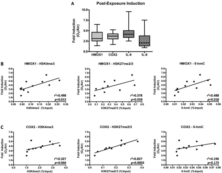
Comparison of Basal Expression and Postexposure Induction of O3-responsive Proinflammatory and Oxidative Stress Genes We assessed the induction of the proinflammatory genes IL-8, COX2, and IL-6, and the oxidative stress gene HMOX1 following O3exposure (Fig.3A). The O3-mediated induction of IL-8 mRNA immediately following exposure (0 h postexposure) ranged from 2.540 to 9.570 with a mean (6SD) of 4.53861.692 (n¼17), COX2 ranged from 1.697 to 5.221 with a mean (6SD) of 3.62160.949 (n¼17), and IL-6 ranged from 0.948 to 7.538 with a mean (6SD) of 2.73361.702 (n¼17). The O3-mediated induction of HMOX1 immediately after exposure ranged from 1.413 to 6.507 with a mean (6SD) of 3.47061.367, (n¼17). The box and whisker plots reflect the heterogeneity in ozone-induced response of cells obtained from different individuals. To determine whether basal gene expression was associated with the magnitude of exposure-mediated induction (“responsiveness”) we compared IL-8, IL-6, COX2, and HMOX1 expression in unexposed cells to the fold induction following O3exposure. There was a negative correlation between basal and ozone-induced IL-8 expression (r2¼0.356, P¼.052), but there were no correlations between basal gene expression and the postexposure fold induction of HMOX1 (r2¼0.006,P¼.823), COX2 (r2¼0.014,P¼.725), and IL-6 (r2¼0.103,P¼.335) (Supplementary Fig. 2).
Baseline Patterns of Specific Chromatin Modifications Correlate with Postexposure Gene Induction
Given the absence of a relationship between basal gene expres-sion and postexposure gene induction and the importance of chromatin modifications as regulators of expression, we sought to determine whether the baseline abundance of specific chro-matin modifications correlated with the pro-inflammatory and oxidative stress responses to O3exposure. We evaluated the rela-tionship between postexposure gene induction and the baseline abundance of the set of six chromatin modifications that we examined in relation to basal gene expression. Ozone-induced expression of HMOX1 had a positive correlation to baseline abun-dance of H3K4me3 (r2¼0.498,P¼.015), H3K27me2/3 abundance (r2¼0.378, P¼.058), and 5-hmC (r2¼0.488, P¼.017) (Fig. 3B). Similarly, postexposure induction of COX2 exhibited a positive
correlation to baseline abundance of H3K4me3 (r2¼0.327,
P¼.066), H3K27me2/3 (r2¼0.827,P¼.0003), and to a lesser extent 5-hmC (r2¼0.196, P¼.17) (Fig. 3C). In contrast, postexposure induction of IL-8 and IL-6 did not correlate with baseline abun-dance of H3K4me3, H3K27me2/3, or 5-hmC (Supplementary Figs. 3CandD). Further, baseline levels of H3K27ac, H4ac, and total H3 did not correlate with the post-exposure induction of HMOX1, COX2, IL-8 or IL-6 (Supplementary Fig. 3). All correlation data between baseline chromatin modification and induced gene expression are shown inSupplementary Tables 5and6. The cor-relations between the specific chromatin modifications that we examined and the induced expression of HMOX1, COX2, IL-8, and IL-6 are summarized inFigure 4B.
Sensitivity Analysis
In contrast to HMOX1 and COX2, postexposure induction of IL-8 and IL-6 was not normally distributed according to the D’Agostino & Pearson omnibus normality test. The Grubb’s test identified a single outlier in each of the IL-8 and IL-6 expression datasets, which originated from the same donor indicating that the lack of normality was driven by data collected from a single donor. The removal of values linked to that donor from all anal-yses resulted in postexposure induction values for all genes being normally distributed and supported our original findings. Omission of the single donor’s data improved the correlation and significance of comparisons between H3K27me2/3 and the postexposure induction of HMOX1 and between H3K4me3, 5-hmC, and H3K27me2/3 and postexposure COX2 induction reported in Figure 3. Following removal of the single donor, O3-induced expression of HMOX1 had a positive correlation to baseline abundance of H3K4me3 (r2¼0.472, P¼.028), 5-hmC (r2¼0.452, P¼.033), and H3K27me2/3 abundance (r2¼0.485,
P¼.037,) and similarly, O3-induced expression of COX2 exhib-ited a positive correlation to baseline abundance of H3K4me3 (r2¼0.677,P¼.004), 5-hmC (r2¼0.494,P¼.023), and H3K27me2/3 (r2¼0.908, P¼.0001). The results of simple linear regression analysis of comparisons between each target gene and specific chromatin modification following removal of the single donor
are shown inSupplementary Table 6. The basal expression of all 4 genes was normally distributed in the total population, which was not affected by the removal of the single donor’s val-ues. Further, removal of the single donor’s values did not impact the outcomes of the comparisons between baseline chromatin modifications and basal gene expression (Supplementary Table 6).
DISCUSSION
The role of chromatin modifications in regulating basal and induced proinflammatory and oxidative stress gene expression in the context of toxicology has received little attention. As the relationship between the epigenome, exposure effects, and sus-ceptibility becomes better defined, the field of epigenetics has the potential to transform our understanding of the mechanisms underlying interindividual variability, establish new risk assess-ment paradigms, and identify modifiable factors that can be leveraged to mitigate the adverse health effects of toxicant expo-sures. To explore this potential, we sought to determine the role that both baseline histone modifications and 5-hmC play in basal and toxicant-mediated induction expression of proinflammatory and oxidative stress genes in a primary cell ALI model of the human airway. Here, we demonstrate that baseline levels of specific chromatin modifications correlate with the interindividual variability in both basal and O3-induced expres-sion of proinflammatory stress genes. The findings presented here demonstrate that (1) the basal and induced expression of certain toxicant-responsive genes exhibit linear relationships with the baseline abundance of specific chromatin modifications, (2) the abundance of H3K4me3 at gene promoters positively cor-relates with the interindividual variability of both basal and induced expression of some, but not all, O3-responsive genes, (3) the abundance of H3K27ac and H4ac at the promoters of specific O3-responsive genes positively correlates with the interindividual variability in their basal, but not induced, expression, and (4) H3K27me2/3 and 5-hmC abundance at the promoters of specific O3-responsive genes positively correlates with the interindividual FIG. 2.Correlations between specific baseline chromatin modification levels and basal gene expression. The abundance of IL-8 (A), IL-6 (B), HMOX1(C) in unstimulated pHBEC was determined as a percentage of ACTB expression and compared to baseline levels of H3K4me3, H3K27ac, and H4ac.r2andPvalues were determined by
variability in their induced, but not basal, expression. These posi-tive correlations indicate that lower and higher baseline abun-dance of specific chromatin modifications at the promoters of certain O3-responsive genes coincide with lower and higher expression, respectively, of O3-responsive genes. These correla-tions exist between several specific chromatin modificacorrela-tions and certain target genes, suggesting that the interindividual vari-ability in postexposure gene induction is modulated by the relative abundance of a set of modifications instead of a single specific modification. Thus the baseline abundance of specific sets of chromatin modifications may be predictive of the interindividual variability in the induction of certain O3-responsive genes. The findings from this study are the first to link the baseline relative abundance of specific histone modifica-tions and DNA hydroxymethylation to the inter-individual varia-bility in proinflammatory and oxidative stress gene induction following toxicant exposure.
The baseline abundance of both H3K4me3 and H3K27me2/3 positively correlated with the postexposure induction of HMOX1 and COX2, but not IL-8 or IL-6; however, both H3K4me3 and H3K27me2/3 were present at all four gene promoters. While FIG. 3.Correlations between specific baseline chromatin modification levels and O3-induced gene expression. (A) Induction of the pro-inflammatory genes COX-2, IL-8,
and IL-6 and the oxidative stress gene HMOX1 were measured in pHBEC immediately following a 2-h exposure to 0.5 ppm O3. Baseline levels of H3K4me3, H3K27me2/3,
and 5-hmC were compared to the postexposure induction of HMOX1 (B) and COX2 (C), in pHBEC. Fold induction is shown as O3/air and was normalized to
correspond-ing fold change in the housekeepcorrespond-ing gene ACTB accordcorrespond-ing to the Pffafl method.r2andPvalues were determined by simple linear regression. The boxes in (B–E)
repre-sent the range of values between 25th and 75th percentiles with the center line reprerepre-senting the median and whiskers indicating the minimum and maximum values observed.n¼17 donors in (A) and 11 (10 donors for H3K27me2/3) in (B) and (C).
FIG. 4.Summary of correlations between specific chromatin modifications and expression of O3-responsive genes. Heat maps summarize correlations between
basal (A) and O3-induced gene expression (B) and the specific chromatin
modifi-cations that were observed in this study. Red and blue coloration indicates posi-tive and negaposi-tive correlation, respecposi-tively.n¼11 donors.
H3K4me3 and H3K27me2/3 are typically associated with active and repressed promoters, respectively, they can be found together on so-called ‘bivalent’ promoters (Bernsteinet al.,2006; Cedar and Bergman, 2011;Mikkelsenet al.,2007). Bivalent pro-moters are poised to be rapidly inducible and often assume a biased active or repressed state after stimulus (Bernsteinet al., 2006;Panet al.,2007). While typically studied in the context of gene regulation during development and differentiation, the principles governing bivalent promoters may also apply to the regulation of toxicant-induced gene expression. All four gene promoters that we examined had detectable levels of H3K4me3 and H3K27me2/3; however, levels of these histone modifica-tions only correlated to the induction of HMOX1 and COX2. This indicates that the presence of a bivalent promoter alone does not guarantee a correlation between levels of H3K4me3 and H3K27me2/3 and post-exposure gene induction. HMOX1 and COX2 also differed from IL-8 and IL-6 in that their induction cor-related with the abundance of 5-hmC at the gene promoters, respectively. The additional correlation of 5-hmC with HMOX1 and COX2 induction lends credence to the notion that both his-tone modifications and DNA methylation states work in concert to shape the exposure response at some, but not all genes. Further, the lack of correlation between H3K4me3, H3K27me2/3, and O3-mediated induction of IL-8 and IL-6 suggests that there are other factors involved in these regulatory processes that have yet to be identified. One such factor may be the more com-monly studied epigenetic modification 5-mC. While we focused on 5-hmC due to its enrichment with gene promoters in the cur-rent study, the role of 5-mC as a predictor of O3-mediated gene induction is intriguing and warrants examination in future studies.
Despite the presence of the activating mark H3K4me3, biva-lent promoters exhibit low activity owing to the additional pres-ence of H3K27me2/3; however, environmental cues could either induce or silence their expression by shifting the balance in favor of H3K4me3 or H3K27me2/3, respectively (Mikkelsenet al., 2007). When coupled with the presence of bivalent promoters at toxicant-responsive genes, environmentally induced changes in the chromatin landscape may provide a mechanistic
explanation for how past exposure history can impact the response to future exposures. Further, exposure-associated dif-ferences in the chromatin landscape may be one of the mecha-nisms underlying combinatorial effects of multiple exposures. The persistence of changes from previous exposures could cre-ate an exposure history in the form of chromatin modification patterns at the promoters of exposure-responsive genes. The patterns of these durable chromatin modifications would then influence the induction of these genes in response to future exposures. Since chromatin modification patterns can be stable across both cellular and organismal generations changes to the epigenome from lifestyle or environmental exposures have been proposed to have multi- and trans-generational effects on susceptibility and disease (Jirtle and Skinner, 2007). Further studies are required to determine whether exposure to inhaled toxicants, such as O3, can change levels of these histone modifi-cations and thus potentially impact the induction of genes such as HMOX1 and COX2 in multiple or repeated exposure scenarios.
Using these principles we have developed an “epigenetic seed and soil” model to explain the interindividual variability in postexposure gene induction that we observed in our donor group (Fig.5). In this model, the chromatin modification states between individuals differ in the relative abundance of sets of specific modifications (the “soil”) that govern how receptive the promoters of certain genes, such as HMOX1 and COX2, are to toxicant-induced cellular signals (the “seed”). When these sig-nals reach the receptive chromatin soil in the cells of a more responsive individual they then result in greater induction of certain toxicant exposure-associated genes. In contrast, when a toxicant-induced signal reaches less receptive chromatin soil the resulting induction of toxicant-responsive genes is lower, thus making that individual less responsive. Differences in the baseline epigenetic “soil” may result from the effects of cumula-tive exposure history, multi- or trans-generational exposure effect, lifestyle, or psychosocial and other factors on the epigenome.
for the role of baseline epigenetic modifications in postexposure gene induction may apply to toxicant exposures in a broad range of cell and tissue types both in vitro and
in vivo.Future studies are needed to test this hypothesis in other exposure models with additional chromatin modifica-tions and with a broad range of toxicants. The epigenetic seed and soil model is a promising prospect; however, our under-standing of how baseline epigenetic profiles influence the tox-icant exposure response is in its infancy. As the relationship between the epigenome, exposure effects, and susceptibility becomes better defined the field of epigenomics has the poten-tial to transform risk assessment paradigms. While compli-cated, the eventual use of epigenetic data, such as the baseline abundance of specific chromatin modifications, to refine dose-response assessments of exposure effects and susceptibility to adverse health outcomes is neither unreasonable nor is it far in the future. In conclusion, our studies reinforce the potential of epigenetic markers to serve as indicators or predictors of responsiveness to pollutants. Given its relationship with life-style and psychosocial factors, we believe that future studies will allow the epigenome to be targeted in susceptible popula-tions to mitigate risk.
FUNDING
This work was supported by the Office of Research and Development’s Pathfinder Innovation Program, a U.S. Environmental Protection Agency intramural funding program.
ACKNOWLEDGMENTS
The authors would like to thank Dr. Marie C. Fortin for her expert opinions during critical review of the manuscript, as well as TRC Environmental Corporation, particularly Mr. Scott Meade, for the design, construction, and maintenance of the
in vitro exposure apparatus. The research described in this article has been reviewed by the Environmental Protection Agency and approved for publication. The contents of this article do not necessarily represent Agency policy, nor does mention of trade names or commercial products constitute endorsement or recommendations for use.
SUPPLEMENTARY DATA
Supplementary data are available online at http://toxsci. oxfordjournals.org/.
REFERENCES
Alexis, N. E., Lay J. C., Hazucha M., Harris B., Hernandez M. L., Bromberg P. A., Kehrl H., Diaz-Sanchez D., Kim C., Devlin R. B., and Peden D. B. (2010). Low-level ozone exposure indu-ces airways inflammation and modifies cell surface pheno-types in healthy humans.Inhal. Toxicol.,22, 593–600.
Baccarelli, A., and Bollati V. (2009). Epigenetics and environmen-tal chemicals.Curr. Opin. Pediatrics,21, 243–251.
Bachman, M., Uribe-lewis S., Yang X., Williams M., and Murrell A. (2014). 5-hydroxymethylcytosine is a predominantly stable DNA modification.Nat. Chem.,6,1049–1055.
Bastain, T. M., Gilliland F. D., Li Y.-F., Saxon A., and Diaz-Sanchez D. (2003). Intraindividual reproducibility of nasal allergic re-sponses to diesel exhaust particles indicates a susceptible phenotype.Clin. Immunol.,109, 130–136.
Bernstein, B. E., Mikkelsen T. S., Xie X., Kamal M., Huebert D. J., Cuff J., Fry B., Meissner A., Wernig M., Plath K.,et al(2006). A bivalent chromatin structure marks key developmental genes in embryonic stem cells.Cell,125,315–326.
Braunstein, M., Rose A. B., Holmes S. G., Allis C. D., and Broach J. R. (1993). Transcriptional silencing in yeast is associated with reduced nucleosome acetylation.Genes Dev.,7, 592–604. Burris, H. H. (2014). Environmental epigenetics: from novelty to
scientific discipline.J. Appl. Toxicol.,34.
Cedar, H., and Bergman, Y. (2011). Epigenetics of haematopoietic cell development.Nat. Rev. Immunol.,11,478–488.
Chen, Y., Jørgensen M., Kolde R., Zhao X., Parker B., Valen E., Wen J., and Sandelin A. (2011). Prediction of RNA polymerase II recruitment, elongation and stalling from histone modifi-cation data.BMC Genomics,12, 544.
Creyghton, M. P., Cheng A. W., Welstead G. G., Kooistra T., Carey B. W., Steine E. J., Hanna J., Lodato M. A., Frampton G. M., Sharp P. A.,et al. (2010). Histone H3K27ac separates active from poised enhancers and predicts developmental state.
Proc. Natl. Acad. Sci. USA,107, 21931–21936.
Davey, C. A., Sargent D. F., Luger K., Maeder A. W., and Richmond T. J. (2002). Solvent mediated interactions in the structure of the nucleosome core particle at 1.9 A˚ resolution.J. Mol. Biol.,
319, 1097–1113.
Dawson, M. a., and Kouzarides, T. (2012). Cancer epigenetics: From mechanism to therapy.Cell,150, 12–27.
Devlin, R. B., McDonnell W. F., Becker S., Madden M. C., McGee M. P., Perez R., Hatch G., House D. E., and Koren H. S. (1994). Ozone-induced release of cytokines and fibronectin by alveo-lar macrophages and airway epithelial cells Ozone-induced release of cytokines and fibronectin alveloar macrophages and airway epithelial cells by. Am. J. Physiol., 266(6 pt 1), L612–L619.
Devlin, R. B., Mckinnon K. P., Noah T., Becker S., Koren H. S., Mckinnon P., Noah T., Koren H. S. (1996). Time-dependent changes of inflammatory mediators in the lungs of humans exposed to 0.4 ppm ozone for 2 hr: A comparison of media-tors found in broncoalveolar lavage fluid 1 and 18 hr after ex-posure.Toxicol. Appl. Pharmacol.,138, 176–185.
Dolinoy, D. C., Weidman, J. R., and Jirtle, R. L. (2007). Epigenetic gene regulation: Linking early developmental environment to adult disease.Reprod. Toxicol.,23, 297–307.
Ficz, G., Branco M. R., Seisenberger S., Santos F., Krueger F., Hore T. A., Marques C. J., Andrews S., and Reik W. (2011). Dynamic regulation of 5-hydroxymethylcytosine in mouse ES cells and during differentiation.Nature,473, 398–402.
Guenther, M. G., Levine S. S., Boyer L. A., Jaenisch R., and Young R. A. (2007). A chromatin landmark and transcrip-tion initiatranscrip-tion at most promoters in human cells. Cell,
130, 77–88.
Holz, O., Jo¨rres R. A., Timm P., Mu¨cke M., Richter K., Koschyk S., and Magnussen H. (1999). Ozone-induced airway inflamma-tory changes differ between individuals and are reproduc-ible.Am. J. Respir. Crit. Care Med.,159, 776–784.
Hou, L., Zhang X., Wang D., and Baccarelli A. (2012). Environmental chemical exposures and human epigenetics.
Int. J. Epidemiol.,41, 79–105.
Jirtle, R., and Skinner, M. (2007). Environmental epigenomics and disease susceptibility.Nat. Rev. Genetics,8, 253–262.
Karlic, R., Chung H.-R., Lasserre J., Vlahovicek K., and Vingron M. (2010). Histone modification levels are predictive for gene ex-pression.Proc. Natl. Acad. Sci. USA,107, 2926–2931.
Kouzarides, T. (2007). Chromatin modifications and their func-tion.Cell,128, 693–705.
Lam, L. L., Emberly E., Fraser H. B., Neumann S. M., Chen E., Miller G. E., and Kobor M. S. (2012). Factors underlying vari-able DNA methylation in a human community cohort.Proc. Natl. Acad. Sci. USA,109, 17253–17260.
Liu, C., Xu J., Chen Y., Guo X., Zheng Y., Wang Q., Chen Y., Ni Y., Zhu Y., Joyce B. T.,et al. (2015). Characterization of genome-wide H3K27ac profiles reveals a distinct PM2.5-associated histone modification signature.Environ. Health,14, 65. Luger, K., Ma¨der A. W., Richmond R. K., Sargent D. F., and
Richmond T. J. (1997). Crystal structure of the nucleosome core particle at 2.8 A resolution.Nature,389, 251–260. McCullough, S. D., Duncan K. E., Swanton S. M., Dailey L. A.,
Diaz-Sanchez D., and Devlin R. B. (2014). Ozone induces a pro-inflammatory response in primary human bronchial epi-thelial cells through MAP kinase activation without NF-jB activation.Am. J. Respir. Cell Mol. Biol.,51, 1–45.
McDonnell, W. F., Horstman D. H., Abdul-Salaam S., and House D. E. (1985). Reproducibility of individual responses to ozone exposure.Am. Rev. Respir. Dis.,131, 36–40.
Mikkelsen, T. S., Ku M., Jaffe D. B., Issac B., Lieberman E., Giannoukos G., Alvarez P., Brockman W., Kim T.-K., Koche R. P.,et al. (2007). Genome-wide maps of chromatin state in plu-ripotent and lineage-committed cells.Nature,448, 553–560. Olden, K., Lin Y.-S., Gruber D., and Sonawane B. (2014).
Epigenome: biosensor of cumulative exposure to chemical and nonchemical stressors related to environmental justice.
Am. J. Public Health,104, 1816–1821.
Pan, G., Tian S., Nie J., Yang C., Ruotti V., Wei H., Jonsdottir G. A., Stewart R., and Thomson J. A. (2007). Whole-genome analysis of histone H3 lysine 4 and lysine 27 methylation in human embryonic stem cells.Cell Stem Cell,1, 299–312.
Pfaffl, M. W. (2001). A new mathematical model for relative quan-tification in real-time RT–PCR.Nucleic Acids Res.,29, p.e45. Ross, A. J., Dailey L. A., Brighton L. E., and Devlin R. B. (2007).
Transcriptional profiling of mucociliary differentiation in human airway epithelial cells.Am. J. Resp. Cell Mol. Biol.,37, 169–185. De Rubeis, S., He X., Goldberg A. P., Poultney C. S., Samocha K.,
Cicek A. E., Kou Y., Liu L., Fromer M., Walker S.,et al. (2014). Synaptic, transcriptional and chromatin genes disrupted in autism.Nature,515, 209–215.
Strahl, B. D., and Allis, C. D. (2000). The language of covalent his-tone modifications.Nature,403, 41–45.
Tahiliani, M., Koh K. P., Shen Y., Pastor W. A., Brudno Y., Agarwal S., Iyer L. M., David R., Aravind L., and Rao A. (2009). Conversion of 5-Methylcytosine to 5-hydroxymethylcytosine in Mammalian DNA by MLL Partner TET1.Science,324, 930–935. Waterland, R. A., Lin J. R., Smith C. A., and Jirtle R. L. (2006).
Post-weaning diet affects genomic imprinting at the insu-lin-like growth factor 2 (Igf2) locus. Hum. Mol.Genetics, 15, 705–716.