Bringing It All Together: Coupling Excision Repair to the DNA
Damage Checkpoint
†Laura A. Lindsey-Boltz
Department of Biochemistry and Biophysics, University of North Carolina School of Medicine, Chapel Hill, North Carolina 27599
Abstract
Nucleotide excision repair and the ATR-mediated DNA damage checkpoint are two critical cellular responses to the genotoxic stress induced by ultraviolet (UV) light and are important for cancer prevention. In vivo genetic data indicate that these global responses are coupled. Aziz Sancar and colleagues developed an in vitro coupled repair-checkpoint system to analyze the basic steps of these DNA damage stress responses in a biochemically defined system. The minimum set of factors essential for repair-checkpoint coupling include damaged DNA, the excision repair factors (XPA, XPC, XPF-ERCC1, XPG, TFIIH, RPA), the 5′-3′ exonuclease EXO1, and the damage checkpoint proteins ATR-ATRIP and TopBP1. This coupled repair-checkpoint system was used to demonstrate that the ~30 nucleotide single-stranded DNA (ssDNA) gap generated by nucleotide excision repair is enlarged by EXO1 and bound by RPA to generate the signal that activates ATR.
Graphical Abstract
Nucleotide excision repair and the ATR-mediated DNA damage checkpoint are two critical cellular responses to UV-induced genotoxic stress and are important for cancer prevention. This review summarizes the in vitro coupled repair-checkpoint system developed by Aziz Sancar and colleagues to analyze the basic steps of these two DNA damage stress responses in a
biochemically defined reaction. They demonstrated that the ~30 nucleotide single-stranded DNA gap generated during the excision repair process by XPA, XPC, XPF-ERCC1, XPG, TFIIH, RPA is enlarged by the exonuclease EXO1 and bound by RPA to generate the signal that activates ATR-ATRIP in the presence of TOPBP1.
HHS Public Access
Author manuscript
Photochem Photobiol
. Author manuscript; available in PMC 2018 January 01.Published in final edited form as:
Photochem Photobiol. 2017 January ; 93(1): 238–244. doi:10.1111/php.12667.
A
uthor Man
uscr
ipt
A
uthor Man
uscr
ipt
A
uthor Man
uscr
ipt
A
uthor Man
uscr
INTRODUCTION
The Sancar lab was a very exciting place to begin a postdoc at the start of the new
millennium. It was six years after his lab had accomplished the heroic feat of reconstituting human nucleotide excision repair in vitro (1), and Aziz had come to the wise decision that the DNA excision repair field was “mature” and that his lab would embark upon the newly emerging field of DNA damage checkpoints. In fact, DNA damage checkpoints were pretty well understood in yeast by this time, in 2001 the Nobel Prize in Medicine was awarded to Leland Hartwell for introducing the concept: cell cycle arrest in response to DNA damage to ensure repair before cell division (2). However, the human checkpoint genes were just being identified, initial Human Genome Project results were published in 2001 (3), and we were eager to characterize the proteins encoded by these genes, with the long-term goal of reconstituting the human DNA damage checkpoint with purified proteins, and the even-longer-term goal of coupling nucleotide excision repair and the DNA damage checkpoint in vitro. It is our hope that research to understand these two cellular responses to DNA damage at a molecular level will ultimately aid in cancer prevention, diagnosis, and treatment.
RECONSTITUTING THE ATR-MEDIATED DNA DAMAGE CHECKPOINT
DNA damage checkpoints are signal transduction pathways induced after DNA damage and are composed of proteins in the four conceptual categories of Sensors, Mediators,
Transducers, and Effectors (4) (Figure 1). The DNA damage checkpoints are broadly defined by two types of damage that induce the response: bulky DNA adducts such as those generated by ultraviolet (UV) light and double strand breaks such as those introduced by ionizing radiation (IR). The DNA damage checkpoints are also defined by the two very large sensor kinases, Ataxia Telangiectasia Mutated (ATM) and ATM and Rad3-related (ATR), which are activated by the DNA damage caused by IR or UV, respectively. These kinases belong to the phosphatidylinositol 3-kinase-related kinase (PIKK) family of Ser/Thr-protein kinases which also include DNA-PKcs, SMG1, TRRAP, and mTOR. Upon activation, the
A
uthor Man
uscr
ipt
A
uthor Man
uscr
ipt
A
uthor Man
uscr
ipt
A
uthor Man
uscr
sensor kinases ATR and ATM phosphorylate and activate the downstream transducer kinases, Chk1 and Chk2, respectively. Considering the historical interest and expertise of the Sancar lab in analyzing DNA damage responses to UV light, we chose to focus on the ATR→Chk1 pathway, while recognizing that there is considerable crosstalk and overlap between these signal transduction pathways.
During the past 16 years we have purified the key human checkpoint proteins in the ATR→Chk1 pathway (Figure 2). ATR is a 300 kDa protein that forms a heterodimer with a 85 kDa protein partner, ATRIP. Initial characterizations of ATR-ATRIP by the Sancar lab were performed with epitope-tagged recombinant proteins (5,6), but we later found that native ATR-ATRIP purified from HeLa cell extracts (7) was more active and consistent, thus we continued to use native protein purified from hundreds of liters of HeLa cells in
subsequent reconstitution experiments (8–15). During the purification of ATR-ATRIP we carefully monitored the ATR-ATRIP-containing fractions for the presence of the other PIKKS, especially the highly abundant DNA-PKcs, since these kinases have similar sequence specificity (preferring S/TQ) and can phosphorylate the same substrates in vitro (16) which complicates data interpretation. Upon activation, ATR phosphorylates itself and other key checkpoint proteins. In our reconstituted system, we initially focused on ATR phosphorylation of Chk1 (7–14), but have also analyzed phosphorylation of other substrates including RPA, p53, and Rad17 (8,13,15) using both quantitative Western blotting with phosphospecific antibodies (7–15) and measuring the incorporation of radiolabeled
phosphate (5,6,10) to analyze the amount of substrate phosphorylation. Chk1 is the ‘classic’ ATR substrate, however we analyzed ATR phosphorylation of RPA and p53 in our repair-checkpoint coupling experiments because Chk1 is expressed at low levels and is not phosphorylated during G0/G1 (15,17,18), and therefore not a physiologically relevant substrate when studying the coupling of excision repair and ATR activation (discussed more below).
My initial project in the Sancar lab was to characterize the human homolog of the fission yeast damage sensor Rad17, which has homology to all five subunits of Replication Factor C (RFC). We found that hRad17 forms a stable complex with the four small subunits of RFC, replacing the large subunit (p140), and we named this checkpoint complex hRad17-RFC (19). RFC loads the polymerase processivity clamp, Proliferating Cell Nuclear Antigen, PCNA, onto DNA during replication. PCNA is a trimer of identical subunits which share homology with the checkpoint proteins Rad9, Rad1, and Hus1, which together form a trimer called the checkpoint 9-1-1 complex. We demonstrated by electron microscopy that the 9-1-1 complex forms a ring-like structure similar to PCNA (20), and that the hRad17-RFC complex loads the 9-1-1 complex onto DNA (21). Although these proteins are genetically required for the ATR-mediated checkpoint in vivo, the loading reaction is inefficient in vitro, and we have not been successful incorporating these checkpoint proteins into our
reconstituted checkpoint system. We suspect that a component may be missing in our system, and indeed, when the 9-1-1-interacting factor RHINO was reported, we re-visited the subject with little success (22). Through collaborations with other labs, we also found that the 9-1-1 complex interacts with and stimulates several enzymes in the Base Excision Repair pathway (23–25) as well as carbamoyl phosphate synthetase/aspartate
A
uthor Man
uscr
ipt
A
uthor Man
uscr
ipt
A
uthor Man
uscr
ipt
A
uthor Man
uscr
transcarbamoylase/dihydroorotase (CAD), a multienzymatic protein required for the de novo pyrimidine synthesis (26).
A huge advance in the checkpoint field was the 2006 discovery by the William Dunphy lab that DNA Topoisomerase 2-Binding Protein 1 (TopBP1) is an activator of ATR-ATRIP (27). We had actually purified and tested TopBP1 in our ATR kinase system before this report, but had not observed the activator effect due to manganese in our reaction buffer. Early reports had indicated that manganese was a requirement for the PIKK class of enzymes (28), and thus our ATR kinase system included manganese, but we found that the combination of magnesium and manganese inhibits TopBP1 activation of ATR-ATRIP. After eliminating manganese, we found TopBP1 to be a potent ATR-ATRIP activator and went on to characterize many properties of its ATR-activating activity including its binding and stimulation by damaged DNA (7,9) (Figure 3), its cooperative mechanism (8), and its binding and stimulation by RPA-coated ssDNA (11).
Long stretches of RPA-coated ssDNA strongly induce the ATR-mediated checkpoint. During S phase the long stretches of ssDNA result from the replication fork stalling and helicase uncoupling that occurs when polymerases encounter the damage (29). Through direct and indirect interactions, RPA recruits key checkpoint proteins to the ssDNA (Figure 4). Direct RPA interactions recruit ATR-ATRIP via ATRIP (30) and the TopBP1 activator via its N-terminus (11). Chk1 is indirectly recruited through interactions with Claspin which interacts with Timeless-Tipin via a direct interaction between Tipin and RPA (31). We have found that p53 and hRad17 interactions with RPA-coated ssDNA also facilitate their
phosphorylation by ATR (13). Interactions with RPA-coated ssDNA effectively concentrate or crowd the checkpoint proteins together such that ATR is in contact with substrates and TopBP1 activator. We have provided evidence for this crowding model both in vitro and in vivo by demonstrating ATR activation through tethering key checkpoint proteins to DNA using the Lac Repressor-Lac Operon system (12,14,22) as well as with light-induced crowding via cryptochrome photobodies (32).
COUPLING NUCLEOTIDE EXCISION REPAIR AND THE ATR-MEDIATED DNA
DAMAGE CHECKPOINT
It had already been demonstrated in some human cell lines that UV-induced ATR activation is dependent on nucleotide excision repair when the cells are in G0 (17,33–35), but two advances in 2010 convinced us that our ultimate goal of coupling nucleotide excision repair and the DNA damage checkpoint with purified proteins would be attainable: we
reconstituted ATR activation with RPA-coated ssDNA in vitro (11) and the Muzi-Falconi lab reported that the UV-induced ATR checkpoint is activated in G0 when EXO1 converts nucleotide excision repair intermediates into long ssDNA gaps in vivo (36,37). After repair of UV damage, EXO1 competes with repair synthesis resulting in a small fraction of the ~30 nucleotide excision repair gaps being resected to generate long stretches of ssDNA. We first performed a ‘proof of principle’ experiment to demonstrate in vitro support for this model. As shown in Figure 5, in in vitro kinase reactions containing purified ATR-ATRIP, TopBP1,
A
uthor Man
uscr
ipt
A
uthor Man
uscr
ipt
A
uthor Man
uscr
ipt
A
uthor Man
uscr
and RPA, we observed RPA phosphorylation only in the presence of ssDNA or when gapped DNA was resected by EXO1 (Figure 5).
Although we had now demonstrated EXO1-dependent ATR activation with model DNA substrates, it took us much longer to optimize the reaction conditions and to generate sufficient quality DNA and excision repair factors to achieve our goal of nucleotide excision repair-dependent ATR activation (15). We determined that DNA damaged with the UV-mimetic N-Acetoxy-2-acetylaminofluorene (AAF) worked better in the reconstitution experiments than DNA damaged with UV. AAF induces bulky AAF-guanine adducts in DNA which are among the best substrates for nucleotide excision repair, whereas the main lesions generated by UV, cyclobutane thymine dimers, are not efficiently excised in in vitro reactions (38). We also determined that the amount of DNA damage per plasmid required optimization, with ~3 AAFs per plasmid (~1/kb) producing the best results. Less AAF damage in the plasmid DNA resulted in less gaps and more AAF damage resulted in more gaps yet blocked EXO1 resection, both conditions resulting in a lower ssDNA to dsDNA ratio which decreased the signal-to-noise ratio.
EXO1 is a very efficient 5′ to 3′ exonuclease on both gapped and nicked DNA. Therefore, any nicks in the DNA generated nonspecifically by any one component resulted in the reaction not being dependent on the other components. DNA nicks introduced in the initial plasmid preparation, introduced during the AAF damage process, or introduced by nuclease contaminants in the repair or checkpoint factors were a problem. This required us to prepare very pure excision repair factors: XPA, XPC-HR23B, XPF-ERCC1, XPG, TFIIH, and RPA (Figure 6).
Once we obtained good quality DNA and proteins, we were finally able to complete the reconstitution experiments. In vitro coupling of nucleotide excision repair and the ATR-mediated DNA damage checkpoint is shown in Figure 7 (Figure 7). ATR activation, as measured by RPA2 phosphorylation, is dependent on the presence of the six excision repair factors (RF), TopBP1, EXO1, and AAF-damaged DNA. We also demonstrated repair-checkpoint coupling as measured by p53 phosphorylation in this study (15).
CONCLUSION
A model summarizing repair-checkpoint coupling is shown in Figure 8 (Figure 8). When DNA is damaged by UV or a UV-mimetic agent, the core excision repair factors (XPA, XPC-HR23B, XPF-ERCC1, XPG, TFIIH, and RPA) excise the damage, and the resulting ~30 nucleotide gap is either filled in by polymerases or the gap is enlarged by EXO1. The resulting long stretch of ssDNA is coated with RPA which recruits ATR-ATRIP, TopBP1, and substrates including p53. This repair-checkpoint coupling work brings together over 25 years of research in the Sancar lab, and as Aziz so elegantly described in his Nobel
Biography (39), this work is “the ultimate in reductionist biochemical research that aims to explain complex cellular phenomenon in a minimalist in vitro system.” It is our hope that by breaking down and rebuilding these repair and checkpoint reactions that we may gain sufficient understanding of the molecular mechanisms of these reactions to inform better
A
uthor Man
uscr
ipt
A
uthor Man
uscr
ipt
A
uthor Man
uscr
ipt
A
uthor Man
uscr
development of tools for diagnosis, prevention, and treatment of cancer and other human disease.
Acknowledgments
The Sancar lab was such a wonderful place to work and study as a postdoc that I could not leave when my postdoctoral training was over. Over the 16 years I have had the great fortune of having Aziz as my mentor, boss, colleague, and friend. By example, he has taught me the value of hard work. I have never been more proud than watching him receive the Nobel Prize in Chemistry. Finally, I would like to acknowledge my appreciation for current and past colleagues in the Sancar lab (students, postdocs, staff) for their dedication, patience, and determination. The author is supported by NIH grant GM118102.
Abbreviations
UV ultraviolet
ssDNA single-stranded DNA
IR ionizing radiation
PIKK phosphoinositol 3-kinase-related kinase
ATM Ataxia Telangiectasia-Mutated
ATR ATM and Rad3-related
PCNA Proliferating Cell Nuclear Antigen
TIM Timeless
TIPIN Timeless interacting protein
CAD carbamoyl phosphate synthetase/aspartate transcarbamoylase/dihydroorotase
TopBP1 Topoisomerase 2-Binding Protein 1
AAF N-Acetoxy-2-acetylaminofluorene
UM unmodified
RF repair factors
References
1. Mu D, Park CH, Matsunaga T, Hsu DS, Reardon JT, Sancar A. Reconstitution of human DNA repair excision nuclease in a highly defined system. J Biol Chem. 1995; 270:2415–2418. [PubMed: 7852297]
2. Hartwell LH. Nobel Lecture. Yeast and cancer. Biosci Rep. 2002; 22:373–394. [PubMed: 12516780] 3. Lander ES, Linton LM, Birren B, Nusbaum C, Zody MC, Baldwin J, Devon K, Dewar K, Doyle M,
FitzHugh W, Funke R, Gage D, Harris K, Heaford A, Howland J, Kann L, Lehoczky J, LeVine R, McEwan P, McKernan K, Meldrim J, Mesirov JP, Miranda C, Morris W, Naylor J, Raymond C, Rosetti M, Santos R, Sheridan A, Sougnez C, Stange-Thomann Y, Stojanovic N, Subramanian A, Wyman D, Rogers J, Sulston J, Ainscough R, Beck S, Bentley D, Burton J, Clee C, Carter N, Coulson A, Deadman R, Deloukas P, Dunham A, Dunham I, Durbin R, French L, Grafham D, Gregory S, Hubbard T, Humphray S, Hunt A, Jones M, Lloyd C, McMurray A, Matthews L, Mercer S, Milne S, Mullikin JC, Mungall A, Plumb R, Ross M, Shownkeen R, Sims S, Waterston RH,
A
uthor Man
uscr
ipt
A
uthor Man
uscr
ipt
A
uthor Man
uscr
ipt
A
uthor Man
uscr
Wilson RK, Hillier LW, McPherson JD, Marra MA, Mardis ER, Fulton LA, Chinwalla AT, Pepin KH, Gish WR, Chissoe SL, Wendl MC, Delehaunty KD, Miner TL, Delehaunty A, Kramer JB, Cook LL, Fulton RS, Johnson DL, Minx PJ, Clifton SW, Hawkins T, Branscomb E, Predki P, Richardson P, Wenning S, Slezak T, Doggett N, Cheng JF, Olsen A, Lucas S, Elkin C, Uberbacher E, Frazier M, Gibbs RA, Muzny DM, Scherer SE, Bouck JB, Sodergren EJ, Worley KC, Rives CM, Gorrell JH, Metzker ML, Naylor SL, Kucherlapati RS, Nelson DL, Weinstock GM, Sakaki Y, Fujiyama A, Hattori M, Yada T, Toyoda A, Itoh T, Kawagoe C, Watanabe H, Totoki Y, Taylor T, Weissenbach J, Heilig R, Saurin W, Artiguenave F, Brottier P, Bruls T, Pelletier E, Robert C, Wincker P, Smith DR, Doucette-Stamm L, Rubenfield M, Weinstock K, Lee HM, Dubois J, Rosenthal A, Platzer M, Nyakatura G, Taudien S, Rump A, Yang H, Yu J, Wang J, Huang G, Gu J, Hood L, Rowen L, Madan A, Qin S, Davis RW, Federspiel NA, Abola AP, Proctor MJ, Myers RM, Schmutz J, Dickson M, Grimwood J, Cox DR, Olson MV, Kaul R, Raymond C, Shimizu N, Kawasaki K, Minoshima S, Evans GA, Athanasiou M, Schultz R, Roe BA, Chen F, Pan H, Ramser J, Lehrach H, Reinhardt R, McCombie WR, de la Bastide M, Dedhia N, Blocker H, Hornischer K, Nordsiek G, Agarwala R, Aravind L, Bailey JA, Bateman A, Batzoglou S, Birney E, Bork P, Brown DG, Burge CB, Cerutti L, Chen HC, Church D, Clamp M, Copley RR, Doerks T, Eddy SR, Eichler EE, Furey TS, Galagan J, Gilbert JG, Harmon C, Hayashizaki Y, Haussler D, Hermjakob H, Hokamp K, Jang W, Johnson LS, Jones TA, Kasif S, Kaspryzk A, Kennedy S, Kent WJ, Kitts P, Koonin EV, Korf I, Kulp D, Lancet D, Lowe TM, McLysaght A, Mikkelsen T, Moran JV, Mulder N, Pollara VJ, Ponting CP, Schuler G, Schultz J, Slater G, Smit AF, Stupka E, Szustakowki J, Thierry-Mieg D, Thierry-Thierry-Mieg J, Wagner L, Wallis J, Wheeler R, Williams A, Wolf YI, Wolfe KH, Yang SP, Yeh RF, Collins F, Guyer MS, Peterson J, Felsenfeld A, Wetterstrand KA, Patrinos A, Morgan MJ, de Jong P, Catanese JJ, Osoegawa K, Shizuya H, Choi S, Chen YJ, Szustakowki J. C. International Human Genome Sequencing. Initial sequencing and analysis of the human genome. Nature. 2001; 409:860–921. [PubMed: 11237011]
4. Sancar A, Lindsey-Boltz LA, Unsal-Kacmaz K, Linn S. Molecular mechanisms of mammalian DNA repair and the DNA damage checkpoints. Annu Rev Biochem. 2004; 73:39–85. [PubMed:
15189136]
5. Unsal-Kacmaz K, Makhov AM, Griffith JD, Sancar A. Preferential binding of ATR protein to UV-damaged DNA. Proc Natl Acad Sci U S A. 2002; 99:6673–6678. [PubMed: 12011431]
6. Unsal-Kacmaz K, Sancar A. Quaternary structure of ATR and effects of ATRIP and replication protein A on its DNA binding and kinase activities. Mol Cell Biol. 2004; 24:1292–1300. [PubMed: 14729973]
7. Choi JH, Lindsey-Boltz LA, Sancar A. Reconstitution of a human ATR-mediated checkpoint response to damaged DNA. Proc Natl Acad Sci U S A. 2007; 104:13301–13306. [PubMed: 17686975]
8. Choi JH, Lindsey-Boltz LA, Sancar A. Cooperative activation of the ATR checkpoint kinase by TopBP1 and damaged DNA. Nucleic Acids Res. 2009; 37:1501–1509. [PubMed: 19139065] 9. Choi JH, Sancar A, Lindsey-Boltz LA. The human ATR-mediated DNA damage checkpoint in a
reconstituted system. Methods. 2009; 48:3–7. [PubMed: 19245835]
10. Lindsey-Boltz LA, Sercin O, Choi JH, Sancar A. Reconstitution of human claspin-mediated phosphorylation of Chk1 by the ATR (ataxia telangiectasia-mutated and rad3-related) checkpoint kinase. J Biol Chem. 2009; 284:33107–33114. [PubMed: 19828454]
11. Choi JH, Lindsey-Boltz LA, Kemp M, Mason AC, Wold MS, Sancar A. Reconstitution of RPA-covered single-stranded DNA-activated ATR-Chk1 signaling. Proc Natl Acad Sci U S A. 2010; 107:13660–13665. [PubMed: 20616048]
12. Lindsey-Boltz LA, Sancar A. Tethering DNA damage checkpoint mediator proteins topoisomerase IIbeta-binding protein 1 (TopBP1) and Claspin to DNA activates ataxia-telangiectasia mutated and RAD3-related (ATR) phosphorylation of checkpoint kinase 1 (Chk1). J Biol Chem. 2011; 286:19229–19236. [PubMed: 21502314]
13. Lindsey-Boltz LA, Reardon JT, Wold MS, Sancar A. In vitro analysis of the role of replication protein A (RPA) and RPA phosphorylation in ATR-mediated checkpoint signaling. J Biol Chem. 2012; 287:36123–36131. [PubMed: 22948311]
14. Hassan BH, Lindsey-Boltz LA, Kemp MG, Sancar A. Direct role for the replication protein treslin (Ticrr) in the ATR kinase-mediated checkpoint response. J Biol Chem. 2013; 288:18903–18910. [PubMed: 23696651]
15. Lindsey-Boltz LA, Kemp MG, Reardon JT, DeRocco V, Iyer RR, Modrich P, Sancar A. Coupling of human DNA excision repair and the DNA damage checkpoint in a defined in vitro system. J Biol Chem. 2014; 289:5074–5082. [PubMed: 24403078]
16. Kemp MG, Lindsey-Boltz LA, Sancar A. The DNA damage response kinases DNA-dependent protein kinase (DNA-PK) and ataxia telangiectasia mutated (ATM) Are stimulated by bulky adduct-containing DNA. J Biol Chem. 2011; 286:19237–19246. [PubMed: 21487018] 17. Matsumoto M, Yaginuma K, Igarashi A, Imura M, Hasegawa M, Iwabuchi K, Date T, Mori T,
Ishizaki K, Yamashita K, Inobe M, Matsunaga T. Perturbed gap-filling synthesis in nucleotide excision repair causes histone H2AX phosphorylation in human quiescent cells. J Cell Sci. 2007; 120:1104–1112. [PubMed: 17327276]
18. Kemp MG, Sancar A. ATR Kinase Inhibition Protects Non-cycling Cells from the Lethal Effects of DNA Damage and Transcription Stress. J Biol Chem. 2016; 291:9330–9342. [PubMed: 26940878] 19. Lindsey-Boltz LA V, Bermudez P, Hurwitz J, Sancar A. Purification and characterization of human
DNA damage checkpoint Rad complexes. Proc Natl Acad Sci U S A. 2001; 98:11236–11241. [PubMed: 11572977]
20. Griffith JD, Lindsey-Boltz LA, Sancar A. Structures of the human Rad17-replication factor C and checkpoint Rad 9-1-1 complexes visualized by glycerol spray/low voltage microscopy. J Biol Chem. 2002; 277:15233–15236. [PubMed: 11907025]
21. Bermudez VP, Lindsey-Boltz LA, Cesare AJ, Maniwa Y, Griffith JD, Hurwitz J, Sancar A. Loading of the human 9-1-1 checkpoint complex onto DNA by the checkpoint clamp loader hRad17-replication factor C complex in vitro. Proc Natl Acad Sci U S A. 2003; 100:1633–1638. [PubMed: 12578958]
22. Lindsey-Boltz LA, Kemp MG, Capp C, Sancar A. RHINO forms a stoichiometric complex with the 9-1-1 checkpoint clamp and mediates ATR-Chk1 signaling. Cell Cycle. 2015; 14:99–108. [PubMed: 25602520]
23. Balakrishnan L, Brandt PD, Lindsey-Boltz LA, Sancar A, Bambara RA. Long patch base excision repair proceeds via coordinated stimulation of the multienzyme DNA repair complex. J Biol Chem. 2009; 284:15158–15172. [PubMed: 19329425]
24. Wang W, Brandt P, Rossi ML, Lindsey-Boltz L, Podust V, Fanning E, Sancar A, Bambara RA. The human Rad9-Rad1-Hus1 checkpoint complex stimulates flap endonuclease 1. Proc Natl Acad Sci U S A. 2004; 101:16762–16767. [PubMed: 15556996]
25. Wang W, Lindsey-Boltz LA, Sancar A, Bambara RA. Mechanism of stimulation of human DNA ligase I by the Rad9-rad1-Hus1 checkpoint complex. J Biol Chem. 2006; 281:20865–20872. [PubMed: 16731526]
26. Lindsey-Boltz LA, Wauson EM, Graves LM, Sancar A. The human Rad9 checkpoint protein stimulates the carbamoyl phosphate synthetase activity of the multifunctional protein CAD. Nucleic Acids Res. 2004; 32:4524–4530. [PubMed: 15326225]
27. Kumagai A, Lee J, Yoo HY, Dunphy WG. TopBP1 activates the ATR-ATRIP complex. Cell. 2006; 124:943–955. [PubMed: 16530042]
28. Chan DW, Son SC, Block W, Ye R, Khanna KK, Wold MS, Douglas P, Goodarzi AA, Pelley J, Taya Y, Lavin MF, Lees-Miller SP. Purification and characterization of ATM from human placenta. A manganese-dependent, wortmannin-sensitive serine/threonine protein kinase. J Biol Chem. 2000; 275:7803–7810. [PubMed: 10713094]
29. Cimprich KA, Cortez D. ATR: an essential regulator of genome integrity. Nat Rev Mol Cell Biol. 2008; 9:616–627. [PubMed: 18594563]
30. Zou L, Elledge SJ. Sensing DNA damage through ATRIP recognition of RPA-ssDNA complexes. Science. 2003; 300:1542–1548. [PubMed: 12791985]
31. Kemp MG, Akan Z, Yilmaz S, Grillo M, Smith-Roe SL, Kang TH, Cordeiro-Stone M, Kaufmann WK, Abraham RT, Sancar A, Unsal-Kacmaz K. Tipin-replication protein A interaction mediates Chk1 phosphorylation by ATR in response to genotoxic stress. J Biol Chem. 2010; 285:16562– 16571. [PubMed: 20233725]
32. Ozkan-Dagliyan I, Chiou YY, Ye R, Hassan BH, Ozturk N, Sancar A. Formation of Arabidopsis Cryptochrome 2 photobodies in mammalian nuclei: application as an optogenetic DNA damage checkpoint switch. J Biol Chem. 2013; 288:23244–23251. [PubMed: 23833191]
33. O’Driscoll M V, Ruiz-Perez L, Woods CG, Jeggo PA, Goodship JA. A splicing mutation affecting expression of ataxia-telangiectasia and Rad3-related protein (ATR) results in Seckel syndrome. Nat Genet. 2003; 33:497–501. [PubMed: 12640452]
34. Marini F, Nardo T, Giannattasio M, Minuzzo M, Stefanini M, Plevani P, Muzi Falconi M. DNA nucleotide excision repair-dependent signaling to checkpoint activation. Proc Natl Acad Sci U S A. 2006; 103:17325–17330. [PubMed: 17088560]
35. Hanasoge S, Ljungman M. H2AX phosphorylation after UV irradiation is triggered by DNA repair intermediates and is mediated by the ATR kinase. Carcinogenesis. 2007; 28:2298–2304. [PubMed: 17615256]
36. Giannattasio M, Follonier C, Tourriere H, Puddu F, Lazzaro F, Pasero P, Lopes M, Plevani P, Muzi-Falconi M. Exo1 competes with repair synthesis, converts NER intermediates to long ssDNA gaps, and promotes checkpoint activation. Mol Cell. 2010; 40:50–62. [PubMed: 20932474]
37. Sertic S, Pizzi S, Cloney R, Lehmann AR, Marini F, Plevani P, Muzi-Falconi M. Human
exonuclease 1 connects nucleotide excision repair (NER) processing with checkpoint activation in response to UV irradiation. Proc Natl Acad Sci U S A. 2011; 108:13647–13652. [PubMed: 21808022]
38. Reardon JT, Sancar A. Recognition and repair of the cyclobutane thymine dimer, a major cause of skin cancers, by the human excision nuclease. Genes Dev. 2003; 17:2539–2551. [PubMed: 14522951]
39. Sancar A. Mechanisms of DNA Repair by Photolyase and Excision Nuclease (Nobel Lecture). Angew Chem Int Ed Engl. 2016; 55:8502–8527. [PubMed: 27337655]
40. Sar F, Lindsey-Boltz LA, Subramanian D, Croteau DL, Hutsell SQ, Griffith JD, Sancar A. Human claspin is a ring-shaped DNA-binding protein with high affinity to branched DNA structures. J Biol Chem. 2004; 279:39289–39295. [PubMed: 15226314]
41. Unsal-Kacmaz K, Chastain PD, Qu PP, Minoo P, Cordeiro-Stone M, Sancar A, Kaufmann WK. The human Tim/Tipin complex coordinates an Intra-S checkpoint response to UV that slows replication fork displacement. Mol Cell Biol. 2007; 27:3131–3142. [PubMed: 17296725] 42. Reardon JT, Sancar A. Purification and characterization of Escherichia coli and human nucleotide
excision repair enzyme systems. Methods Enzymol. 2006; 408:189–213. [PubMed: 16793370]
A
uthor Man
uscr
ipt
A
uthor Man
uscr
ipt
A
uthor Man
uscr
ipt
A
uthor Man
uscr
Figure 1.
The DNA damage checkpoint pathways. The pathways consist of damage sensors, mediators, signal transducers, and effectors. DNA damage by UV and UV-mimetic agents activates the ATR→Chk1 pathway. Ionizing Radiation (IR) induces double-strand breaks which activates the ATM→Chk2 pathway. Modified from (4).
A
uthor Man
uscr
ipt
A
uthor Man
uscr
ipt
A
uthor Man
uscr
ipt
A
uthor Man
uscr
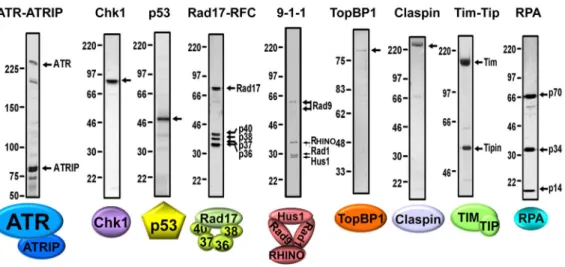
Figure 2. Purified Checkpoint Factors
Different subunits in the complexes are indicated with arrows. The checkpoint factors were purified as described: ATR-ATRIP (7), Chk1 (9), p53 (8), Rad17-RFC (19), 9-1-1-RHINO complex (22), TopBP1 (7), Claspin (40), Timless-Tipin (41), RPA (11).
A
uthor Man
uscr
ipt
A
uthor Man
uscr
ipt
A
uthor Man
uscr
ipt
A
uthor Man
uscr
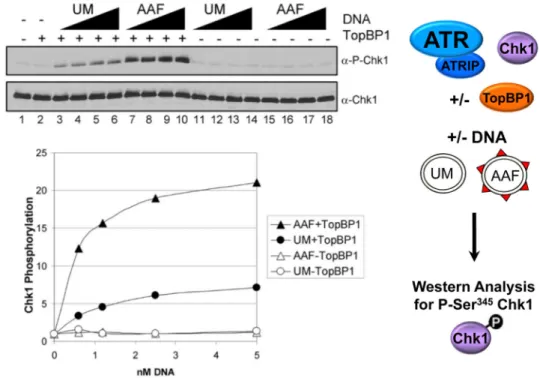
Figure 3. TopBP1-dependent stimulation of ATR kinase activity by AAF-damaged DNA
Kinase assays were performed as pictured on the right with ATR–ATRIP, His-Chk1-kd, GST-TopBP1-His, and with 0.62–5 nM unmodified (UM) or AAF-damaged (AAF) pUC19 plasmid DNA. The left shows immunoblotting of the reactions for phosphorylated Chk1 (P-Chk1, which is phosphorylated at S345) (Upper) and total Chk1 (Lower). The average levels of Chk1 phosphorylation from four independent experiments are quantitated in the graph below. Reproduced with permission from (7).
A
uthor Man
uscr
ipt
A
uthor Man
uscr
ipt
A
uthor Man
uscr
ipt
A
uthor Man
uscr
Figure 4. Model of ATR kinase activation by RPA-coated ssDNA
RPA directly interacts with ATRIP, the N-terminus of TopBP1, Tipin (Tip), Rad17, Rad9, and p53. The consequence of all of these interactions on long stretches of ssDNA is to localize ATR kinase with its TopBP1 activator and substrates. The arrow indicates
phosphorylation of the checkpoint signal transduction kinase, Chk1, by ATR. In addition to Chk1, ATR phosphorylates nearly all of the other proteins depicted, but these
phosphorylation events are not shown for simplicity.
A
uthor Man
uscr
ipt
A
uthor Man
uscr
ipt
A
uthor Man
uscr
ipt
A
uthor Man
uscr
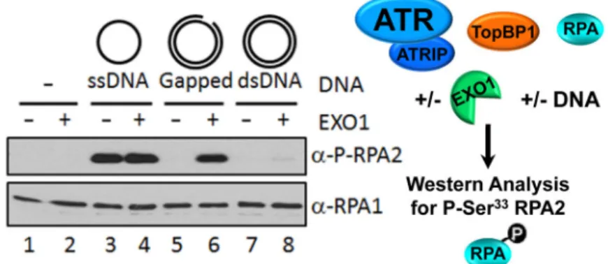
Figure 5. Model System for Repair-Checkpoint Coupling
ATR kinase reactions were carried out as depicted on the right with ATR-ATRIP, TopBP1, RPA, and EXO1 as indicated. Single-stranded DNA (ssDNA), plasmid DNA (dsDNA), or gapped DNA was added to the reaction as indicated. The left shows immunoblotting of the reactions for phosphorylated RPA2 (P-RPA2, which is phosphorylated at S33) (Upper) and total RPA with antibodies against RPA1 (Lower). Reproduced with permission from (15).
A
uthor Man
uscr
ipt
A
uthor Man
uscr
ipt
A
uthor Man
uscr
ipt
A
uthor Man
uscr
Figure 6. Purified Nucleotide Excision Repair Factors
Different subunits in the complexes are indicated with arrows. The checkpoint factors were purified as described (42).
A
uthor Man
uscr
ipt
A
uthor Man
uscr
ipt
A
uthor Man
uscr
ipt
A
uthor Man
uscr
Figure 7. Reconstituted Repair-Checkpoint Coupling
Kinase reactions, as diagrammed on the right, contained ATR-ATRIP, TopBP1, RPA, EXO1, and different concentrations of unmodified (UM) or AAF DNA from excision reactions with or without repair factors (RF) as indicated. Reactions were analyzed by immunoblotting for phospho-RPA2 and RPA1 for loading. The graph below shows the relative levels of phosphorylated RPA2 from three identical repeats. Reproduced with permission from (15).
A
uthor Man
uscr
ipt
A
uthor Man
uscr
ipt
A
uthor Man
uscr
ipt
A
uthor Man
uscr
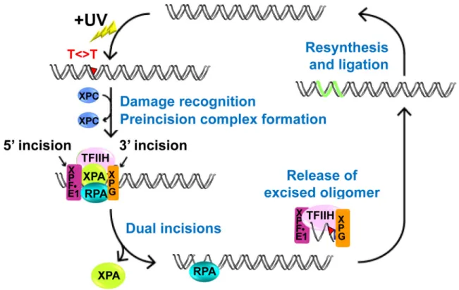
Figure 8. Model of Repair-Checkpoint Coupling
When DNA is damaged by UV, the core excision repair factors excise a ~30 nucleotide oligomer containing the damage. The resulting gap is either filled in by polymerases or the gap is enlarged by EXO1. The resulting ssDNA is coated with RPA which recruits ATR-ATRIP, TopBP1, and substrates including p53. Modified from (15).