Original Research Article
Foreign bodies in esophagus
Anchal Gupta
1, Apurab Gupta
2*, Padam Singh Jamwal
3INTRODUCTION
The presence of a foreign body in the gastrointestinal tract is a challenging problem. Its management depends on a number of factors, such as anatomic location, shape and size of the foreign body, and duration of impaction. Foreign bodies retained in the esophagus are by far the most dangerous. Perforations are common and may result in death.1,2 Foreign bodies are normally located in 1 of the 3 physiologic narrowings of the esophagus, most frequently at the cricopharyngeal level but also at the aortic arch and at the gastroesophageal junction. The foreign body must be extracted within 24 hours to avoid necrosis in the esophageal wall or complications such as
esophageal perforation, para or retropharyngeal abscesses, aorto or bronchoesophageal fistula, pulmonary edema, empyema, esophageal diverticula, or lobe atelectasis.3,4
Children have a tendency to put objects into their mouths, and therefore, foreign body ingestion is more common in this population group. Among adults, it occurs mainly in people with psychiatric disorders or mentally challenged individuals, as well as in prisoners and alcoholics.
The type of foreign body ingested has been related to patient age- coins, fish bones, and dentures are the most commonly swallowed objects by children, adults, and the ABSTRACT
Background: The presence of a foreign body in the esophagus is a challenging problem. Perforations may result in
death. Impaction mandates immediate extraction.
Methods: A retrospective chart review was made of all patients hospitalized in Department of ENT, SMGS Hospital
with a diagnosis of foreign bodies in the gastrointestinal tract between July 2017 to February 2018. Forty patients were identified. The charts were reviewed for the following: patient demographics, preoperative diagnosis, kind and location of the foreign body, timing of the procedure and the length of hospitalization.
Results: The youngest patient in our study was 8 months old while the oldest was 60 years old. The male: female ratio of 2.3:1. The most common site of impaction was cricopharynx (55%) followed by thoracic esophagus (40%) and lower end of esophagus (5%). The most common foreign body was coin (50%) and all were seen in children upto 7 years of age. 25 (62.5%) patients were children of age less than 10 years. 20 children showed coin ingestion, 1 child of 8 months presented with impaction of fruit seed and 4 children with battery button ingestion. The length of retention of foreign body ranged from 2 hours to 6 days. All the patients were managed with rigid esophagoscopy under general anesthesia all within 12 hours of admission.
Conclusions: Rigid endoscope as the instrument of choice for extracting foreign bodies from the esophagus as delay
in extracting foreign bodies from the esophagus may lead to retention of foreign body and hence perforation.
Keywords: Esophagus, Foreign body, Perforation
1
Senior Resident, 2Lecturer, 3Professor, Department of ENT, Head and Neck Surgery, SMGS Hospital, Government Medical College, Jammu, Jammu and Kashmir, India
Received: 02 April 2019
Revised: 16 April 2019
Accepted: 18 April 2019
*Correspondence:
Dr. Apurab Gupta,
E-mail: [email protected]
Copyright: © the author(s), publisher and licensee Medip Academy. This is an open-access article distributed under
the terms of the Creative Commons Attribution Non-Commercial License, which permits unrestricted non-commercial use, distribution, and reproduction in any medium, provided the original work is properly cited.
elderly, respectively. Although most foreign bodies are passed through the gastrointestinal tract without complications, between 10% and 20% need to be extracted by nonsurgical procedures, whereas less than 1% require surgery.1-4
The method chosen for extracting a foreign body from the esophagus depends on patient age, clinical condition, size, shape, and texture of the object as well as its anatomical location.4 Although rigid esophagoscopy has fallen almost completely out of use since the advent of flexible esophagoscopy, we agree with Holinger in that rigid endoscopy is indicated for foreign bodies lodged at the level of the pharynx and cricopharyngeus muscle, mostly when objects are large and sharp or penetrating.5,6 Rigid esophagoscopy success rates range from 94% to 100%, the estimated incidence of esophageal or pharyngeal perforation is 0.34%, and mortality is 0.05%.6,7
This study was conducted with an objective to find out the most common type of foreign body ingested, the site and age of presentation and the time of presentation as early intervention is required which if delayed may cause complication like perforation
METHODS
The current study was conducted at Department of ENT, SMGS Hospital, Government Medical College, Jammu, from July 2017 to February 2018. Approval for this study was obtained from our Institutional Ethical Committee, and individual consent was waived.
A retrospective chart review was made of all patients Hospitalized in Department of ENT, SMGS Hospital with a diagnosis of foreign bodies in the esophagus between July 2017 to February 2018. The selection criteria of patients was are as follows: (i) each patient had a history of FB ingestion and the FB was located in esophagus; (ii) the FB was found impacted in the esophageal tract; (iii) the FB was found physically impacted or disrupting the esophageal wall by doing ray soft tissues neck and X-ray chest PA view; and (iv) they received emergency rigid esophagoscopy after admission.
Forty patients were identified. The charts were reviewed for the following: patient demographics, preoperative diagnosis, kind and location of the foreign body, timing of the procedure and the length of hospitalization.
Data collection and analysis
The following demographic data were collected and analysed: age, sex, type, location, duration, removal and complications. The variables were compared using the Pearsons’s square test, the continuity correction chi-square test or Fischer’s exact test as appropriate. Additionally, p<0.05 was selected to indicate a
significant difference. The data were analysed using SPSS version 22.
RESULTS
The current study was conducted at Department of ENT, SMGS Hospital, Government Medical College, Jammu, from July 2017 to February 2018. Forty patients were included in the study. The following observations were made.
Age and sex distribution of patients
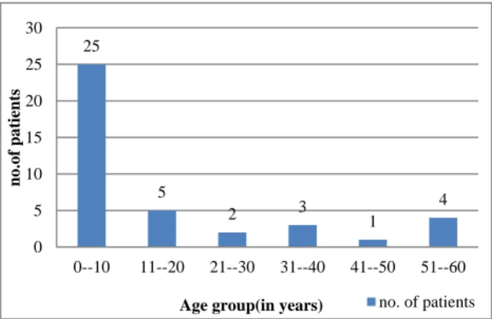
The youngest patient in our study was 8 months old while the oldest was 60 years old. The mean age was 18 years. Among them 25 patients were in the age group of 8 months to 7 years.
Figure 1: Age distribution of patients.
Figure 2: Sex distribution of patients.
There were 28 male and 12 female patients with male: female ratio of 2.3:1 (Figure 1 and 2).
Type of foreign body and site of impaction of foreign body
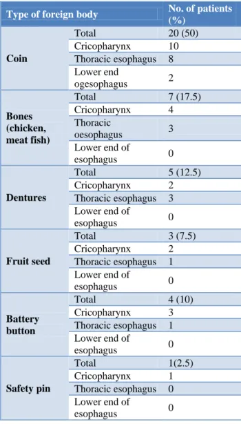
The most common site of impaction was cricopharynx (55%) followed by thoracic esophagus (40%) and lower end of esophagus (5%). The most common foreign body was coin (50%) and all were seen in children upto 7 years
25
5
2 3 1 4
0 5 10 15 20 25 30
0--10 11--20 21--30 31--40 41--50 51--60
no
.of
pa
tient
s
Age group(in years) no. of patients
28 12
Sex distribution of patients
male
of age. There were 7 patients in which bones were impacted out of these 2 were fish bones impacted in cricopharynx, 3 were meat bones (1 in cricopharynx and 2 in thoracic esophagus), 2 were chicken bones (1 in cricopharynx, 1 in thoracic esophagus).
Twenty five (62.5%) patients were children of age less than 10 years. 20 children showed coin ingestion, 1 child of 8 months presented with impaction of fruit seed and 4 children with battery button ingestion. This high incidence of foreign body ingestion in children was due to their habit of putting things in mouth while playing (Table 1, Figure 3 and 4).
Table 1: Distribution of type of foreign bodies and their site of impaction.
Type of foreign body No. of patients
(%)
Coin
Total 20 (50)
Cricopharynx 10
Thoracic esophagus 8 Lower end
ogesophagus 2
Bones (chicken, meat fish)
Total 7 (17.5)
Cricopharynx 4
Thoracic
oesophagus 3
Lower end of
esophagus 0
Dentures
Total 5 (12.5)
Cricopharynx 2
Thoracic esophagus 3 Lower end of
esophagus 0
Fruit seed
Total 3 (7.5)
Cricopharynx 2
Thoracic esophagus 1 Lower end of
esophagus 0
Battery button
Total 4 (10)
Cricopharynx 3
Thoracic esophagus 1 Lower end of
esophagus 0
Safety pin
Total 1(2.5)
Cricopharynx 1
Thoracic esophagus 0 Lower end of
esophagus 0
Duration of impaction of foreign body
The length of retention of foreign body ranged from 2 hours to 6 days. The mean duration was 20 hours.
All the patients were managed with rigid esophagoscopy under general anesthesia all within 12 hours of
admission. The maximum delay of 6 days was due to delay in seeking medical advice.
Figure 3: Site of impaction of foreign body.
Figure 4: Distribution of patients according to type of foreign body ingested.
DISCUSSION
The ingestion of foreign bodies is a common emergency of the upper GI tract and the esophageal foreign body can easily cause impaction or even perforation of the esophageal wall. The prevalence of ingested foreign bodies depends mainly on patient age, cooking methods, and the food culture of the population. It is well known that the esophagus is the narrowest portion of the alimentary canal, except for the vermiform appendix, and it has four areas of physical narrowing. Moreover, esophageal perforation is also affected by the strong contraction of the hypopharyngeal and cricoesophageal muscles.
Our study showed that the most common site of impaction was cricopharynx (55%) followed by thoracic esophagus (40%) and lower end of esophagus (5%). There is an apparent predominance of certain types of foreign bodies in specific groups of patients. Coins and toys are a relatively common finding in children.8,9 In our study also, the most common foreign body was coin (50%) and all were seen in children upto 7 years of age. There were 7 patients in which bones were impacted out of these 2 were fish bones impacted in cricopharynx, 3
22 16
2
Site of impaction
cricopharynx
thoracic esophagus
lower end of esophagus
20
7 5
3 4
1 0
5 10 15 20 25
coin bone denture fruit seed battery
button
safety pin
no
.
o
f
a
ptient
s
were meat bones (1 in cricopharynx and 2 in thoracic esophagus), 2 were chicken bones (1 in cricopharynx, 1 in thoracic esophagus). 25 (62.5%) patients were children of age less than 10 years. 20 children showed coin ingestion, 1 child of 8 months presented with impaction of fruit seed and 4 children with battery button ingestion. It is attractive to assume that emergence of typical groups predisposed to swallowing certain types of foreign bodies might help prevent such accidents
Our findings coincide with the findings by Ray and Vinson whose report is concerned with the 584 cases in which the foreign body was located in the esophagus.10 In their study of the patients whose records were reviewed, 269 were under 10 years of age, while 315 were 10 years of age or older. The youngest patient was 2 months old, and the oldest was 94 years. The absence of teeth is a major factor in the impaction of a foreign body in the esophagus. In adults inadequate mastication and the loss of sensation in the mouth which results from artificial dentures are responsible for bones and meat sticking in the esophagus. Dentists should warn all patients, when they are provided with partial or complete dentures, of this hazard and should explain that care should be taken in the preparation and mastication of food.
Retention leads to perforation, which is only a matter of time.11-13 Therefore, all foreign bodies retained in the esophagus should be removed as soon as diagnosed.
The presence of a perforation in association with a foreign body and a mediastinal inflammatory mass should be treated by extraction of the foreign body, enteric but no oral feeding, and antibiotics until healing has occurred as demonstrated by contrast esophagogram.14
In planning the extraction, one of the important points to considered is the proper choice of the instruments. This is particularly important in the case of sharp and pointed foreign bodies, such as denture with protruding hooks, shaving blades, and open safety pins, which increase the danger of perforation. Extraction of these objects requires special attention and experience. Some may have to be drawn, sometimes only partially, into the lumen of the rigid esophagoscope, to enable their manipulation and extraction while protecting the esophageal mucosa.15 This protection is not possible with the flexible instrument.
Historically, the initial method of management of esophageal foreign bodies was extraction through the rigid esophagoscope.
The success rate with the use of rigid instrument ranges between 94% and 100%.16-18 The estimated incidence of esophageal perforation is 0.34% with a 0.05% mortality rate.19
We always use the rigid esophagoscope and a variety of forceps. The wide lumen of the rigid instrument is of
great help in manipulating the foreign body and extracting it, and we believe that this should be the instrument of choice. This idea is not isolated and has been suggested by several authors.
CONCLUSION
Our experience indicates that the use of a rigid esophagoscope is safe and reliable. Based on this experience and that of other authors, we recommend the use of the rigid endoscope as the instrument of choice for extracting foreign bodies from the esophagus as delay in extracting foreign bodies from the esophagus may lead to retention of foreign body and hence perforation.
Funding: No funding sources Conflict of interest: None declared
Ethical approval: The study was approved by the Institutional Ethics Committee
REFERENCES
1. Yee KF, Schild JA, Hollinger PH. Extraluminal foreign bodies (coins) in the food and air passages. Ann Otol. 1975;84:619–23.
2. Bloom RR, Nakano PH, Gray SW, Skandalakis JE. Foreign bodies in the gastrointestinal tract. Am Surg. 1986;52:618–21.
3. Athanassiadi K, Gerazounis M, Metaxas E, Kalantzi N. Management of esophageal foreign bodies: a retrospective review of 400 cases. Eur J Cardiothorac Surg. 2002;21:653-6.
4. Matern U, Aschendorff A, Krebs A, Kohlberger E, Rückauer KD. A new method for extracting wooden foreign bodies from the upper esophagus. Endoscopy. 2000;32:1002-3.
5. Holinger LD. Management of sharp and penetrating foreign bodies of the upper aerodigestive tract. Ann Otol Rhinol Laryngol. 1990;99:684-8.
6. Roffman E, Jalisi S, Hybels R, Catalano P. Failed extraction of a sharp esophageal foreign body with a flexible endoscope. Arch Otolaryngol Head Neck Surg. 2002;128:1096-8.
7. Berggreen PJ, Harrison ME, Sanowski RA, Ingebo K, Noland B, Zierer S. Techniques and complications of esophageal foreign body extraction in children and adults. Gastrointestinal Endosc. 1993;39:626-30.
8. Silverberg M, Tillotson R. Esophageal foreign body mistaken for impacted button battery. Pediatr Emerg Care. 2006;22:262–5.
9. Little DC, Shah SR, St Peter SD, Calkins CM, Morrow SE, Murphy JP, et al. Esophageal foreign bodies in the pediatric population: our first 500 cases. J Pediatr Surg. 2006;41:914–8.
10. Ray ES, Vinson RP. Foreign Bodies Removed From the Esophagus: A Statistical Study. Current Review of literature 1958;85:61-4.
12. Bloom RR, Nakano PH, Gray SW, Skandalakis JE. Foreign bodies in the gastrointestinal tract. Am Surg 1986;52:618–21.
13. Medina HM, Garcia MJ, Velasquez O, Sandoval N. A 73-year-old man with chest pain 4 days after a fish dinner. Chest. 2004;126:294–7.
14. Naidoo RR, Reddi AA. Chronic retained foreign bodies in the esophagus. Ann Thorac Surg. 2004;77:218–20.
15. Roffman E, Jalisi S, Hybels R, Catalano P. Failed extraction of a sharp esophageal foreign body with a flexible endoscope. Arch Otolaryngol Head Neck Surg. 2002;128:1096–8.
16. Chaikhouni A, Kratz JM, Crawford FA. Foreign bodies of the esophagus. Am Surg. 1985;51:173–9.
17. Vizcarrondo FJ, Brady PG, Nord HJ. Foreign bodies of the upper gastrointestinal tract. Gastrointest Endosc. 1983;29:208–10.
18. Brady PG. Endoscopic removal of foreign bodies. In: Silvis SE, ed. Therapeutic gastrointestinal endoscopy. New York: Igaku-Shoin; 1990.
19. Giordano A, Adams G, Bois L Jr, Meyerhoff W. Current management of esophageal foreign bodies. Arch Otolaryngol. 1981;107:249 –51.
Cite this article as: Gupta A, Gupta A, Jamwal PS.