Dr Sandeep Chahande et al JMSCR Volume 06 Issue 10 October 2018 Page 886
A Study of Clinico-Pathological Profile of Nasal Masses
Authors
Dr Sandeep Chahande
1, Dr Ved Prakash Narve
2*1
Senior Resident, Department of E.N.T., Gajra Raja Medical College, Gwalior, M.P. 474009 Email: [email protected], Ph no. +917756928614
2
Professor, Department of E.N.T., Gajra Raja Medical College, Gwalior, M.P. 474009 *Corresponding Author
Dr V. P. Narve (M.S.)
Professor, Department of E.N.T., Gajra Raja Medical College, Gwalior, M.P. 474009, India Email: [email protected], Ph. No. +919826240262
Abstract
Nasal mass is a very frequently encountered complaint in OPD of Otorhinolaryngology. Clinical and radiological evaluation of these lesions help a lot to reach a diagnosis but histopathological examination is a must to reach a final diagnosis and start appropriate treatment.
Objectiveof this study was to study the clinical presentation of various types of nasal masses, to study their incidence in various age groups and their sex distribution and histopathological evaluation of these nasal masses.
Method: The study was conducted in Gajra Raja Medical College, Gwalior, Department Of Otorhinolaryngology and Head and Neck Surgery from September 2014 to August 2018. After taking the consent for surgery, patients with nasal masses on clinical evaluations were selected for excision or biopsy of nasal masses.
Result: Out of 102 cases studied 79 cases were of non neoplastic masses and 23 were neoplastic. Among non neoplastic lesions nasal polyps were the most common histopathological types and neoplastic were further divided into benign and malignant lesions. Capillary haemangiomas formed the most common benign lesions and squamous cell carcinoma was the most commonly occurring malignant lesion. Nasal obstruction was the most common presenting symptom followed by nasal discharge, headache, epistaxis and voice change.
Conclusion:Histopathological evaluation of nasal masses is very important part of diagnosis and treatment of nasal masses. Since all nasal masses present with overlapping symptoms, so it becomes important to identify and differentiate non neoplastic, benign and malignant lesions for radical cure of the disease.
Introduction
We encounter a lot of patients having complaints related to nose in the Otorhinolaryngology OPD. Apart from providing aesthetic significance, nose has very important role in maintaining physiological functions of the body. It is
concerned with perception of smell and not only acts as a conduit for air passage but along with paranasal sinuses it also regulates the temperature, humidity and filtration of air we breathe. Since nasal cavity is directly exposed to environmental air changes, dust, and chemicals, a variety of
www.jmscr.igmpublication.org Impact Factor (SJIF): 6.379
Index Copernicus Value: 79.54 ISSN (e)-2347-176x ISSN (p) 2455-0450
Dr Sandeep Chahande et al JMSCR Volume 06 Issue 10 October 2018 Page 887 diseases are encountered which are associated
with nose. Nasal masses are one of the common clinical presentations in routine ENT OPD. Patients presenting with nasal masses usually have complaints of nasal obstruction, sneezing, discharge, occasional nasal bleeding and headache, and rarely may complain facial pain and facial deformity. Clinically these nasal masses may show variability. They may be single or multiple, white, pale, or they may appear red, or may be sessile or pedunculated. Though the presenting symptoms and clinical evaluation with proper history might help in reaching a proper diagnosis but a histopathological examination is a must in making the definitive diagnosis. A variety of nasal masses are encounterd which range from polyps (allergic or inflammatory), infective lesions (like rhinosporodiosis and rhinoscleroma), rhinoliths, neoplastic lesions like capillary angiomas, squamous cell carcinoma , inverted papilloma, adenocarcinoma and so on. The prevalence of nasal obstruction due to nasal polyps is about 4%. On the other hand malignancies of nose and paranasal sinus accounts for not more than 3% of head and neck cancers and less than 1% of all malignant lesions.
Thus due to such a variability of nasal masses histopathological evaluation has become an important diagnostic tool along with clinical features for proper diagnosis and treatment of nasal masses.
The aim of this study was to evaluate the clinical features of various types of nasal masses, to categorize them into different histopathological types and to find out their incidence.
Materials and Methods
The study was conducted in the Department of Otorhiolaryngology and Head and Neck surgery, Gajra Raja Medical College, Gwalior for the period of September 2014 to August 2018. After taking the consent for surgery, patients with nasal masses on clinical evaluations were selected for excision or biopsy of nasal masses. Ethmoidal polyps that could be treated conservatively,
extensive vascular masses (angiofibromas) and patients not willing for surgery or biopsy were not included in the study. Demographic data regarding age, sex, chief complaints, clinical examination and histopathological reports were collected. A detailed history and clinical examination was done in a systemic manner and noted on a specially designed proforma.
Results
A total of 102 patients were selected for the study with nasal masses. Youngest patient in our study was of 5 years who was diagnosed with embryonal rhabdomyosarcoma and oldest patient was of 75 years of age, who was diagnosed with maxillary squamous cell carcinoma.
Table 1 Age Distribution
S. No. Age Group No. of Patients Percentage
1. <10 years 9 8.8%
2. 11-20 years 33 32.3%
3. 21-30 years 19 18.6%
4. 31-40 years 13 12.74%
5. 41-50 years 14 13.7%
6. 51-60 years 6 5.8%
7. 61years and above
8 7.8%
8. TOTAL 100 100%
The peak age distribution was observed in the age group of 11-20 years group followed by 21-30 years age group. In our study least incidence of nasal masses was observed in the age group of 51-60 years. Maximum incidence of benign neoplastic masses was seen in 41-50 year age group whereas maximum incidence of malignant neoplastic masses was seen in 21-30 years age group and 61 years and above age group.
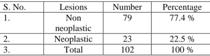
Table 2 Neoplastic vs non-neoplastic lesions
S. No. Lesions Number Percentage
1. Non
neoplastic
79 77.4 %
2. Neoplastic 23 22.5 %
Dr Sandeep Chahande et al JMSCR Volume 06 Issue 10 October 2018 Page 888
Fig. 1 Age distribution of benign nasal masses
Fig.2 Age distribution of malignant nasal masses
A male preponderance was noted in our study, with 64 male patients and 38 females who presented with nasal masses. Male to female ratio came out to be 1.68:1.
Table 3 Sex distribution of nasal masses in study population
S. No. Gender No. of Patients Percentage
1. Male 64 62.7%
2. Female 38 37.2%
3. Total 102 100%
Table 4 Unilateral vs Bilateral
S. No Laterality Number Percentage
1. Unilateral
A) Right sided mass B) Left sided mass
44 46
43.1% 45.1%
2. Bilateral 12 11.8%
3. Total 102 100%
Out of the total 102 cases 90 cases of nasal mass were unilateral whereas 12 cases were bilateral. Out of these bilateral nasal masses most of the cases were of inflammatory nasal polyps, whereas one case was of inverted papilloma and another case was Wegner’s granulomatosis.
Nasal obstruction (92.15 %) was most common presenting complaint followed by nasal discharge (75.4%). Headache (54.9%), epistaxis (22.5%), voice change (25.4%), sneezing (13.7%), loss or decreased smell (9.8%) and facial swelling (7.8%) were other common symptoms. Post nasal discharge was seen in 76.4% of cases. Mass seen on posterior rhinoscopy and sinus tenderness were seen in 25.4% and 16.6% of cases respectively. Among the 102 cases under study, 77.4% of cases were non neoplastic masses, 11.8% were benign neoplastic and 10.8% were neoplastic malignant. Inflammatory polyps (including allergic polyps) were the most commonly observed masses that constituted 67.6 %. 3 cases were of rhinosporodiosis and 2 cases of suspecting nasal masses were found to be rhinolith. One case of bilateral nasal mass was histopathologically confirmed as Wegner’s granulomatosis.
Fig. 3 Presenting signs and symptoms of nasal masses
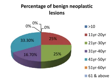
Benign neoplastic masses constituted 11.8% of all the nasal masses. Cappillary haemangioma was the most common benign neoplastic lesion (7.8% of all nasal masses), followed by inverted papilloma (2.9%) and angiofibroma (0.98%). 0%
25%
25% 16.70%
33.30%
0% 0%
Percentage of benign neoplastic lesions
>10
11yr-20yr
21yr-30yr
31yr-40yr
41yr-50yr
51yr-60yr
61 & above
9.10%
9.10%
27.30%
9.10%
0% 18.10%
27.30%
Percentage of maliganant neoplastic lesions in various age groups
>10 11yr- 20yr 21yr-30yr 31yr-40yr
41yr-50yr 51yr-60yr 61yr & above
0 10 20 30 40 50 60 70 80 90 100
Dr Sandeep Chahande et al JMSCR Volume 06 Issue 10 October 2018 Page 889 11 cases were diagnosed histologically as
malignant masses which covered 10.8 % of all nasal masses. Out of these, squamous cell carcinoma was most common (3.9%) followed by lymphoma (2.9%). One case of each adenocarcinoma, olfactory neuroblastoma, rhabdomyosarcoma and osteoblastoma was diagnosed in our study.
Discussion
In most of the cases in our study, the clinical diagnosis came out to be same as
histopathological results, wherever non-neoplastic masses and benign neoplastic masses were concerned but in case of suspected malignant lesions the histopathological examination of masses/biopsies helped in reaching the final diagnosis.
Nasal masses, in our study showed predilection for males, as male to female ratio came out to be 1.68:1. It was similar to a study done by Zafar et al1 where it was 1.7:1 and was comparable to study by A. Lathi et al2 where it came out to be 1.5:1.
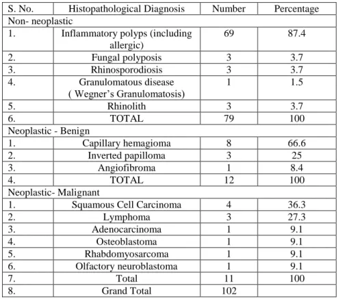
Table 5 Histopathological diagnosis
S. No. Histopathological Diagnosis Number Percentage
Non- neoplastic
1. Inflammatory polyps (including
allergic)
69 87.4
2. Fungal polyposis 3 3.7
3. Rhinosporodiosis 3 3.7
4. Granulomatous disease
( Wegner’s Granulomatosis)
1 1.5
5. Rhinolith 3 3.7
6. TOTAL 79 100
Neoplastic - Benign
1. Capillary hemagioma 8 66.6
2. Inverted papilloma 3 25
3. Angiofibroma 1 8.4
4. TOTAL 12 100
Neoplastic- Malignant
1. Squamous Cell Carcinoma 4 36.3
2. Lymphoma 3 27.3
3. Adenocarcinoma 1 9.1
4. Osteoblastoma 1 9.1
5. Rhabdomyosarcoma 1 9.1
6. Olfactory neuroblastoma 1 9.1
7. Total 11 100
8. Grand Total 102
The most common age group presenting with nasal masses was 11-20 years and 21-30 years group which constituted 32.3% and 18.6% of cases, which is similar to study by Dinesh Garg et al3. Zafar et al1 had a mean age of 22.5 in their study.
Maximum incidence of benign neoplastic lesions was seen in 41-50 years group (33.3%) and that of malignant lesions was seen in 6th and 7th decades (27.3%). In a study Banerjee A. et al4 reported maximum benign and malignant lesions in 31-40 and 51-60 years age group respectively.
In our study 90 cases were of unilateral masses, 44 were right sided and 46 were left sided. nasal masses. 12 cases presented with bilateral masses. Uma R and Meharaj Banu8 reported similar observations in their study of 110 cases of nasal masses, where 91 cases were unilateral and 19 cases were bilateral. In contrast Bakari et al9 reported 44.7% bilateral sinonasal masses and 55.3% of unilateral nasal masses.
Dr Sandeep Chahande et al JMSCR Volume 06 Issue 10 October 2018 Page 890 Kumar10 also reported similar findings in his study
where nasal obstruction (91%) was most common complaint with nasal discharge (70%). Other presenting complaints included headache (54.9%), anosmia (9.8%), epistaxis (22.5%), voice change (25.4%) and sneezing (13.7%).
Nasal masses were found in almost all patients on clinical examination and post nasal discharge was seen in about76.4 % cases. Facial swelling (7.84%), sinus tenderness (16.66%) and mass seen on posterior rhinoscopy (25.49%) were other common signs seen on examination.
In our study we divided the cases histologically into non-neoplastic masses and neoplastic masses. Neoplastic lesions were futher divided into benign and malignant lesions. Among the non-neoplastic lesions nasal polyps (inflammatory and allergic) were the most common (67.6%). Tondon et al5 (64%) and Dasgupta et al6 (62.5%) too reported similar findings in their study.
Nasal polyps (inflammatory and allergic) were the most common histopathological types (67.6%) among all nasal masses, and among the non neoplastic nasal masses (87.6%). These values were in concordance with studies of Tondon et al5. (64%) and Dasgupta et al6 (62.5%). Nasal masses with fungal sinusitis were seen in 3 cases which was comparable to study by Dinesh Garg et al3 and Modh et al7. Dinesh Garg et al3 had reported 5 cases of fungal sinusitis in their one year study of nasal masses.
Rhinosporodiosis is a chronic granulomatous disease caused by Rhinosporidium seeberi that present as granulomatous nasal masses often associated with epistaxis. It is common in Indian subcontinent and in Sri Lanka. 3 cases were seen during our study. Pradhananga et al11 has reported one case in their study whereas 2 and 5 cases were reported by Lathi et al2 and A. Banerjee et al4 respectively.
Single case of Wegner’s Granulomatosis was seen during our study as similar results were reported by A. Banerjee et al4 in their seven year retrospective study. We included 3 cases of rhinolith in our study as they presented with
similar clinical manifestations of nasal masses until histopathological reports confirmed their diagnosis.
Among the neoplastic nasal lesions, benign lesions observed were haemangiomas, papillomas and angiofibroma. Capillary haemangiomas were the most common benign bleeding nasal masses observed in our study. 8 cases of capillary haemangiomas were noted that constituted 66.6% of all benign neoplastic lesions. A Banerjee et al4 57.14% of benign neoplastic as capillary haemangioma whereas these lesions constituted 47.3% in study of Lathi Et al2.
Dr Sandeep Chahande et al JMSCR Volume 06 Issue 10 October 2018 Page 891 All these cases were given appropriate treatment
and care. All the cases of sinonasal polyps were treated by avulsive polypectomy and wherever required Cald Well Luc surgery was done. Other benign lesions were excised radically and proper follow up was done to ensure no recurrence. In case of suspected malignant lesions pre operative radiological examinatios were carried out and diagnosis was confirmed by biopsies and histopathological examinations. Malignant cases were treated with chemo-radiotherapy and proper follow up was done wherever required.
Conclusion
Most of the sinonasal masses have almost similar clinical manifestations like nasal obstruction, nasal discharge, sneezing, headache and epistaxis. Clinical examination and radiological investigations help to make a provisional diagnosis but histopathological evaluation is utmost important for appropriate treatment of these lesions. In our Hospital we try our best to cure the patient with available facilities. Ethmoidal polyps that could be treated conservatively and angiofibromas with severe extension, were not included in our study, this might have created minor differences in the data of our study.
References
1. Zafar U, Khan N, Afroz N, et al. Clinico-pathological study of non-neoplastic lesions of nasal cavity and paranasal sinuses. Indian J Pathol Microbiol 2008;51:26-9
2. Lathi A., Syed M.M.A, Kalakoti P et al.
Clinico-pathological profile of sinonasal masses: a study from tertiary care hospital of India. Acta Otorhinolaryngologica Italica 2011;31:372-377.
3. Garg D, Mathur K. Clinico-pathological study of space occupying lesions of nasal cavity, paranasal sinuses and nasopharynx. Journal of Clinical and
Diagnostic Research, 2014 Nov, Vol-8 (11): FC04-FC07
4. Banerjee A, Ghosh S, Histopathological patterns of nasal masses: A seven year study. Indian Journal of Basic and Applied Medical Research; March 2017; Vol-6, Issue-2, P. 491-497.
5. Tondon PL, Gulati J, Mehta N,
Histological study of polypoidal lesions in nasal cavity. Indian J Otolaryngol. 1971; 13:3-11.
6. Dasgupta A, Ghosh RN, Mukherjee C.
Nasal polyps- Histopathologic spectrum. Indian J otolaryngol Head Neck Surg. 1997;49:32-37.
7. Modh SK, Delwadia KN, Gonsai RN. Histopathological spectrum of sinonasal masses- A study of 162 cases. Int J Cur Res Rev. 2013;5(03);83-91.
8. Uma R, Meharaj Banu O A.
Histopathological study of nasal mass- A study of 110 cases. International Journal of Recent Trends in Science and Technology. May 2016;19(1): 98-102.
9. Bakari A, Afolabi OA, Adoga AA, et al. Clinico-pathological profileof sinonasal masses: an experience in national ear care cemtre Kaduna, Nigeria. BMC Research Notes 2010;3:186.
10.Ambrish Kumar. Lesions of nasal cavity, paranasal sinuses and nasopharynx- a clinicopathological study. International Journal of Medical Research and Review; August 2016/ Vol-4/Issue-8:1302-1306. 11.Pradhananga RB, Adhikari P, Thapa NM,
Shrestha A, Pradhan B. Overview of nasal masses. J Inst Med.2008:30:13-6.
12.Alpesh M Maru, Umang V. Patel et al.
Histopathological study of nasal masses in patients coming to a tertiary care hospital: A study of 70 cases. Medical journal of Dr D. Y. Patil University; July-August-2015, Vol8, Issue 4.
Dr Sandeep Chahande et al JMSCR Volume 06 Issue 10 October 2018 Page 892
study of sinonasal masses. Bangladesh J Otorhinolaryngol 2010;16:15-22.
14.Panchal L, Vaideeswar P, et al. Sinonasal epithelial tumours-A pathological study of 69 cases. J Postgrad Med . 2005;1(1):30-34.
15.N. Khan, U. Zafar, et al. Masses of nasal cavity, paranasal sinuses and nasopharynx: A clinicopathological study.
Indian Journal of Otolaryngology and Head and Neck Surgery. Vol 58, No. 3, July-September 2006:259-263.