Original Article
Impacts of SB203580 on the spatial memory and the
expression of phosphorylated p38 MAPK and Homer1a
in the hippocampus in rats with diffuse brain injury
Cheng-Jing Xue1, Ya-Ning Zhao2, Jian-Min Li1, Chang-Xiang Chen1, Shu-Xing Li1, Ai-Jun Fu1
1Affiliated Hospital of North China University of Science and Technology, Tangshan, China; 2Department of
Reha-bilitation, North China University of Science and Technology, Tangshan, China
Received November 3, 2015; Accepted January 5, 2016; Epub March 1, 2016; Published March 15, 2016
Abstract: Objectives: The impacts of SB203580 on the spatial memory and the expression of phosphorylated p38 MAPK and Homer1a in the hippocampus in rats with diffuse brain injury were investigated. Methods: In the Neuro-surgery Center Laboratory of North China University of Science and Technology, the DBI rat models were built fol-lowing Marmarou’s method. One hundred and forty nine male Sprague-Dawlley rats were divided into three groups:
sham operation (SO) group, DBI group and DBI+SB203580 intervention group (i.p. 0.01 μg/kg). The water maze
experiment was carried out to test the spatial memory of the animals. An electron microscope was used to observe the morphological changes of the hippocampus neurons. The expression levels of phosphorylated p38 MAPK and Homer1a were detected with western blot. Results:Compared with the SO group, nerve cells and synaptic injury in the hippocampi, higher expression of Homer1a and phosphorylated p38 MAPK, a prolonged latent period in the animals’ searching for the safety island, and a lower frequency of crossing the initial platform were found in the DBI group; compared with the DBI group, a lower degree of morphologic and structural injury in brain tissues, a lower
expression level of phosphorylated p38 MAPK, and a significantly higher expression level of Homer1a, a shorter
latent period in the animals’ searching for the safety island, and a higher frequency of crossing the initial platform were found in the DBI+SB203580 group. Conclusion: SB203580 can promote the rehabilitation of the learning and memory function of DBI rats, which is associated with the inhibition of p38 MAPK phosphorylation and the up-regulated expression of Homer1a.
Keywords: Diffuse brain injury, Homer1a, rat, MAPK, memory
Introduction
Diffuse brain injury (DBI) is a common neuro-surgical disease with a high lethality, and survi-vors otherwise often suffer from attention and memory disorders [1, 2]. The pathological me- chanism of DBI includes the excitatory amino acid activating oxygen free radicals, calcium
overloading, inflammatory cytokines, electro -lyte disturbance, apoptosis, etc., resulting in secondary brain injury [3]. SB203580 is a
pyri-dine and imidazole-containing aromatic hetero
-cyclic compound with wide application, working as a specific inhibitor of p38 mitogen activat-ed protein kinase (MAPK). Studies show that
SB203580 can reduce the volume of cerebral infarction in cerebral ischemia-reperfusion ra- ts, reduce intracerebral aggregation of
gluta-mate, inhibit inflammatory responses, etc. [5,
6], with promising development prospects. Ho- mer protein family is a group of signal transduc-tion proteins primarily situated in the central
nervous system, playing a significant role in sig -nal transduction, synapse formation and cellu-lar location of receptors. Three categories of Homer have been found, Homer1, Homer2 and
Homer3. Homer1a is the first to be identified
phosphory-lated p38 MAPK and Homer1a in the hippo-campus, and neuron apoptosis in the rats were observed, providing a new perspective into the treatment of DBI.
Materials and methods
Subject grouping and model preparation
Ninety six male Sprague-Dawlley rats were divided into three groups: control group (n=24), DBI group (n=40) and DBI+SB203580 inter-vention group (n=32). And each group was fur-ther divided into four subgroups based on the time points: 6 h, 24 h, 48 h and 72 h.
The DBI rat models were built following Mar- marou’s method [9]: the animals were
ether-ized for 70~150 s; a copper bar, 18 mm in
diameter, plummeted from 1.5 m and hit ag- ainst the stainless steel pad placed in the cen-ter of the coronal-sagittal suture, and then a DBI rat model was created. The control group
was only subjected to etherization without inju -ry. DBI+SB203580 inhibitor group: SB203580
was dissolved with DMSO in advance (0.4 μg in 1% DMSO), and injected i.p. (0.01 μg/kg) to the
subjects 1 h before injury. During the model preparation, 16 subjects died in the DBI group and eight died in the DBI+SB203580 inhibitor group.
Observation of cerebral ultrastructure (elec-tron microscope)
One rat was selected from each subgroup of each group, and immediately decapitated to collect brain tissues. Bilateral hippocampi were separated on the glacial table, cut into 1×1×1
mm blocks, and immediately fixed with 4% glu -taraldehyde. After washed twice by cacodylate
buffer (0.1 mol/L), these blocks were immobi
-lized by 1% osmium tetroxide. Then after
wa-shed by the buffer again, they were subjected to dehydration in a graded acetone series, ep-
oxy soakage, embedding, ultrathin section and
staining by uranyl acetate and lead citrate. The changes of cerebral ultrastructure were ob- served with a transmission electron microsc- ope (H-7650, Japan).
Homer1a immunohistochemistry
Five rats were selected from each subgroup of
each group and narcotized with 0.4% sodium
pentobarbital. The heart was exposed after
open-chest operation and subjected to cardiac perfusion with 4% paraformaldehyde. The brain was collected after decapitation and cut open in a coronal plane 1 mm and 6 mm behind the optic chiasma. The central part was collected
and fixed with 4% paraformaldehyde, and then subjected to paraffin embedding, sectioning (5 μm) and immunohistochemical staining. Steps: the sections were deparaffinized by the
conv-entional method and subjected to microwave repair with citrate; Homer1a antibody (1:200) or phosphorylated p38 MAPK antibody (1:150) was added; incubated at 4°C in a wet box over-night; IgG antibody-HRP multimer was added (PV two-step method); incubated at 37°C for 30
min; after DAB coloration, the tissue blocks
were dehydrated, cleared and sealed. PBS was
used in place of the first antibody in the nega -tive controls. The microscope was used for observation and photographing. Quantitative
analysis of the positive rate: five sections were
selected from each sample; using an optical
microscope with a micrometer (200×), five same visual fields in the hippocampus were
randomly selected in each section, and the numbers of the positive cells in the hippocam-pus and the total cells were counted; the results were shown with the average positive cell rate
in each visual field (the number of Homer1a
positive cells or phosphorylated p38 MAPK positive cells to the number of total cells ratio %).
Test on the learning and memory function
The rats of the DBI and the DBI+SB203580 groups were in a poor mental state after injury, and their appetite was on the decline within 24 h but improved at 48 h. According to the
meth-od of Smith et al. [6], the Morris water maze
was employed in this study to test the learning and memory function of the subjects 72 h after injury. The safety island was placed at the
sec-ond quadrant of the water maze, and water was
added up to 2-3 cm above the safety island with the temperature remaining 22-25°C. The camera and the computer automatically traced
and filmed the rats and statistically analyzed the track in search of the safety island, the
latency value and the times of crossing the
platform. Data collecting: five selected subjects
were trained three times and tested three times
respectively before sacrifice, and the escape
Statistics
The data were processed with SPSS17.0 soft-ware. Analysis of variance was carried out under a factorial design. The data were showed as mean ± standard deviation (_x ± S). P<0.05
indicated statistical significance.
Results
Morphologic and structural changes in brain tissues
Petechiae were generally seen in all the post-injury rats’ brain of the DBI and the DBI+ SB203580 inhibitor groups, but no obvious lac-eration was found. Disarrangement and
swell-ing of axons, bubblswell-ing, infoldswell-ing and layerswell-ing of
myelin, structural irregularity of neurofilament
in axons (denaturation), axonotmesis, etc. were observed in the brain tissues of the two groups using the electron microscope. Moreover, peri-capillary edema, massive organelle accumula-tion in swelling and denatured neurons, etc. were detected. The animal models were suc-cessfully built based on the mortality and the morphologic changes of the tissues [8].
In the SO group, regular nuclei, clear nucleoli, uniform nucleoplasm, smooth nuclear mem-branes, distinct borders, abundant organelles including Golgi apparatus, rough endoplasmic reticuli, polyribosomes, mitochondria, lysoso- mes, etc. and normal structure were observed in the neurons of the hippocampi. And integral and clear synaptic structure was also observed.
While nuclear chromatin fragmentation, signifi -cant loss of mitochondria and glycogen gran-ules, disappearance of organelles and
uniden-tifiable synaptic vesicles were found in the hippocampi of the DBI group. A significantly
lower degree of cerebral ultrastructural injury was found in the SB203580 intervention group,
[image:3.629.99.531.79.332.2]and the synapses were recognizable and abun -dant (See Figure 1).
Figure 1. Morphological changes of the neurons and the synapses in the hippocampi of each group (electron micro-scope ×20,000). A-C. Morphological changes of the neurons in the hippocampi of the control group, the DBI group (24 h), and the SB203580 intervention group (24 h); D-F. Morphological changes of the synapses in the hippocampi of the control group, the DBI group (24 h), and the SB203580 intervention group (24 h).
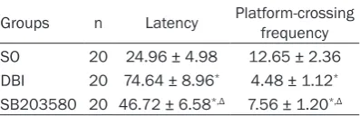
Table 1. Intergroup comparison of the results
of Morris water maze (_x ± S)
Groups n Latency Platform-crossing frequency
SO 20 24.96 ± 4.98 12.65 ± 2.36
DBI 20 74.64 ± 8.96* 4.48 ± 1.12* SB203580 20 46.72 ± 6.58*,Δ 7.56 ± 1.20*,Δ
Note: compared with the SO group, *P<0.05; compared
[image:3.629.101.300.427.491.2]Evaluation on spatial memory
Compared with the SO group, a prolonged escape latency and a lower platform-crossing frequency were found in the DBI group; com-pared with the DBI group, a shorter escape latency and a higher platform-crossing frequen-cy were found in the DBI+SB203580 inhibitor group (P<0.05) (See Table 1).
Results of the western blot on Homer1a and phosphorylated p38 MAPK proteins
The bands of Homer1a and phosphorylated p38 MAPK were clear. With the absorbance
value of β-actin as the internal reference, the
absorbance values of the bands in each group were revised and subjected to semiquantitative analysis: compared with the controls, a higher Homer1a content was detected at each time point in the DBI group, and the content reached
the peak at 24 h, and decreased at 48 h and 72
h, though higher than the controls (P<0.05);
compared with the DBI group, a higher Homer1a content was detected at each time point in the DBI+SB203580 inhibitor group and the
differ-ence was statistically significant (P<0.05, Fi- gure 2; Table 2). Compared with the controls, a higher phosphorylated p38 MAPK content was detected at each time point in the DBI group,
and the content reached the peak at 24 h, and
decreased at 48 h and 72 h, though higher than the controls (P<0.05); compared with the DBI group, a lower phosphorylated p38 MAPK content was detected at each time point in the DBI+SB203580 inhibitor group (P<0.05, Figure 2; Table 3).
Discussion
MAPKs signal transduction pathway is the cen-tral signal pathway connecting most extracellu-lar signals and membrane receptors, regulating transcription factors and genes. Extracellular
[image:4.629.102.533.79.202.2]signal-regulated kinase (ERK), C-Jun N-terminal kinase (JNK), p38 MAPK, etc. are included in
[image:4.629.100.531.268.323.2]Figure 2. Homer1a protein and phosphorylated p38MAPK in rat cortex from various. (A: Control group; B: DBI group; C: SB203580).
Table 2. Intergroup comparison of the Homer1a protein expression in the hippocampi (_x ± S)
Groups n 1 h 6 h 24 h 48 h 72 h
SO 15 0.011 ± 0.010 0.010 ± 0.009 0.011 ± 0.010 0.010 ± 0.010 0.010 ± 0.010 DBI 15 0.096 ± 0.020* 0.144 ± 0.026* 0.172 ± 0.030* 0.136 ± 0.023* 0.114 ± 0.020* SB203580 15 0.156 ± 0.026*,Δ 0.198 ± 0.029*,Δ 0.246 ± 0.038*,Δ 0.154 ± 0.021*,Δ 0.132 ± 0.012*,Δ
Note: compared with the SO group, *P<0.05; compared with the DBI group, ΔP<0.05.
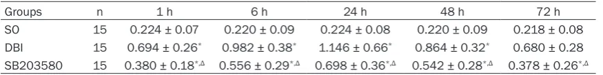
Table 3. Intergroup comparison of the expression of phosphorylated p38 MAPK protein in the hippo-campi (_x ± S)
Groups n 1 h 6 h 24 h 48 h 72 h
SO 15 0.224 ± 0.07 0.220 ± 0.09 0.224 ± 0.08 0.220 ± 0.09 0.218 ± 0.08
DBI 15 0.694 ± 0.26* 0.982 ± 0.38* 1.146 ± 0.66* 0.864 ± 0.32* 0.680 ± 0.28 SB203580 15 0.380 ± 0.18*,Δ 0.556 ± 0.29*,Δ 0.698 ± 0.36*,Δ 0.542 ± 0.28*,Δ 0.378 ± 0.26*,Δ
[image:4.629.98.536.380.435.2]this family. Studies have verified the intracere -bral activation of p38 MAPK signal by brain injury. This activation primarily displays nega-tive regulation on the central nervous system, which directly causes damage to synaptic plas-ticity of hippocampal neurons and leads to inju-ry of long-term potentiation (LTP) via mediating
multiple inflammatory and pathogenic factors,
and consequently results in impairment of spa-tial learning ability [10, 11]. Hippocampal
neu-rons cultured with kainic acid (KA) manifest
degenerative changes, swelling, rough mem-brane surface with humps and memmem-brane rup-ture, and p38 MAPK signal pathway engages in this process. Inhibition on p38 MAPK signal pathway protects hippocampal neurons from toxic injury [12, 13]. In this study, we found that
p38 mitogen activated protein kinase (MAPK)
inhibitor SB203580 could relieve the ultra-structural injury of hippocampal neurons, and perform satisfying therapeutic effects on the learning and memory function impairment of DBI rats.
In 1997, Brakeman was the first to report
Homer protein family, and Homer1a was the
first to be identified in this family. Early research -ers observed the relationship between Homer and animal behavior using the virus vector technology and the transgenic technology. For instance, the recombinant adeno-associated virus gene delivery system was used to overex-press exogenous Homer1a protein in the hip-pocampi of adult rats, causing hippocampus-related memory impairment [14]; impairment of the motor function and the ability of coordi-nated motion were found in the transgenic mice overexpressing Homer1a, and repeating compulsive behavior could also exist [15, 16]. Recently, the long-term loss of Homer proteins (including Homer1a) has been found to be the critical factor leading to degenerating cognitive function of the mice with post-traumatic stress injury in the process of neurodegenerative dis-eases [16]. Our results showed that SB203580 could up-regulate the Homer1a expression in the hippocampus of rats. The author considers that the pretreatment of SB203580 increases the Homer1a expression in the hippocampus. And Homer1aon one hand lessens the nerve injury induced by calcium overloading, and on the other hand regulates synaptic transmission in the hippocampus via the regulation on the distributions and the quantities of
metabo-tropic and inometabo-tropic glutamate receptors as
well as the speed of calcium influx [17, 18],
which is one of the possible mechanisms of SB203580 improving animals’ learning and memory function.
SB203580 can up-regulate the expression of Homer1a, indicating the participation of Ho- mer1a in the activation of p38 MAPK which mediates the nerve injury after DBI. Researches show that the activation of Homer1a relates to the excitatory effect of glutamate and calcium
influx [18]; excitotoxicity of glutamate can pro -mote the rapid activation of p38 MAPK, and p38 MAPK activation is indispensable in this process. The high concentration of neuronic glutamate can be decreased by the inhibition of p38 MAPK pathway by SB203580 [19]. Therefore, the author considers that SB203580 impacts the expression of Homer1a via its reg-ulation on the local concentration of glutamate
and the flow or the speed of calcium influx.
Furthermore, study also shows that the MAPKs signal can regulate the ubiquitin-proteasome system, the degeneration system of Homer1a, consequently to impact the expression of Ho- mer1a [20]. The relationship between the MA- PKs signal and Homer1a in the DBI pathologi-cal process remains to be further studied. In conclusion, SB203580 can promote the re- habilitation of DBI rats’ learning and memory function, which is related to the inhibition of p38 MAPK phosphorylation and the up-regulat-ed expression of Homer1a.
Disclosure of conflict of interest
None.
Address correspondence to: Dr. Jian-Min Li, North China University of Science and Technology, 78 Jian She South Road, Tangshan 063000, Hebei Province, China. Tel: +86+15081978570; E-mail: [email protected]
References
[1] Zhao YN, Li SX and Li JM. The condition of re-covery executive dysfunction in patients with craniocerebral injury investigation and analy-sis. Zhong Hua Hu Li Za Zhi 2010; 254-256. [2] Ji JX, Jiang ZL, He DJ and You YP. Basal ganglia
[3] Urban RJ, Harris P and Masel B. Anterior hypo-pituitarism following truamatic brain injury. Brain Inj 2005; 19: 349-358.
[4] Mcpeak LA, Stries WM and Cope VN. Disability
evaluation following traumatic brain injury. Phys Med Rehabil Clin N Am 2001; 12: 587-601.
[5] Piao CS, Kim JB, Han PL and Lee JK. Adminis-tration of the p38 MAPK inhibitorSB203580 affords brain protection with a wide therapeu-tic window against focal ischemic insult. Neu-rosci Res 2003; 73: 537-44.
[6] Tang ZH, Liao ZB, Shi QH, Xie YF, He ZH and
Zhan Y. Blocking p38 signal pathway lowers
MMP-9 expression and reduces brain edema in rats with traumatic brain injury. Nan Fang Yi Ke Da Xue Xue Bao 2012; 07: 928-931. [7] Bottai D, Guzowski JF, Schwarz MK, Kang SH,
Xiao B, Lanahan A, Worley PF and Seeburg PH. Synaptic activity induced conversion or intron-ic to exonintron-ic sequence in Homer1 immediate early gene expression. Neurosci 2002; 22: 167-175.
[8] Sato M and Suzuk S. NMDA receptor stimula -tion and brain-derived neurotrophic factor up-regulate homer 1a mRNA via the
mitogen-acti-vated protein kinase cascade in cultured
ce-rebellar granule cells. Neurosci 2001; 21911: 3797-3805.
[9] Marmarou A, Foda MA, van den Brink W,
Campbell J, Kita H and Demetriadou K. A new model of diffuse brain injury in rats. Part I: Pathophysiology and biomechanics. Neuro-surg 1994; 80: 291-300.
[10] Yuan H, Yang S, Zhou WX and Zhang YX. MAPK cascade signaling pathways and enhanced long time history. Zhong Guo Yao Li Xue Tong Bao 2006; 22: 769-774.
[11] Butler MP, O’Connor JJ and Moynagh PN. Dis-section of tumor-necrosis factor-alpha inhibi-tion of long-term potentiainhibi-tion (LTP) reveals a
p38 mitogen-activated protein kinase-depen -dent mechanism which maps to early-but not late-phase LTP. Neuroscience 2004; 124: 319-26.
[12] Izumi Y, Tokuda K and Zorumski CF. Long-term
potentiation inhibition by low-level N-methyl-D-aspartate receptor activation involves calci-neurin, nitric oxide, and p38 mitogen-activated
protein kinase. Hippocampus 2008; 18:
258-65.
[13] KI YW, Park JH, Lee JE,Shin IC,Koh HC. JNK and p38MAPK regulate oxidative stress and
the inflammatory response in chlorpyrifos-in -duced apoptosis. Toxicol Lett 2013; 218: 235-245.
[14] Klugmann M, Symes CW, Leichtlein CB, Klaussner BK, Dunning J, Fong D, Young D and During MJ. AAV-mediated hippocampal expres-sion of short and long Homer 1 proteins
dif-ferentially affect cognition and seizure activity
in adult rats. Mol Cell Neurosci 2005; 28: 347-360.
[15] Tappe A and Kuner R. Regulation of motor per-formance and striatal function by synaptic scaffolding proteins of the Homer1 family. Proc Natl Acad Sci USA 2006; 103: 774-779. [16] Szumlinski KK, Lominac KD, Kleschen MJ, Ole
-son EB, Dehoff MH, Schwarz MK, Seeburg PH,
Worley PF and Kalivas PW. Behavioral and neu-rochemical phenotyping of Homer1 mutant
mice: possible relevance to schizophrenia.
Genes Brain Behav 2005; 4: 273-288. [17] Herrmann L, Ionescu IA, Henes K, Golub Y,
Wang NX, Buell DR, Holsboer F, Wotjak CT and
Schmidt U. Long-Lasting Hippocampal Synap-tic Protein Loss in a Mouse Model of Posttrau-matic Stress Disorder. PLoS One 2012; 7: 1-7. [18] Salm EJ and Thayer SA. Homer proteins
accel-erate Ca2+ clearance mediated by the plasma membrane Ca2+ pump in hippocampal neu-rons. Biochem Biophys Res Commun 2012; 424: 76-81.
[19] Chaparro-Huerta V, Flores-Soto ME,
Gudiño-Cabrera G, Rivera-Cervantes MC, Bitzer-Quin -tero OK and Beas-Zárate C. Role of p38 MAPK
and pro-inflammatory cytokines expression in
glutamate induced neuronal death of neonatal rats. Int J Dev Neurosci 2008; 26: 487-495. [20] Li Y, Krogh KA and Thayer SA. Epileptic