Co-expression and purification of the RadA recombinase with the
1RadB paralog from
Haloferax volcanii
yields heteromeric ring-like
2structures.
34
Authors: 5
Bushra B. Patoli1, a,*, Jody A. Winter1, bAtif A. Patoli1, a, Robin M. Delahay2and Karen A. 6
Bunting1, c 7
8 9
Affiliations: 10
1School of Biology, Queens Medical Centre, University of Nottingham, Nottingham, UK, NG7 11
2UH. 12
2School of Medicine, University of Nottingham, Nottingham, UK, NG7 2UH. Email: 13
14
aInstitute of Microbiology, University of Sindh, Jamshoro, Pakistan (Present address). Email: 15
16
bNottingham Trent University, Nottingham, UK, NG11 8NS (Present address). Email: 17
18
cAlbumedix Ltd, Nottingham, UK, NG7 1FD (Present address). Email: 19
20 21 22 23 24 25 26
*Correspondence:Bushra B. Patoli, Institute of Microbiology, University of Sindh, Jamshoro, 27
Pakistan. Tel.: +92 3113005252. [email protected]
28 29 30
Keywords:Homologous recombination, RadA,Haloferax volcanii, Co-expression, Halophilic 31
protein purification. 32
33 34
Abbreviations:Immobilized metal affinity chromatography (IMAC); Discrete Optimised 35
Protein Energy (DOPE); ATPase domain (AD); N-terminal domain (NTD); Size Exclusion 36
Chromatography (SEC); Electron Microscopy (EM) 37
Abstract
40 41
The study of archaeal proteins and the processes they contribute to poses particular challenges due 42
to the often extreme environments in which they function. DNA recombination, replication and 43
repair proteins of the halophilic euryarchaeon,Haloferax volcanii(Hvo) are of particular interest 44
as they tend to resemble eukaryotic counterparts in both structure and activity and genetic tools 45
are available to facilitate their analysis. In the present study, we show using bioinformatics 46
approaches that the Hvo RecA-like protein RadA is structurally similar to other recombinases 47
although is distinguished by a unique acidic insertion loop. To facilitate expression of Hvo RadA 48
a co-expression approach was used, providing its lone paralog, RadB, as a binding partner. At 49
present, structural and biochemical characterization of Hvo RadA is lacking. Here, we describe for 50
the first time co-expression of Hvo RadA with RadB and purification of these proteins as a 51
complex under in vitro conditions. Purification procedures were performed under high salt 52
concentration (>1 M sodium chloride) to maintain the solubility of the proteins. Quantitative 53
densitometry analysis of the co-expressed and co-purified RadA-B complex estimated the ratio of 54
RadA to RadB to be 4:1, which suggests that the proteins interact with a specific stoichiometry. 55
Based on a combination of analyses, including size exclusion chromatography, Western blot and 56
electron microscopy observations we suggest that RadA multimerizes into a ring-like structure in 57
the absence of DNA and nucleoside cofactor. 58
Introduction
63 64
Homologous recombination is one of the principal pathways to repair deleterious DNA damage 65
such as double-strand breaks. The basic mechanism of homologous recombination, comprising 66
homologous base-pairing and a strand exchange reaction, is similar in all forms of life, however it 67
varies in complexity and enzymology. Various enzymes participate in the recombination process, 68
including the key recombinase which belongs to the RecA family of proteins (1). These enzymes 69
are well conserved in all three domains of life and are termed RecA in bacteria (2), RadA in 70
archaea (3), and Rad51 in eukaryotes (4). The strand exchange protein identified in archaeal RadA 71
is more similar to the eukaryotic Rad51 protein (~40% amino acid identity) than to its prokaryotic 72
counterpart RecA in eubacteria (~20% amino acid identity) (3, 5, 6). Crystallographic structure 73
analysis of archaeal RadA has contributed significantly to our understanding of the structure and 74
function of eukaryotic Rad51 (7, 8). Various accessory proteins also participate in the strand 75
exchange reaction and these are usually termed recombination mediators. Many recombination 76
mediators are encoded by genes that arise as a result of a duplication event within the genome, and 77
are termed paralogs. In yeast, the paralogous proteins Rad55 and Rad57 share some similarity to 78
Rad51 and function as a heterodimeric complex which may stabilize the Rad51-assembled 79
nucleoprotein filament (9). Five Rad51 paralogs, XRCC2, XRCC3, RAD51B/RAD51L1, 80
RAD51C/RAD51L2, and RAD51D/RAD51L3, are found in eukaryotes. These paralogs form 81
various heteromeric complexes, mostly dimers such as RAD51B-RAD51C, RAD51D-XRCC2, 82
RAD51C-XRCC3 or tetramers such as RAD51B-RAD51C-RAD51D-XRCC2 (10). 83
The identification and characterisation of RecA-like proteins in both crenarchaea and euryarchaea 84
diversity. This may reflect that the archaea demonstrate an evolutionary mixture of replication, 86
repair and recombination functions in between simple bacterial and more complex eukaryotic 87
forms of life. 88
RadB was the first RadA paralog to be characterised in archaea and its presence is confined to the 89
Euryarchaeota. RadB lacks the N-terminal domain of RadA (11) and strand exchange activity (12). 90
Interaction of RadB with RadA has been demonstrated in Pyrococcus furiosus (Pfu) and it was 91
proposed that RadB functions in a manner analogous to the yeast Rad55-57 proteins in the strand 92
exchange reaction (12). Genetic analysis ofradBfrom the euryarchaeonHaloferax volcanii(Hvo) 93
demonstrates that RadB functions in the homologous recombination pathways in concert with 94
RadA (13, 14). 95
Hvo RadB has been successfully over-expressed inEscherichia coli(15) and both Hvo RadA and 96
RadB proteins have been purified after conditional over-expression in their native host (16, 17). A 97
recent study has confirmed the in vivo interaction of Hvo RadA and RadB, exploiting mass 98
spectrometry to identify co-purifying proteins following IMAC chromatography (18). However, 99
both quantity and purity of the resulting proteins has been insufficient for structural and 100
biochemical analyses previously described for other DNA-associated halophilic proteins such as 101
Hvo PCNA and RPA3 (19, 20). Additionally, when over-expressed inE. coli, recombinant Hvo 102
RadA has also been found to co-purify with DNA, providing further impediments to downstream 103
analyses since conventional approaches to remove contaminating DNA, such as denaturation and 104
nuclease treatment, are likely to be detrimental to subsequent characterization [17]. 105
The study of recombination proteins in the hyperthermophilic crenarchaeon, Sulfolobus tokodaii
106
(St),has shown that over-expression and characterization of StRad55B, a paralog of StRadA, was 107
expression of Hvo RadA with RadB as both a novel strategy for maintenance and optimisation of 109
RadA protein stability in the non-halophilic host,E. coli,and as a potential means to limit RadA 110
interaction with host DNA to facilitate its purification. Using this approach we successfully 111
demonstrate soluble over-expression of the Hvo RadA-RadB complex, provide clear evidence for 112
RadA-RadB protein-protein interaction and present robust methodology to enable their further 113
characterization and study. 114
115
Materials and Methods
116 117
Bacterial strains, plasmids and growth conditions. 118
E. coli chemically competent strains DH5α™ (Invitrogen) and Rosetta 2 (DE3) (Novagen) were
119
used for cloning and gene expression, respectively. The PCR-amplified target gene was cloned 120
first into the Zero Blunt PCR vector (Invitrogen) and sequenced to exclude amplification errors. 121
The pET11 expression vector (Novagen) was used when single overexpression of radAorradB
122
was desired. For co-expression and purification of RadA and RadB, the E. coliRosetta 2 (DE3) 123
strain was transformed with pET-Duet-1-based plasmid constructs (Novagen). 124
Bacterial cultures were grown at 37°C in Luria-Bertani (LB) broth in a shaking incubator or on 125
LB agar plates supplemented with ampicillin (100 μg ml-1), chloramphenicol (34 μg ml-1) or 126
kanamycin (34 μg ml-1) as appropriate. 127
128
Knock out of thearnAgene fromE. coliRosetta2 (DE3) expression strain 129
A mutant of the Rosetta2 (DE3) expression strain carrying a kanamycin resistance cassette (kanR) 130
(22). Donor strain, obtained from theE. coliGenetic Stock center (CGSC), was grown at 37C to 132
an OD650 of 0.3-0.4 to add the P1 phage stock (~107-108PFU). After 3-4 h incubation, purified 133
lysate was used to transduce the kanRmarker into recipient Rosetta2 (DE3) cells. Recipient strain 134
was grown at 37C to OD650 of 0.8, pelleted, re-suspended in 0.1 M MgSO4, 5 mM CaCl2 and 135
incubated with purified lysate for 25 minutes at 37C. After addition of sodium citrate to a final 136
concentration of 0.5 M to chelate calcium ions and prevent further total lysis of the recipient strain, 137
transductants were plated and sub-cultured to purity on LB agar containing kanamycin (34 μg ml -138
1) and chloramphenicol (34 μg ml-1). 139
140
Molecular methods 141
HvoradAgene (Accession number: U45311) was PCR-amplified from theH. volcaniiwild-type 142
DS2 strain (23). The forward primer (radA6HisF) contained an NdeI restriction site (underlined) 143
and a 6xHis tag
144
5’GACCTCATATGCATCACCATCACCATCACATGGCAGAAGACGACCTC-3’ and the
145
reverse primer (radABamR) encoded a BamHI restriction site (underlined) 5’-146
GCAATGGATCCTTATTACTCGGGCTTGAGACCGGCGTCCTG-3’. The His-tagged radA
147
gene was cloned first into the Zero Blunt PCR vector for sequencing and then sub-cloned for 148
overexpression either into pET11 (using NdeI and BamHI restriction sites) or pET-Duet1 plasmid 149
at multiple cloning site 2 (MCS-2) (using the NdeI and EcoRV restriction sites). The Hvo radB
150
coding sequence (690bp) was excised from plasmid pCPG42 using the NdeI and BamHI restriction 151
sites (15) . After sequence verification in a sub-cloning vector, radBwas cloned into MCS-1 of 152
pET-Duet1 using the NcoI and HindIII restriction sites to yield the pBPRAD2 construct. The 153
155
Over-expression, purification and analysis of RadA and RadB proteins 156
Rosetta2 (DE3) or Rosetta2 (DE3) ΔarnA E. colitransformed with the pBPRAD2 construct were 157
used for over-expression and purification of co-expressed His-tagged RadA and un-tagged RadB 158
proteins. An overnight culture was inoculated into baffle flasks containing 2.4 L LB broth 159
supplemented with antibiotics as appropriate, and grown at 37°C. Over-expression ofradA-Bwas 160
induced with 0.2 mM isopropyl β-D-1-thiogalactopyranoside (IPTG) at an OD600of 0.4 – 0.6 with 161
incubation for a further 3 hrs in a shaking incubator at 30°C. The cell pellet was harvested by 162
centrifugation and lysed by sonication on ice in buffer A (50 mM HEPES, pH7.0, 10 mM 163
imidazole and 1 M NaCl) containing EDTA-free protease inhibitor cocktail (Roche). The cell 164
lysate was clarified by centrifugation at 16,000 x g for 30 minutes at 4°C to remove insoluble 165
debris. 166
Soluble proteins in the supernatant were used for downstream purification of the complex using 167
Cobalt-based immobilized metal affinity chromatography (IMAC). A Liquid Chromatography 168
Column (2.5 cm x 10 cm, Sigma-Aldrich) containing 5 mL of Talon® metal affinity resin 169
(Clontech) was equilibrated with 20 column volumes of buffer A (50 mM HEPES pH 7.0, 1 M 170
NaCl and 30 mM imidazole). The clarified soluble fraction was loaded and incubated with rolling 171
for 10 minutes at room temperature. The flow-through and two 20 mL buffer A washes were 172
retained separately in fresh tubes for SDS-PAGE gel analysis. Bound proteins were eluted with 13 173
mL of buffer B (50 mM HEPES pH 7.0, 1 M NaCl and 300 mM imidazole) following rolling for 174
5 minutes at room temperature, prior to collection of the eluate. The eluate was loaded onto a 26/60 175
Superdex 200 (S200) preparative column (GE Healthcare) using an ÄKTA Prime Plus system. 176
mM HEPES pH 7.0 and 1 M NaCl). The proteins in each IMAC and SEC fraction were analyzed 178
by SDS-PAGE gel. SEC fractions were also analysed by agarose gel electrophoresis to select 179
fractions free from contaminating DNA. Samples containing low concentrations of protein were 180
first concentrated using StrataClean resin (5 μl ml-1) (Stratagene) as previously described (24) 181
prior to gel loading. All samples were adjusted to maintain equivalent loading in all gels. 182
The identity of RadA and RadB proteins was confirmed using MALDI-TOF-MS analysis of 183
excised bands by the Biopolymer Synthesis and Analysis Unit (BSAU), University of Nottingham. 184
Quantitative reflectance densitometric analysis of Coomassie Brilliant Blue-stained proteins was 185
performed using a BioRad GS-800 calibrated densitometer and Quantity One® Software. Western 186
blots were blocked with 5%, w/v, milk powder in phosphate buffered saline (PBS)-Tween 20, then 187
probed with 10 μL (1/1000 diluted) of alkaline phosphatase conjugated-Mouse Anti-Hexa-His 188
antibodies (Sigma) for 1 h and developed using BCIP/NBT substrate according to manufacturer’s 189
instructions (Sigma). 190
191
Electron microscopy 192
Co-expressed, purified RadA-B proteins were concentrated to 1 mg ml-1using a Vivapore 10/20 193
7500 Da cutoff (Vivascience) and were maintained in 1 M NaCl buffer to preserve the soluble and 194
native state of the proteins. Protein concentrations were determined using the Qubit protein assay 195
kit (Invitrogen). Protein samples were applied to a carbon-Formvar grid (Agar Scientific) and 196
allowed to settle for 10 minutes. The sample was then stained with either 0.5 or 1%, w/v, 197
phosphotungstic acid (PTA; pH 7.0) for 1 minute. The grids were then imaged using a JEOL 198
JEM1010 transmission electron microscope at 100, 000X or 200, 000X magnification. 199
Homology Modelling 201
Homology modelling of the Hvo RadA primary sequence was performed using the PyMod 2.0 202
plugin module for PyMol (25, 26) as a convenient interface to Modeller 9v4 (27). The Pfu RadA 203
structure (1PZN) chain A was used as the highest scoring template and aligned against the Hvo 204
RadA sequence using Clustal Omega. Modeller parameters were adjusted to accommodate 205
automated building of disulfide bridges and the highest level of optimization and refinement, with 206
additional energy minimisation performed on resulting models. Output models with corresponding 207
low scoring Discrete Optimised Protein Energy (DOPE) profiles, indicative of limited modelling 208
errors, were further inspected for agreement with secondary structural elements and diversity in 209
loop disposition prior to final model selection. 210
211 212
Results
213 214
Conservation of the ATPase domain of RadA from both halophilic and non-halophilic 215
species 216
Structural comparison of RadA from P. furiosus with RadA homologs in other domains of life 217
reveals similarity in the ATPase domain (AD) although differences are apparent at amino and 218
carboxyl terminal domains of the proteins (Fig. S1a). Conservation of amino acid residues at the 219
Walker A, Walker B and DNA binding (L1 and L2) motifs is also apparent in a protein sequence 220
alignment of Hvo RadA with RadA homologs (Fig. S1b), and the Hvo RadB protein. Aside from 221
this conserved motif organization, Hvo RadA and RadB proteins are otherwise dissimilar, sharing 222
smaller protein, due to lack of a RadA-equivalent N-terminal domain (NTD) (Fig. S1c). 224
Along with a characteristic negative charge distribution on the protein surface, a distinct preference 225
for certain amino acids is also a hallmark of halophilic proteins. This is apparent in Hvo RadA as 226
a comparative increase in the acidic residue, aspartate, which is the most common adaptation 227
relative to mesophilic and thermophilic counterparts (Fig. S2). However, whereas a marked 228
reduction in the lysine content and increases in serine and alanine have been reported in other 229
halophilic proteins (28) this is not similarly the case for Hvo RadA. This disparity reflects the lack 230
of any currently known universal form of halophilic adaptation. 231
It is of particular interest to understand how halophilic proteins have adapted to interact with DNA, 232
given the involvement of basic residues in DNA binding of the phosphate backbone. Guy and 233
others (15) demonstrated that residues in a conserved basic patch (the KHR triplet) were crucial 234
for DNA binding in Hvo RadB. To gain some insight into how preference for particular amino 235
acid usage in Hvo RadA, would likely affect the surface character of Hvo RadA, we built a 236
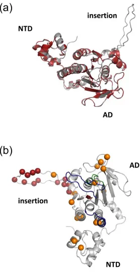
structural model in Modeller 9v4 for comparative analysis. Superposition of the Hvo RadA model 237
with RadA homologs shows the expected correspondence with the principal structural elements in 238
the N-terminal and ATPase domains (Fig. 1a) although, unlike other RadA proteins, an extensive 239
insertion loop found only in the Hvo RadA sequence (159-182) is also apparent (Fig. 1a and Fig. 240
S1b). 241
Fig. 1(b) shows the conserved elements of RadA in the Hvo RadA homology model in relation to 242
lysine residues within the molecule. The L1 and L2 loops within the ATPase domain are involved 243
in ssDNA binding. Chen and others (29) identified a number of key positively charged residues in 244
Sso RadA L1 (R217, R223 and R229) that are also conserved in Hvo RadA. Conversely, of the 245
equivalent of K27 is retained in Hvo RadA, with K60 being substituted with as aspartate residue 247
(D52). However, Hvo RadA possesses an alternative lysine at position 42. The majority of the 248
lysine residues within Hvo RadA are located on the face of the molecule containing the NTD, L1 249
and L2 motifs, suggesting their retention may be related to involvement in DNA-binding. 250
The large insertion sequence seemingly unique to Hvo RadA (residues 159-182) is of potential 251
interest (Fig. 1 and Fig S1b). Given the lack of homology to other family members the 252
conformation of the modelled loop remains speculative but it can be assumed that it is positioned 253
on the face of the molecule as indicated in Fig. 1(b). The loop region comprises a large number of 254
acidic residues, both aspartate and glutamate, which, in the context of the uncommonly higher 255
proportion of lysines in Hvo RadA relative to other halophilic proteins, may function to maintain 256
solubility of the molecule or promote/stabilize RadA interactions with other proteins. 257
258
Differential co-expression and purification of soluble RadA-RadB proteins 259
His-tagged Hvo radA and un-tagged radB were co-expressed from pBPRAD2 in the Rosetta 2 260
(DE3)E. colistrain in order to minimise issues with codon usage that can occur when expressing 261
archaeal proteins in a heterologous host (30). SDS-PAGE gel analysis of clarified lysates of 262
induced cultures identified overexpressed proteins at the anticipated molecular mass for 6xHis-263
RadA (~ 40 kDa) and RadB (~25 kDa) (Fig. 2). However, by comparison with the broadly 264
equivalent amounts of RadA and RadB observed when each protein was expressed from pET11 265
individually (Fig 2. lanes 2 & 4 respectively), RadB, when co-expressed with RadA, appeared 266
substantially diminished in abundance. This may suggest a possible issue with bicistronic 267
expression, although DUET vectors are engineered for independent promoter control of both 268
RadB expression or stability; a previous analysis of the RadA:RadB ratio inP. furiosuscell extract 270
showed the cellular amount of RadB protein to be ~200 times lower than that of RadA (12). 271
Consequently, the lack of equimolar expression observed may better reflect a more physiological 272
ratio of RadA and RadB proteins and, as such, was not considered to be an impediment to further 273
analysis. 274
H. volcaniirequires 2 M sodium chloride to maintain internal osmotic balance in artificial media 275
(23). Consideration is therefore required around buffer selection to maintain salt levels to promote 276
solubility of halophilic proteins and avoid aggregation.Structural characterization of Hvo PCNA 277
at 2 Å resolution demonstrated the correct folding of the trimeric protein with a root mean square 278
deviation of 1.3-1.7 Å compared with other archaeal and eukaryotic PCNA molecules (19, 20). 279
Similarly, the Hvo RPA3 DNA-binding protein was shown to be competent to bind DNA in buffers 280
containing 1 M KCl, but not at the lower concentration tested (0.2 M KCl) (19, 20). Both proteins 281
were expressed inE. coli and were principally purified in buffers containing 1M NaCl, therefore 282
this concentration was maintained in this study. 283
IMAC was used as a first step for the purification of His-tagged RadA and un-tagged RadB using 284
a cobalt-based metal affinity resin. The cobalt-based resin was observed to bind His-tagged 285
halophilic RadA protein with a higher specificity compared to nickel-based resins, reducing non-286
specific binding of host proteins (data not shown). 287
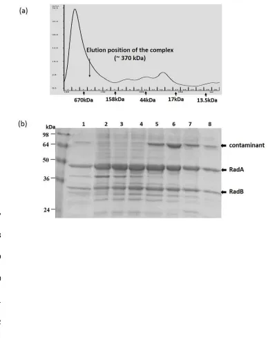
Co-eluted His-tagged RadA and un-tagged RadB proteins were observed at a similar ratio as before 288
at their expected molecular mass positions by SDS-PAGE analysis (Fig. 2b), indicating specific 289
interaction. Eluted fractions were subsequently subjected to SEC for further purification and to 290
provide an initial indication of the stoichiometry of RadA-RadB interaction. Both proteins eluted 291
in protein ratio was apparent in any elution fraction suggesting that complex formation may be 293
constrained by a particular stoichiometric configuration (Fig. 3b). Absence of contaminating DNA 294
was confirmed by agarose gel electrophoresis. 295
296
Optimisation of the heterologous expression system 297
Whereas previous experiments demonstrated successful purification of a stable RadA-RadB 298
complex, subsequent analysis was hampered by a limitation of the purification protocol to remove 299
all non-specific host proteins. One major persistent contaminant (~64 kDa) that could not be 300
removed by standard optimisation approaches without significant concomitant decrease in yield 301
of the RadA-RadB complex was identified by MALDI analysis to be the product of thearnAgene 302
(bi-functional polymyxin resistance protein, ArnA). The primary sequence of ArnA demonstrates 303
an abundance of histidine residues (31) likely accounting for its capture by IMAC resins. Similarly, 304
the ~74 kDa ArnA protein is reported to form a hexameric structure consisting of a dimer of trimers 305
(31, 32) with a predicted molecular mass of ~440 kDa, close to the observed ~370 kDa RadA-B 306
complex. 307
In view of this, bacteriophage-mediated P1 transduction was performed to delete the arnA gene 308
from the Rosetta 2 (DE3) expression strain. The resulting Rosetta 2 (DE3) ΔarnAmutant strain 309
was found to be equivalent to the parent strain in respect of growth characteristics and over-310
expression of RadA-RadB. Subsequently, RadA-RadB complex expressed from the modified 311
strain was purified to ~95% homogeneity following the previous two-step purification protocol 312
(data not shown). 313
314
Recombinase proteins characteristically oligomerise into ring structures or helical nucleoprotein 316
filaments. Electron microscopy of P. furiosus RadA revealed that the protein forms dimers of 317
heptameric ring structures in solution in the absence of DNA (8) whereas, inS. solfataricus, RadA 318
forms an octameric ring bound to DNA (33). Since Hvo RadA-RadB is consistently purified as a 319
large ~370 kDa stable complex, some form of RadA oligomerization is clearly indicated. 320
Preliminary densitometric analysis of co-purified RadA and RadB protein bands estimates the ratio 321
of RadA co-purified with RadB to be 4:1. Therefore the observed ~370 kDa RadA-RadB SEC 322
complex would most closely correspond to eight molecules of RadA in complex with two 323
molecules of RadB although this estimation is limited by the resolution of the S200 SEC column 324
employed in this study. 325
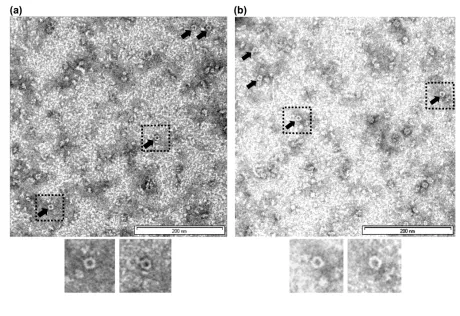
Subsequent electron microscopy observation of our purified Hvo RadA-RadB protein complex 326
confirms that Hvo RadA-RadB complex also possesses a tendency to self-associate into 327
multimeric structures in the absence of DNA (Fig. 4). In order to distinguish protein structures 328
from electron microscopy (EM) artifacts resulting as a consequence of high salt levels, several 329
control grids were prepared and imaged with either buffer only or buffer plus stain at 100,000X 330
magnification. The images captured showed ring-like structures throughout the field which 331
suggests that Hvo RadA-RadB complex in its native state similarly exists as a ring-like structure 332
(Fig. 4). 333
334
335
Discussion
The expression and purification of halophilic proteins, particularly from heterologous host 337
overexpression systems presents several challenges, principally due to the particular 338
physicochemical properties of these proteins which constrain conventional approaches. 339
Halophilic proteins are adapted to maintain solubility and function in hyper saline (1-4 M) 340
conditions (34). The presence of high salt in halophilic organisms affects the gross molecular 341
conformation of both proteins (35) and DNA (36). Mesophilic DNA-binding proteins typically 342
make heavy use of positively charged residues to mediate electrostatic interactions with DNA (15, 343
29, 37). In the majority of organisms the presence of higher concentrations of salt interferes with 344
DNA-binding and leads to aggregate formation in mesophilic proteins. The situation is reversed 345
in halophilic counterparts. Halophilic proteins are generally decorated with negatively charged 346
amino acid residues which allow the binding of surplus water and salt to build up a hydrated 347
solvent network on the surface of proteins. Structural studies have shown that binding of hydrated 348
cations (provided by excess salt and water molecules) around the negatively charged residues on 349
the protein surface reduces the electrostatic repulsive forces between polyanionic DNA and protein 350
molecules (19, 38). At lower concentrations of salt, the protective effect is lost and the repulsive 351
forces between the acidic residues lead to unfolding and inactivation of the protein (28). 352
Consistently, H. volcanii RadA has an overall abundance of acidic amino acids, particularly 353
aspartate which, based on homology modelling of RadA, contributes to the overall predicted 354
negative surface charge of the protein. Accounting for this, we successfully adapted a protocol for 355
purification of soluble RadA under moderate salt conditions. Further protocol modifications were 356
introduced to enable purification from persistent contaminants including both DNA and particular 357
host proteins. Most notably, we overcame the previously observed deleterious effect of RadA over-358
purification as a DNA-free complex with RadB using a relatively facile two-step purification 360
protocol. Based on our observations we speculate that RadA interacts with a small proportion of 361
RadB in a particular stoichiometry to stabilize it as a binding partner. 362
H. volcaniiRadA is similar to other characterised RadA homologues. Consistent with its function 363
as a recombinase, ATP and DNA binding motifs are apparent which have a well-conserved 364
sequence composition despite the common skews in overall amino acid usage typically observed 365
in halophilic proteins (Fig. 1 and Fig. S1). The abundance of acidic amino acids such as aspartate 366
in Hvo RadA could potentially result in electrostatic repulsion with the negatively charged DNA 367
backbone underin vitroconditions (Fig. 1). The crystal structure analysis of Hvo PCNA indicates 368
that this protein compensates for the reduction in positively charged surface residues by employing 369
cation binding (19). However, the interaction in this instance is largely topological in nature rather 370
than via interaction of specific residues. A conserved basic patch on the surface of RadB has been 371
shown to be crucial in DNA binding (15) and number of similarly conserved positively charged 372
residues are retained in Hvo RadA. The presence of a highly negatively charged loop uniquely 373
present in Hvo RadA may compensate for the retention of lysine residues that presumably are 374
maintained to enable direct DNA-binding (Fig. 1a). 375
The failure to observe DNA binding activity in Hvo RadA across a range of methods (refer to 376
supplementary text, Fig. S3 and Table S1) suggests that, for this protein, the protein-DNA 377
interaction may require additional, as yet unidentified, factors. It is also possible that under the 378
conditions of our assays, RadA and RadB interaction occluded the DNA binding site or prevented 379
an active conformational state of the proteins required for DNA binding and ATPase activity. It is 380
also possible that additional co-factors are required for enzymatic activity of the complex.
A recently published study exploring the role of Hvo RadB in homologous recombination also 382
confirmed interaction of RadA with RadB when the proteins were individually over-expressed in 383
H. volcanii, identifying co-purifying proteins by mass spectrometry [39]. Genetic analyses 384
identified point mutations in RadA that suppress theradBphenotype, supporting the hypothesis 385
that RadB functions as a recombination mediator, potentially by inducing conformational changes 386
within RadA. Additionally, the authors noted thatin vitroassays performed did not demonstrate 387
functional DNA binding, as found in this study, also noting that additional co-factors may be 388
required. 389
Recombinases have been generally observed to self-assemble into ring structures (six to eight 390
protomers) in their native state and monomerize to interact with DNA to form the helical 391
nucleoprotein filaments. So far the precise function of the ring structures is not clear in the process 392
of homologous base pairing. The toroidal form in recombinases has been suggested to function as 393
an inactive storage form of protein, which is likely utilized to occlude the polymerization motif, 394
preventing unwanted interaction of the protein with DNA. EM analysis of the purified protein 395
complex showed that RadA-RadB also multimerizes into ring-like structures, which we consider 396
may be of the order of a heptamer or octamer (Fig. 4), similar to its RadA/Rad51 homologs. On 397
the basis of our collective observations, further studies to understand the dynamics of halophilic 398
protein-protein and protein-DNA interactions in H. volcanii, including gross structural 399
determination and robust biophysical characterization can now be anticipated. 400
401
Funding
This work was supported by the University of Sindh, Jamshoro and the Higher Education 403
Commission of Pakistan. K.B. and J.W. were supported by a Wellcome Trust RCDF award to K.B. 404
(Grant no. 076556/Z/05/Z). 405
406
Conflicts of interest
407
None. 408
409 410
References
411 412
1. Kowalczykowski SC, Eggleston AK.Homologous pairing and DNA strand-exchange 413
proteins.Annu Rev Biochem.1994;63:991-1043. 414
415
2. Story RM, Weber IT, Steitz TA.The structure of the E. coli recA protein monomer and 416
polymer.Nature.1992;355(6358):318-25. 417
418
3. Sandler SJ, Satin LH, Samra HS, Clark AJ.recA-like genes from three archaean 419
species with putative protein products similar to Rad51 and Dmc1 proteins of the yeast 420
Saccharomyces cerevisiae.Nucleic Acids Res.1996;24(11):2125-32. 421
422
4. Ogawa T, Shinohara A, Nabetani A, Ikeya T, Yu X, et al. RecA-like recombination 423
proteins in eukaryotes: functions and structures of RAD51 genes.Cold Spring Harb Symp Quant
424
Biol. 1993;58:567-76. 425
426
5. Shinohara A, Ogawa H, Ogawa T. Rad51 protein involved in repair and recombination 427
in S. cerevisiae is a RecA-like protein.Cell.1992;69(3):457-70. 428
429
6. Komori K, Miyata T, Daiyasu H, Toh H, Shinagawa H, et al.Domain analysis of an 430
archaeal RadA protein for the strand exchange activity.J Biol Chem. 2000;275(43):33791-7. 431
432
7. Ariza A, Richard DJ, White MF, Bond CS.Conformational flexibility revealed by the 433
crystal structure of a crenarchaeal RadA.Nucleic Acids Res.2005;33(5):1465-73. 434
435
8. Shin DS, Pellegrini L, Daniels DS, Yelent B, Craig L, et al.Full-length archaeal Rad51 436
structure and mutants: mechanisms for RAD51 assembly and control by BRCA2.EMBO J.
439
9. Sung P, Klein H.Mechanism of homologous recombination: mediators and helicases 440
take on regulatory functions.Nat Rev Mol Cell Biol. 2006;7(10):739-50. 441
442
10. Symington LS.Role of RAD52 epistasis group genes in homologous recombination and 443
double-strand break repair.Microbiol Mol Biol Rev.2002;66(4):630-70. 444
445
11. Sandler SJ, Hugenholtz P, Schleper C, DeLong EF, Pace NR, et al.Diversity of radA 446
genes from cultured and uncultured archaea: comparative analysis of putative RadA proteins and 447
their use as a phylogenetic marker.J Bacteriol.1999;181(3):907-15. 448
449
12. Komori K, Miyata T, DiRuggiero J, Holley-Shanks R, Hayashi I, et al.Both RadA 450
and RadB are involved in homologous recombination in Pyrococcus furiosus.J Biol Chem.
451
2000;275(43):33782-90. 452
453
13. Allers T, Ngo HP.Genetic analysis of homologous recombination in Archaea: Haloferax 454
volcanii as a model organism.Biochem Soc Trans.2003;31(Pt 3):706-10. 455
456
14. Haldenby S, White MF, Allers T.RecA family proteins in archaea: RadA and its 457
cousins.Biochem Soc Trans. 2009;37(Pt 1):102-7. 458
459
15. Guy CP, Haldenby S, Brindley A, Walsh DA, Briggs GS, et al.Interactions of RadB, a 460
DNA repair protein in archaea, with DNA and ATP.J Mol Biol.2006;358(1):46-56. 461
462
16. Allers T.Overexpression and purification of halophilic proteins in Haloferax volcanii. 463
Bioeng Bugs.2010;1(4):288-90. 464
465
17. Allers T, Barak S, Liddell S, Wardell K, Mevarech M.Improved strains and plasmid 466
vectors for conditional overexpression of His-tagged proteins in Haloferax volcanii.Appl
467
Environ Microbiol.2010;76(6):1759-69. 468
469
18. Wardell K, Haldenby S, Jones N, Liddell S, Ngo GHP, et al.RadB acts in homologous 470
recombination in the archaeon Haloferax volcanii, consistent with a role as recombination 471
mediator.DNA Repair(Amst). 2017;55:7-16. 472
473
19. Winter JA, Christofi P, Morroll S, Bunting KA.The crystal structure of Haloferax 474
volcanii proliferating cell nuclear antigen reveals unique surface charge characteristics due to 475
halophilic adaptation.BMC Struct Biol.2009;9:55. 476
477
20. Winter JA, Patoli B, Bunting KA.DNA binding in high salt: analysing the salt 478
dependence of replication protein A3 from the halophile Haloferax volcanii.Archaea.
479
2012;2012:719092. 480
481
21. Sheng D, Li M, Jiao J, Ni J, Shen Y.Co-expression with RadA and the characterization 482
of stRad55B, a RadA paralog from the hyperthermophilic crenarchaea Sulfolobus tokodaii.Sci
483
485
22. Sternberg N.Bacteriophage P1 cloning system for the isolation, amplification, and 486
recovery of DNA fragments as large as 100 kilobase pairs.Proc Natl Acad Sci U S A. 487
1990;87(1):103-7. 488
489
23. Mullakhanbhai MF, Larsen H.Halobacterium volcanii spec. nov., a Dead Sea 490
halobacterium with a moderate salt requirement.Arch Microbiol.1975;104(3):207-14. 491
492
24. Ziegler J, Vogt T, Miersch O, Strack D.Concentration of dilute protein solutions prior 493
to sodium dodecyl sulfate-polyacrylamide gel electrophoresis.Anal Biochem. 1997;250(2):257-494
60. 495 496
25. Janson G, Zhang C, Prado MG, Paiardini A. PyMod 2.0: improvements in protein 497
sequence-structure analysis and homology modeling within PyMOL.Bioinformatics. 2016. 498
499
26. DeLano WL.The PyMol Molecular Graphics System.DeLano Scientific LLC, Palo
500
Alto, Calif, USA.2008. 501
502
27. Webb B, Sali A.Comparative Protein Structure Modeling Using MODELLER. Curr 503
ProtocBioinformatics.2014;47:5 6 1-32. 504
505
28. Lanyi JK.Salt-dependent properties of proteins from extremely halophilic bacteria. 506
Bacteriol Rev.1974;38(3):272-90. 507
508
29. Chen LT, Ko TP, Chang YW, Lin KA, Wang AH, et al.Structural and functional 509
analyses of five conserved positively charged residues in the L1 and N-terminal DNA binding 510
motifs of archaeal RADA protein.PLoS One.2007;2(9):e858. 511
512
30. Poidevin L, MacNeill SA.Biochemical characterisation of LigN, an NAD+-dependent 513
DNA ligase from the halophilic euryarchaeon Haloferax volcanii that displays maximal in vitro 514
activity at high salt concentrations.BMC Mol Biol.2006;7:44. 515
516
31. Gatzeva-Topalova PZ, May AP, Sousa MC.Crystal structure and mechanism of the 517
Escherichia coli ArnA (PmrI) transformylase domain. An enzyme for lipid A modification with 518
4-amino-4-deoxy-L-arabinose and polymyxin resistance.Biochemistry.2005;44(14):5328-38. 519
520
32. Breazeale SD, Ribeiro AA, McClerren AL, Raetz CR.A formyltransferase required 521
for polymyxin resistance in Escherichia coli and the modification of lipid A with 4-Amino-4-522
deoxy-L-arabinose. Identification and function oF UDP-4-deoxy-4-formamido-L-arabinose.J
523
Biol Chem.2005;280(14):14154-67. 524
525
33. Yang S, Yu X, Seitz EM, Kowalczykowski SC, Egelman EH.Archaeal RadA protein 526
binds DNA as both helical filaments and octameric rings.J Mol Biol.2001;314(5):1077-85. 527
528
34. Dym O, Mevarech M, Sussman JL.Structural features that stabilize halophilic malate 529
531
35. Madern D, Ebel C, Zaccai G.Halophilic adaptation of enzymes.Extremophiles.
532
2000;4(2):91-8. 533
534
36. Schlick T, Li B, Olson WK. The influence of salt on the structure and energetics of 535
supercoiled DNA.Biophys J.1994;67(6):2146-66. 536
537
37. Lee CD, Wang TF.The N-terminal domain of Escherichia coli RecA have multiple 538
functions in promoting homologous recombination.J Biomed Sci.2009;16:37. 539
540
38. Bergqvist S, Williams MA, O'Brien R, Ladbury JE.Halophilic adaptation of protein-541
DNA interactions.Biochem Soc Trans. 2003;31(Pt 3):677-80. 542
543
39. Hartman AL, Norais C, Badger JH, Delmas S, Haldenby S, et al.The complete 544
genome sequence of Haloferax volcanii DS2, a model archaeon.PLoS One. 2010;5(3):e9605. 545
546
547
548
549
550
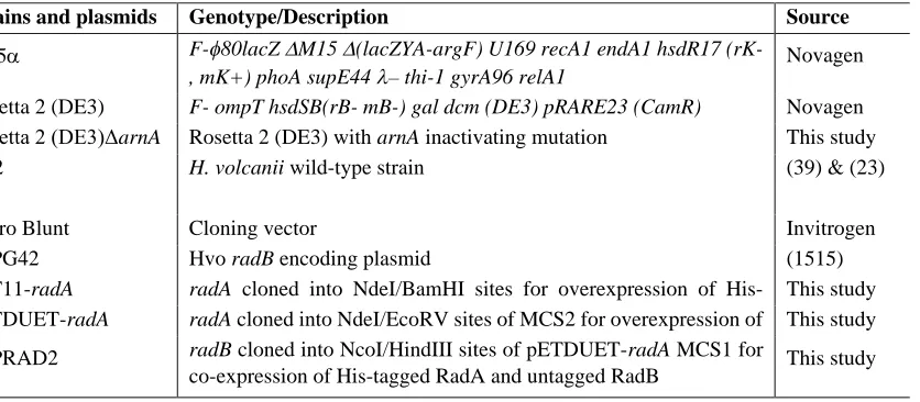
Table
[image:22.595.91.509.174.357.2]566 567
Table 1. List of strains and plasmids. 568
Strains and plasmids Genotype/Description Source
DH5 F-80lacZM15(lacZYA-argF) U169 recA1 endA1 hsdR17
(rK-, mK+) phoA supE44– thi-1 gyrA96 relA1
Novagen
Rosetta 2 (DE3) F- ompT hsdSB(rB- mB-) gal dcm (DE3) pRARE23 (CamR) Novagen
Rosetta 2 (DE3)ΔarnA Rosetta 2 (DE3) witharnAinactivating mutation This study
DS2 H. volcaniiwild-type strain (39) & (23)
pZero Blunt Cloning vector Invitrogen
pCPG42 HvoradBencoding plasmid (1515)
pET11-radA radA cloned into NdeI/BamHI sites for overexpression of His-tagged RadA
This study pETDUET-radA radAcloned into NdeI/EcoRV sites of MCS2 for overexpression of
His-tagged RadA
This study
pBPRAD2 radBcloned into NcoI/HindIII sites of pETDUET-radAMCS1 for
co-expression of His-tagged RadA and untagged RadB This study
Figure legends
585 586
Fig. 1(a). Superposition of the Hvo RadA homology model with the Pfu RadA crystal 587
structure. The Hvo RadA homology model is shown in grey and the Pfu RadA structure 588
(1PZN:Chain A) in red (cartoon representation). Good correspondence is apparent between major 589
structural elements, although Hvo RadA can be distinguished by an additional large loop insertion. 590
(b). Cartoon representation of the backbone of the modelled Hvo RadA structure.The Walker 591
A motif (green), Walker B motif (red) and DNA binding loops (blue) are indicated. The C 592
position of the lysine residues in Hvo RadA are indicated with orange spheres. The Cposition of 593
acidic residues located in an insertion loop in Hvo RadA relative to related sequences (see 594
supplementary Fig. S1) are indicated with red spheres. NTD, AD and the insertion loop are 595
labelled. 596
597
Fig. 2(a). Coomassie Brilliant-Blue stained SDS-PAGE gel demonstrating co-over-expression 598
of Hvo RadA and RadB inE. coli. Broadly equivalent levels of recombinant RadA and RadB 599
were observed in whole cell lysates when expressed independently (lanes 3 & 4, respectively) 600
compared with reduced levels of RadB observed when co-expressed with RadA (lane 2, arrow). 601
Lane 1 shows the pre-induction sample. RadA and RadB positions are indicated in (b). Molecular 602
mass markers are indicated (kDa). (b) Purification of co-expressed RadA-RadB proteins by 603
cobalt-based immobilized metal affinity chromatography. Untagged RadB is observed to co-604
purify with His-tagged RadA. 605
Fig. 3(a). Purification of RadA-RadB complex by Size Exclusion Chromatography. 607
Chromatogram monitoring UV absorbance at 280 nm indicating the elution of the RadA-RadB 608
complex at 130-170mL volume (arrow). Fractions as collected are indicated on the x-axis. The 609
relative elution peaks for a series of molecular mass standards are shown with arrows. (b). 610
Coomassie Brilliant-Blue stained SDS-PAGE gel representing the purified proteins after 611
SEC.Lane 1 showing 10l Talon purified load prior to purification on a S200 column. All S200 612
purified fractions corresponding to the UV peak at 100-180 mL (lanes 2-9) show RadA and RadB 613
co-purification at their estimated MW markers. An ~64kDa contaminating host protein (ArnA) 614
consistently co-eluted with the RadA-B complex as indicated in lanes 5-8. 615
616
Fig. 4. Transmission Electron Microscopy (TEM) images of co-expressed RadA-B complex. 617
TEM images of co-expressed RadA-B complex at 100,000X magnification. All of the protein 618
samples were negatively stained with either (a) 1% or (b) 0.5% phosphotungstic acid (PTA). 619
Consistently sized (~13nm diameter) ring-like RadA-RadB complexes are indicated with arrows. 620
Boxed ring-like structures are shown enlarged in inset images. 621
Figure 1
630
Figure 2
632
Figure 3
646
Figure 4
657