1 Responsive cell-material interfaces
Hala S. Dhowre,1,2 Sunil Rajput,1,2 Noah A. Russell,1 Mischa Zelzer*2,3,4
1
University of Nottingham, Neurophotonics Lab, Faculty of Engineering, Nottingham NG7 2RD, UK.
2
University of Nottingham, School of Pharmacy, Boots Science Building, University Park, Nottingham NG7 2RD, UK.
3
Interface and Surface Analysis Centre, Boots Science Building, University of Nottingham, University Park, Nottingham, NG7 2RD, UK.
4
National Physical Laboratory, Teddington, Middlesex, TW11 0LW.
* Corresponding author:
[email protected] +44 (0) 115 74 84519
Abstract
Major design aspects for novel biomaterials are driven by the desire to mimic more varied and complex properties of a natural cellular environment with man-made materials. The development of stimulus responsive materials makes considerable contributions to the effort to incorporate dynamic and reversible elements into a biomaterial. This is particularly challenging for cell-material interactions that occur at an interface (biointerfaces); however, the design of responsive biointerfaces also presents opportunities in a variety of applications in biomedical research and regenerative medicine. This review will identify the requirements imposed on a responsive biointerface and use recent examples to demonstrate how some of these requirements have been met. Finally, the next steps in the development of more complex biomaterial interfaces, including multiple stimuli responsive surfaces, surfaces of 3D objects and interactive biointerfaces will be discussed.
Keywords
2 1. Introduction
Engineering the cellular environment with synthetic materials has been a long standing challenge for biomedical research.1 The desire to engineer new tissue in the lab, encourage self-regeneration of damaged tissue, replace damaged tissue with synthetic materials or to further our understanding of cellular processes all involve the interaction of cells with artificial materials such as implants, scaffolds, biomedical devices and cell culture surfaces. A frequently encountered design element for these materials is mimicry of one or more characteristics of the natural cellular environment, with cell adhesion and survival being primary considerations alongside more specific aspects such as proliferation, migration or differentiation.2 In nature, these cellular responses are influenced by the cellular environment3; on solid biomaterials (i.e. excluding hydrogels and similar biomaterials where cells ingress into the material) the surface properties of the material define the nature of the cell-material interactions.4
The natural cellular environment is highly complex and dynamic5–8, making its understanding and emulation a considerable challenge. It has been recognised that merely presenting properties (chemistry, topography, stiffness) similar to those present in the natural extracellular matrix (ECM) by a material surface is not sufficient to address current challenges in biomaterial and healthcare research.5,6 To better understand cell-material interactions and develop biomaterials for advanced applications in regenerative medicine dynamic elements will have to be incorporated within the biointerface.
Responsive materials, i.e. materials that change one or more of their properties when exposed to a stimulus, continue to attract considerable attention; significant efforts are placed in developing novel stimulus-responsive materials and surfaces with enhanced control and variation over the material response; overviews over progress in this area can be found in a number of recent reviews.7,9–16 Most of these reviews discuss responsive materials in general; fewer focus on responsive surfaces and interfaces.14–16
This review aims to outline the design characteristics required and the challenges involved in engineering a new generation of responsive cell-material interfaces (biointerfaces) and highlights current strategies and recent advances in this field.
2. Cell-material interactions
3 2.1.The cellular environment
The interaction of cells with their natural environment is highly complex and dynamic, involving a host of interconnected signalling pathways.3,5,8 To mimic even part of these interactions the composition and properties of the natural cellular environment and the communication pathways between the cell and its surrounding have to be understood.
2.1.1. ECM composition and interactions
The environment of a cell is heterogeneous and dynamic, including a large number of proteins that form the extracellular matrix (ECM), as well as freely diffusing molecules, which mediate interactions with other, neighbouring cells.
The interaction of cells with their local environment is essential for the cohesion and structure of tissue and affects cellular processes such as spreading, migration, proliferation and differentiation.17 Cells interact with the ECM and other cells through proteins and carbohydrates present in the cell membrane and by secreting soluble factors into the extracellular environment, e.g. via exocytosis. While interactions involving membrane bound molecules are restricted to the immediate surroundings of the cell, secreted, soluble factors may interact with both the local and the wider cellular environment. Neighbouring cell-cell interactions are mediated by cell junction proteins such as ephrins and cadherins that are involved in cell-cell adhesion, signal transduction and mechanical coupling between cells.18,19
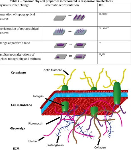
The ECM is a complex protein based hydrogel. Some of the main components that constitute the ECM are shown at the bottom of Figure 1. The ECM’s main constituents are collagens, a diverse family of proteins whose helical fibrils provide structural support and binding sites for other proteins.20 Through the formation of elastic fibres the protein elastin provides elasticity as well as structural support to the ECM.21 Proteins such as fibronectin, vitronectin and laminins display binding sites for other proteins, including growth factors and adhesion molecules from the cell membrane.20,22
The interaction of cells with the ECM is mediated by membrane receptors. One class of such receptors are integrins (see Figure 1), heterodimeric membrane proteins composed of α- and β-subunits whose specific pairing determines the affinity of the integrin to ligands presented by ECM proteins.23 One of the most studied ECM ligands is the peptide sequence RGD that was first identified in fibronectin.24 While RGD is possibly the most well known integrin binding motif, other integrin binding peptides have been reported, among them PHSRN, REDV and LDV in fibronectin and IKVAV, YIGSR and PDSGR in laminin.19
In addition to membrane proteins, membrane bound carbohydrate conjugates also contribute to the interaction of cells with their surroundings. The carbohydrate conjugates (glycoproteins, glycolipids and proteoglycans) in the cell membrane form a layer of varying composition and thickness around the cell’s exterior, the glycocalyx, that is indicated by the proteoglycans shown in Figure 1.25 Through these membrane bound carbohydrates, cells are able to establish connections with other cells and the ECM.
4
This dynamic behaviour is an integral part of a functioning biological system and underlies essential biological processes including stem cell differentiation and cell migration26–28 as well as the development of diseases.7
Cells do not only respond to cues presented by their environment, they also actively remodel it.3Through exocytosis and other pathways cells secrete molecules that reshape the ECM and interact with other cells. Prominent examples are matrix-metalloproteinases (MMPs), cell secreted enzymes that degrade the ECM through proteolysis.29 Other cell-secreted proteins such as collagen and fibronectin contribute to the construction the ECM and thus influence ECM structure and composition.30
2.1.2. Cell adhesions
Adhesion dependent cells rely on the formation of cell adhesions (mechanisms by which the cell can anchor itself to its surrounding) to survive and function. Understanding how cell adhesions form and mediate communication between the cell and its environment is essential to designing and engineering functional biointerfaces. It should be noted that most insight regarding the formation of cell adhesions stem from investigations on 2D surfaces; it has been suggested that the role of cell adhesions is different in a 3D cell culture environment.31
The formation of cellular adhesions has been described in four distinct stages, the surface recognition, the early attachment stage, the intermediate attachment stage and the late adhesion or cell spreading stage.6 These ultimately result in the formation of mature adhesions (molecular complexes referred to as focal adhesions) through which integrins establish connections between the cell and their surroundings. These processes are dynamic and reversible. Cell migration, for example, requires the controlled assembly and disassembly of focal adhesion complexes to enable the cell to make and break adhesions with its surrounding.32,33
The cytoplasmic components of integrin molecules are connected to the actin cytoskeleton via intermediate proteins such as talin.34 Focal adhesion complexes thus contribute to both the formation and regulation of cell-material interactions and translate information about mechanical, chemical and topographical cues from the extracellular to the intracellular environment.35 The properties of the cytoskeleton affect essential cellular processes such as elasticity, migration, division and differentiation. The focal adhesion mediated connection between the cytoskeleton and the local cellular environment provides a direct link between the cell and the ECM.32
2.2.Biointerface properties
5
When brought into contact with live cells, the material surface is rapidly coated by proteins.4 Cells therefore rarely interact with the material surface directly, the cell-material interaction is typically mediated by a surface adsorbed protein layer.36,37 The composition and properties of the protein layers adsorbed onto a surface is determined by a multitude of factors such as solution protein composition and concentration, protein size and the specific protein-surface interaction.38 On cell adhesive surfaces, the resulting protein layer typically includes important cell adhesion promoting proteins such as fibronectin, vitronectin, laminin and fibrinogen.36
The cell response to a surface strongly depends on the properties (type, concentration, distribution and motility) of the surface adsorbed proteins30,39. By modifying the substrate surface properties (e.g. chemistry, topography), 36,40–42 the nature of the protein layer and thus the response of cells to the biomaterial can be controlled.43,44
Three surface properties have been identified as main determinants of cell behaviour and cell fate: surface chemistry, surface topography and surface elasticity or stiffness.5 The latter two are both physical characteristics and will therefore be classed together in this review. While we will discuss these surface properties separately, they are not mutually exclusive and various combinations of them may give rise to synergistic or different biological responses.
2.2.1. Chemical
Variations in the chemical composition of material surfaces have been shown to affect a broad range of cell properties, including adhesion45–48, spreading46,49, migration46,50, proliferation51 and differentiation.51–53 Cell responses have been related to both specific (e.g. specific molecules or molecular structures) and non-specific (hydrophilicity/hydrophobicity, charge) surface chemical properties.
Non-specific chemical properties such as charge46 and wettability46,54 have been shown to affect cell adhesion55 or phenotype and functionality of mesenchymal stem cells.56. As the surface of cells is typically negatively charged57, the presence of positive surface charges often promotes cell adhesion. Similarly, moderately hydrophilic molecules also tend to promote cell adhesion to a surface.55 In particular cell adhesion on surfaces containing amine functionalities has been shown to be increased when compared to other functionalities such as carboxyl (negatively charged), methyl (hydrophobic), and hydroxy (neutral and hydrophilic) groups.58
Biomolecules can be immobilised on a surface to exploit the specific interaction of cell-surface ligands with the ECM. Immobilised proteins (e.g. laminin), peptide sequences (e.g. RGD) and carbohydrates (e.g. galactose) have been used to control cell behaviour.47,59 The fibronectin derived peptide sequence RGD has received particular attention to promote integrin mediated cell adhesion to artificial surfaces.60
2.2.2. Physical
6
respond to them.62 Micron-sized features that match the dimensions of whole cells may not be recognised by cell-surface receptors34, whereas nanoscale features that are similar in size to cell receptors have been shown to have a major impact on the response of cells to a surface.34For example, topographical features with several tens of nanometres in depth can affect cell adhesion63,64 and differentiation.65
It is not only the feature size that affects cell response, the way nanoscale features are organised on a surface equally impacts cell fate. Disordered arrangements of circular pits (100 nm deep, 120 nm diameter) caused increased osteoblastic differentiation of mesenchymal stem cells compared to ordered patterns (square or hexagonal alignments) of pits with the same dimension.61
Besides surface topography, matrix elasticity (or stiffness) of a material has been recognised as another physical property that directs stem cell fate.66 The lineage of mesenchymal stem cells was shown to correlate with variations in the stiffness of the material. The differentiated cell type obtained on a material with a particular stiffness corresponded to the natural stiffness of the relevant tissue.
2.3.Biomaterial evolution
Since the emergence of biomaterial research more than 50 years ago, the definitions and requirements for a biomaterial have changed considerably;67–69 Figure 2 shows a list of biomaterial classes and the interactions they undergo with cells and external environments. Initially, the focus in biomaterial selection was on biocompatibility, with the goal to completely suppress or reduce cell-material interactions to minimise undesired responses (toxicity, inflammation) of cells or tissue to the material.70 An improved understanding of cell-material interactions led to the design of bioactive (or instructive) biomaterials that engage with the host tissue and positively enhance material integration.71 An on-going need to improve the performance of biomaterials and mimic the dynamic processes within the ECM more closely led to the emergence of responsive biomaterials (also referred to as smart biomaterials) that are able to change their properties dynamically – and in some cases reversibly – in the presence of an external stimulus.67,72 In this case, information flow proceeds from the environment through the material to cell. While not strictly part of this review, it should be noted that the inverse process, where information about the cell is collected by the material and transduced to an external processing unit is the key concept of biosensors.
7
other system. For the design of interactive interfaces, we distinguish two types. If external stimuli are able or indeed required to control the interfacial interactions, it is externally regulated. In contrast, autonomous interfaces are able to perform their function completely self-sustained and independent from external factors.
It is becoming clear that the next generation of biomaterials should include significant elements of interactivity with the surrounding cells to provide increased biomaterial functionality through a more seamless integration of the artificial materials with the biological surrounding. While this development has already begun for bulk materials, the design of biologically responsive surfaces able to provide an interactive cell-material interface (interactive biomaterials) will present major challenges and opportunities.
3. Designing responsive biointerfaces
There are three main material properties that have to be considered for the fabrication of a responsive biointerface. These are indicated by the three components in Figure 2. Firstly, the material has to elicit a predefined biological response in a dynamic manner. Secondly, the biological response of the material has to be linked to a controllable change in the surface properties of the material. Thirdly, these dynamic surface properties have to respond to specific stimuli in a predefined fashion. These three surface properties have to be causally connected to produce the chain of events that leads from a stimulus triggered change in surface properties to the induction of differential biological responses. Ultimately, these events aim to mimic the dynamic elements of natural biological processes.8,77
3.1.Biological response
From a biological perspective, the material surface involved in the generation of a biointerface has to provide chemical and physical cues that promote both general and specific biological responses, not necessarily simultaneously but in a temporally or spatially controlled manner.
Like biocompatible and bioactive materials, responsive biointerfaces have to be non-toxic and support cell adhesion and survival in at least one of the material states that the surface can adopt. A number of biocompatible materials including collagen, alginate, poly(glycolic acid), poly(lactic acid) and poly(lactic-co-glycolic acid) are known and have been approved by the FDA for clinical applications78 and many others are used in biomedical research to study cell material interactions and develop new biomedical materials.
In addition to the general material properties, the material surface properties have to be able to adapt two different states that elicit differential cell responses, effectively mimicking the extracellular environment by changing the properties of the artificial cell environment at a specific point in time or space.8 Dynamic control over a variety of cell responses would be desirable. Control over cell adhesion and/or survival on a surface can be used to aid biomaterial integration in a living tissue,79 detach laboratory cell cultures for further processing80 or control spatial cellular organisation to create more complex artificial tissue.81 Stimulated cell migration and polarisation may also aid the design of more complex tissue constructs and control the population of porous 3D objects with cells. Materials that control stem cell fate, including proliferation and differentiation, by recreating the stem cell niche in
8
and interaction of a material surface with a number of other cellular processes, including cell division, cell-cell communication and metabolic processes may have interesting biotechnological and biomedical applications.
3.2.Material response
To present surfaces with different biological properties within the same biointerfacial environment, the material surface has to be able to change some of its physical or chemical properties in response to the presence of a stimulus. These dynamic surface changes occur in a time dependent manner but may additionally be spatially restrained.
The stimuli induced material changes can affect generic surface properties such as wettability82–84 or charge85, for example by changing the ionisation state or morphology of the material surface. To mimic the dynamic variation of the display of biomolecules such as cell adhesion receptor ligands in the ECM and modulate more specific cell-material interactions, the presentation of biomolecules on material surfaces may also be varied in response to a stimulus.74,86–89Finally, to mimic the ability of the ECM to dynamically change its structure, surfaces with stimuli induced changes in topography and elasticity are desirable.90–93
Stability within the complex biological environment is another important aspect and is of major importance if long-term bio-interfaces are to be established.37 Enzymatic degradation, non-specific hydrolysis, protein adsorption and other biological processes may affect the stability and performance of the surface over time. The ability of the material surface to withstand these effects has to be tuned to match the intended biological application.
3.3.Stimuli
To enable control over the material and cell response, the change in the material surface properties has to be linked to the presence of a specific triggering event (stimulus). The choice of an appropriate stimulus depends on the application requirements, the ability to match a particular material change to a specific stimulus and the response characteristics of the material/stimulus interaction.
9
To enable a specific, targeted interaction, the material response has to be matched to a specific triggering event and avoid non-specific material responses to other factors. Non-natural, externally applied stimuli such as light of specific wavelengths97 and electrochemical potentials98 can be matched specifically to a particular material response, while biomolecules that are used as stimuli can be expected to be less specific. The cellular environment is very diverse and able to provide biomolecules that could trigger a material response.99 While this provides opportunities to design cell-responsive biointerfaces, it also presents challenges as biomolecule responsive materials may respond less specifically than other responsive biointerfaces. In particular enzymes100 such as proteases may cause undesired responsiveness or degradation of peptide based interfaces.37
The response characteristics of the material to the stimulus are varied; stimulus exposure times, on/off times of the material response and reversibility of the response determine the performance of a responsive biointerface.101 The speed at which a material property can be switched depends on both the speed at which the stimulus is made available and on how fast the material responds to these changes (on/off time). Some stimuli such as light provide extremely fast exposure times but can be limited by the speed of the molecular rearrangements on the surface.102 For irreversible stimuli responses (e.g. photolytic or enzymatic degradation) only the ‘on’ response time has to be considered. For reversible materials, in principle two scenarios are possible: a sustained response wherein the surface change only persists for the duration of the presence of the stimulus (e.g. pH or temperature) and permanent response that persists even after the stimulus (e.g. enzymes, electrochemical potentials) has been removed. These differences may not always be strictly applicable as some materials such as photoresponsive molecules (e.g. azobenzene, spiropyran) may undergo spontaneous transitions back to the thermodynamically stable state over a prolonged period of time even without any additional stimulation.103,104
Finally, the action of a stimulus can be universal, affecting the whole interface, or locally constrained at specific areas of the material. The nature of the stimulus typically determines the range of its effect. If general changes in the environment such as pH and temperature are employed as trigger events, localisation is generally not possible. Other soluble factors (biomolecules) are also difficult to focus spatially. Microfluidic devices and electrode arrays provide strategies to operate some stimulus exposure events on a moderately local scale105,106. High spatial resolution of stimulus application can be achieved with light97.
The choice of the stimulus is important as it imposes specific constraints on various aspects of the properties of the responsive interface; the chosen stimulus therefore has to be well matched to the intended application.
4. Engineering responsive biointerfaces
10 4.1.Stimuli
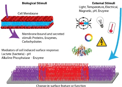
To approach the interactive qualities that define the natural interactions between cells and the ECM with a man-made biointerface, it is necessary to control not only the way material surface respond to stimuli, but the stimulus mechanisms itself has to be carefully designed and incorporated into the interface. Several comprehensive reviews exist that discuss the types of stimuli used to design responsive materials and surfaces.7,9–16 Here, we will briefly discuss the stimuli used thus far to design responsive biointerfaces in the context of the origin of the stimulus as illustrated in Figure 3, as this determines the material’s scope for applications in regenerative medicine.
4.1.1. External stimuli
In the present context, external stimuli will be defined as stimuli that originate from outside the biointerfacial environment by an event in the wider surroundings that is decoupled from the biological system. As this comprises signals that are readily controlled by a human or machine, it is the most prevalent way that surface responses have been triggered to date. Figure 3 shows a representation of external stimuli; artificial changes in temperature,91,92,107 the application of an electrical potential,85,108–110 the application of light87,88,111,112 and mechanical forces93,113 have all been used to trigger a dynamic change in either chemical or physical surface properties to affect the behaviour of cells. In addition, biomolecules that are naturally present in a biological environment – enzymes74,76 and carbohydrates89 for example – have been artificially added to systems to trigger a response of the biointerface.
Thermoresponsive materials used for biointerfaces are typically polymers with a temperature dependent variation of the miscibility of the polymer chains with a solvent (e.g. water). A change in temperature results in a change in the hydration state of the material.114,115 If applied as surface films, this will affect the wettability and the morphology of the polymer film. Typical examples of temperature responsive materials are poly(N-isopropylacrylamide) (PNIPAM)116 and elastin derived polypeptides.117Other materials such as poly(ε-caprolactone) were used as shape-memory materials to prepare surfaces with dynamically changing topographical features.91
pH responsive biointerfaces are mainly based on poly(acrylic acid) (PAA) and poly(methacrylic acid) PMAA.118,119 They undergo changes in surface charge and water content similar to thermoresponsive surfaces. Integration of these materials with high aspect ratio topographical features allows the use of pH responsive gels to reversibly modulate the orientation of surface topographical features.90 In these instances, the swelling and contraction of the hydrogel exerts mechanical forces on the topographical features and thus places them under mechanical strain, causing them to bend.
Electrical potentials have been more widely used to alter biointerface properties; their effects range from reorganisation of the molecular conformation of molecules on the surface85,98,109 to the induction of chemical reactions that modulate the presence of surface chemical groups.110,120–122 These materials rely on the presence of an electrically conducting substrate material.
host-11
guest complexes with α-cyclodextrin was also exploited to be able to reversibly attach chemical functionalities to a α-cyclodextrin modified surface via self-assembly processes.88
Magnetoresponsive materials have been employed to modify cell-surface interaction through the incorporation of magnetic nanoparticles into a polymeric hydrogel matrix. Application of a magnetic field caused distortion of the hydrogel surface, resulting in an alteration of the mechanical properties of the surface of the hydrogel.124
Mechanoresponsive biointerfaces were prepared from polymer films that alter physical properties such as porosity and topography of the surface. While the former was used to modulate the availability of biomolecules to the interface125, the latter could be employed to reversibly present topographical patterns on the surface.93
4.1.2. Biological stimuli
For the present purpose, biological stimuli are defined as stimuli that can be provided by a natural cellular environment. As indicated in Figure 3, these stimuli can be either soluble factors that are secreted by cells into their local environment, or they can be molecules immobilised in the cell membrane. Even though, for technical reasons, most biological stimuli are currently supplied externally, they do have the potential to interact directly with cells and dynamically respond to changes in the biological environment, thus moving closer towards the design of an autonomous, interactive cell-material interface.
It is well established that biological stimuli can affect bulk material properties. Notably, it was shown that acrylate based hydrogels that contain peptides as cross linkers could be degraded by cell secreted matrix metalloproteinases.73 Recently, first evidence has been reported that cell secreted enzymes (alkaline phosphatase) may also alter the properties of phosphorylated peptide surfaces.76 As an alternative to enzymes that can act with high specificity and selectivity, the triggering of a surface response through cell mediated changes of a bulk property (pH) is also possible.126 These strategies now open up the possibility to design cell responsive biointerfaces in analogy to the already existing cell responsive bulk materials.
Biomolecule responsive surfaces have been prepared to interact with carbohydrates, peptides or enzymes. Carbohydrate interactions typically occur through the formation of reversible bonds with surface bound boronic acids89 while peptides have been attached to complementary peptide strands that are immobilised on a surface via non-covalent interactions;86,127 in both cases, competitive replacement by the stimulus (another carbohydrate or peptide) causes a change in the chemical surface composition. Nucleotide and carbohydrate responsive materials have been shown to modulate the wettability of a surface.83,84 Enzymes have been used to alter the surface chemistry by irreversible cleavage of specific covalent bonds.74,76
4.2.Material response
12 4.2.1. Dynamic chemical properties
Surface chemistry is a major determinant of cell-material interactions. As the chemistry presented by the ECM to the cell is not static (for example, the protein composition, conformation and structure is subject to continued change), the ability to dynamically modulate biologically relevant chemical cues at the interface between the cell and a man-made material is attractive to mimic biological behaviour.
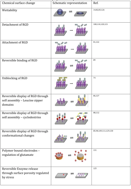
Biointerfaces with dynamic surface chemistries have been obtained by changing surface wettability and by modulating the availability of specific chemical groups on the surface. The latter mostly focuses on the presentation of RGD based peptides on the surface, but modulation of the surface presentation of some other biomolecules has also been reported. An overview of the strategies discussed here to induce chemical surface changes in a biointerface is given in Table 1.
4.2.1.1.Wettability
The wettability of a surface can be altered in a responsive and reversible manner by controlling the water content of a polymer film. The degree of swelling (and hence the water content) of polymers such as pNIPAM or PMAA can be altered by changing environmental properties such as temperature or pH.107,126 The change in environmental conditions affects the ability of the polymers to undergo intramolecular interactions and thus modulates the amount of water that can be taken up within the polymer film.14 The change in water content subsequently alters the wettability of the polymer surface. This stimuli responsive surface wettability can be used to modulate the cell adhesive properties of the material and has found prominent use for the fabrication of cell sheets. 80,107
In addition to pH and temperature, biomolecules can also be used to change the wettability of a polymer surface. A reversible change of surface wettability from a super-hydrophobic (water contact angle of 150o) to a super-hydrophilic state (water contact angle of 0o) was accomplished using a multi component polymer film system consisting of either a nucleotide responsive co-polymer of PNIPAM, phenylthiourea and phenylboronic acid83 or a carbohydrate responsive co-polymer of PNIPAM and 3-(acryloyl thioureido) phenylboronic acid.84
4.2.1.2.Presentation of RGD
13
Detachment. The detachment of RGD based peptides from a surface has been accomplished using electrical potentials as stimuli. Direct desorption of RGD terminated alkanethiols from gold and ITO surfaces has been accomplished by applying electrical potentials to the interfaces in the presence of cells (fibroblasts).108,110 This resulted in the removal of the complete thiol-peptide conjugate from the surface, causing detachment of individual cells, spheroids and cell sheets. An alternative to the desorption of the full peptide conjugate is the cleavage of a redox sensitive linker between the peptide and the surface tether.121,128 Quinones can be reversibly converted into hydroquinones through application of an electrochemical potential. Derivatives of these molecules bearing peptides through ester or silyl bonds can be electrochemically induced to cleave the link with the peptide and release the peptide from the surface, removing the cell adhesive ligand form the surface and causing cell detachment.
Attachment. Quinones are able to undergo Diels-Alder reactions with cyclopentadiene, such that peptides conjugated to cyclopentadiene can be immobilised on a quinone surface under physiological conditions. With a hydroquinone surface, this reaction can be triggered on demand via an electrical stimulus that causes the oxidation of hydroquinone to quinone.122 This approach was used to dynamically change the cell adhesiveness of a patterned surface. Adhesion of fibroblasts cultured on a surface that was patterned with fibronectin (a cell adhesion promoting protein) was originally constrained to the protein pattern. In situ activation of the surrounding surface via electrically induced quinone formation enabled attachment of peptide-cyclopentadiene conjugates in the presence of cells, rendering the whole surface cell adhesive and allowing the fibroblasts to populate the complete surface122,128. Although not strictly performed in a stimulus responsive manner, it should be mentioned that a similar strategy was used to control the position of two different cell types on a surface. On an azide terminated surface, patterns of fibronectin were created for the adhesion of the first cell type.81 The azide surface can undergo a catalyst free cyclo addition with a cyclo-octyne bicyclo[6.1.0]nonyne-peptide conjugate (a click reaction), rendering the remaining surface cell adhesive and allowing spatially defined culture of a second cell line on the surface.
Reversible covalent binding. Covalent bond formation can also be used to reversibly modify a surface with RGD sequences. The diols present in carbohydrates have a high affinity for boronic acids and attach to them through the reversible formation of boronic esters. Hence a carbohydrate containing polymer conjugated to an RGD based peptide can be immobilised onto a surface that displays polymer brushes decorated with phenylboronic acid.89This cell adhesive surface was shown to support the adhesion of MG63 cells which could subsequently be removed from the surface by the addition of carbohydrates (glucose or fructose) that competitively displace the carbohydrate polymer and remove RGD from the material surface.
14
fluorenylmethyloxycarbonyl (Fmoc) group.74 Osteoblasts did not adhere to the Fmoc-terminated peptide surface, but when the blocking group was removed by enzymatic (elastase) cleavage of the terminal alanine-alanine sequence, the RGD sequence became accessible and cell adhesion was possible.
Reversible display through self-assembly. The chemical surface changes discussed so far were focused on the modification of covalent bonds and therefore typically non-reversible. The use of carefully designed self-assembling materials enables the design of surfaces with reversible chemistries. Mimicking the ability of proteins and natural peptides to partake in specific, non-covalent interactions that form supramolecular structural motifs, it is possible to custom design polypeptides with specific amino acid sequences that form heterodimeric complexes, so-called zipper molecules. If one of the complementary polypeptide zipper molecules is terminated by an RGD sequences and attached to a surface, dimerisation with the second zipper sequence enables non-covalent, reversible modulation of the surface chemistry.86,127 If the complementary strand is conjugated to a PEG chain, dimerisation will cause the RGD sequence on the surface to be masked by the PEG, rendering the surface non-cell adhesive and causing detachment of fibroblasts. Cell adhesiveness of the surface can be restored by adding unmodified complementary zipper sequences that competitively replace the PEG conjugates.86
A different self-assembly approach to reversibly trigger the display of an RGD based peptide exploits host-guest chemistry wherein a smaller molecule is embedded within a larger, cage-like molecule to form a supramolecular complex. Azobenzene is a photoresponsive molecule able to partake in such a host-guest interaction with α-cyclodextrin when in its trans state; if the conformation of azobenzene is changed to its cis state (through exposure to light), the resulting conformational change prevents azobenzene from entering the cavity in α-cyclodextrin. By coupling GRGDS to azobenzene, it is therefore possible to modulate the chemical functionalization of an α-cyclodextrin surface in response to light.88,112 When azobenzene is in its trans state and attached to the surface, the displayed RGD sequence enables attachment of HeLa cells. Irradiation of the peptide modified at 365 nm breaks the supramolecular complex, removing the peptide sequence form the surface and causing cell detachment.88
15
Conformational surface changes to alter surface chemistry have also been induced by applying an electrical potential to the surface. It was shown that self-assembled monolayers (SAMs) can be reoriented by applying a electrochemical potential to the surface that attracts the charged endgroups of the SAMs to the substrate surface, thus changing the charge as well as the chemical functionalities displayed at the surface.129 This principle was employed to control protein (neutravidin)98and bacterial (M. hydrocarbonolasticus)85attachment to a surface, notably not by modulating the display of RGD based peptides but by changing the charge and/or hydrophilicity of the surface. To control the display of a peptide (GRGDS) on a surface, the same electrochemically controlled mechanism was employed in a mixed SAM that contained peptide conjugates and molecules with charged endgroups (sulfates and tertiary amines).109 In the absence of an electrical potential, the charged molecules were extended and shielded the peptides from cells cultured on these surfaces, preventing cell adhesion. Application of an electrical potential caused the charged molecules to reorient towards the surface, making the peptide available for cellular interaction and promoting the adhesion of endothelial and differentiated HL60 cells.
Even though not reversible per se, it should be noted that electrochemical potentials can also be used to repeatedly change the cyclisation of a peptide sequence. A surface bound peptide sequence terminated with an oxyamine can undergo electrochemically stimulated cyclisation with surface bound hydroquinone similar to the reaction discussed above.130 Electrochemical reduction of the resulting oxyimino-quinone leads to the opening of the cyclic structure. This surface is therefore able to switch from a linear to a cyclic and then back to a linear peptide structure and it was shown these structural changes affect the spreading and migration of fibroblasts.
4.2.1.3.Presentation of other biomolecules
The importance of integrins in cell adhesion and cell signalling processes has without doubt contributed to the prevalence of dynamic surfaces that target RGD as chemical modifier for the properties of the biointerface. Here we shall shortly discuss the control of the availability of two other biomolecules that are essential in the regulation of cellular processes; the neurotransmitter glutamate and the enzyme alkaline phosphatase.
In an attempt to design an artificial synapse that is able to interface with a biological counterpart, polypyrrole based molecular imprinted polymers were prepared on an electrode surface to act as reservoir for glutamate.131 Glutamate possesses an overall negative charge under physiological conditions; by altering the electrostatic potential of the polymer coated electrode, it was shown that glutamate could be dynamically released and bound to the polymer on demand. So far, this approach is in a concept stage and has not yet been realised in an interface with an actual biological system.
16
system, it does display traits that are similar to essential biological processes employed by cells to interact with their environment.
4.2.2. Dynamic physical properties
It is now well established that the influence of both topography and stiffness of the extracellular environment are essential in determining how cells interact with their surroundings. Considerable effort has been placed in designing 3D structures, in particular hydrogels, to control these physical parameters dynamically in materials suitable for cell culture and thus advance our ability to determine cell fate. While some of these bulk hydrogel materials may be transferrable as films onto other substrates to generate biointerfaces, surfaces with in situ tuneable topography and/or stiffness are attractive for example for in vitro control of stem cell fate or neuron guidance. An overview of the approaches discussed below is given in Table 2.
4.2.2.1.Topography generation
Grooves on the surface of cell culture substrates have been used extensively to achieve directional alignment or migration of cells.12 Reversible formation of micrometer sized grooves can be accomplished by compressing an oxidised polymer film (Epo Tek, a propriety material) that was supported on PDMS.93 The formation of topographical features caused the orientation of C2C12 myoblasts along the grooves; markedly, this cell orientation was reversible, the cells took up a random orientation again after the topographical features were removed. A similar effect was observed when mesenchymal stem cells were cultured on surfaces with reversible micron-scale groove topography.91 These materials were prepared by creating memory shape effects into a poly(ε-caprolactone) surface. Transition from one shape to another was accomplished in a thermoresponsive manner by exposing the material to 40°C for 10 min. The reversible formation of nanosized topographical features on a surface has in principle been demonstrated with a conductive polymer film.132 Nanosized features were written into an electrodeposited polybithiophene film with the aid of an atomic force microscopy (AFM) tip. These features could be smoothed out by electrochemically oxidising the polymer film. The ensuing uptake of counter ions (perchlorate) caused the polymer film to swell and smooth out the nanoindentations. Whilst this work has not yet been applied in a biological context, it demonstrates the feasibility to dynamically control topographical features of biologically relevant dimensions.
4.2.2.2.Topography reorientation
17
Furthermore, by using asymmetric topographical structures or by patterning the hydrogel in specific areas on the surface, unidirectional and locally restricted reorientation of the surface microtopography was accomplished.90,135 These topographical changes are reversible and repeatable and the ability to tune the stimulus response by careful design of the hydrogel film makes it attractive for applications in a biological environment.
4.2.2.3.Topographical shape
The memory shape effect mentioned above to form or remove topographical features on a surface can also be used to realize shape transitions. By first imprinting a poly(ε-caprolactone) surface with a primary shape during cross linking and then imprinting a secondary shape under mechanical strain at high temperature followed by cooling under strain, two separate topographies can be imprinted within the same surface.91 It was shown that this procedure can be used to reversibly switch between these two micron-sized shapes (e.g. between hexagons and squares or L-shapes and circles) under conditions amenable for cell culture.
4.2.2.4.Topography and stiffness
The temperature responsive properties of poly(ε-caprolactone) can also be used to simultaneously modulate the overall stiffness and topography of a surface. By varying the degree of cross-linking within the polymer film, it was shown that the temperature responsiveness can be tuned such that the stiffness / roughness transition at a biologically acceptable temperature (33°C).92 Below the transition temperature, the material is relatively stiff (50 MPa) and displays a rough surface; elevation of the temperature above 33°C reduces the stiffness (1 MPa) and produces a smooth surface topography. It was shown that this transition caused myoblasts that were cultured on the poly(ε-caprolactone) surface to take on a round morphology and eventually detach from the surface. In contrast, fibroblasts showed a much weaker response to physical changes of the surface properties, with only 20% of the cell changing their morphology.
Magnetoresponsive hydrogels are another way of eliciting reversible transitions in physical surface properties. Magnetic particles incorporated in a hydrogel (2-hydroxy-ethyl-methacrylate, ethylene glycol dimethacrylate and styrene maleic anhydride copolymer) enable distortion of the hydrogel surface if placed in a magnetic field. Due to the magnetic forces acting on the entrapped particles, the hydrogel is placed under mechanical strain that results in a distortion of the material surface as well as a change in the stiffness of the hydrogel. Mesenchymal stem cells cultured on these hydrogel surfaces have been shown to differentiate in to cartilage in response to the magnetoresponsive change in physical properties of the hydrogel.124
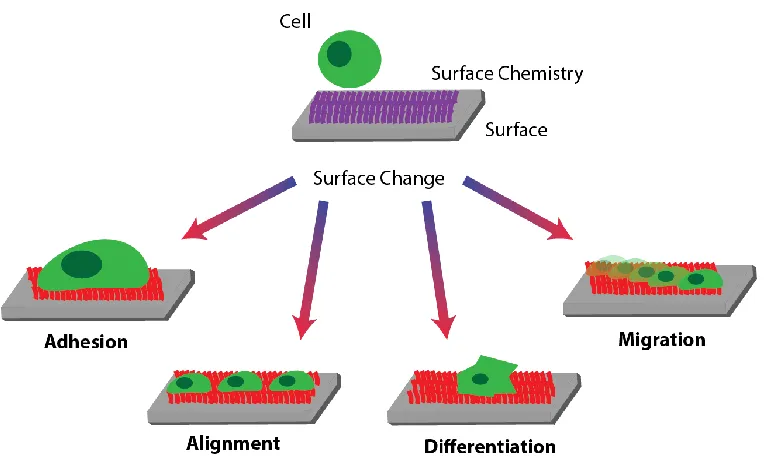
4.3.Biological response
18
that have already been observed as a result of a dynamically changing surface property are captured in Figure 4 and will be discussed shortly in the following sections.
The most prevalent biological response that has been reported for responsive surfaces is the modulation of cell adhesion. By dynamically changing the presentation of RGD based peptides on the surface, the adhesion of osteoblasts,74,87 fibroblasts,86,108,110,113,123,127 endothelial109 and HeLa cells88,112 has been reported. In several cases, cell adhesiveness of the surface could be changed repeatedly and reversibly.86–88,109,111,112,127 In analogy to the pNIPAM based thermo-responsive surfaces that were designed to harvest cell sheets,136,137 many of these cell-adhesion modulating surfaces were put forward as alternative routes to harvest cells. Notable other applications focus on the ability to spatially control the adhesion of cells to enable co-cultures of different cell types in predefined patterns81.
Another biological response that was achieved on dynamic surfaces is the alignment of mesenchymal stem cells91, and myoblasts93 to dynamic micron-sized topographical features. Cell alignment is important for example for neuronal networks and muscles; reversible cell alignment at an interface may therefore be attractive as biointerfacial actuators or to support directional growth of tissue.
The differentiation of mesenchymal stem cells into cartilage was accomplished on a hydrogel surface after altering the surface shape and stiffness by exposing the material to a magnetic field.124 Finally it was shown that the spreading and migration of fibroblasts can be changed in response to dynamic changes in the conformation of peptide sequences presented on a surface.130
It is evident that the progress made in static surfaces to control specific cellular responses has not yet been translated into dynamic surfaces to the same extent. While we build our understanding on which surface properties modulate particular biological responses, it will be necessary to consider how these properties can be incorporated into biointerfaces in a dynamic manner. This will allow us to approach the dynamic and complex interactions of natural biological interfaces and enable advanced regulation of cell-material interactions for regenerative medicine applications such as stem cell therapies or implant devices.
5. Current developments
Most literature on responsive surfaces for biological applications focuses on flat, 2D surfaces that respond to one type of stimulus which is externally provided. These surfaces have already shown great promise in the dynamic control of cell behaviour; however, they are still far removed from the complex interactions that take place between cells and the ECM. In biology, several stimuli interact with both the cell and the ECM in a dynamic and interdependent manner within a complex 3D structure. Some progress has been made to incorporate some of these aspects in an artificial interface, as well; these advances will be reviewed here and discussed in context of the design of a new generation of biointerfaces.
5.1.Multi stimuli responsive materials
19
mechanisms. Simplification and isolation of single responses enabled us to design surfaces that mimic these interactions on a rudimentary level; however, it does ignore any potential synergistic effects that arise from the multi-responsive nature of cell-ECM interactions. To advance artificial cell-material interactions, it will be necessary to design multi-stimuli responsive surfaces.5
The concept of multi-stimuli responsive materials is emerging as one of the coming challenges for the responsive materials community. A recent review discussed progress towards multi-responsive polymers and highlights a number of multi-responsive systems that have been reported over the last decade.138 These materials typically consist of block- or copolymers of two or more different monomers that display responsiveness to different stimuli. Notably, it is possible to design multi-responsive surfaces that convert the input signal (initial stimulus) to a meaningful output signal. A triple stimuli responsive random copolymer based on pH sensitive N,N-Dimethylaminopropyl acrylamide (DMAPAM), temperature responsive N-t-butylacrylamide (NTBAM) and solvent polarity sensitive 4-N-(2-acryloyloxyethyl)-N-methylamino-7-N,N-dimethylaminosulfonyl-2,1,3-benzoxadiazole (DBD-AE) showed a change in fluorescence (light response) due to a pH and temperature induced alteration of the polarity of the copolymer.139
Multiple stimuli responsive polymers should, in principle, be transferrable onto a solid substrate to prepare multi-stimuli responsive surfaces. Only few such surfaces have been reported, in particular for biological applications. Dual stimuli responsive surfaces have been prepared as copolymers of NIPAM (temperature responsive) and acrylic acid or methacrylic acid (pH responsive) via surface initiated polymerisations.118,119 The materials were characterised in terms of wettability118 and swelling characteristics119, displaying changes in these properties in response to either temperature or pH. Biological applications of such materials remain to be demonstrated.
There is a clear application potential for surfaces that respond to multiple stimuli in a biological surrounding to explore synergistic effects of the interaction with a more complex system. In addition, it would also be attractive to design materials with multiple responses to mimic the versatility of the ECM more closely. Multi-responsive surfaces that respond to multiple stimuli orthogonally, e.g. produce two different responses to two different stimuli would bring us significantly closer to approaching more complex cell-material interactions.
5.2.Responsive surfaces of 3D materials
The term ‘surfaces of 3D materials’ includes both the surfaces of materials with topographical features and the internal surfaces of porous, 3D objects. Both represent significant challenges in terms of surface modification and characterisation but are of interest as materials for regenerative medicine because they provide environments that, to a degree, mimic structural features present in the ECM in addition to presenting biologically relevant chemical cues.
20
Solid 3D scaffolds are widely used as supports for tissue engineering applications. Modification of the internal surface of such 3D structures is attractive to create responsive 3D structures that simultaneously simulate the chemical and structural dynamics of the ECM.141 A step towards this is the release of bacterial inclusion bodies through biodegradation in polymer matrices made from poly(caprolactone), poly(lactic acid) or chitosan.142 Even though this example relies on degradation and thus may not fully fit with the specific stimuli/response interactions of materials discussed here, it highlights the possibility of addressing the responsiveness of surfaces in porous 3D materials.
While a variety of surface modification approaches for 3D materials exist,143–145 uniform surface modification of internal 3D structures is not straightforward and compounded by limitations regarding the analysis of the surfaces of complex 3D structures. This makes the preparation of responsive 3D surfaces challenging. Recent advances in label free, chemical 3D analysis146,147may address some of these issues and we anticipate that the emergence of methods capable of characterising internal surfaces of 3D structures will contribute significantly to the development of strategies that enable the design of responsive surfaces on 3D structures.
5.3.Interactive biointerfaces
To fully integrate man-made materials in a biological environment, materials are required that do not only provide biologically relevant cues, but that are able to respond to biologically stimuli presented by the cells. In recent literature, the vision of interactive and more complex cell-material interfaces, i.e. surfaces that dynamically respond to biological stimuli in a biologically relevant manner, has begun to emerge6,13 and a few systems have been designed that address certain challenges towards the design of interactive biointerfaces.
In an attempt to expand responsive interfaces to a more dynamic, biology based system, bacteria have been used to modify material surfaces and generate what the authors termed a ‘living interface’ as an intermediate between mammalian cells and the material surface. The non-pathogenic bacterium Lactococcus lactis has been genetically modified to express the RGD containing fibronectin fragment FNIII7-10148 and thus affect the adhesion and morphology of C2C12 cells.149 While the bacteria-modified surface was not shown to be directly cell-responsive, one can envision that the bacteria film would be responsive to changes in the biological environment and may be able to respond to changes in its surroundings that are brought about by the mammalian cells culture on the surface.
Cell responsiveness of a surface to a stimulus provided by a cell has been accomplished by employing the formation of metabolic products as trigger event. Lactoccocus lactis produces lactic acid which changes the pH of its surrounding and is thus able to affect changes in a pH responsive surface.126 pH induced expansion of a triblock copolymer of polybutadiene, poly(methacrylic acid) and quaternized poly(2-(dimethylamino)ethyl methacrylate) consequently resulted in the self-induced detachment of the bacteria from the surface.
21
that cell-secreted alkaline phosphatase may be able to induce dephosphorylation without the need to add other external stimuli, promoting the concept of a cell-responsive material surface.
Another emerging aspect of cell-material interaction is the interface with internal cellular processes. Cell invasive techniques are well established to measure properties such as membrane potentials.150 The measurement of intracellular parameters, for example by perforating the cell membrane with carbon nanotube based electrochemical sensors,151 is increasingly attracting attention. It has been shown that nanometre sized electrodes can be seamlessly integrated into the cell membrane.152 Consequently, hollow tubes (approx. 100 nm diameter) were fabricated on surfaces and connected to a liquid reservoir.153 These tubes were inserted into HeLa and CHO cells and it was shown that fluorescent dyes could be delivered directly into the intercellular space. Combining both intracellular sensing and intracellular delivery of materials, one can envision that a combined system that may enable intracellular cell-material interfaces may be possible in the near future.
Interactive biointerfaces have great potential to advance regenerative medicine. More dynamic tissue culture platforms would allow finer control over in vitro cell cultures, enabling the fabrication of more complex artificial tissue with higher complexity and functionality. Cell responsive surfaces can be used to monitor biological processes either to design sensors or to gain an improved understanding of how cells interact with their environment and/or each other. Dynamically responsive surfaces on implants could be more seamlessly integrated within the host tissue by not only instructing the biological environment but also responding to the requirements of the host tissue. Promising advances have been made on various fronts that are likely to enable the emerging of this technology in the near future as a powerful tool for regenerative medicine but a number of challenges have to be addressed before a truly viable, interactive biointerface can be designed.
6. Future perspectives
The ultimate goal of advanced responsive surfaces that aim to interface with a biological environment must be to approach an increased level of integration of the artificial material with living cells. The nature of this integration could take many forms and will depend on the final application that the biointerface will be designed for. While mimicking even parts of the complex and dynamic interactions present in biology may seem a daunting task with present technology, it also presents a large number of exciting opportunities.
The identification of appropriate stimuli for a responsive interface is of crucial importance as it determines subsequent considerations regarding the material properties and its design. A wide variety of stimuli and material responses able to change biologically relevant material surface properties have already been explored. More frequently used stimuli such as light, electrical potentials, temperature and pH have already proven valuable to externally control biointerfaces.
22
between material surfaces and cells is likely to become instrumental in the design of more seamlessly integrated biointerfaces. An important prerequisite for this is a detailed understanding of the natural role of the stimulus-enzyme in the biological environment. The development of a much more thorough understanding of natural processes that are regulated by biomolecules, e.g. the involvement of enzymes in disease states, will be vital to the design of new generations of responsive biointerfaces.
The design of surfaces that respond to biological stimuli will require increased attention if the cell-material interaction is to be translated into a biologically meaningful response and a responsive biointerface suitable for practical biomedical applications. It will therefore be necessary to translate and expand responsive material technology that has been developed for bulk materials to the biointerface. New material surfaces modification will be required that not only respond to biomolecules (or other stimuli) but also translate this response to a measurable or functional change in either the surface or the bulk material properties.
To date, only a limited number of materials have been used to prepare responsive surfaces for biological applications, thereby possibly limiting the types of cell responses that were observed so far. As biomolecules are likely to become central to the design of stimuli responsive biointerfaces, it can be expected that biomolecule based surfaces may attract increasing interest in future due to their versatility and their ability to interact with biological stimuli.
As many biological interactions are reversible, the design of reversibly responsive biointerfaces may well make use of similar concepts, for example by using the ability of enzymes to catalyse reactions in both directions or by exploiting the use of two different enzymes that catalyse opposite reactions of the same substrates. Ultimately, this concept could lead to the generation of reversible biointerfaces in which changes in one or both directions could be made fuel dependent, e.g. they only occur in the presence of other cofactors. Such biointerfaces would represent a significant step towards mimicking the natural complexity and diversity of biological processes.
These advances can ultimately be envisioned to lead to the generation of more seamless cell-material interfaces, in which cells not only respond to cues from the surface, but where surfaces also respond to stimuli presented by cells. Such interactive biointerfaces would be highly attractive for self-controlled or autonomous biointerfaces and would open up exciting application potentials to recreate more complex cell niches in which, for example, stem cell fate can be controlled in an unprecedented temporal and spatial manner or where nerve cells can be interfaced more seamlessly with an artificial material.
For practical reasons, most biointerfaces have thus far been designed on ‘flat’ substrates and with the ability to respond to a single, externally provided stimulus. To mimic the complexity of a biological system, response to several stimuli, including those provided by a cell and modification of surfaces of 3D objects will be desirable, and strategies have begun to emerge to address these challenges.
23
A major challenge in the design of such complex and dynamic biointerfaces is their monitoring and analysis. Advances in this area will heavily rely on the accessibility of suitable analysis tools that enable monitoring of the cell-material interface with high spatial and temporal resolution in a complex surrounding, ideally in the presence of live cells. While recent advances in surface and interface analysis such as the increasingly powerful capability to perform label free 3D surface analysis of organic materials addresses parts of this challenge, further development will be necessary to access dynamic changes at the biointerface with minimal perturbance of the system.
The recent advances in the literature discussed here clearly show that the design of responsive interfaces is gaining impetus. Literature on the design of responsive bulk materials is already extensive and responsive surfaces are on the way to develop a similar toolset. Based on the considerable potential and interest in the area, we are likely to see a number of exciting developments in the near future that will advance the integration of artificial materials with cells considerably and open up a large number of new applications.
Acknowledgements
24 References
1. Custódio C A, Reis R L & Mano J F. Engineering biomolecular microenvironments for cell instructive biomaterials. Adv. Healthc. Mater. 3, 797–810 (2014).
2. Von der Mark K, Park J, Bauer S & Schmuki P. Nanoscale engineering of biomimetic surfaces: cues from the extracellular matrix. Cell Tissue Res. 339, 131–53 (2010).
3. Daley W P, Peters S B & Larsen M. Extracellular matrix dynamics in development and regenerative medicine. J. Cell Sci. 121, 255–64 (2007).
4. Roach P, Eglin D, Rohde K & Perry C C. Modern biomaterials: a review - bulk properties and implications of surface modifications. J. Mater. Sci. Mater. Med. 18, 1263–77 (2007).
5. Murphy W L, McDevitt T C & Engler A J. Materials as stem cell regulators. Nat. Mater. 13, 547–57 (2014).
6. Ventre M, Causa F & Netti P A. Determinants of cell-material crosstalk at the interface: towards engineering of cell instructive materials. J. R. Soc. Interface 9, 2017–32 (2012).
7. Kim J & Hayward R C. Mimicking dynamic in vivo environments with stimuli-responsive materials for cell culture. Trends Biotechnol. 30, 426–39 (2012).
8. Martino S, D’Angelo F, Armentano I, Kenny J M & Orlacchio A. Stem cell-biomaterial interactions for regenerative medicine. Biotechnol. Adv. 30, 338–51 (2012).
9. Holzapfel B M, Reichert J C, Schantz J-T, et al. How smart do biomaterials need to be? A translational science and clinical point of view. Adv. Drug Deliv. Rev. 65, 581– 603 (2013).
10. Higuchi A, Ling Q D, Kumar S S, et al. External stimulus-responsive biomaterials designed for the culture and differentiation of ES, iPS, and adult stem cells. Prog. Polym. Sci. 39, 1585–1613 (2014).
11. Chan A, Orme R P, Fricker R A & Roach P. Remote and local control of stimuli responsive materials for therapeutic applications. Adv. Drug Deliv. Rev. 65, 497–514 (2013).
12. Rashidi H, Yang J & Shakesheff K M. Surface engineering of synthetic polymer materials for tissue engineering and regenerative medicine applications. Biomater. Sci. 2, 1318 (2014).
13. Skorb E V. & Andreeva D V. Surface Nanoarchitecture for Bio-Applications: Self-Regulating Intelligent Interfaces. Adv. Funct. Mater. 23, 4483–4506 (2013).
25
15. Mendes P M. Cellular nanotechnology: making biological interfaces smarter. Chem. Soc. Rev. 42, 9199–9576 (2013).
16. Leal-Egaña A, Díaz-Cuenca A & Boccaccini A R. Tuning of Cell-Biomaterial Anchorage for Tissue Regeneration. Adv. Mater. 25, 4049–4057 (2013).
17. Cai L & Heilshorn S. Designing ECM-mimetic materials using protein engineering. Acta Biomater. 10, 1751–1760 (2014).
18. Maître J-L & Heisenberg C-P. Three functions of cadherins in cell adhesion. Curr. Biol. 23, R626–33 (2013).
19. Shekaran A & Garcia A. Nanoscale engineering of extracellular matrix-mimetic bioadhesive surfaces and implants for tissue engineering. Biochim. Biophys. Acta 1810, 350–360 (2011).
20. Kim S-H, Turnbull J & Guimond S. Extracellular matrix and cell signalling: the dynamic cooperation of integrin, proteoglycan and growth factor receptor. J. Endocrinol. 209, 139–51 (2011).
21. Kielty C M, Sherratt M J & Shuttleworth C A. Elastic fibres. J. Cell Sci. 115, 2817–28 (2002).
22. Ruoslahti E. Fibronectin and its receptors. Annu. Rev. Biochem. 57, 375–413 (1988).
23. Bellis S. Advantages of RGD peptides for directing cell association with biomaterials. Biomaterials 32, 4205–4210 (2011).
24. Pierschbacher M & Ruoslahti E. Cell attachment activity of fibronectin can be duplicated by small synthetic fragments of the molecule. Nature 309, 30–33 (1984).
25. Tateno H, Uchiyama N, Kuno A, et al. A novel strategy for mammalian cell surface glycome profiling using lectin microarray. Glycobiology 17, 1138–46 (2007).
26. Cavalcanti-Adam E A, Volberg T, Micoulet A, et al. Cell spreading and focal adhesion dynamics are regulated by spacing of integrin ligands. Biophys. J. 92, 2964–2974 (2007).
27. Santos E. Cell-Biomaterial Interaction: Strategies To Mimic The Extracellular Matrix.
28. Hynes R O. Extracellular matrix: not just pretty fibrils. Science 326, 1216–1219 (2009).
29. Nagase H, Visse R & Murphy G. Structure and function of matrix metalloproteinases and TIMPs. Cardiovasc. Res. 69, 562–73 (2006).
30. Frantz C, Stewart K M & Weaver V M. The extracellular matrix at a glance. J. Cell Sci. 123, 4195–200 (2010).