ACCELERATION
*,1
Rosaline Tina Paul
ARTICLE INFO ABSTRACT
New approaches to shorten treatment time with areas of
are some examples. 3.Post lag
as compression of periodontal ligament, bone deformation and to certain biochemical
compression of the periodontal ligament leads to the release messengers. Hormones
neurotransmitters such as substance P and vasoactive intestinal polypeptide
receptors present on the cell surface of target cells and initiate a process of intracellular cellular
calcium. Second messengers inside the cells is believed osteoblasts w
producing of stimulating
It involves fluxes of ions, such as Ca2+, Mg membrane
adenosine triphosphate (ATP) (cAMP) and guanosine 3’, 5’ considered to be the
target cells. All three substances serve as co acceleration by physical stimulatio
the mechanical
bone surface lining cells endothelial and epithelial helpful in
tooth movement
tooth movement .The combined effect of force differentiation
expression.
medullary bone results in a temporary burst of rebuilds
and remineralisation in phenomenon movement.
Copyright©2017, Rosaline Tina Paul et al. This is an open access article distributed under the Creative Commons Att use, distribution, and reproduction in any medium, provided the original work is properly cited.
INTRODUCTION
New approaches to shorten treatment time without foregoing optimal results, has become a primary goal of all areas of orthodontics. Low friction and self-ligating bracket systems, preformed archwires, alveolar corticotomies are some examples. (Krishnan and Davidovitch, 2009)
moved because cells around their roots are enticed by the mechanical force to remodel the tissues around them.
*Corresponding author: Rosaline Tina Paul,
PSM Dental College, India
ISSN: 0975-833X
Article History:
Received 20th December, 2016 Received in revised form 16th January, 2017
Accepted 04th February, 2017 Published online 31st March,2017
Key words:
Acceleration of tooth movement, Regional acceleratory phenomenon, Electric,
Mechanical, Magnetic, Laser, Chemical,
Hyperbaric oxygen therapy.
Citation: Rosaline Tina Paul, Dr. Ligil, A. R. and Biswas,
International Journal of Current Research, 9, (03), 47484
REVIEW ARTICLE
ACCELERATION OF TOOTH MOVEMENT
Tina Paul,
1Dr. Ligil, A. R. and
2Biswas, P. P.,
1Dr. Lijo
and
1Dr. Primal T. Francis
1
PSM Dental College, India
2Royal Dental College, India
ABSTRACT
New approaches to shorten treatment time without foregoing optimal results,
areas of orthodontics. Low friction and self-ligating bracket systems, preformed archwires, alveolar corticotomies are some examples. The phases of normal tooth movement are (Burstone
lag /continuous phase. When a force is applied onto a tooth, it results in a number of biophysical as compression of periodontal ligament, bone deformation and tissue injury. These biophysical events in tu to certain biochemical reactions at a cellular level which bring about bone remodelling.
compression of the periodontal ligament leads to the release of some extra gers. Hormones such as parathormone, local chemical mediat neurotransmitters such as substance P and vasoactive intestinal polypeptide
receptors present on the cell surface of target cells and initiate a process of intracellular cellular signalling results in formation of second messengers, which include cyclic
calcium. Second messengers inside the cells is believed to initiate formation of bone cells namely osteoclasts and osteoblasts which are responsible for bone remodelling. Mechanical forces, at present the onl
producing orthodontic tooth movement, are but one way to activate cells. Chemical and of stimulating bone cells to perform specific functions. Cellular activation occurs in o
involves fluxes of ions, such as Ca2+, Mg2+, Na+, K+, Cl, and inorganic
membrane-bound enzymes adenylate cyclase and guanylate cyclase. Enzymes act upon their respective substrates, adenosine triphosphate (ATP) and guanosine triphosphate (GTP), to produce adenosine 3’,
(cAMP) and guanosine 3’, 5’-monophosphate (cGMP). These latter substances, together with Ca2+, are sidered to be the intracellular “second messengers” which mediate the effects of external
target cells. All three substances serve as co-factors in enzymatic phosphorylation reactions.
acceleration by physical stimulation concomitant with orthodontic forces is done in order to augment the effect of the mechanical forces. Tissue remodeling effect which is mediated by a var
bone surface lining cells endothelial and epithelial cells and different kinds of leukocytes
helpful in increasing the speed of tooth movement. The chemical methods to accelerate the rate of orthodontic tooth movement includes application of pharmacologic agents have shown good results in
tooth movement .The combined effect of force application and the exogenous administration of an osteoclast differentiation factor has an effect on the rate of orthodontic tooth movement and
expression. In surgical method of corticotomy were linear cuts and bur holes are made medullary bone results in a temporary burst of localized soft and hard tissue remodel rebuilds the bone back to its normal state. Rapid tooth movement was due to transient
and remineralisation in the bone due to wound healing. This phenomenon is known as the regional acceleratory phenomenon (RAP). This article presents a detailed description of the va
movement.
is an open access article distributed under the Creative Commons Attribution License, which use, distribution, and reproduction in any medium, provided the original work is properly cited.
New approaches to shorten treatment time without foregoing optimal results, has become a primary goal of all areas of ligating bracket systems, preformed archwires, alveolar corticotomies are some , 2009) Teeth can be moved because cells around their roots are enticed by the
the tissues around them.
The phases of normal tooth movement are (Burstone
1. Initial phase- period of very rapid movement seen immediately after force application
2. Lag phase-relatively low rates of tooth movement characterised by hyalinization of periodontal ligament (4-20 days). Necrotic tissue removal and bone resorption from adjacent marrow spaces (indirect resorption) and from direction of viable p
ligaments (undermining) allows resumption of tooth movement (Hu et al., 2013
3. Post lag /continuous phase
Available online at http://www.journalcra.com
International Journal of Current Research Vol. 9, Issue, 03, pp.47484-47495, March, 2017
INTERNATIONAL
OF CURRENT RESEARCH
Tina Paul, Dr. Ligil, A. R. and Biswas, P. P., Dr. Lijo K. Jose and Dr. Prmal T. Francis, 2017.
47484-47495.
z
Dr. Lijo K. Jose
out foregoing optimal results, has become a primary goal of all ligating bracket systems, preformed archwires, alveolar corticotomies (Burstone 1962) 1. Initial phase, 2. Lag phase lts in a number of biophysical events such tissue injury. These biophysical events in turn lead ng about bone remodelling. Bone deformation and of some extra-cellular signaling molecules called first ical mediators such as prostaglandins and neurotransmitters such as substance P and vasoactive intestinal polypeptide (VIP). The first messengers bind to receptors present on the cell surface of target cells and initiate a process of intracellular signalling. The
intra-signalling results in formation of second messengers, which include cyclic AMP, cyclic GMP and to initiate formation of bone cells namely osteoclasts and Mechanical forces, at present the only clinical means for Chemical and physical agents are capable . Cellular activation occurs in or through the cell membrane. 2+, Na+, K+, Cl, and inorganic phosphate as well as activation of the Enzymes act upon their respective substrates, and guanosine triphosphate (GTP), to produce adenosine 3’, 5’-monophosphate These latter substances, together with Ca2+, are intracellular “second messengers” which mediate the effects of external stimuli on their enzymatic phosphorylation reactions. The rational of orthodontic forces is done in order to augment the effect of forces. Tissue remodeling effect which is mediated by a variety of cells like fibroblasts, root and d different kinds of leukocytes. Thus this method is The chemical methods to accelerate the rate of orthodontic includes application of pharmacologic agents have shown good results in increasing orthodontic application and the exogenous administration of an osteoclast factor has an effect on the rate of orthodontic tooth movement and periodontal ligament gene In surgical method of corticotomy were linear cuts and bur holes are made extending 0.5mm into the localized soft and hard tissue remodeling (i.e., regeneration) which h movement was due to transient localised demineralization This phenomenon is known as the regional acceleratory tion of the various methods of acceleration of tooth
ribution License, which permits unrestricted
The phases of normal tooth movement are (Burstone, 1962)
period of very rapid movement seen force application (24hrs-2days). relatively low rates of tooth movement characterised by hyalinization of periodontal ligament Necrotic tissue removal and bone resorption from adjacent marrow spaces (indirect resorption) and from direction of viable periodontal ligaments (undermining) allows resumption of tooth
., 2013).
Post lag /continuous phase-rate of movement gradually
INTERNATIONAL JOURNAL OF CURRENT RESEARCH
or suddenly increases characterized by removal of necrotic tissue (20-30days) Third and fourth phase-acceleration and linear phase respectively lasts for 40 days after initial force application. (Krishnan and Davidovitch, 2009)
When a force is applied onto a tooth, it results in a number of biophysical events such as compression of periodontal ligament, bone deformation and tissue injury. These biophysical events in turn lead to certain biochemical reactions at a cellular level which bring about bone remodeling. Bone deformation and compression of the periodontal ligament leads to the release of some extra-cellular signalling molecules called first messengers. Hormones such as parathormone, local chemical mediators such as prostaglandins and neurotransmitters such as substance P and vasoactive intestinal polypeptide (VIP). The first messengers bind to receptors present on the cell surface of target cells and initiate a process of intracellular signaling (Krishnan and Davidovitch, 2009). The intra-cellular signalling results in formation of second messengers, which include cyclic AMP, cyclic GMP and calcium. Second messengers inside the cells is believed to initiate formation of bone cells namely osteoclasts and osteoblasts which are responsible for bone remodelling. Mechanical forces, at present the only clinical means for producing orthodontic tooth movement, are but one way to activate cells. Chemical and physical agents are capable of stimulating bone cells to perform specific functions. Cellular activation occurs in or through the cell membrane. It involves fluxes of ions, such as Ca2+, Mg2+, Na+, K+, Cl, and inorganic phosphate as well as activation of the membrane-bound enzymes adenylate cyclase and guanylate cyclase. Enzymes act upon their respective substrates, adenosine triphosphate (ATP) and guanosine triphosphate (GTP), to produce adenosine 3’, 5’-monophosphate (cAMP) and guanosine 3’, 5’-monophosphate (cGMP). These latter substances, together with Ca2+, are considered to be the intracellular “second messengers” which mediate the effects of external stimuli on their target cells. All three substances serve as co-factors in enzymatic phosphorylation reactions. The rational of acceleration by physical stimulation concomitant with orthodontic forces is done in order to augment the effect of the mechanical forces. Tissue remodeling effect which is mediated by a variety of cells like fibroblasts, root and bone surface lining cells endothelial and epithelial cells and different kinds of leukocytes. Thus this method is helpful in increasing the speed of tooth movement (Hu et al., 2013).
The chemical methods to accelerate the rate of orthodontic tooth movement includes application of pharmacologic agents have shown good results in increasing orthodontic tooth movement. The combined effect of force application and the exogenous administration of an osteoclast differentiation factor has an effect on the rate of orthodontic tooth movement and periodontal ligament gene expression. (Hu et al., 2013) In surgical method of corticotomy were linear cuts and bur holes are made extending 0.5mm into the medullary bone results in a temporary burst of localized soft and hard tissue remodeling (i.e., regeneration) which rebuilds the bone back to its normal state. Rapid tooth movement was due to transient localised demineralisation and remineralisation in the bone due to wound healing. This phenomenon is known as the regional acceleratory phenomenon (RAP). The regional acceleratory phenomenon (RAP) is a local response of tissues to noxious stimuli by which tissue regenerates faster than normal in a
regional regeneration/remodeling process. Bone deformation and compression of the periodontal ligament leads to the release of some extra-cellular signalling molecules called first messengers. They include hormones such as Parathormone, local chemical mediators such as prostaglandins and neurotransmitters such as substance P and vasoactive intestinal polypeptide (VIP). This is followed by the cascade of cellular events resulting in faster tooth movement (Thomas et al., 2008).
Electric method of acceleration of tooth movement
Electric method of accelerating tooth movement Electric fields are generated around particles that bear electric charge. A group of charges moving in the same direction is called an “electric current.” Electric charges with opposite signs (positive and negative) attract each other, while charges with the same sign repel each other. These forces of attraction or repulsion are carried through space from charge to charge by the electric field. Electric fields are easily shielded, they may be weakened, distorted or blocked by conducting objects such as earth, trees, and buildings. The electric field is measured in volts per meter (V/m) or in kilovolts per meter (kV/m) while the electric current is measured in “amperes”(amps).The strength of both electric fields decrease as one moves away from the source of field (Vinod and Zeev, 2006).
Piezoelectric phenomenon
Piezoelectricity is a phenomenon observed in many crystalline materials in which a deformation of the crystal structure produces a flow of electric current as a result of displacement of electrons from one part of the crystal lattice to the other. In the same way a small electric current is generated when bone is mechanically deformed (Vinod and Zeev, 2006). Sources of the electric current in the bone are
1. Collagen 2. Hydroxyapatite
3. Collagen-hydroxyapatite interface
4. The mucopolysaccharide fraction of the ground substance
The first three sources are in a crystalline state but the last source is not crystalline but possess the ability to generate electric current when deformed. When a crystal structure is deformed, electrons migrate from one location to another resulting in an electric charge. (Loius, 1974) When the force is maintained, the crystal structure is stable and no further electric effect is observed but when the force is released the crystals return to their original shape and a reverse flow of electrons is observed. (Vinod and Zeev, 2006)
Ideal requirement for current generation
1. The current may be applied directly or induced indirectly but must be D.C. rather than A.C.
2. The current range may vary from l microampere to as high as 20 microampere. Osteogenic electrical energy is likely to be a unidirectional signal, otherwise it might mediate both apposition and resorption alternately, yielding a net lack of effect.
Cellular events that take place in an electric field
The manipulation of the current density value may be the critical factor in producing initial morphological changes in "repair cells" and alteration of bone surface. An electric field attracts "repair molecules" such as proline and glycine, the basic amino acid building blocks for collagen, which are negatively charged at a neutral pH. These molecules rapidly migrate in an oriented electric field. Directly adjacent to the positive dipoles are areas of negative potential or the cathode. These cathodes actively repel local oxygen. Low local tissue oxygen tension is a favorable environment for new bone and cartilage growth while hyperoxia initiates bone resorption. Low tissue p02 activates mitochondrial release of calcium
[image:3.595.63.262.506.699.2]which, in turn, become the "seed" to initiate the local calcification process. These charges may be altered by the superimposition of direct or induced current flow on the membrane surface through the electrolyte environment in which it dwells. Ions may move across the cell membranes .Critical chemical concentration gradients may change, turning on (activating) various biological pump mechanisms. Receptor sites may be activated by the change in charge so that low concentrations of normal circulating hormone-like molecules may cause an enzymatic cellular response. Alternately, inhibitors of some important biochemical process such as the calcium-dependent current on the cell membrane may temporarily lose their ability to bind or dam a potential effect, setting a cascade of events into motion (Loius, 1974). It is likely that within milliseconds, cation flux changes involving Na+ and K+ take place. Changes produced by ATPase are detectable within minutes after perturbation. Changes in cyclic AMP concentration and Ca++ flux have been noted at about the same time. The charge apparently changes the physical characteristics of the cell surface, because changes in cell adherence have also been detected. This may be due to changes in the charge on the cell surface or to changes in matrix production with an increase in fibro-nectins or other nectin-like products (Zeev et al., 1980).
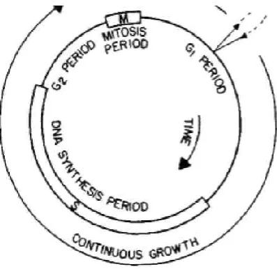
Figure 1. Diagrammatic representation of cell cycle
There are changes or shifts in the cell cycle .Cells in G2 are
recruited into the M phase of the cell cycle. Cells are also mobilized from G1 expressed as increased numbers of cells entering the DNA synthesis phases as measured by H-thymidine uptake studies Cell proliferation may be enhanced by bioelectric perturbation (Zeev et al., 1980). Since cell
proliferation is enhanced, one might be concerned about induction of neoplasia from bioelectric perturbation. The pre-dominant effect appears to be at the cell membrane through activation of cytosolic processes leading to increased proliferation, without affecting the process of DNA replication itself. The quantification and integration of these components of the matrix are often interdependent .A change in a highly charged molecule may eventually affect all the others. Changes in proteoglycan structure lead to local changes in pH, pO2, and
ultimately in calcification. (Zeev et al., 1980)
Events occurring at the Cathode
Increase the rate of cGMP accumulation in most osteoblasts. Cyclic nucleotide is subsequently extruded from many of the cells, thus decreasing the number of intensely stained cells.
Events occurring at the Anode
Inflammation – related migratory activities, secretion of acids and lysosomal enzymes, substances essential for the degradation of the organic and inorganic components of bone.
Events occurring at the at periosteal surface
Electrically active "repair" response apparently operates according to Wolff's law. Bone contains oriented fibers of collagen which, if stressed, generate negative electric potential toward the side of the applied stress. This electrical imbalance creates a minicircuit. The accompanying electrical field vectors orient tropocollagen and ultimately collagen in a direction which resists the lines of stress. This matrix supports osteogenesis and the resulting new bone is formed on the concave side and is oriented longitudinally and parallel to the force vectors. (Zeev et al., 1980)
Advantages of bioelectric stimulation
The mucosa on the electrically stimulated side appeared healthier than comparable control tissue. The composition of organisms in the microbiological flora of the mucosa also differed (Zeev et al., 1980).Applied orthodontic force is well below the maximum that could be sustained by the alveolar bone.
Side effects of large magnitude of electric current
[image:3.595.371.481.670.786.2]1. The electrically positive environment could initiate root resorption and cause root distortion stimulating piezoelectric induced cementumclasia. “The threshold of resorbability between bone and cementum may lie in their relative susceptibility to piezoelectric induction (Zeev et al., 1980).
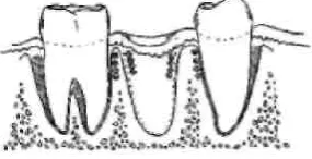
Figure 2. Electric positive and negative charge on tooth surface
2. Untoward sequelae of orthodontic extraction cases are clefting of alveolar bone and opening of the crown contact points at the extraction site. The root movements of the mesial and distal teeth into extraction sites are accompanied by varying degrees of tipping and concave bending of bone. (Zeev
[image:4.595.72.215.144.217.2]et al., 1980)
Figure 3. Two rapidly moving teeth
This means that two negatively charged bony surfaces move toward each other in rapidly moving teeth. The like-charged opposing piezoelectric fields may interfere with bone cell nutrition and osteogenesis producing a concentration of calcium ions and highly calcified tissue. Like-charged surfaces may repel some of the organic matrix molecules. Upset the normal bony architecture causing a disturbance in the buccal and lingual plate form. A radiopacity can often be seen at the bone of the closed extraction site. This density may be a thickened buccal and lingual plate. When bone is mechanically compressed by root paralleling mechanics, the "repaired bone" may act as a compressed spring and cause the local clinical relapse. In conclusion, electric currents, when applied noninvasively to gingival tissues, are capable of activating many PDL cells and neighboring osteoblasts. This effect is localized to well-defined zones near the electrodes and, therefore, may be used in areas where bone remodeling is desired, such as in the case of orthodontic treatment. Microscopic examination revealed a significant increase in the number of alveolar bone osteoblasts and PDL cells intensely stained for cAMP and cGMP, a uniform staining pattern of cells in areas near the anode and cathode, and bone apposition near the cathode (Norton et al., 1984).
Application of Static magnetic field
The area around a magnet within which magnetic force is exerted, is called a magnetic field. It is produced by moving electric charges. The presence and strength of a magnetic field is denoted by “magnetic flux lines”. The direction of the magnetic field is also indicated by these lines. The closer the lines, the stronger the magnetic field and vice versa. When iron particles are placed over a magnet, the flux lines can be clearly seen. Magnetic fields also generate power in particles which come in contact with it. A moving charge always has both a magnetic and an electric field, and that’s precisely the reason why they are associated with each other. They are two different fields with nearly the same characteristics. Wherever there is electricity, there are also electric and magnetic fields generated which is accompanied by invisible lines of force created by the electric charges. Electric fields result from the strength of the charge while magnetic fields result from the motion of the charge, or the current. A group of charges moving in the same direction is called an “electric current.” When such charges move they create additional forces known as a “magnetic field.” The use of electromagnetic energy to stimulate biologic systems has recently received a considerable amount of attention in both the basic science and clinical areas
of medicine and dentistry. (Norton et al., 1984) Electromagnetic fields have been used successfully to induce healing in fractures of human long bones that have proved resistant to conventional treatment and have frequently required amputation, before the development of electrobiologic therapy. Osteogenesis or chondrogenesis was frequently observed at the cathode and was attributed to the effects of the direct current. (Norton et al., 1984) Because of the inconvenience and clinical impracticality of implanting electrodes, efforts were made to find alternative methods of applying electrical energy to living tissue, this lead to the introduction of powerful samarium-cobalt (SmCO3) magnets in 1968. Since these magnets can be
made sufficiently small, their use to provide orthodontic forces is ideal.
Figure 4. Magnetic field
Biological Effects of PEMF
Apoptosis of bone marrow osteoprogenitor and tendon fibroblast cells. Reduces growth and development. Interferes with hormone receptor interactions on the cell surface (Luben
et al., 1982). (Thomas and Peter, 1987) Recent investigations
on PEMF on the activities of cells in the periodontium is that it induces faster bone resorption and deposition. This is confirmed by the fact that the cells associated with hard tissue resorption and elimination of the necrotic tissues are positive for tartrate acid EMFs enhance DNA, RNA and protein proliferation in cultures
Short-term EMF application is suggested to cause accelerated calcium uptake in cartilage.
Such effects may contribute to the reported therapeutic 'Benefits of EMF on fracture healing and bone metabolism during tooth movement. (Thomas and Peter, 1987)
Clinical applications in orthodontics
1. The principle of intermittent application of an external perturbation above a certain threshold value to provide maximum bony response is used clinically for tooth movement
2. Intermittent force application which provides the tissue with rest periods results in the most physiologic tooth movement with the minimum of undesired sequelae. (Thomas and Peter, 1987)
[image:4.595.311.564.226.330.2]membranes, PEMF-produced pulsating currents are capable of penetrating the cell membrane. These stimuli could act either at the level of the cell membrane or directly affect intracellular organelles. Orthodontic mechanical forces can trigger an inflammatory response in the adjacent periodontal tissues, leading to the discharge by mast cells of serotonin and histamine resulting in increased vascular permeability of the periodontal ligament. Both prostaglandins and blood macrophages have also been implicated as potential sources of enzymes that can stimulate bone resorption.
Application of mechanical loads to accelerate
tooth movement
Mechanical signals can positively influence physiologic processes critical to human health from accelerating tooth movement and craniofacial repair to preventing
osteoporosis Mechanical signals can be potent regulators of bone mass, morphology, and material properties .
mechanical loads are applied to intact tissues
the tissues usually become distorted (strained).Loads such as gravity prompt cells to arrange the architecture of the bony structural features in a way that would resist redundant loads. This phenomenon is known as "Wolff's Law," outlined by Julius Wolff in 1892. Bone bending is suggested as an important mechanism linking the cellular and m
[image:5.595.49.288.397.588.2]associated with tooth movement due to orthodontic and Sven, 1991)
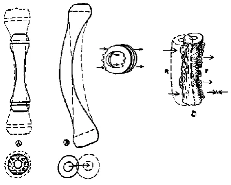
Figure 5. Wolffes law of bone transformation
When bone cells are subjected to non-redundant loads such as orthodontic forces, the cells are activated and remodeling of the alveolar process ensues, which facilitates tooth movement.
9
Mechanically- induced bone remodeling
Responses to orthodontic tooth movement are the resorption of bone in areas under "pressure" and deposition in areas of "tension." The cells have a unique behavior of distinguishing between tension and compression-this statement is proved observing the form and the structure of bon fundamental idea of the trajectoral theory
-cancellous bone at the proximal end of the human femur are orientated along the lines of principal stress corresponding with the lines of maximum and minimum loading
47488 International Journal of Current Research,
produced pulsating currents are capable of the cell membrane. These stimuli could act either at the level of the cell membrane or directly affect intracellular Orthodontic mechanical forces can trigger an inflammatory response in the adjacent periodontal tissues, ge by mast cells of serotonin and histamine resulting in increased vascular permeability of the periodontal ligament. Both prostaglandins and blood-borne macrophages have also been implicated as potential sources of
mechanical loads to accelerate
Mechanical signals can positively influence physiologic human health from accelerating tooth movement and craniofacial repair to preventing and treating Mechanical signals can be potent regulators of morphology, and material properties .9 When mechanical loads are applied to intact tissues in vivo or in vitro,
the tissues usually become distorted (strained).Loads such as arrange the architecture of the bony structural features in a way that would resist redundant loads. This phenomenon is known as "Wolff's Law," outlined by Bone bending is suggested as an cellular and metabolic activity associated with tooth movement due to orthodontic force. (Sten
Wolffes law of bone transformation
redundant loads such as orthodontic forces, the cells are activated and remodeling of the alveolar process ensues, which facilitates tooth movement.
h movement are the resorption of bone in areas under "pressure" and deposition in areas of behavior of distinguishing this statement is proved by observing the form and the structure of bones .The - the trabeculae of cancellous bone at the proximal end of the human femur are orientated along the lines of principal stress corresponding with the lines of maximum and minimum loading (Harvold, 2003).
Orthodontically induced tooth movement is based on the principle of adaptive remodeling of the periodontal ligament and alveolar bone, after the application of a force. The force is applied to the crown of the tooth by brackets or springs. force results in distortion of the tooth and periodontal tissues with subsequent mechanical deformation of the surrounding ground substance and subsequently the cells. The alveolar process during orthodontic tooth movement
resorption of bone depends on the cytokines produced locally by mechanically activated cells
connective tissue remodeling include the interleukins, tumor necrosis factors, interferon’s, polypeptide growth factors, and colony stimulating factors. Stretch activated channels have also been considered in specific cells as a mechanism of intracellular triggering. Such channels have now been identified in a variety of tissues and cell types including skeletal muscle cells; vascular endothelial cells; epith airway cells; osteoblast-like cells; human skin fibroblasts. Since ion channels have been found in many cells, it has been hypothesized that the opening or closing of certain channels may induce changes in tissues after the application of a tipping force, has provided the first experimental evidence to support this hypothesis. Molecular basis for cell to cell communication during mechanically induced remodeling Small charged particles can move through the membrane down their concentration gradients by simple passive diffusion, even though it is a relatively slow process.
Transport proteins provide a more significant and rapid path for ionic exchange. A proportion of transport proteins are simply a channel with an aqueous center that provide a pathway for passive diffusion of ions. Some channels are continuously open, whereas others, which are referred to as gated channels, can be open or closed, depending on the ambient conditions. Such channels can be divided into voltage activated and chemically activated channels.
have recently been found that are gated by physiologic levels of tension. These mechanosensitive channels open and close in response to distortion of the cell Voltage activated channels are controlled by alterations in memb
channels form the basis of the action potential by which electrical signals are transmitted along nerve and muscle cells. During action potential sodium, potassium and calcium channels are opened by depolarization of the membrane. Chemically activated channels can be opened by many types of ligand including endocrines.
identified such channels in human
al 1990 found three classes of mechanosensitive ion channels in osteoblast-like cells. Some appeared to be nonselective for cations but, other channels were potassium selective. After the detection of mechanosensitive ion channels Davidson proposed that these channels may mediate the response of bone to mechanical loading.
Second messenger systems activated by mechanical forces
The behavior of all cells is modulated by internal signaling systems, which translate a wide array of external stimuli, such as hormones or mechanical forces, into a narrow range of internal signals or second messengers.
Sutherland et al 1958 noticed that free glucose appeared under these conditions and postulated that adrenaline was acting as the "first" messenger, binding to a receptor and stimulating the production of a powerful c
(Harvold, 2003) The second messenger associated with
International Journal of Current Research, Vol. 9, Issue, 03, pp.47484-47495, March, 2017
Orthodontically induced tooth movement is based on the principle of adaptive remodeling of the periodontal ligament and after the application of a force. The force is applied to the crown of the tooth by brackets or springs. Such a sults in distortion of the tooth and periodontal tissues with subsequent mechanical deformation of the surrounding ground substance and subsequently the cells. The alveolar ring orthodontic tooth movement. The formation or nds on the cytokines produced locally by mechanically activated cells. Cytokines that can influence connective tissue remodeling include the interleukins, tumor necrosis factors, interferon’s, polypeptide growth factors, and etch activated channels have also been considered in specific cells as a mechanism of intracellular triggering. Such channels have now been identified in a variety of tissues and cell types including skeletal muscle cells; vascular endothelial cells; epithelial like cells; human skin fibroblasts. Since ion channels have been found in many cells, it has been hypothesized that the opening or closing of certain channels may induce changes in tissues after the application of a tipping orce, has provided the first experimental evidence to support Molecular basis for cell to cell communication during mechanically induced remodeling Small charged particles can move through the membrane down their simple passive diffusion, even process.
Transport proteins provide a more significant and rapid path for ionic exchange. A proportion of transport proteins are simply a channel with an aqueous center that provide a for passive diffusion of ions. Some channels are continuously open, whereas others, which are referred to as gated channels, can be open or closed, depending on the ambient conditions. Such channels can be divided into voltage vated channels. Protein channels have recently been found that are gated by physiologic levels of tension. These mechanosensitive channels open and close in response to distortion of the cell Voltage activated channels are controlled by alterations in membrane potential. These channels form the basis of the action potential by which electrical signals are transmitted along nerve and muscle cells. During action potential sodium, potassium and calcium channels are opened by depolarization of the membrane. mically activated channels can be opened by many types of Stockbridge and French1988 identified such channels in human skin fibroblasts. Davidson et
1990 found three classes of mechanosensitive ion channels in e cells. Some appeared to be nonselective for cations but, other channels were potassium selective. After the detection of mechanosensitive ion channels Davidson et al. proposed that these channels may mediate the response of bone
Second messenger systems activated by mechanical forces
The behavior of all cells is modulated by internal signaling a wide array of external stimuli, such as hormones or mechanical forces, into a narrow range of or second messengers. (Harvold, 2003) 1958 noticed that free glucose appeared under these conditions and postulated that adrenaline was acting as the "first" messenger, binding to a receptor and stimulating the production of a powerful chemical "second" messenger. The second messenger associated with
mechanical force transduction is adenosine 3',5' cyclic monophosphate (cAMP). Cyclic AMP was first identified in 1960 after initial observations of the way liver slices behaved when exposed to adrenaline. Somjen et al (1980) found that direct application of physical forces to cultured bone cells stimulated cyclic adenosine-5-monophosphate (AMP) and DNA synthesis. The authors believed these changes were mediated by prostaglandins, specifically prostaglandin E2.
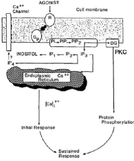
[image:6.595.321.547.359.638.2]Salzstein and Pollack (1987) considered that the fluid flow in bone would lead to the phenomenon of streaming potentials. These locally developed potential differences are, however, very small compared with the potential difference changes generated by muscle activity. Sandy et al (1989) demonstrated that the phosphoinositide pathway is activated in addition to the cyclic AMP pathway. They found that inositol phosphate production increased with mechanical stretch when examining osteoblasts. (Harvold, 2003) The importance of phospholipids as a messenger system was not fully appreciated until the 1980s when Streb et al. demonstrated that products of inositol lipid break down could cause release of intracellular calcium ([Ca2+] 14 The hydrolysis of phosphatidylinositol 4,5-bis- phosphate in response to activation of cell surface receptors, leads to inositol triphosphate [Ins(l,4,5)P3] formation and the release of Ca2+ from intracellular stores. Phosphorylation of Ins (l,4,5) P3 yields Ins (l,3,4,5) P4 which may control calcium entry at the plasma membrane through calcium channels. Ins (l,4,5) P3 is also a proven mediator of mitogenesis in a variety of cell types.
Figure 6. Second messenger system activated by mechanical forces
Second messengers other than cAMP, such as, eicosanoids and the phospholipid metabolites inositol phosphates and diacylglycerol, could mediate the effects of mechanical deformation. Ca2+ changes previously reported by Harell and co-workers might have occurred as the result of a cAMP-dependent Ca2+ flux through cell membranes. Thus the effects of hormones or mechanical stress on cellular metabolism cannot be explained by cAMP elevation alone. The PI pathway could certainly account for many of the changes seen in mechanically deformed tissues including an elevation of Ca2+ and an increase in DNA synthesis. Recently demonstrated that
murine calvarial osteoblasts elevate inositol phosphates, as well as cAMP in response to PGE2 and parathyroid hormone
(PTH). Many years external loading of skeletal structures has been known to induce changes in the architecture of bone at cell and tissue level This has been utilized by orthodontists in the treatment of malocclusions and by orthopedic surgeons as an aid to fracture repair.
Shape change as a basis for the transduction of mechanical forces
Folkman and Moscona showed that flattened cells synthesize more DNA than rounded cells, and Aggeler and coworkers developed the concept that catabolic rather than anabolic events are associated with rounded cells. They found that agents such as phorbol diesters (which stimulate protein kinase C) caused a rounding up of synovial fibroblasts resulting in an increase of collagenase and plasminogen activator production, but a reduction in the synthesis of the matrix proteins collagen and fibronectin. Similar findings were made in epithelial cells by Hong and Brunette and by Unemori and Werb where shape change was induced by nonchemical means. That the critical factor in triggering production of procollagenase was reorganization of the cytoskeletal protein actin, rather than change in cell shape (Harvold, 2003).
Figure 16. Cytoskeletal matrix interactions
The cytoskeleton presents a number of possibilities for transducing mechanical forces acting on cells and/or their adjacent matrices. The three main components of the cytoskeleton are microtubules, micro-filaments, and intermediate filaments. Microfilaments are perhaps the best situated of the three systems to detect these changes. (Jonathan
et al., 1993) The major subunit protein of the microfilaments is
[image:6.595.57.272.382.641.2]These tight adhesions are known as focal contacts, adhesion plaques or focal adhesions. Many of the extracellular matrix proteins responsible for cell adhesion contain a common peptide sequence Arg-Gly-Asp (RGD), which is essential for the cell-binding properties of these proteins. These RGD "sites" are recognized by a family of membrane integral proteins termed integrins that span the cell membrane from the cytoplasm to the extracellular matrix. Integrins do not bind directly to microfilaments, such as actin, but are dependent on associated proteins for this function (e.g., fibronectin extracellularly and talin intracellularly). Actin and vinculin bind to this talin-integrin complex. It is possible therefore to visualize a complex set of events that might arise either from distorting a cell, cell membrane, or extracellular matrix mechanically, or by inducing a change in cell shape with hormones and growth factors. (Jonathan et al., 1993) Transforming growth factor beta (TGF-3) induces shape change in osteoblasts with an inhibition of phenotypic expression (alkaline phosphatase activity), an effect that can be mimicked by addition of fibronectin. This suggests that the activity of TGF-3 may be mediated through interactions between the extracellular matrix and cytoskeletal elements. (Jonathan et al., 1993)PGE1 PGE2 and PTH also induce shape
change in bone cell cultures that is associated with changes in the microfilament system rather than microtubules. Disruption of microtubules in osteoblasts with colchicine can induce shape change and an increase in PG synthesis thought to be independent of the alteration in cell shape. Banes et al. found a decrease in tubulin and suggested that this may have a role in mediation of mechanical stress. One has to consider that shape change by cultured cells in vitro in response to various hormones and growth factors may not necessarily occur in vivo. (Jonathan et al., 1993)
Clinical evidence that mechanical signals are anabolic
Cross-sectional studies in humans -bone morphology can change markedly in response to long-term exercise in professional tennis players, the cortical wall thickness of the humerus in the dominant playing arm can be up to 45% larger than the non-racquet arm. Similar evidence of bone hypertrophy has been reported in a range of athletes and locations from ballet dancing to soccer, weightlifting, speed skating, squash, dancing/gymnastics, or physical activity. Exercise can rapidly and effectively produce large increases in bone mass, much of the data have been equivocal. Mechanical signal are perceived as osteogenic by resident bone cells populations, such as osteocytes, osteoblasts, osteoclasts, or bone marrow cells. Bone formed due to high-load, high-impact exercises using few repetitions (weightlifting or jumping) may be superior to those exercises using lower loads with high repetitions (swimming or walking). (Jonathan et al., 1993) However, inevitable factors could be difficult to control, such as genetics, gender, body habitus,, nutrition, compliance, or interactions between stress response and the local, site-specific mechanical adaptations. Normal strains cause volumetric changes in the tissue Shear strains cause angular deformations. When changes in remodeling events were compared between loading regimes inducing predominantly shear or predominantly normal strains, it became clear that bone tissue can readily differentiate between different kinds of deformation. Even though bone cells were responsive to both normal and shear strains, only normal strains increased the degree of juxtacortical turnover. It is critical to realize that dynamic but not static strains have osteogenic potential.
Recent studies have indicated that the manner in which loading cycles are distributed plays an important role in defining the magnitude of the anabolic response. The mechanisms by which the sensitivity of cells to mechanical signals is increased by rest inserted loading may be associated with high cell refractory periods, enhanced "bone fluid" flow, synchronized osteocytic activity, and/or enhanced cellular communication. (Jonathan et al., 1993)Skeletal sensitivity to low-level high-frequency mechanical signals. The weak correlation of new bone formation with the specific sites of peak strain magnitudes suggests that other mechanical factors may also be relevant for defining bone mass and morphology. There exists a non-linear interdependence between cycle number, strain frequency, and strain magnitude. The reduction in strain threshold could be associated with an increase in cycle number, it is likely that the increase in frequency at which loading occurred played a large role. Bone can sense and respond to even extremely small mechanical signals if they are applied at high frequencies. Application of resonance vibration during orthodontic tooth movement affects the acceleration of tooth movement by increasing the activity of the cells in the periodontal ligament. (Zengo et al., 1973)
Vibrations can produce high-quality bone and decrease resorptive activity
To investigate potential changes in indices of bone formation, adult mice were subjected to brief daily periods of whole body vibrations at 0.3 g, 45 Hz. The mechanical stimulus increased bone formation rates by 32%. Any increase in formative activity becomes structurally relevant only if the material properties of the newly formed bone are of high quality (Jonathan et al., 1993). Analysis of collagen and mineral content and composition was performed on newly formed metaphyseal cortical and trabecular bone by synchronous infrared micro-spectroscopy No significant differences in the major chemical constituents were found between control and vibrated mice, suggesting that the mechanical treatment improved bone's structural strength. (Zengon et al., 1973) Similarly after a 3-week exposure to the low-level vibrations, osteoclastic activity in the trabecular metaphysis and epiphysis of the tibia was 30% lower in vibrated mice than in age-matched controls. Thus the output of different cells residing in bone can be modulated by extremely low-level vibrations. (Zengon et al., 1973)
Vibrations enhance the musculoskeletal system
Bone's anabolic and catabolic activity can be altered by the vibratory mechanical signal, its impact on the musculoskeletal system was investigated in an eight-week-old mice subjected to the mechanical treatment described above had a 14% greater trabecular bone volume in the tibial metaphysis while periosteal bone area, bone marrow area, cortical bone area, and the moments of inertia of this region were all significantly greater (up to 29%). (Judex et al., 2009) The soleus muscle also realized gains with up to 29% greater total cross-sectional area as well as type I and type II fiber area. The small magnitude and brief application of the non-invasive intervention emphasized that the mechano-sensitive elements of the musculoskeletal system are not necessarily dependent on strenuous long-term activity to initiate a structurally relevant response in the adolescent musculoskeletal system. If maintained into adulthood, the beneficial structural changes in cortical bone, and muscle may serve to decrease the incidence of
osteoporotic fractures and sarcopenia later in life. (Judex et al., 2009)
Acceleration of tooth movement using Low Level Laser irradiation
Laser in an acronym for LIGHT AMPLIFICATION BY STIMULATED EMISSION OF RADIATION. Some of the clinically useful lasers are carbon-dioxide lasers which work well in soft tissue, Erbium lasers which work well in both soft and hard tissue.
Interaction of laser to biological tissue
1. Photochemical interaction –This include biostimulation which describes the stimulatory effect of laser light on biochemical and molecular process that normally occur in tissues such as healing and repair
2. Photothermal interaction –This is manifested clinically as photo ablation or the removal of tissue byvaporisation and super heating of tissue fluids, coagulation and haemostasis and photopyrolysis
3. Photomechanical interaction-In this interaction photodisruption or photodissociation which is breaking apart of structures by laser light and photoacoustic interaction which involve the removal of tissues with shock wave generation
4. Photoelectrical interaction-Here photoplasmolysis, which describes how tissue is removed through the formation of electrically charged ions and particles that exists in a semi gaseous, high energy state.
Laser therapy has an accelerating stimulus for dental movement. The cascade of events required to induce dental movement, could involve osteoclast differentiation via RANK/ RANKl. Normal human osteoblast cells (NHOs) are sensitive to low-level laser irradiation with Photon LASER (As-Ga-AI) in early stages of culture, causing an enhancement of cell proliferation from the first day. Low level laser accelerates orthodontic tooth movement – the stimulatory action occurs during the proliferation and differentiation stages of bone cellular precursors but not during later stages of cellular proliferation (Angela et al., 2009). The number of blood vessels in the PDL was increased by OTM and daily LLL irradiation. The cell reaction to laser irradiation was not characterized by an increment of proliferation, but by an increment of temperature and intracellular calcium concentration. These effects of laser therapy acts as an accelerating stimulus for dental movement. Low level laser therapy has shown positive effects on bone remodelling, optimising tooth movement, fibroblast proliferation, collagen synthesis and organization. Er Yag laser- increase in its cognate enzyme, COX2 mRNA levels leading to PGE2 production in human gingival fibroblast cells. PGE2 production by Er Yag laser was suppressed to a lower level by a selective inhibitor of COX2 (Angela et al., 2009)Low level laser irradiation facilitates the velocity of tooth movement by the expression of matrix metalloproteinase-9,cathepsin K and alpha(V)Beta(3)integrins in rats which also facilitates tooth movement. (Pourzarandian et al., 2005) Biostimulation mechanisms of laser irradiation in bone tissue cells, such as osteoblasts and osteoclasts, do not provide strong evidence for the therapeutic application of laser in orthodontics. This system is being used as a therapeutic tool without sound scientific evidence of the effects on cells involved in dental movement. The present results suggest that
low-level laser irradiation of human osteoblasts stimulates cell proliferation for up to 6 days, This increment is significantly higher than that observed in nonirradiated control groups. The detailed mechanism of laser-cell interactions is still a matter of research, and information about the effect of low-level laser irradiation on proliferation and is far from complete in the available literature. In clinical orthodontics, undesirable outcomes such as a movement of anchor teeth during tooth movement and relapse of well aligned teeth to their original positions after treatment sometimes occur. If these phenomena could be prevented, excellent treatment results with long-term stability could be achieved and maintained.
Acceleration by chemical means
The rate-limiting step occurs during the lag phase, as osteoclasts are recruited from the bone marrow. Osteoclasts in the periodontal ligament originate from bone marrow precursors which mature into preosteoclasts that fuse and become activated to form multinucleated bone-resorbing osteoclasts. Application of various chemicals that causes the differentiation of the osteoclasts in the form of gels, injections and liposomal formulation which in turn augments tooth movement Some of the chemicals that are used to augment tooth movement are
Macrophage colony stimulating factor (Patricia et al., 2011)
Parathormone (Soma et al., 2000)
Insulin like growth factor (Ong et al., 2001)
Epidermal like growth factor (Karina et al., 2008)
Hyperbaric oxygen therapy (Sila et al., 2008)
Acceleration of tooth movement by surgical methods (corticotomy and corticision)
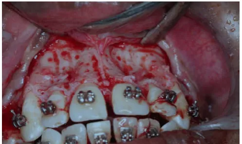
cuts that extend to the marrow. (Krishnan et al., 2013) The duration of RAP depends on the type of tissue, and usually lasts about 4 months in human bone. This phenomenon causes bone healing to occur 10–50 times faster than normal bone turnover.
Figure 6. Corticotomy cuts
This reduces the biomechanical resistance and enables rapid tooth movement through trabecular bone. Transient osteopenia may be prolonged with loading orthodontic application, (estimated 3–4 months). This is why it is imperative to adjust the orthodontic appliance every 2 weeks. Hyalinization (tissue necrosis) is caused by excessive compression of the PDL as a result of excessive pressure, which suppresses blood supply, although it may appear even with light force. This hyalinized tissue attracts neutrophil granulocytes and macrophages by chemotaxis and must be removed and remodeled before starting bone resorption by osteoclasts and subsequent orthodontic tooth displacement. Vascular access of osteoclasts to the PDL–laminadura interface is limited when the PDL is compressed. Thus, extensive and prolonged hyalinization of the PDL results in slower tooth movement. After Reitan’s studies, this period of hyalinization has been designated as the lag phase or arrest phase of orthodontic tooth movement, after the initial phase and before the acceleration or post-lag phase.
Rate of tooth movement which is infuenced by hyalinization, is directly related to
1. Force magnitude,
2. Type of movement (bodily or tipping movement), 3. Bone metabolic capacity (bone density, systemic and
genetic factors)
It has been proved that during retention, clinical outcomes of periodontal AOO patients improved and did not relapse. In essence, the AOO procedure is in vivo tissue engineering, highlighting the ability to morph bone with orthodontic tooth movement, periodontal bone activation, and alveolar augmentation.
Precautions to be taken
1. Maintain the vitality of the hard and soft tissue -Mobilisation of the outlined single tooth blocks of bone is contraindicated intrapulpal and intraosseous morbidity
2. Green – stick fracturing and luxation of dentoalveolar structure since these segments lose their structural integrity (osteopenia)
3. Luxation and interefere with the integrity of neurovascular bundle exiting at the apex and result in nonvitality
4. Overcompression of PDL can lead to hyalinisation the removal of which leads to root resorption (Kyu et al., 2001)
As long as the root surface is vital-no apical epithelial migration .The alveolar deficiency can be corrected by augmentation .Only resorbable grafts are used .Medications that reduce turnover rate and increase Calcium uptake are problematic-NSAID and Bisphosphonates. An unaesthetic so-called "black triangle" is often observed postoperatively where arch length discrepancies are treated or where injudicious or inopportune manipulation of the incisive papilla is employed. Generally, to prevent or predict this esthetic complication, the roots should be no further apart than 3 mm, and the distance from the crest of alveolar bone to the apical extent of the interdental contact area should not exceed 5 mm. Buccal and lingual vertical tension releasing incisions should be made from the principal sulcular incision into the depth of the buccal vestibule. Lingual releasing incisions should be avoided. The releasing incisions are designed to provide improved visualization over the "envelope-flap" that does not have vertical releases. The tension releasing incision also provides a surgical guide for the linear decortication of the canine-lateral incisor cortical bone. Suya (1991) most orthodontic treatment should be completed in the first three to four months after corticotomy –linear interproximal decortication Wilco 1 2001 developed a patented technique which is called Periodontally Accelerated Osteogenic Orthodontics (PAOO)-Selective Alveolar Decortication –along with placement of a resorbable graft was placed over the surgical site resulted in 3-4 times faster tooth movement. Wilco 3 reported (reversible osteopenia) reduced mineralization of the alveolar bone surrounding the involved teeth during orthodontic movement. (World J Orthod 2003) As the Wilcko brothers— an orthodontist and a periodontist—reported a 1/2 to 1/3reduction in traditional orthodontic treatment time. Their publications and conference presentations aroused intense curiosity, mainly because they were based solely on case reports. Frost – 2003 a direct correlation between severity of bone corticotomy and /or osteotomy and the intensity of the healing response .This lead to accelerated bone turnover at the surgical site - this was designated “regional accelerated phenomenon.”(RAP) Fergusson - 2006 defined RAP as an osteopenic process – anabolic activity increased by 150% in 3weeks. Sebaoun 2006 reported 200% catabolic activity and 400% anabolic activity. Kelsons reported in 2006-apposition of bone at lamina dura increased by 46% at 4 weeks in the same animal model. Duker investigated how corticotomy affected the vitality of the teeth and the margin. Rearrangement of the teeth within a short time after corticotomy damaged neither the pulp nor the periodontal ligament (PDL). He supported the idea of preserving the marginal crest bone in relation to inter-dental cuts; these cuts must always be left at least 2 mm short of the alveolar crestal bone level periodontium in beagle dogs. (Krishnan et al., 2013) The first messengers bind to receptors present on the cell surface of target cells and initiate a process of intra-cellular signalling. The intra-cellular signalling results in formation of second messengers, which include cyclic AMP, cyclic GMP and calcium. Initiate formation of bone cells namely osteoclasts and osteoblasts which are responsible for bone remodelling. An intensifed bone response (increased osteoclastic and osteoblastic activity, and increased levels of local and systemic infammation markers)-Neutrophils,
Macrophages, granulocytes, cytokines, prostaglandines, leukotrienes and thromboxane A2 in areas around cuts that extend to the marrow.
This response varies directly in duration, size, and intensity with the magnitude of the stimulus, and it is considered a physiological “emergency” mechanism, which accelerates the healing of injuries that could affect survival. The cancellous portion of the alveolar bone gets induced into a more pliable, transient, reversible, demineralized state called osteopenia. Osteopenia is a state of calcium depletion, occurring because of two processes—osteoclasis (surface resorption) and osteocytic osteolysis (osteon remodeling). A catabolic process, a resorption response resulting in decreased bone density, but with no change in alveolar bone volume. With demineralization, bone matrix transportation occurs and the remaining collagenous soft tissue matrix of the bone is transported with the root in the direction of movement. The demineralization is followed by the anabolic process, a formation response wherein new bone is deposited and the osteoid matrix gets remineralized. (Krishnan et al., 2013)As long as tooth movement continues, RAP is prolonged. When RAP dissipates, osteopenia disappears. The RAP commences a few days after surgery, peaks between 1 and 2 months when catabolic and anabolic responses are threefold higher, dissipates to a normal steady state by 11 weeks after surgery, and takes approximately 6 to 24 months to resolve completely. The perforations in SAD made were as effective as the alternative circumscribed corticotomy cuts. Since these perforations provided the necessary bleeding points and communications with the softer inner medullary bone. These communications then act as pathways through which new blood vessels and pluripotential cells migrate from the medullary bone into the cortical plates. Pluripotential cells remove old bone and create new bone and make the cortical plates more vital and responsive to the forces of tooth movement. Bone morphogenetic protein infuences the primitive uncommitted stem cells to become more specifc cell types in bone morphogenesis. The growth protein component in the soft tissue matrix of the bone stimulates an increase in the osteoblastic activity, resulting in remineralization of the soft tissue matrix. Bone luxation is contraindicated because it could lead to intrapulpal and intraosseous morbidity and can jeopardize the integrity of the neurovascular bundle exiting the apices of the fenestrations over root prominences, bony dehiscence formation can be effectively repaired with this teeth, resulting in devitalization. The structural integrity of the periodontium is enhanced with periodontal alveolar augmentation with a bone graft, thus producing an environment resistant to relapse. Histological study in dogs showed increased turnover of alveolar spongiosa immediately adjacent to the decortication areas, without any orthodontic force being applied. Trabecular bone surface area decreased by half and PDL surface area increased by two-fold. Marked increase in inflammatory cells. Increase number of pluripotential stem cells. Increase in number of osteoclasts, osteoblasts. There is an initial stage of woven bone formation which begins in the periosteal area and then extents to medullar bone, reaching its maximal thickness on day. (Krishnan et al., 2013)
This cortical bridge of woven bone is a fundamental component of RAP, providing mechanical stability of bone after injury. From day 7, the woven bone in the cortical area begins to undergo remodeling to lamellar bone, but woven
bone in the medullary area undergoes resorption, which means transitory local osteopenia. From day 7, the woven bone in the cortical area begins to undergo remodeling to lamellar bone. But woven bone in the medullary area undergoes resorption, which means transitory local osteopenia -usually peaks at 1-2 months. (Fischer, 2007)Lasts from 6 to 24 months to subside completely. Clinically, alveolar bone exposure after refection of soft tissue flaps is known to cause some degree of bone resorption, mainly around teeth or dental implants. This RAP has been observed not only after corticotomy of the alveolar bone but also after full-thickness mucoperiosteal flap rejection without touching the bone. A study in the mandibles of rats showed transitory trabecular bone resorption after flap refection. (Cho et al., 2007) The degree of resorption was greater if lingual and buccal flaps were used, was greater in the lingual plate, and peaked with maximum resorption at 3 weeks after surgery, which is equivalent to 3 months in humans. Transient osteopenia may be prolonged with loading orthodontic application, (estimated 3–4 months). This is why it is imperative to adjust the orthodontic appliance every 2 weeks. Tooth movements were accomplished in 2 weeks with AOO procedure, as compared with conventional orthodontics in 6- to 8-weeks interval
Advantages
1. Reduced treatment time 2. Less root resorption
3. More support due to additional bone graft 4. Low relapse rate
Disadvantages
1. Extra surgical cost
2. Post surgical crestal bone loss and recession may occur 3. Pain, swelling and possibility of infection
4. Proper case selection is very important since it is not applicable to all cases
The reasons for this accelerated velocity are, most likely, the expansion and enhancement of the local inflammation, and the associated healing process. This fact is particulary evident in patients subjected to alveolar decortication. Here, in addition to inflammatory cells, stem cells may migrate out of alveolar bone marrow cavities, and contribute to the augmentation of the tissue remodeling. It may be concluded that the velocity of tooth movement may be accelerated by focusing on the paradental tissues with combinations of physical stimuli, including mechanical forces. RAP introduced by Corticision
Kim et al developed an interesting technique that eliminated
flap reflection methods. They used transmucosal incision “corticision,” wherein a reinforced scalpel was used as a thin chisel to separate the interproximal cortices transmucosally, without a surgical flap reflection. Such transmucosal manipulation of alveolar bone, although minimized morbidity, failed to recruit significant RAP (Young et al., 2013).
Laser corticotomy