Primary open-angle glaucoma: on the
development of novel therapeutic
approaches
A Thesis Submitted to Trinity College Dublin for the Degree of
Doctor of Philosophy
by Darragh Crosbie
Supervised by Professor Peter Humphries
Smurfit Institute of Genetics
Trinity College Dublin
Declaration
I declare that this thesis has not been submitted as an exercise for a degree at this or any other university and it is entirely my own work except where noted. I agree to deposit this thesis in the University’s open access institutional repository or allow the Library to do so on my behalf, subject to Irish Copyright Legislation and Trinity College Library conditions of use and acknowledgement.
Acknowledgements
I would like to take this opportunity to thank every person that has helped me throughout the course of my PhD study. Firstly, to Prof. Pete Humphries, thank you so much for the opportunity to embark on this challenging project and for your continued support. I am also hugely grateful to Dr. Lawrence Tam who offered continued guidance throughout this project, meeting any stumbling blocks along the way. Special thanks to Dr Sophie Kiang, Dr Marian Humphries, Dr. Ester Reina-Torres, Dr Matthew Campbell, and Dr James Keaney. In particular to James for guidance early on in my studies and for contributing hugely to the MRI project. I must offer sincere thanks to Marian for developing any AAV constructs and offering her expertise in any PCR related problems, and Sophie for looking out for me ever since I entered the lab. To Dr. Paul Kenna I offer my gratitude for any help with animal work, and thanks to all those working in the animal unit Dave, Charlie and Caroline for taking care of my animals. I would like to thank Rustam
Rakhmatullin and Christian Kerskens, who run the mouse MRI facility. This work was greatly helped by our collaborators, Prof Colm O’Brien, Dr. Darryl Overby, Dr. Joseph Sherwood, Prof. Daniel Stamer, and Kristin Perkumas. Dr. Joseph Sherwood established the eye perfusion system for our group and Prof. Daniel Stamer
supplied primary human cells with the help of Kristin Perkumas.
To all my friends I must offer thanks for keeping life fun throughout my PhD studies. Firstly to Paul, Jeff we had some adventures and some fantastic
Summary
Glaucoma is one of the most prevalent forms of preventable blindness, affecting more than 60 million people worldwide. While normotensive forms of the disease do exist, the majority of cases are caused by elevations in intraocular pressure. The open-angle form of the disease is the most common and the primary standard of care is the use of topically-applied pressure reducing medications. Such
medications work either by slowing down the production of aqueous humour, or increasing its outflow from the eye. Interestingly, none of these medications target the major route of aqueous humour outflow, the conventional outflow pathway, comprised of the trabecular meshwork and Schlemm’s canal. Current therapies often fail to meet adequate IOP reduction and surgical intervention is required in these cases, which can result in significant side effects. There is, therefore, a distinct unmet clinical need for therapies primarily targeting the conventional outflow pathway and the work presented in this thesis directly addresses this issue using the mouse as an animal model.
Chapter 2 (after the general introduction of Chapter 1) describes work in
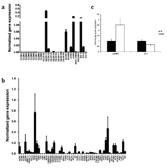
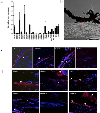
which tight junctions of the eye are studied as targets for glaucoma therapy. The inner wall of SC is an intact barrier joined by tight junctions that contributes to AH outflow resistance (Overby et al. 2009). The tight junction components joining SC
endothelial cells were characterized in in vitro and in vivo studies inhuman,
primate and mouse with a view to then down-regulating these via siRNA delivery to the mouse AC (results of overall project reported in Tam et al., (2017).
Furthermore, a similar approach was used down-regulate claudin-5 and occludin tight junction proteins in endothelial cells of the inner retina in the DBA/2J
glaucoma mouse, resulting in increased clearance of soluble amyloid-β (1-40) from the ganglion cell layer.
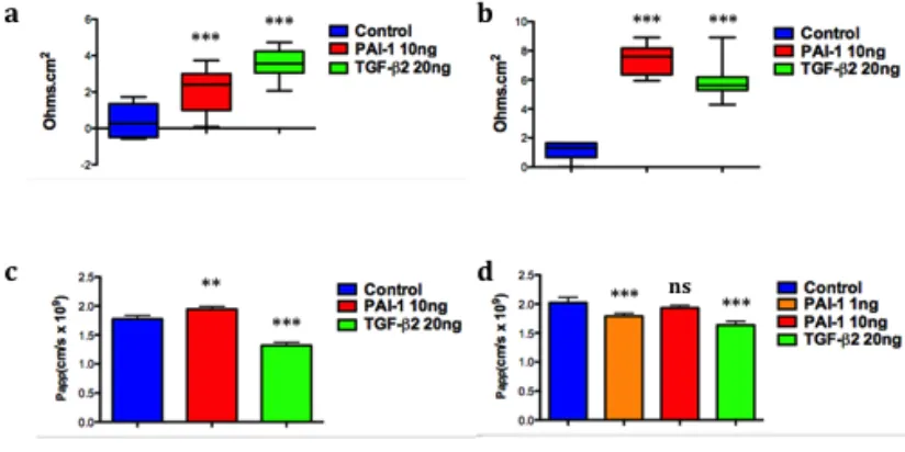
As part of a group effort, this virus was further used to deliver MMP-3 to the outflow pathway, enhancing aqueous humour outflow, as reported in O’Callaghan, Crosbie, et al. (2017). Increased plasminogen activator inhibitor-1 (PAI-1) levels were observed in POAG patient AH. PAI-1 is a downstream effector of TGF-β2, which has a role in glaucomatous pathogenesis (Fuchshofer & Tamm 2012). PAI-1 increased the trans-endothelial electrical resistance across both SC endothelial cell (SCEC) and TM cell monolayers, increasing ECM deposition and cell contractility. Previously, virally expressed TGF-β2 has been used to create a mouse model of glaucoma (Shepard et al. 2010), and so mouse recombinant PAI-1 was placed into the AAV-2/9 vector and expressed from the mouse corneal endothelium with the goal of creating a novel glaucoma model. After 12-weeks no increase in IOP or decrease in outflow facility was evident in AAV-PAI-1treated eyes, potentially due to low PAI-1 secretion.
Assessment of AH dynamics in a model is important in novel treatment discovery. Magnetic resonance imaging (MRI) is a non-invasive technique that allows real-time measurement of morphology and fluid movement (Townsend et al. 2008). Chapter 4 reports data on the use of MRI to evaluate the glaucoma mouse eye. The effect of the glaucoma medication, latanoporst, was measured using Gd-MRI, with treated eyes displaying enhanced clearance of contrast agent. Age-related changes in aqueous humour dynamics, morphology and blood-aqueous barrier integrity were detected in the DBA/2J pigmentary glaucoma mouse model. Use of MRI in this scenario is in its infancy, and the results obtained clearly
Table of contents
DECLARATION ... I
ACKNOWLEDGEMENTS ... II
SUMMARY ... III
TABLE OF CONTENTS ... V
ABBREVIATIONS ... X
CHAPTER 1: GENERAL INTRODUCTION ... 2
GLOBAL BLINDNESS AND GLAUCOMA ... 2
OVERVIEW OF GLAUCOMA ... 3
Glaucomatous optic neuropathies ... 3
POAG and increased outflow resistance ... 6
THE CONVENTIONAL OUTFLOW PATHWAY ... 8
The trabecular meshwork (TM) and juxtacanalicular tissue (JCT) ... 8
Schlemm’s canal ... 10
GENERATION OF OUTFLOW RESISTANCE ... 12
Outflow resistance at the juxtacanalicular tissue ... 14
Outflow resistance at the Schlemm’s canal inner wall ... 15
SEGMENTAL AH OUTFLOW ... 15
GENETICS OF POAG ... 17
Myocillin ... 17
Optineurin ... 18
GWAS and POAG ... 19
THE MOUSE AS A MODEL FOR GLAUCOMA ... 19
Myocilin mouse model of glaucoma ... 20
DBA/2J mouse model of glaucoma ... 21
Ad-TGF-β2 and Ad-CTGF mouse models ... 23
Induced hypertension mouse models: vein occlusion ... 23
Induced hypertension mouse models: microbeads ... 24
Induced hypertension mouse models: glucocorticoid induced glaucoma ... 24
Mouse models of retinal ganglion cell death ... 25
GLAUCOMA MEDICATIONS ... 25
Prostaglandin analogues ... 26
Carbonic anhydrase inhibitors (CAIs) and cholinergics ... 28
IOP-LOWERING GLAUCOMA SURGERIES ... 29
Laser trabeculoplasty ... 29
IOP-lowering glaucoma surgeries: trabeculectomy and stent surgeries ... 30
NOVEL MEDICATIONS: TARGETING THE CONVENTIONAL OUTFLOW PATHWAY ... 30
Rho-kinase (ROCK) inhibitors ... 30
Adenosine receptor agonists ... 31
Novel prostaglandin analogues ... 31
Actin depolymerisation agents ... 32
OBJECTIVES OF THIS STUDY ... 33
CHAPTER 2: ON MANIPULATION OF OCULAR ENDOTHELIAL TIGHT JUNCTIONS: POTENTIAL APPLICATIONS IN TREATMENT OF OCULAR HYPERTENSION AND RETINAL DISEASE PATHOLOGY. ... 36
INTRODUCTION ... 36
Summary ... 36
The blood-eye barriers ... 37
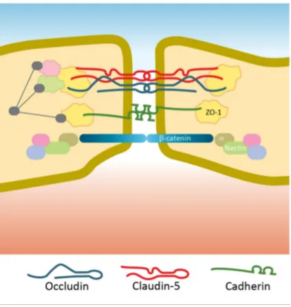
Component of the tight junction complex ... 37
The blood-retina barrier (BRB) ... 40
The blood-aqueous barrier (BAB) ... 41
RESULTS ... 44
Characterization of the tight junction proteins in human Schlemm’s canal endothelial cells (SCECs). ... 44
siRNA treatment of SCECs ... 47
Characterization of tight junctions in the mouse and monkey SC endothelia ... 48
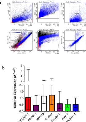
An attempt to isolate mouse SC endothelial cells ... 51
iBRB modulation in the DBA/2J mouse model of glaucoma ... 55
DISCUSSION ... 59
STATEMENT OF COLLABORATION ... 65
CHAPTER 3: AAV-MEDIATED DELIVERY OF RECOMBINANT PROTEINS TO THE OUTFLOW TISSUES. ... 67
INTRODUCTION ... 67
Summary ... 67
Adeno-associated virus (AAV) ... 68
Drawbacks of AAV ... 70
AAV in the AC ... 72
Cytokines and glaucoma ... 74
TGFβ-2 pathways ... 74
TGF-β2 in the TM and SC ... 75
RESULTS ... 78
AAV transduction of the AC tissues ... 78
PAI-1 and TGF-β2 in POAG AH ... 84
Effect of recombinant PAI-1 and TGF-β2 on SCEC and TM monolayers ... 86
Tight junction and ECM expression on PAI-1/TGF-β2 treatment ... 90
The effect of PAI-1/TGF-β2 on cytoskeletal structure ... 95
AAV-mediated secretion of PAI-1 to the mouse AH ... 99
DISCUSSION ... 102
STATEMENT OF COLLABORATION ... 112
CHAPTER 4: DETECTION OF AGE-RELATED CHANGES IN EYE MORPHOLOGY AND AQUEOUS HUMOUR DYNAMICS IN DBA/2J MICE USING CONTRAST-ENHANCED OCULAR MRI ... 114
INTRODUCTION ... 114
Summary ... 114
Methods of AH flow measurements in vivo. ... 115
Magnetic Resonance Imaging (MRI) ... 116
MRI in the eye ... 117
MRI in glaucoma ... 118
Overview ... 120
RESULTS ... 122
Gd-MRI in the mouse AC ... 122
Gd-MRI used to assess AH dynamics ... 125
Age-related changes in the DBA/2J mouse ... 127
DISCUSSION ... 134
STATEMENT OF COLLABORATION ... 138
CHAPTER 5: MATERIALS AND METHODS ... 140
5.1CELL CULTURE ... 140
5.2ANIMALS ... 140
5.3HUMAN TIGHT JUNCTION PCR ARRAY ... 141
5.4MOUSE TIGHT JUNCTION PCR ARRAY ... 141
5.6RETINAL DISSECTION ... 143
5.7WESTERN BLOTTING:RETINAL LYSATES ... 143
5.8IMMUNOHISTOCHEMISTRY: RETINA ... 144
5.9PLASMA/BRAIN TISSUE ISOLATION AND ELISA ... 144
5.10IMMUNOHISTOCHEMISTRY:AC FLATMOUNT (PECAM-1) ... 145
5.11MOUSE AC DISSOCIATION AND FACS ANALYSIS ... 145
5.12PATIENT AQUEOUS HUMOUR SAMPLES ... 146
5.13CYTOKINE ARRAY OF AH SAMPLES ... 146
5.14TRANSENDOTHELIAL ELECTRICAL RESISTANCE (TEER) MEASUREMENT ... 147
5.15PERMEABILITY ASSESSMENT BY FITC-DEXTRAN FLUX ... 147
5.16CELL VIABILITY ... 148
5.17TOTAL PAI-1 QUANTIFICATION ... 149
5.18WESTERN BLOTTING OF AH LEVELS OF TGF-Β2 ... 149
5.19IMMUNOCYTOCHEMISTRY ECM COMPONENTS ... 149
5.20IMMUNOCYTOCHEMISTRY TIGHT JUNCTION PROTEINS ... 150
5.21WESTERN BLOTTING: TIGHT JUNCTIONS ... 150
5.22WESTERN BLOTTING:ECM COMPONENTS ... 151
5.23IMMUNOHISTOCHEMISTRY FOR FROZEN SECTIONS:AC AND SCHLEMM’S CANAL ... 152
5.24IMMUNOHISTOCHEMISTRY FOR PARAFFIN EMBEDDED SECTIONS ... 153
5.25IMMUNOHISTOCHEMISTRY AAV TREATED MOUSE AC ... 153
5.26AAVS ... 154
5.27INTRACAMERAL INJECTION ... 155
5.28INTRAVITREAL INJECTIONS ... 156
5.29SUB-RETINAL INJECTIONS AND RETINAL FLATMOUNT ... 156
5.30IOP MEASUREMENTS:AAV-PAI-1 ... 157
5.31IOP:DBA/2J-SIRNA ... 157
5.32IOP: PRIOR TO MRI ... 158
5.33MEASUREMENT OF OUTFLOW FACILITY:AAV-PAI-1 ... 158
5.34MRI: MOUSE PREPARATION ... 162
5.35MRI SCAN PROTOCOL ... 162
5.36MRI:OCULAR ANATOMY ... 163
5.37ASSESSMENT OF T1-WEIGHTED GD-DTPA ENHANCEMENT ... 163
5.38STATISTICAL ANALYSIS ... 163
CONCLUDING REMARKS AND FUTURE STUDIES ... 166
APPENDICES ... 208 APPENDIX I:TAM ET AL.,2017.ENHANCEMENT OF OUTFLOW FACILITY IN THE MURINE EYE BY TARGETING SELECTED TIGHT-JUNCTIONS OF SCHLEMM’S CANAL ENDOTHELIA.SCIENTIFIC REPORTS.
... 208 APPENDIX II:CAMPBELL ET AL.,2017.MANIPULATING OCULAR ENDOTHELIAL TIGHT JUNCTIONS: APPLICATIONS IN TREATMENT OF RETINAL DISEASE PATHOLOGY AND OCULAR HYPERTENSION.
Abbreviations
AAV Adeno-associated virus
AAV-null AAV2/9 with no transgene
AAV-PAI-1 AAV2/9 with mouse PAI-1 transgene
AAV2/2 AAV with AAV2 genome and AAV2
capsid
AAV2/5 AAV with AAV2 genome and AAV5
capsid
AAV2/9 AAV with AAV2 genome and AAV9
capsid
AV Adenovirus
AC Anterior chamber
AH Aqueous humour
ALT Argon laser trabeculoplasty
AMD Age-related macular degeneration
APP Amyloid precursor protein
AV Adenovirus
Aβ Amyloid-beta
BBB Blood-brain barrier
BOLD Blood oxygenation level dependent
BRB Blood-retinal barrier
CAI Carbonic anhydrase inhibitors
CB Ciliary body
CNV Choroidal neovascularisation
Cr Reference facility
CTGF Connective tissue growth factor
DN Double negative
DP Double positive
dsDNA Double-stranded deoxyribonucleic acid
FACS Fuorescence-activated cell sorting
FSC-W Forward scatter width
GAG Glycosaminoglycan
Gd-BOPTA Gadobentate dimeglumine
Gd-DTPA Gadopentetic acid
Gd-MRI Gadolinium enhanced MRI
GV Giant vacuoles
H&E Hematoxylin and eosin
ICAM-1 Intercellular adhesion molecule-1
indAAV2/9 AAV2/9 with a doxycycline inducible
promoter
IOP Intraocular pressure
ITR Inverted terminal repeat
JAM Junctional adhesion molecule
JCT Juxtacanalicular tissue
LLC Large latent complex
MAD Median absolute dispersion
MRI Magnetic resonance imaging
MSP1 Mucopolysaccharidosis type I
NMDA N-methyl-D-aspartate
NTG Normal tension glaucoma
ONL Outer nuclear layer
OCT Optical coherence tomography
PACG Primary angle closure glaucoma
PAI-1 Plasminogen activator inhibitor-1
PI Propidium iodide
POAG Primary open angle glaucoma
PXG Pseudoexfoliative glaucoma
rAAV recombinant Adeno-associated virus
RGC Retinal ganglion cell
RPE Retinal pigment epithelium
RT-PCR Real-time polymerase chain reaction
SC Schlemm’s canal
virus
scAAV2/2 scAAV with scAAV2 genome and AAV2
capsid
scAAV2/5 scAAV with scAAV2 genome and AAV5
capsid
scAAV2/9 scAAV with scAAV2 genome and AAV9
capsid
SCEC Schlemm’s canal endothelial cell
SLT Selective laser trabeculoplasty
SPARC Secreted protein acidic and rich in
cysteine
SSC-A Side scatter area
SSC-W Side scatter width
ssDNA Single-stranded deoxyribonucleic acid
t-PA Tissue plasminogen activator
TGF-β2 Transforming growth factor-β2
TJ Tight juncion
TUNEL Terminal deoxynucleotidyl transferase
dUTP nick-end labeling
u-PA Urokinase plasminogen activator
ZO-1 Zonula occludens-1
ZONAB Zolnula occludens-1 associated nucleic
Chapter 1
Chapter 1: General Introduction
Glaucoma is a progressive optic neuropathy that accounts for 2.1 million cases of blindness worldwide. The most common form of glaucoma is primary open angle glaucoma (POAG), which is characterized by glaucomatous retinal damage with an open iridocorneal angle. In POAG the major risk factor is increased intraocular pressure (IOP), caused by a dysregulation in aqueous humour (AH) production and drainage (Bourne et al. 2016; Tham et al. 2014; Braunger et al. 2015). AH drainage occurs via two pathways, the uveoscleral and the conventional pathway. Drainage of AH occurs primarily through the conventional outflow pathway with a smaller fraction of AH outflow draining through the uveoscleral pathway. The fraction of uveoscleral drainage decreases with age (Goel et al. 2010). Despite this, the majority of current glaucoma therapies that increase AH outflow primarily target the uveoscleral pathway.
The overall focus of this thesis has been to explore, through murine
systems, concepts relating to the discovery of novel glaucoma therapies that target the conventional AH outflow pathway. In this introductory chapter, the prevalence of glaucoma worldwide is outlined, together with an overview of different clinical forms of disease. The regulation of AH homeostasis that controls IOP is discussed, along with a detailed appraisal of the anatomy and structure of the conventional outflow pathway. Resistance to outflow facility is key in increased IOP, and
therefore the role of the tissues of the conventional outflow pathway in resistance generation is explored. Use of animal models in glaucoma is central to
understanding biological reactions to IOP- lowering therapies, and the suitability of the mouse as a model is covered. The final section of this introduction focuses on current therapies, their efficacies and modes of action, along with recent progress in the development of novel glaucoma medications.
Global blindness and glaucoma
is defined as any visual impairment caused by cataract, uncorrected refractive error, trachoma, glaucoma or diabetic retinopathy. Cataract is the leading cause of blindness worldwide, being responsible for 33.4% of recorded blindness, with uncorrected refractive error being the second largest cause. Both of these are largely preventable by surgery in the case of cataract, and by spectacles, contact lenses and refractive surgery in relation to uncorrected refractive error and hence these conditions are more prevalent in the developing world where there is less access to medical intervention. Outside of these, glaucoma is the next leading cause of preventable blindness, responsible for 6.6% of global blindness – more in fact than trachoma and diabetic retinopathy combined. From 1990 to 2010 the number of cases of blindness caused by cataract reduced from 12.3 to 10.8 million. In contrast, the proportion of people affected by glaucoma rose from 4.4% to 6.6%, relating to an increase from 1.3 million in 1990 to 2.1 million cases in 2010 (Bourne et al. 2013; Bourne et al. 2016; Congdon 2003). The number of patients presenting with glaucoma in 2013 was estimated at 64 million, predicted possibly to increase to 112 million by 2040, most of these increases being attributable to Asia and Africa (Tham et al. 2014).
Overview of glaucoma
Glaucomatous optic neuropathies
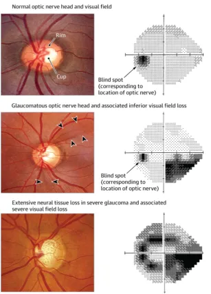
Fig. 1.1. Normal, glaucomatous, and severe glaucomatous optic nerve heads and visual field tests.
In the normal optic nerve head the pink area of neural tissue forms the
neuroretinal rim and the empty central space is the ‘cup’. In the glaucomatous
optic nerve head the retinal rim is thinning, with cup enlargement. The
arrowheads point to retinal nerve fibre layer defect, which appears as a wedge -shaped dark area emanating from the optic nerve head. The superior neural loss corresponds to the inferior defect (black scotoma) seen on the visual field. In the severe glaucomatous eye there is more extensive neural loss with severe
main categories; open- and closed- angle. With open-angle glaucoma the
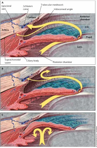
iridocorneal angle is unobstructed, whereas in closed-angle glaucoma the angle is obscured by the iris, blocking the outflow tissues (Fig. 1.2.b-c). Both of open-angle and closed-angle glaucoma can occur as primary or secondary conditions (Leske 2007).
The global prevalence of glaucoma is 3.54% with 3% of this consisting of patients with primary open angle glaucoma (POAG) and 0.5% patients with
primary angle closure glaucoma (PACG). Regional analysis of glaucoma shows that the highest prevalence of POAG is found in Africa (4.79%) and of PACG in Asia (1.09%). Similarly, between ethnic groups, prevalence of POAG is highest in those of African ancestry (5.40%) and PACG prevalence is highest in people of Asian ancestry (1.20%) (Tham et al. 2014). Clinically, in closed angle glaucoma the AC angle is occluded around more than 180° of the circumference of the eye causing a severe rise in IOP. In the case of acute angle closure, patients experience a sharp increase in IOP, that can lead to sudden and painful loss of vision, which can be accompanied by nausea and vomiting due to the iris completely covering the entire trabecular meshwork (Gupta & Chen 2016).These individuals must get immediate medical intervention to avoid permanent vision loss. PACG has a high prevalence among Asian populations and risk factors include: small cornea, shallow AC, thick lens, anteriorly shifted lens position and short axial length (Sun et al. 2017). In the majority of cases angle closure is caused by pupillary block. Briefly, increased resistance at the iris-lens junction to the flow of AH from the posterior to the anterior chamber forms a pressure gradient across the iris and ultimately results in forward bowing of the iris and AC angle closure (Fig. 1.2c).
results in increased IOP, ocular deformation and RGC death. Though primary congenital and juvenile forms of POAG exist, the most common form is adult onset (>35 years); a chronic disease that is often linked with high IOP. However, in some cases the disease progresses and patients can develop optic neuropathy without ever exhibiting high IOP. This is known as normal-tension glaucoma (NTG) and patients generally have consistent IOP measurements of less than 21mmHg. The work presented in this thesis focuses on the mechanisms and treatments
associated with POAG involving high IOP.
POAG and increased outflow resistance
POAG is characterized as any nerve damage that meets the criteria discussed above and decreased AH outflow, but without angle closure (Fig. 1.2b) (Foster et al. 2002). There are numerous risk factors associated with POAG such as myopia, increased age, a family history of glaucoma, race, and ethnicity. With age, other risk factors associated with glaucoma increase, such as blood pressure, cardiovascular disease and thinner central corneas. POAG prevalence is 5 times higher in those of African descent in comparison to other ethnic groups (Harasymowycz et al. 2016; Hollands et al. 2013). In addition to the above, high IOP is one of the most common risk factors for development for POAG and reduction thereof is the main mode of treatment to retard disease progression (Harasymowycz et al. 2016).
IOP is controlled by the homeostatic regulation of AH production and drainage. AH is produced in the processes of the ciliary body (CB) and is an active process. The ciliary arterial supply is derived from the major arterial circle of the iris and it delivers water, ions, proteins and plasma components to the fenestrated capillaries of the CB. The AH components are extracted from the blood, by
uveoscleral pathways (Freddo 2013; Kiel et al. 2011). The conventional pathway drains AH through the TM/SC and out through collector ducts and back into the vascular system to the episcleral veins, whereas in the unconventional pathway, the AH passes through the base of the CB into the suprachoroidal space (see Fig. 1.2a)(Alm & Nilsson 2009; Tamm 2009).
AH that passes through the unconventional pathway passes through the interstitial spaces between the longitudinal ciliary muscle bundles and into the supraciliary spaces. In other words, the unconventional pathway must pass through the ciliary muscle and as a result, factors affecting its contraction or relaxation affect outflow. For example pilocarpine (which is used to treat acute ACG) causes ciliary muscle contraction, decreasing the spaces between these muscle fibres thereby reducing unconventional outflow. Conversely atropine acts as a cholinergic antagonist, relaxing the ciliary muscle and increasing
unconventional outflow. The effect of CB muscle tone on unconventional outflow appears to influence AH drainage with age. The unconventional pathway accounts for approximately 10-35% of AH drainage in humans, which has shown to
decrease with age. The high variances in outflow estimates are due to indirect measurements of human AH outflow. In eyes at age 60, there is a significant increase in connective tissue in the anterior section of the ciliary muscle, which reduces interstitial space, and this may well contribute to the unconventional outflow reduction associated with increasing age. Another feature of
unconventional outflow is that it is relatively pressure insensitive and for this reason it was originally thought that the amount of flow through the
The conventional outflow pathway
The trabecular meshwork (TM) and juxtacanalicular tissue (JCT)
The majority of AH outflow in humans occurs through the conventional pathway located at the iridocorneal angle (Fig. 2a). AH is filtered from the AC through the TM, the juxtacanalicular tissue (JCT) and finally the inner wall of the SC; from here it drains through collector channels back into the limbal vasculature (Keller et al. 2011; Floyd et al. 1985). This pathway is pressure dependent and outflow
resistance changes dynamically in healthy tissue to help maintain IOP within a physiologically safe range. Most of the evidence suggests that the majority of outflow resistance, the primary determinant of IOP, is generated at the inner wall of the SC and the connective tissue of the JCT (Fig. 1.3) (Overby et al. 2009; Llobet et al. 2003).
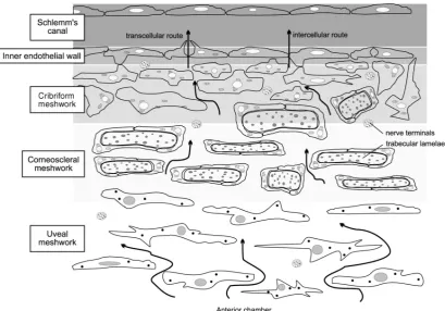
The TM is a highly organised tissue that is located proximal to the AC at the corneoscleral angle. The connective tissue of the TM is composed of prolongations arising from the iris and CB; the inner section consists of columns of glycoproteins, collagen, hyaluronic acid and elastic fibers covered by flattened TM cells (Llobet et al. 2003). It can be separated into regions based on anatomical location, and these differ in structure and function. From the inner to outermost regions, the TM is composed of the uveal meshwork (the first tissue encountered by exiting AH), the corneoscleral meshwork, and finally the JCT (Fig. 1.3). The uveal meshwork is a highly fenestrated tissue formed by trabecular beams made of elastic fibres, collagens and glycoproteins, associated with endothelial TM beam cells. These trabecular beams form a netlike structure with large interstitial spaces that have very little effect on outflow resistance generation. Next is the corneoscleral meshwork. This layer continues for a depth of around 100μm between the uveal meshwork and the outer JCT layer. Similarly to the inner uveal layer, the
corneoscleral layer is composed of highly irregular layers of trabecular beams that form a meshwork of extracellular matrix (ECM), covered and maintained by a continuous layer of TM cells. As this layer continues to the outer edge, the openings between these beams gradually become smaller, that acts as a filter, funneling the AH towards SC.
Fig. 1.2. Normal and abnormal aqueous humour (AH) flow.
and merges with the basement membrane of the SC inner wall, extending on average 10μm from the SC endothelial cells. In contrast to the uveal and corneoscleral network, the JCT is not organized into a layered structure but is composed of an amorphous ECM with a discontinuous scattering of several layers of JCT cells embedded into it. These JCT cells are separate from the SC inner wall but can form processes that make contact with SC endothelial cells and TM cells of the corneoscleral layer (Keller & Acott 2013; Acott & Kelley 2009; Overby et al. 2009; Gong et al. 2002; Llobet et al. 2003).
The ECM support of the TM is a highly dynamic structure that is regulated by its environment, with changes in the extracellular meshwork being linked to actin cytoskeletal changes in the resident cells, thus effecting cell rigidity (Fletcher & Mullins 2010). Interestingly, although the JCT region is both functionally and structurally different to the uveal and corneoscleral regions, the only known biomarker that differentiates the respective cells is the stress protein αB-crystallin (Fuchshofer et al. 2006; Siegner et al. 1996). Though often termed endothelial cells due to their flat nature, the cells of the TM show epithelial cell characteristics and are in fact derived from ocular mesenchymal cells, originating from the neural crest (Cvekl & Tamm 2004; Braunger et al. 2015).
The cells of the uveal and corneoscleral regions are actively phagocytic and act as filters, removing debris such as pigment granules, erythrocytes and
degraded ECM from the AH before reaching the less porous JCT region (Saccà et al. 2016). They can also act as mechanosensors, expressing several known
mechanotransduction channels. In vivo, high IOP has been shown to increase the
pressure gradient across the TM causing cells to stretch in response. In
conjunction with these observations, TM cells in vitro display a diverse response to
mechanical stretching including alterations in ECM, cytoskeleton and induction of cytokine production (Hirt & Liton 2017). These responses are all involved in regulation of outflow resistance and in fact the region around the JCT and SC inner wall is where the majority of outflow resistance is generated (Keller & Acott 2013).
Schlemm’s canal
elliptical canal. This flattened channel encircles the cornea with an average meridional diameter of 233μm and is connected to collector channels along its length, these channels ultimately drain AH into the episcleral veins. Collector channels separate into 2 different categories, those with simple oval openings and those with complex orifice structures forming bridge-like structures or tethered flaps, which may act as vascular valves that inhibit AH backflow (Bentley et al. 2016). Due to unidirectional influx of AH, not all SC endothelia share the same properties, and are separable into inner and outer wall endothelial cells. Along the canal, the inner and outer wall cells are joined by septa, especially clustered at regions with a high proportion of collector channels, and owing to their positioning these septa may prevent collapse of the canal structure. The cells of the inner and outer SC wall differ in morphology, cell biomarkers, specialized cellular organelles, and functions. Whether these differences are due to the vastly different
biomechanical environment these cells experience or due to the effect of the neighbouring cells is unclear (Overby et al. 2009; C. Dautriche et al. 2015).
SC inner wall cells differ to most endothelial cells. Although they are of endothelial origin, during differentiation they acquire some lymphatic
characteristics while retaining their identity as endothelial cells. Interestingly, they express both the endothelial marker PECAM-1 and the lymphatic marker PROX-1 (Park et al. 2014b). Distinct from other endothelial cells, which experience
pressure in the apical to basal direction, pressure is exerted on SC cells basally to apically, i.e. from the AC to the SC lumen. Thus SC endothelial cells have unique barrier properties that allow them to regulate outflow resistance, including the formation of giant vacuoles (GV) and the presence of micron-sized pores that cross the inner wall (Johnson 2006).
exposing the cytoplasmic space. In addition to the transcellular pores, the
continuous SC endothelium is broken by paracellular pores that form between the cell junctions (Johnson 2006). A higher density of paracellular pores has been
linked with areas of increased flow rate in ex vivo eyes and the overall pore density
of the inner wall has been shown to be decreased up to 5-fold in glaucomatous tissues (Braakman et al. 2015a).
Generation of outflow resistance
It is universally recognized that elevated IOP seen in POAG is as a result of
increased outflow resistance, at the conventional outflow tissues. Despite this, it is still unknown how resistance is generated in both glaucomatous and normal eyes (from here on the term glaucomatous will refer to POAG). The uveoscleral pathway carries less than 10% of total AH outflow and thus does not contribute significantly to outflow resistance in the human eye (Johnson 2006; Overby et al. 2009). In the conventional pathway it is generally accepted that very little outflow resistance is attributable to the uveal and corneoscleral meshworks. Both of these structures are incredibly porous; interstitial spaces range from 25-75μm in the inner regions of the uveal meshwork to 2-15μm within the deeper regions of the corneoscleral meshwork (Overby et al. 2009).
These opening have no effect on resistance, as demonstrated by the fact that removal of the proximal TM tissues in enucleated human eyes has no effect on outflow resistance (McEwen 1958; Grant 1963). The lumen of SC carries the total flow of AH that passes through the conventional pathway and is open at low IOPs. However, due to TM expansion during pressure increase the canal has been shown to collapse experimentally (Johnstone & Grant 1973). Collapse of the canal in this manner could potentially lead to the generation of outflow resistance. Yet outflow resistance in normal, as compared to glaucomatous eyes, at high pressure is still much lower, despite canal collapse in the both (Johnson 2006).
Fig. 1.3. Schematic diagram of the conventional outflow pathway. Arrows indicate the flow of aqueous humor (AH) from the anterior chamber (AC) through the trabecular meshwork (TM) toward the Schlemm’s canal (SC) lumen. The separate regions of the TM are labeled; beginning with the innermost layer, the uveal meshwork, the corneoscleral meshwork, and the cribriform meshwork or juxtacanalicular tissue (JCT) region. The final barrier in this pathway is the SC inner wall, where AH flows through both
trabeculectomy, which includes removal of the SC inner wall and surrounding TM, there still remains a minimum of 25% of outflow resistance distal to the SC
(Rosenquist et al. 1989; Van Buskirk 1977). Despite this, it is unlikely that distal tissues play a role in increased resistance in POAG. An early study showed that all glaucomatous related increases in resistance were eliminated in glaucomatous eyes, subsequent to complete trabeculectomy (Grant 1963; Johnson 2006).
Outflow resistance at the juxtacanalicular tissue
Experimental evidence suggests that the bulk of conventional outflow resistance is generated within the vicinity of the inner SC wall, within 14μm (Mäepea & Bill 1989; Mäepea & Bill 1992). This includes the area around the JCT, the SC inner wall endothelium and the discontinuous membrane of this endothelium. The basement membrane has the potential to create significant flow resistance. Nonetheless, examination by quick freeze/deep etch electron microscopy indicates that the basement membrane is discontinuous, thereby limiting its capacity for resistance generation (Johnson 2006; Gong et al. 2002). The JCT forms a meshwork proximal to the SC inner wall and contains micron-sized pores dotted throughout, lacking visible ECM ultrastructure and presenting a rather tortuous pathway for AH to follow on its route from the AC. However, if the current model of JCT structure holds true, then the size of these spaces is too large for the JCT to offer significant impact to outflow resistance (Ethier et al. 1986; Murphy et al. 1992). Either the JCT is not the region of primary resistance generation in the conventional pathway or techniques used for visualizing this tissue do not preserve the ECM present in these pores (Johnson 2006).
known what fraction of outflow resistance is influenced by the JCT.
Outflow resistance at the Schlemm’s canal inner wall
The final tissue capable of generating outflow resistance is the endothelium of SC, these cells being joined by tight junctions (Gong et al. 1996). AH flows through the inner wall of the canal both through paracellular routes and intracellular pores. However, based on pore density it has been estimated that the SC inner wall only contributes to about 10% of total outflow resistance (Bill & Svedbergh 1972). It should be noted however that the fixative used to visualize these pores may increase the number of apparent pores than would be present physiologically. Moreover, there is a decrease in SC pore density in glaucomatous eyes and it is likely that the portion of outflow resistance attributable to the SC inner wall is greater than previously calculated (Johnson et al. 2002; Overby et al. 2009). We are thus left with a paradox in relation to outflow resistance in the AC - the majority of outflow resistance is generated within the area surrounding the SC inner wall, but none of the relevant tissues generate sufficient resistance to account for this, even taking additive effects into account (Mäepea & Bill 1989; Mäepea & Bill 1992; Overby et al. 2009).
This has led to the proposal of a synergistic model of outflow resistance generation, in which the AH is funneled toward SC pores. SC pores are relatively widely spaced apart, with around 20 – 30μm of separation, and presumably create non-uniform flow patterns in the JCT. This funneling effect would reduce the effective filtration area of the JCT and so increase the resistance generation in this tissue (Overby et al. 2009). In this way the funneling effect would create outflow resistance greater than the combined resistance of each tissue. Supporting this model is the effect of SC endothelium disruption by EDTA, which results in greater reduction in outflow resistance than would be estimated from the pore
morphology alone (Hamanaka & Bill 1987). Thus, according to the funneling model, IOP-reducing treatments targeting the SC inner wall have the greatest hope of significant outflow resistance reduction.
Segmental AH outflow
and across the conventional outflow pathway are regions of active and inactive outflow (Carreon et al. 2017). Eyes perfused with cationic ferritin or fluorescent microspheres show high and low flow tracer regions within the TM (Hann et al. 2005). Differential flow patterns are not seen when these same tissues are bathed in tracer, suggesting that these differences are caused by regions of variable flow rate in the conventional pathway. In humans the conventional tissues are generally composed of one third low-flow, one third medium-flow and one third high-flow regions (Vranka, Bradley, et al. 2015). Segmental outflow has been demonstrated in a recent study to be preserved in the tissues distal to the TM, such as SC and the episcleral veins. In that study the TM had an effective filtration area 86% of its total length, whereas the SC and episcleral veins had effective filtration areas of 35% and 41% respectively (Cha et al. 2016). As of yet it is unknown what factors influence segmental outflow, though there is evidence to suggest tracers
accumulate preferentially in regions of the TM proximal to collector channels (Hann & Fautsch 2009; Cha et al. 2016).
Another factor may be the funneling effect in action. It has been suggested that untethering of the SC and TM will relieve outflow resistance by negating the funneling effect and variable tethering of these tissues along the eye circumference influences segmental outflow (Overby et al. 2009). Bovine eyes treated with the rho-kinase inhibitor (Y-27632) demonstrated an increase in total high-flow region area, in conjunction with increased separation between the SC inner wall and JCT. Rho-kinase inhibitors target the SC inner wall and reduce cell-cell interactions (Lu et al. 2008).
removal (Sabanay et al. 2004).
Segmental flow is also influenced by ECM composition and regulation of ECM turnover. The expression of ECM associated glycoprotein versican is inversely related to labeling of high flow regions in the TM (Keller et al. 2011). In another instance, the most highly expressed gene product of the TM is secreted protein acidic and rich in cysteine (SPARC), which is a matricellular glycoprotein involved in ECM remodeling. SPARC null mice show a 15-20% decrease in IOP and outflow is much more uniform across the circumference than in wild-type mice (Haddadin et al. 2017; Swaminathan et al. 2017). In summary, the conventional outflow pathway is composed of regions of variable AH flow, dependent on SC inner wall tethering, TM and SC cell contractility and ECM deposition.
Genetics of POAG
POAG is a complex multifactorial disease that exhibits some heritability that is very dependent on polygenic and gene-environmental interactions. Linkage studies have so far have identified 20 chromosomal loci linked with POAG, however these genes together account for <10% of cases in the general population (Abu-amero et al. 2015). More recently, genome wide association studies (GWAS) have found 16 gene/loci that are significantly associated with POAG in European Caucasian and Asian populations (Wiggs & Pasquale 2017). Mendelian forms of the disease do exist but account for a very small fraction of the cases. Mutations within four
specific disease-causing genes having been found in myocilin (MYOC), cytochrome
P450 family 1 subfamily B polypeptide 1 (CYP1B1), optineurin (OPTN), and
TANK-binding kinase 1 (TBK1) (Liu & Allingham 2017; Zhou et al. 2017).
Myocillin
Mutations in MYOC were first identified in families with autosomal dominant
POAG. The gene located on chromosome 1q, was known to be expressed in the CB and the TM (Sheffield et al. 1993; Stone et al. 1997). Mutations in this gene are associated with juvenile glaucoma, generally manifesting between 3 and 20 years of age, and account for 3-5% of POAG cases worldwide (Wang & Wiggs 2015).
MYOC encodes an extracellular protein, myocilin, of unknown function that is
2009). Most disease-causing mutations occur in the olfactomedin domain of this gene, which inhibits protein secretion from TM cells (Jacobson et al. 2001).
Dominantly-mutated MYOC expressed in TM cells leads to accumulation of the
misfolded protein, leading to disruption of cellular protein trafficking and ER stress (Kasetti et al. 2016). Overexpression, reduction or complete loss of myocilin in mice does not lead to a glaucomatous phenotype, suggesting it does not
normally play a role in IOP regulation (Liu & Allingham 2017). However, transgenic mice expressing the human or mouse Tyr437His variant develop glaucomatous features, including elevated IOP, progressive RGC loss and axonal degeneration. Some of the retinal degeneration phenotype may be
IOP-independent in these animals, since myocilin is also expressed at the optic nerve head (Chou et al. 2014).
Optineurin
Mutations in the OPTN gene are associated with normotensive glaucoma (NTG)
(Child et al. 2002; Ariani et al. 2006). OPTN is involved in membrane trafficking, protein secretion, cell division, autophagy, and host defense against pathogens (Liu & Allingham 2017). The most common variant found in this gene is the missense mutation E50K, associated with very early onset disease progression (Aung et al. 2005). The mutation may be involved in an enhancement of the interaction between OPTN and TBK1, leading to accumulation of the insoluble OPTN and ER
stress in cells (Minegishi et al. 2013). Mutations in the TBK1 gene have also been
associated with hereditary forms of glaucoma. TBK1 encodes a serine/threonine
kinase involved in the regulation of inflammatory responses to foreign agents, specifically regulating the expression of genes in the NF-κB signaling pathway
(Fingert 2011). Duplication of the TBK1 gene has been associated with ~1% of all
NTG cases, sharing its pathway of pathogenesis with OPTN. TBK1 is expressed in
the RGCs, with duplication of TBK1 in mice leading to progressive RGC loss without
increased IOP (Fingert et al. 2016). While the exact mechanism by which OPTN and
GWAS and POAG
Other genes discovered by association analysis with POAG include WDR36, NTF4,
ASB10, EFEMP1 and IL20RB. Genome-wide association studies (GWAS) have increased the extent to which we can understand the genetic component of
glaucoma. It must be noted that all GWAS studies to date have been in populations of Asian and European derivations despite the high prevalence of POAG in those of African descent (Liu & Allingham 2017). Of the genes to come from these studies,
the most examined are the caveolins, CAV1 and CAV2. Caveolins are integral
membrane proteins that are the principal components of caveolae membranes. They are involved in cellular transport, mechanotransduction, cell proliferation, and signal transduction, and are expressed in the retina, CB, TM and SC endothelial cells (Gu et al. 2017; Liu & Allingham 2017). Reduced expression of caveolins in glaucomatous tissues may be indicative of their pathological role in POAG
(Surgucheva & Surguchov 2011). Interestingly, Cav-/- mice display ocular
hypertension, with decreased AH outflow. In these mice TM and SC cells lack caveolae and display a greater susceptibility to rupture through mechanical stress (Elliott et al. 2016). More than 50 other genes have been identified using the GWAS
approach including those with significant associations: CDKN2BAS, TMCO1, SIX6,
8q22, AFAP1, ABCA1, GMDS, TGFBR3, FNDC3B, ARHGEF12,TXNRD2, ATXN2, PMM2,
GAS7, FOXC1, . These genes are involved in a variety pathways such as actin signalling, ER stress, cytokine signalling, membrane trafficking, mitochondrial function, cell division and ocular development all of which could have a role in the progression of the disease (Abu-amero et al. 2015; Liu & Allingham 2017; Wiggs & Pasquale 2017; Bailey et al. 2016).
The mouse as a model for glaucoma
AH formation, regulation and outflow, have similar responses to IOP-lowering drugs, and also have no washout rate, a trait so far known only to be shared between mouse and human eyes (Fernandes et al. 2015). In relation to the AC, the differences between mice and primates include; a larger AC in relation to total eye size and a narrower angle with a thinner and more posteriorly placed TM
containing fewer collagen beams (Chowdhury et al. 2015).
Retinal damage in human glaucoma begins at the optic nerve head. In mice, increased IOP causes similar glaucomatous retinal damage with dysfunction of optic nerve transport. Though the mouse optic nerve head is comparable to
primates there are some important differences, particularly at the lamina cribrosa. In the human optic nerve head the bundled axons pass through the lamina
cribrosa, a meshwork of astrocyte-covered connective tissue beams. The mouse has a poorly developed collagenous lamina cribrosa, but is supported by
transversely orientated astrocytes (Abe et al. 2015; Morrison et al. 2012). Overall, mouse models are a valuable resource in glaucoma research with structurally similar outflow pathways, similar IOP regulatory mechanisms, and hypertension-induced RGC death.
Myocilin mouse model of glaucoma
Myocilin is an extracellular protein of unknown function and mutation of the
MYOC gene is associated with autosomal dominant juvenile POAG (Menaa et al.
2011). Mutations in MYOC result in the prevention of myocilin secretion from TM
cells, resulting in an intracellular aggregation thereof and ER stress (Kasetti et al. 2016). Myocilin is highly expressed in mouse TM cells and RGCs, which is similar to humans (Takahashi et al. 1998; Tamm 2002). The first use of a mouse model to
investigate the role of myocilin in glaucoma was a MYOC knockout mouse, which
had no IOP increase, morphological abnormalities or any other glaucomatous phenotype (Kim et al. 2001). Similarly, overexpression of the gene had no
phenotype related to glaucoma (Fernandes et al. 2015). Mutated mycocilin mouse mutants have also been used to recapitulate glaucomatous phenotypes.
Strain-dependent differences were evident, with some strains lacking any glaucomatous phenotype and others developing a slight increase in IOP and increased RGC death in aged mice. This is despite a lack of myocilin secretion in all transgenic mice (Gould et al. 2006; Senatorov et al. 2006). However, with the use of an adenoviral vector expressing a mutated Tyr437His human myocilin gene that targeted the iridocorneal angle, Shepard et al. were able to demonstrate a very large increase in IOP. This study demonstrated the importance of the peroxisomal targeting sequence, PTS1, which is exposed in the human mutant protein (Shepard et al. 2007).
Transgenic mice expressing the human mutant have since been developed. However, only mice with the human gene under control of a CMV promoter display a distinct glaucomatous phenotype (Zode et al. 2011; Zhou et al. 2008). This CMV-driven model demonstrates that the myocilin mutant elicits an unfolded protein response in the mouse TM, and is associated with a loss of TM cells (Zode et al. 2011). It has been established that these mice have increased deposition of ECM proteins, fibronectin and laminin in the conventional outflow tissues, visible from
3 months of age (Kasetti et al. 2016). These studies only use a single MYOC
mutation but recently novel mutations under evaluation suggest other pathways could be involved in the pathogenesis of glaucoma (McKay et al. 2013; Itakura et al. 2015).
DBA/2J mouse model of glaucoma
The DBA/2J mouse is the best-described congenital mouse model of glaucoma (Fernandes et al. 2015). These mice develop a pigment-dispersing iris disease that leads to increases in IOP and RGC loss, as well a severe age-related anterior
segment anomalies including iris atrophy, peripheral anterior synechiae and pigment dispersion (John et al. 1998). The iris disease is split into two main components: iris pigment dispersion and iris stromal atrophy. The iris pigment dispersion is characterized by a deterioration of the posterior iris pigment
epithelium which is mediated by a premature stop codon in the Gpnmb gene. A
recessive mutation in the Tyrp1 gene is responsible for iris stromal atrophy which
these mice, however on other mouse strain backgrounds they are not sufficient to initiate IOP increase or RGC death (Anderson et al. 2006). This indicates that other factors influence the glaucomatous phenotype in these animals, since the two mutations are sufficient to cause iris disease but not glaucoma on different strains.
In the DBA/2J mouse, as disease progresses there is a breakdown of the blood-aqueous barrier and increased leukocyte infiltration to the AC. This phenotype, along with iris dispersion, are all rescued by the transplantation of C57BL/6J bone marrow into the DBA/2J animal (Mo et al. 2003; Anderson et al. 2008). This demonstrates the possibility of a role for the immune response in the DBA/2J disease, yet to date such a role has yet to be elucidated (Nair et al. 2014). The DBA/2J mouse model shows characteristics of chronic glaucoma, with the disease progressing more rapidly in females than males. IOP begins to rise in females at ~6 months, comparative to ~8 months in males, and reaches a peak at 10 months in both sexes (Libby et al. 2005). DBA/2J animals display disruption in RGC axon transport prior to RGC cell death, as occurs in glaucoma. At 13 months of age there is no significant decrease in RGC density, however 75% of all RGCs are deficient for axonal transport. At 18 months of age these animals have significantly reduced RGC density (Buckingham et al. 2008). Despite these late changes in RGC density, apoptotic RGCs are found in the DBA/2J retina from 9 months of age (Reichstein et al. 2007; Schuettauf et al. 2004).
paracellular permeability and clearance of amyloid-β1-40 from the retina into the peripheral circulation (see Chapter 2).
Ad-TGF-β2 and Ad-CTGF mouse models
TGF-β2 is associated with glaucoma; levels are increased in POAG AH, and exogenous TGF-β2 causes changes in TM and SC cell permeability (Fuchshofer & Tamm 2012). Adenoviral vectors have been used to transduce the cells of mouse outflow tissues, including the TM and the SC (Li et al. 2013). Expression of a spontaneously active human TGF-β2 from such vectors results in a significant IOP increase at day 7 post-injection, which slowly declines but is still moderately high at day 29, with a concomitant decrease in outflow facility (Shepard et al. 2010).
The TGF-β2 mouse model has been used to demonstrate the role of Secreted Protein Acidic and Rich in Cysteine (SPARC) in glaucoma pathogenesis. SPARC is a matricellular protein of the TM and null mice have a 15-20% lower IOP compared to WT. These mice also are resistant to Ad-TGF-β2-induced IOP
increases and inhibit ECM deposition in the outflow tissues (Swaminathan et al. 2014). Connective tissue growth factor (CTGF) is up-regulated in the TM after TGF-β2 treatment and mediates much of the TGF-TGF-β2-induced ECM synthesis. Increase in CTGF expression in the mouse TM results in an increase in IOP that lasts up to 63 days post injection. At day 63, there is a significant reduction in optic nerve axon density. Following these results, a transgenic mouse expressing increased CTGF in the TM was developed. These mice have increased IOP up to 3 months of age and develop reduced optic nerve axon density (Junglas et al. 2012). Adenoviral mouse models develop high IOP with axon degeneration, similar to POAG, and the phenotype develops at an early age, allowing the use of a contralateral eye as a control. This being said, the AV expression is transient and these models are therefore not suitable as models of chronic glaucoma.
Induced hypertension mouse models: vein occlusion
this technique also results in ocular inflammation and is not suitable for research on outflow tissues owing to the damage caused. Glaucoma models have also been generated by targeting the episcleral veins via an occlusion model or with the intravenous injection of saline. In occlusion models IOP is increased by
photocoagulation of the episcleral veins which results in a decrease in AH outflow. Models of this sort also cause complications, including ocular inflammation and ocular surface damage. In the saline-induced model, hypertonic saline is injected into the limbal vessels, bringing about an increase in AH outflow resistance and as a result, IOP elevation. Disadvantages of this approach are the transience of IOP elevation and the difficult technical aspect of microneedle insertion into mouse limbal vessels (Ishikawa et al. 2015).
Induced hypertension mouse models: microbeads
The microbead model of ocular hypertension is an attractive option, being cost-effective, not technically demanding, rapid in onset and applicable in a high percentage of animals. Broadly, IOP is increased by injection of microbeads
(ranging in size from 1-6μm) into the AC. The beads migrate into the TM lodging in these tissues and increase outflow resistance. Such mice demonstrate increased IOP and RGC death; interestingly, RGC loss is strain-specific, with increased RGC death in the CD1 mouse compared to the C57BL/6J mouse after microbead induced IOP elevation (Morgan & Tribble 2015). The principal disadvantages in this model include physical disruption of the TM, which is inconsistent with POAG, and the fact that microbeads can be lost from the AC angle. In regard to the latter, it is interesting to note that new techniques have been developed using magnetic microbeads that can be directed via magnets to the angle (Ishikawa et al. 2015).
Induced hypertension mouse models: glucocorticoid induced glaucoma
administration of the glucocorticoid dexamethasone, results in ocular
hypertension that can be modulated using classical glaucoma therapies (Zode et al. 2011; Whitlock et al. 2010). These models are characterized by an increase in outflow resistance with increased ECM deposition in the JCT tissue (Overby, et al. 2014). In mixed genetic backgrounds, a proportion of the population are non-responders and do not develop ocular hypertension, implying a non-elucidated genetic component (Whitlock et al. 2010). Also, on systemic use of dexamethasone, mice can experience adverse effects including weight loss (Overby, et al. 2014).
Mouse models of retinal ganglion cell death
Several methods have been developed that are designed to induce rapid and uniform RGC death in mice. These include intraocular delivery of various
compounds inducing RGC death such as staurosporine or N-methyl-D-aspartate
(NMDA), an analog of glutamate. Mouse RGCs are susceptible to glutamate toxicity and demonstrate a dose-dependent loss upon NMDA administration (McKinnon et al. 2009). In addition to these, RGC loss can also be induced in mice via direct optic nerve (ON) injury. Complete intraorbital optic nerve crush, or transection, both produce clean and replicable models of RGC degeneration. Both techniques result in two phases of RGC death, with an abrupt loss of 80-95 % of RGCs within 7-12 days post-transection. The second phase is more protracted, with 1-2 % RGC survival at the latest timepoints.
Topographically, these models have diffuse loss of RGCs throughout the retina, consistent with total ON lesion. In comparison, RGC degeneration in ocular hypertensive models is more gradual with patchy loss of RGCs throughout the retina, consistent with damage of ON bundles near the optic nerve head (Vidal-Sanz et al. 2017). These techniques offer quick and reproducible models of RGC death after ON injury, useful for testing neuroprotective treatments. However, RGC loss shows differences in temporal and spatial progression to that seen in ocular hypertensive models, and lack the major risk factor of POAG, increased IOP.
Glaucoma medications
this, IOP remains the only treatable risk factor in glaucoma. POAG can ultimately, only be diagnosed after the detection of neuronal loss at the optic nerve head combined with visual field defect testing (Weinreb et al. 2014). Therefore, in the majority of cases treatment can only begin after RGCs have already been damaged. In one study, an average IOP reduction of 25% resulted in a 17% decrease in patients with progression of visual field defects (Heijl 2002). Treatment goals for IOP reduction vary greatly on an individual basis but in general, a target IOP reduction of 25-30% is desired. Treatments generally include pressure-reducing eye drops that either reduce AH production or increase outflow. In more severe cases surgery is required (Harasymowycz et al. 2016).
The first line of treatment in early glaucoma is the use of medications, most commonly including prostaglandin analogues, β-blockers, α-2 agonists and
carbonic anhydrase inhibitors (Gupta & Chen 2016). Data obtained from a recent meta-analysis indicated that the most efficacious of these are the prostaglandin analogues such as latanoprost, bimatoprost and travoprost (T. Li et al. 2016). These medications reduce IOP by increasing outflow primarily through the
uveoscleral pathway, although some evidence indicates that conventional outflow is also increased during treatment (Bahler et al. 2008; Toris et al. 2008).
Prostaglandin analogues
Prostaglandins are autacoids, hormones that are released and act locally, and are produced natively in the AC from cells of the outflow pathway (Weinreb et al. 2002). These hormones and their analogues affect AH outflow and IOP, and though the exact mechanism is yet to be elucidated, binding of the FP receptor plays a significant role. Remodeling of the ECM in the uveoscleral pathway is the most well-studied effect of prostaglandin treatment. Long-term treatment of monkey eyes with various prostaglandin drugs resulted in enlargement of all the tissue spaces within the ciliary muscle bundles increasing fluid pathways (Nilsson et al. 2006). Increases in interstitial tissue space are due to dissolution of collagen types I and III within the ECM of the ciliary muscle by stimulation of matrix
increased outflow (Bahler et al. 2008; Toris et al. 2008). These medications have some side effects including conjunctival hyperemia, increased pigmentation of the iris, eyelid and eyelash, and eyelash thickening. In the majority of these cases the side effects do not result in treatment discontinuation (Cracknell & Grierson 2009).
β-blockers
β-blockers have been in use clinically for over 30 years, though they are
contraindicated in patients with cardiac or pulmonary conditions and have known side effects. They block β-adrenoceptor binding, reducing AH production by
competitive inhibition of the sympathetic pathway of the CB. Typically β-blockers are selective or non-selective. Selective inhibitors specifically target β1-receptors whereas non-selective inhibitors can target both β1- and β2- adrenoceptors, found within the ciliary processes (Brooks & Gillies 1992; Trope & Clark 1982). The resulting decrease in AH production is likely due to the inhibition of
catecholamine-stimulated synthesis of cAMP in the ciliary epithelium (Marquis & Whitson 2005). Timolol, the most widely used β-blocker, is non-selective. Some reports claim that an added benefit to treatment using selective β-blockers may be enhanced retinal blood flow, decreasing optic nerve axon loss (Collignon-Brach 1992; Messmer et al. 1991). Despite this, the role of retinal blood flow in glaucoma treatment is not yet understood (Noecker 2006).
Topical applications of these β-blockers results in systemic absorption that
can lead to severe side effects. Blocking β1-receptors of the heart by Timolol may lead to bradycardia, arrhythmia, bronchospasm, and congestive heart failure. Furthermore, Timolol can pass the blood-brain barrier and block serotonin receptors in the CNS. It may also cause depression, weakness, fatigue and impotence (Noecker 2006). Nonetheless, Timolol is widely used in glaucoma treatment often in combination with prostaglandin analogues.
α-2 agonists
α-2 adrenergic agonists decrease IOP by the dual action of decreased AH
glaucoma therapies, but these had too many serious side effects such as systemic hypotension. Modern treatments use very selective α-2 agonists like brimonidine, which has an IOP-lowering effect comparative to the β-blockers, and is often used as an adjunctive with latanoprost and Timolol (Fudemberg et al. 2008; T. Li et al. 2016). Side effects of the selective α-2 agonists locally include allergic
blepharoconjunctivitis, a systemically dry mouth, headache, and fatigue (Marquis & Whitson 2005).
Carbonic anhydrase inhibitors (CAIs) and cholinergics
Carbonic anhydrase inhibitors (CAIs) relieve ocular hypertension through
reduction of AH formation by inhibiting the catalysis of the reversible hydration of
CO2 by carbonic anhydrasein the ciliary epithelium (Civan & Macknight 2004).
CAIs were initially administered systemically, with acetazolamide first used for glaucoma therapy in 1954 (Gloster & Perkins 1955). Systemically, CAIs are
effective IOP-lowering agents but have some severe side effects amongst which are paraesthesias of the hands and feet, nausea, vomiting, fatigue, weight loss, and off-target effects to the kidney. Due to these issues, oral medications are generally only used as a temporary therapy, such as before surgery. Topically-applied CAIs such as dorzolamide and brinzolamide, are less effective in comparison to
acetazolamide, but they are additive in their IOP-lowering effect when used with other therapies such as Timolol, and therefore are often used as an adjunct treatment (Marquis & Whitson 2005). The topical CAIs cause significantly fewer adverse effects; systemically bitter taste has been reported in ~25% of patients, and locally, effects include stinging, burning and itching (Swenson 2014).
Lastly are the cholinergic drugs, which had reduced clinical use in recent years and are now not commonly used in POAG. These drugs enhance conventional outflow by stimulating CM muscle contraction, thus increasing the TM/SC angle area (Overby, et al. 2014). This however, has the added effect of reducing
IOP-lowering glaucoma surgeries
Laser trabeculoplasty
Generally, glaucoma is a chronic condition. Medical treatment is approached in a stepwise manner to reach the optimal treatment combination for a desirable outcome. In some cases where medical therapy does not reach optimum
therapeutic goals, or if non-compliance becomes an issue, treatments such as laser therapy or incisional surgery may be required (Weinreb et al. 2014; Marquis & Whitson 2005).
Argon laser trabeculoplasty (ALT) was first proposed in 1979 and involves regularly-spaced laser burns to the anterior trabecular meshwork (Wise & Witter 1979). ALT improves outflow through the TM leading to very high initial success rate. After 5 years however, the success rate is reduced to only 50%. Complications include transient IOP elevation, inflammation of the iris and the formation of peripheral anterior synechiaes (adhesion of the iris to the AC angle) (Marquis & Whitson 2005).
Selective laser trabeculoplasty (SLT) is a similar technique that transmits energy only to the pigmented TM cells, lowering the off-target effect of the laser burn (Kagan et al. 2014). Histological examination of tissue post-ALT indicates that the ECM layers and collagen beams surrounding the TM endothelial cells, as well as the cells themselves, are disrupted and show coagulation damage. Conversely, post-SLT treatment, the disruption was far more focused to the TM cells
specifically and the tissues lacked coagulation damage (Kramer & Noecker 2001; Rodrigues et al. 1982). The mechanism of SLT action has not been fully elucidated; it may be due to the protease/cytokine release that occurs after a laser burn. Outflow from the conventional pathway can be increased via ECM remodeling, and
studies show that TM cells in vitro and in explant models show increased
thickness and corneal hysteresis (Zhou & Aref 2017).
IOP-lowering glaucoma surgeries: trabeculectomy and stent surgeries
Incision surgeries are also commonly used in glaucoma treatment, the most frequent one being trabeculectomy. The latter involves generating a scleral flap with excision of a minute amount of the TM, forming a channel from flap to the AC and allowing the AH to exit the subconjunctival space in a bleb. Long-term scarring along the channel can lead to decreased outflow and hence this form of surgery could be greatly improved by using anti-fibrotic agents. Complications may include hypotony, cataract formation, choroidal effusion, haemorrhage, and inflammation of the eye (Weinreb & Khaw 2004; Marquis & Whitson 2005). Modern methods of glaucoma surgery utilize stent insertion to create a path for the AH to bypass the areas of greatest resistance. These stents are minimally invasive and can bypass the TM to the SC, or suprachoroidal space. Stent surgeries offer different
advantages, some having fewer complications but lesser IOP-lowering effect, hence the type of stent to be used must be chosen on a case-to-case basis (Manasses & Au 2016).
Novel medications: targeting the conventional outflow pathway
Rho-kinase (ROCK) inhibitors
The goal in developing novel glaucoma therapies is to target the cells of the conventional outflow pathway, specifically those in the resistance-generating region around the SC and JCT, with the goal of lowering IOP. The Rho-kinase (ROCK) inhibitors have IOP-lowering effects in humans and have been evaluated for clinical safety and efficacy. Two ROCK inhibitor compounds, ripasudil and fasudil, have been advanced to phase 3 clinical studies (Rao et al. 2017). Ripasudil has been approved for treatment in cases where other treatments are ineffective and shows good tolerability and IOP-lowering effect after 1 year in patients (Garnock-Jones 2014; Inazaki et al. 2017). Rho-kinases are serine/threonine kinases that are involved in the regulation of actin cytoskeletal dynamics, cell adhesion, cell stiffness, and ECM reorganization (Fukata et al. 2001).
Tanihara 2013). On ROCK inhibitor treatment, SC cells show a marked increase in permeability, alter the integrity of tight and adherens junctions, and result in F-actin depolymerisation (Kaneko et al. 2016; Kameda et al. 2012). Recently, work has commenced on a new class of ROCK inhibitors, Netarsudil, that also has activity against the norepinephrine transporter, the drug having been shown to lower IOP in preclinical studies. Netarsudil increases outflow facility, in
conjunction with an increase in the active filtration area along the SC, and increases JCT thickness, which may be associated with cell relaxation (Ren et al. 2016; G. Li et al. 2016).
Adenosine receptor agonists
Adenosine receptor agonists reduce IOP by increasing outflow through the
conventional pathway. There are four known adenosine receptor subtypes; A1, A2A,
A2b, and A3 (Lu et al. 2017). Adenosine is produced in the extracellular space from
ATP, and elevated AH levels of adenosine have been associated with PACG (Li et al. 2011; Andrés-Guerrero et al. 2017). Adenosine-receptor agonists reduce IOP by early reduction in AH secretion, followed by an increase in outflow through
promotion of expression of MMP-2 via the A1 receptor, which results in ECM
degradation and turnover at the TM (Zhong et al. 2013). Trabodenson is an
adenosine agonist with high affinity and specificity for A1 receptors, and has been
assessed in a Phase III clinical trial, showing well-tolerated toxicity profiles along with possible neuroprotective effect (Myers et al. 2016; Laties et al. 2016).
Novel prostaglandin analogues