Declaration
I declare that this thesis has not been submitted as an exercise for a degree at this or any other university and it is entirely my own work, except where otherwise acknowledged. I agree to deposit this thesis in the University’s open access institutional repository or allow the Library to do so on my behalf, subject to Irish Copyright Legislation and Trinity College Library conditions of use and acknowledgement.
Acknowledgements
I would like to thank my supervisor, Professor Pete Humphries, for giving me the opportunity to carry out this project, and for his support over the course of my PhD. Special thanks to Dr. Lawrence Tam for his supervision, guidance, and encouragement throughout my research. Massive thank you to Dr. Ester-Reina Torres, Dr. Joseph Sherwood, Professor Darryl Overby, and Professor Dan Stamer for all their expertise. Thank you also to Dr. Anna-Sophia Kiang, Dr. Marian Humphries, and Dr. Matthew Campbell for their continued help and advice. Thanks to Dr. Matthew Lawrence, RxGen, St Kitts, for generosity with his time and resources. Thank you to the European Research Council for funding my studies.
Summary
Primary open-angle glaucoma (POAG) is one of the leading causes of blindness worldwide, affecting an estimated 44.1 million people, with the number of affected individuals predicted to rise to 79.8 million by 2040, largely due to rapidly increasing aging populations (Tham et al. 2014). POAG is characterised by elevated intraocular pressure (IOP) due to increased resistance to the outflow of aqueous humor (AH) through the conventional outflow pathway, comprising the trabecular meshwork (TM) and Schlemm’s canal (SC), through which the majority of AH drainage occurs in humans. This elevated IOP causes damage to and loss of the axons of the optic nerve, with subsequent retinal ganglion cell (RGC) death in the retina, together termed glaucomatous optic neuropathy. As RGC function to transmit visual signals from the retina to the brain, this damage leads to a progressive, irreversible loss of visual field. The most common current treatments for POAG are topically applied medications that aim to reduce IOP through lowering the production rate of AH, or increasing the outflow rate of AH through the unconventional outflow pathway. There is an unmet need for therapies that target the site of increased outflow resistance in POAG, the conventional outflow pathway.
attempted in wildtype primates, with no effect on AH outflow or IOP detected. Later analysis showed that downregulation of target TJ proteins had been unsuccessful in these animals. Potential improved RNAi delivery mechanisms are discussed.
Glossary of abbreviations
AAV Adeno-associated virus
AH Aqueous humor
AQP Aquaporin
ASB10 Ankyrin repeats and suppressor of
cytokine signalling box-containing protein-10
ASC Apoptosis-associated speck-like protein
containing a caspase recruitment domain
BBB Blood-brain barrier
BCF Brain capillary fraction
C Conventional outflow facility
CARD Caspase recruitment domain
CLAN Cross-linked actin network
Cr Reference conventional outflow facility at
8 mmHg
DAMP Damage-associated molecular pattern
DEX Dexamethasone
ECM Extracellular matrix
EM Electron microscopy
ER Endoplasmic reticulum
FITC Fluorescein isothiocyanate
GC Glucocorticoid
GUK Guanylate kinase
GV Giant Vacuole
GWAS Genome-wide association study
HUVEC Human umbilical vein endothelial cell
iBRB Inner blood-retinal barrier
IHC Immunohistochemistry
IL-18 Interleukin-19
IOP Intraocular pressure
JAM3 Junctional adhesion molecule-3
JAMA Junctional adhesion molecule A
JCT Juxtacanalicular tissue
LC Lamina cribrosa
LCM Laser capture microdissection
MMP Matrix metalloprotease
MYOC Myocilin
NF-kB Nuclear factor kappa-B
NK Natural Killer
NLR NOD-like receptor
NLRP3 NACHT, LRR and PYD domain
containing protein-3
NO Nitric oxide
NT Non-targeting
NTG Normal tension glaucoma
OHT Ocular hypertension
ONC Optic nerve crush
ONH Optic nerve head
OPTN Optineurin
PACG Primary angle-closure glaucoma
PAMP Pathogen-associated molecular pattern
PDG Pigment dispersion glaucoma
PDZ PSD95-DLGA-ZO-1 homology
POAG Primary open-angle glaucoma
PRR Pattern recognition receptor
PXG Pseudo-exfoliative glaucoma
PYD Pyrin domain
RGC Retinal ganglion cell
RNAi RNA interference
ROS Reactive oxygen species
SC Schlemm’s Canal
SEM Scanning electron microscopy
SH3 SRC homology 3
shRNA short hairpin RNA
sIOP Spontaneous IOP
siRNA Short interfering RNA
T Targeting
TBK1 TANK-binding kinase -1
TEER Trans-endothelial electrical resistance
TEM Transmission electron microscopy
TIGR Trabecular meshwork induced
glucocorticoid response (See MYOC)
TIMP Tissue inhibitor if matrix metalloprotease
TIR Toll-IL-1 receptor
TJ Tight Junction
TJP1 Tight junction protein-1 (ZO-1)
TLR Toll-like receptor
TM Trabecular meshwork
VEGF Vascular endothelial growth factor
Table of contents
Declaration ... I
Acknowledgements ... II
Summary ... III
Glossary of abbreviations ... V
Table of contents ... VIII
Chapter 1: Introduction: On aqueous humor outflow dynamics and
glaucoma ... 1
Anatomy of the eye in brief ... 1
Aqueous humor flow dynamics ... 3
Aqueous humor formation ... 5
Unconventional outflow pathway ... 6
Conventional outflow pathway ... 6
Schlemm’s canal inner wall pores ... 9
Isolation of cells of the conventional outflow pathway ... 10
Generation of outflow resistance in the conventional pathway ... 11
Features of glaucoma ... 14
Intraocular pressure and ocular hypertension ... 14
The optic nerve head ... 15
Glaucomatous optic neuropathy ... 15
Types of glaucoma ... 17
Primary angle-closure glaucoma ... 17
Primary open-angle glaucoma ... 18
Normal tension glaucoma ... 18
Pseudoexfoliative glaucoma ... 19
Pigment dispersion glaucoma ... 19
Secondary glaucoma ... 20
Epidemiology and risk factors of glaucoma ... 20
Genetics of primary open angle glaucoma ... 21
Current treatments for primary open-angle glaucoma ... 22
Pharmacological treatment ... 23
Surgical intervention ... 25
Chapter 2: Characterisation and modulation of Schlemm’s canal tight
junction proteins ... 27
Introduction ... 27
Structure and composition of tight junctions ... 27
Tight junction mediated control of paracellular permeability ... 29
Tight junctions of Schlemm’s canal ... 32
Aims ... 33
Results ... 34
Characterisation of tight junction expression in human SC endothelial cells ... 34
Validation of tight junction siRNAs ... 40
Discussion ... 44
Statement on collaboration ... 46
Chapter 3: Investigating the therapeutic potential of RNAi mediated downregulation of tight junction proteins in Schlemm’s canal ... 47
Introduction ... 47
Modulation of tight junction protein expression ... 47
Pressure dependence of aqueous humor outflow ... 49
Measuring aqueous humor outflow facility ... 50
Aims ... 52
Results ... 53
Perfusion Systems ... 53
Effect of siRNA modulation of tight junctions on murine conventional outflow ... 58
Effect of siRNA modulation of tight junctions on murine intraocular pressure ... 62
Ultrastructural analysis of Schlemm’s Canal after siRNA modulation of selected tight junction components ... 63
Adaptation of iPerfusion system to facilitate primate in vivo conventional outflow measurement ... 66
Analysis of effect of delivery of siRNA targeting tight junction components on protein levels in dissected primate outflow tissues ... 73
Mouse in vivo measurement of spontaneous IOP ... 73
Discussion ... 78
Statement on collaboration ... 83
Chapter 4: Investigation of a murine model of ocular hypertension ... 84
X
The DBA/2J mouse ... 85
Induced models of ocular hypertension ... 86
Models of ocular hypertension with increased outflow resistance in the trabecular meshwork ... 86
Aims ... 89
Results ... 90
Evolution of elevated intraocular pressure in dexamethasone treated mice ... 90
Potential reasons for high baseline IOP readings ... 92
Effect of siRNA treatment on intraocular pressure in dexamethasone treated mice ... 94
Effect of siRNA treatment on outflow facility in dexamethasone treated mice ... 96
Discussion ... 99
Statement on collaboration ... 102
Chapter 5: The effect of IL-18 on Schlemm’s canal endothelial permeability and conventional outflow facility ... 103
Introduction ... 103
The innate immune system and sterile inflammation ... 103
The NLRP3 inflammasome ... 104
Interleukin-18 ... 106
Sterile inflammation and primary open-angle glaucoma ... 108
Potential relevance of interleukin-18 to outflow facility ... 108
Aims ... 109
Results ... 110
Effect of interleukin-18 on Schlemm’s canal endothelial paracellular permeability ... 110
Effect of interleukin-18 on Schlemm’s canal endothelial cell viability ... 112
Effect of interleukin-18 on Schlemm’s canal endothelial cell tight junction expression ... 113
Effect of interleukin-18 on conventional outflow facility ... 115
Conventional outflow facility in knockout mice deficient in active interleukin-18 ... 115
Interleukin-18 levels in aqueous humor in a model of axonal damage ... 118
Interleukin-18 levels in human glaucomatous aqueous humor ... 120
Discussion ... 121
Conclusion ... 124
Materials and Methods ... 129
Cell culture ... 129
Human tight junction PCR array ... 129
Western blotting ... 130
Immunohistochemistry ... 131
siRNA ... 132
Cell viability ... 132
Pump based perfusion ... 133
iPerfusion ... 133
Intracameral injection ... 134
Rebound tonometry ... 135
Transmission electron microscopy ... 135
Primate in vivo iPerfusion ... 136
Murine spontaneous IOP measurement ... 136
Dexamethasone micro-osmotic pump implantation ... 137
Trans-endothelial electrical resistance measurement ... 137
Cell permeability assay using FITC-dextran ... 138
Optic nerve crush ... 138
ELISA ... 139
References ... 140
Chapter 1: Introduction: On aqueous humor outflow
dynamics and glaucoma
This chapter will provide an outline of the anatomy of the human eye, together with an explanation of the dynamics of aqueous humor outflow and the generation of outflow resistance. The clinical features of glaucoma, its epidemiology, and contributory genetic factors will be discussed, as well as a summary of the currently available treatments for glaucoma, where the need for improved and more targeted therapies is made clear.
Anatomy of the eye in brief
The anatomy of the eye can be divided grossly in to an anterior and posterior chamber, with the iris demarcating the boundary between the two, fig 1.1. The anterior chamber is bounded by the cornea at its anterior aspect, and the iris at its posterior aspect, with the pupil in the middle of the iris allowing the passage of light through to the retina. The region where the iris and cornea meet is referred to as the iridocorneal angle. The anterior chamber is filled with a fluid called aqueous humor (AH), which has a high rate of turnover, providing nutrients to the avascular tissues of the front of the eye, and mechanical support to the eye through maintaining intraocular pressure (IOP). The lens lies behind the iris, held in place by suspensory ligaments, and functions to focus light before it reaches the retina.
2 of different neural cell types which perceive light, and convert this perception to an electrical signal to be carried to the brain, as shown in fig 1.2.
Figure 1.1: The anatomy of the eye.
The major tissues of the whole human eye are shown labeled here, with the anterior chamber to the right of the diagram and the posterior chamber to the left. From the National Eye Institute image bank.
Figure 1.2: The structure of the retina.
(A) Representation of the overall structure of the retina at the posterior of the eye, with the bottom of the diagram representing the anterior direction.
(B) Diagram of the neural circuitry of the retina, showing a three- neuron chain for transmission of visual signals to the brain, from photoreceptor cells, through bipolar cells, to the retinal ganglion cells. (Purves et al. 2001)
Aqueous humor flow dynamics
4 ciliary epithelium, which line the processes of the ciliary body, and moves forward through the iris into the anterior chamber. In the anterior chamber, temperature differentials between AH near the cooler corneal surface and warmer interior of the eye cause convective flow patterns to arise (Heys & Barocas 2002).
[image:16.595.98.466.320.571.2]From the anterior chamber, AH can leave the eye via two distinct pathways. The first, known as the unconventional, or uveoscleral outflow pathway, involves the drainage of AH through the ciliary muscle, exiting the eye through the suprachoroidal space and the sclera. The second route, known as the conventional, or trabecular, outflow pathway involves drainage of AH through the trabecular meshwork (TM), into the lumen of a canal, called Schlemm’s canal (SC). SC runs around the circumference of the iridocorneal angle, with AH exiting the lumen through collector channels which drain into the episcleral veins.
Figure 1.3: Anatomy of the iridocorneal angle and aqueous humor flow pathways. A. The structure of the iridocorneal angle and the route taken by aqueous humor, from its formation at the ciliary processes, movement around the iris, entering the anterior chamber through the pupil, and finally moving in to the angle itself.
Aqueous humor formation
AH is derived from peripheral blood and is secreted by the epithelial cells of the ciliary processes into the AC, representing a blood-aqueous barrier (BAB). AH must pass through several tissue barriers before reaching the interior of the eye, these barriers being the capillary walls, the stroma of the ciliary body, and the epithelial cell layer. Both passive and active processes are involved in the formation of AH, with passive processes leading to the accumulation of AH components in the stroma of the ciliary body, while active secretion is necessary to complete AH formation (Goel et al. 2010). Passive diffusion allows solutes, in particular lipid soluble molecules, to enter the stroma through across the lipid membranes of capillaries in a concentration dependent manner. Similarly, passive ultrafiltration occurs to move water and water-soluble solutes to the stroma, through fenestrations between the vascular endothelial cells of the capillaries, driven by an osmotic gradient and a differential pressure gradient. This ultrafiltration is size and charge selective, mediated by the fenestrated pores in the capillary endothelium (Civan & Macknight 2004).
Active secretion accounts for 80 – 90 % of AH formation, with this secretion occurring at the non-pigmented epithelial cells of the ciliary processes (Mark 2010). Protein transporters in the cell membranes of these cells mediate selective transcellular movement of water, charged ions, and other molecules. Aquaporins (AQPs) form molecular water channels, allowing bulk flow of water against an osmotic gradient, and AQP1 and AQP4 have been demonstrated to contribute to the formation of AH (Yamaguchi et al. 2006). Other specialized channels actively transport ions including Cl-, HCO3-, and Na+, as well as biomolecules such as ascorbic acid, and amino acids,
across the epithelial layer (Goel et al. 2010). AH turnover occurs at a rate of 1 – 1.5 % of the anterior chamber volume per minute, at an average of 2.4 µL/min in healthy individuals (Brubaker 1997).
6 Unconventional outflow pathway
The existence of the unconventional outflow pathway was inferred through early tracer studies showing the accumulation of tracer molecules in the ciliary body, choroid and sclera following perfusion of these molecules into the anterior chamber. Later studies carried out by Anders Bill in the 1960s introduced radiolabeled tracers into the anterior chamber, which were then shown to accumulate in the same tissues as mentioned above. Bill showed that the tracer accumulated at a constant rate, differing from the diffusion rate of the tracer, leading him to conclude that an AH flow pathway existed out of the anterior chamber through the ciliary muscle and in to the suprachoroidal space (Johnson et al. 2015; Bill 1965).
In the unconventional pathway, AH enters the trabecular meshwork as normal, and from there can move laterally, directly in to the interstitial spaces of the longitudinal bundles of the ciliary muscle, as there is no epithelial barrier blocking access of AH to these spaces (Alm & Nilsson 2009). From the ciliary muscle AH then enters the suprachoroidal space where it can exit the eye by several different pathways, with selective drainage of different AH components occurring through different exit routes. Transcleral drainage can occur through perivascular spaces, accommodating the drainage of larger proteins across the sclera, while smaller molecules and water can exit across the sclera independently of sclera pores through direct diffusion (Jackson et al. 2006; Krohn & Bertelsen 1997). Removal of AH can also occur through osmotic driven forces in the choroid, with drainage though the uveovortex veins (Johnson et al. 2015)
The proportion of AH draining from the anterior chamber via the unconventional outflow pathway exhibits high interspecies variability. In humans, reported values of the amount of outflow occurring through the unconventional pathway vary from 12 – 54 % of total AH outflow, with similar values reported in primates, while in cats, values as low as 3 % are reported (Alm & Nilsson 2009). In certain mice strains, values as high as 80 % of total outflow have been reported (Aihara et al. 2003a).
Conventional outflow pathway
the scleral sulcus. The TM is located at the inner portion of this sulcus, and is the region of the outflow pathway that AH first reaches, while SC is located past the TM at the interior, fig 1.4.
The TM consists of several layers of structural lamellae, beams of connective tissue composed of collagenous and elastic fibres, covered by TM cells. The beams are attached anteriorly to Descemet’s membrane, and extend in a posterior direction where they branch and attach to the scleral spur and ciliary body stroma. The posterior branching of the lamellae gives the TM a triangular shape with a narrow anterior area and wide posterior (Tamm 2014).
The TM can be classified into three main regions, fig 1.4. The inner layer, called the uveal meshwork, consists of up to three layers of TM cell-covered beams originating from the ciliary body at their anterior side. This layer is loosely organized and has large extracellular spaces. The middle layer, the corneoscleral meshwork, is composed of 8 – 15 layers of beams covered in TM cells, which are thicker than uveal beams and originate at the scleral spur (Tamm 2014). The extracellular spaces of the corneoscleral meshwork become progressively narrower as they approach Schlemm’s canal (Abu-Hassan et al. 2014). These two layers together form a filtering meshwork that acts to clear debris from the aqueous humor as it drains out of the AC, with the cells in these TM layers displaying high phagocytic activity removing cellular debris from AH as it passes through, to prevent clogging of the meshwork downstream (Llobet et al. 2003; Abu-Hassan et al. 2014). The final layer, the cribriform meshwork, or juxtacanalicular tissue (JCT), is the narrowest of the three layers. The JCT does not possess any beams, and is instead a loose connective tissue composed of 2 – 5 layers of cells surrounded by an extensive fibrillary extracellular matrix (ECM). The JCT has the smallest extracellular spaces in the TM (Tamm 2009). The TM cells of the JCT form elongated processes, attaching to other TM cells, or to the endothelial cells of the SC inner wall, forming a complex 3-dimensional cellular network.
8 outflow range in value from 40 % to as high as 90 % of total AH outflow, while in mice values are lower, ranging from 20 – 40 % (Braunger et al. 2015a; Lei et al. 2011; Crowston et al. 2004).
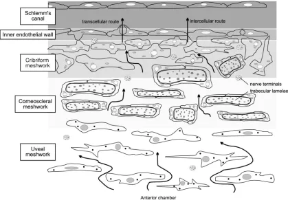
Figure 1.4: The structure of the conventional outflow pathway
Arrows here show the direction of aqueous humor flow through the conventional outflow pathway, beginning at the uveal meshwork, and moving through the corneoscleral meshwork, to the cribriform, or juxtacanalicular, meshwork. From there, flow passes across the inner endothelial wall of Schlemm’s canal by either a transcellular or intercellular, also known as paracellular, route, to the canal lumen (Llobet et al. 2003).
indicating that ECM expression may have a role in segmental outflow (Vranka et al. 2015; Keller et al. 2011).
Schlemm’s canal inner wall pores
Isolation of cells of the conventional outflow pathway
The two main cell types of the conventional outflow pathway, TM cells and SCECs, have been isolated from donor human eyes, and cultured in vitro, allowing further study of their cellular and molecular properties.
The first of these cell types to be isolated were TM cells, isolated through dissection of TM strands and outgrowth of cells from the explant onto a culture plate (Polansky et al. 1981; Polansky et al. 1979). These cells appeared as broad, flat cells, exhibiting cellular branching, and growing in monolayers (Alvarado et al. 1982). TM cells were shown to have several TM specific features, such as expression of a low-density lipoprotein receptor, absence of von Willebrand factor, a vascular endothelial cell marker protein, and high extracellular levels of tissue plasminogen activator (Chang et al. 1991; Snyder et al. 1993). TM cells were also shown to express myocilin in response to treatment with glucocorticoids (Polansky et al. 2000). While this isolation process achieved culture of TM cells, the cellular yield was low, and the process inherently selected for peripheral cells and cells with faster migration rates, giving rise to non-representative populations of TM cells. Improvements came through using a modified protocol intended for isolation of microvascular endothelial cells. This process consisted of the dissection of the TM explants from human donor eyes, and in vitro digestion of the ECM surrounding TM cells, creating a suspension of human TM cells. In this manner, TM cellular yield was greatly increased, and a more representative population of TM cells cultured (Stamer et al. 1995). This same ECM digestion process was utilized to isolate TM cells from donor eyes of patients with primary open-angle glaucoma (POAG) (Stamer et al. 2000).
as further proteins which differentiate SCECs from TM cells (Perkumas & Stamer 2012). Analysis of SCECs derived from glaucomatous donor eyes showed altered pore formation as compared to cells from healthy eyes, exhibiting significantly reduced paracellular and transcellular pore density when perfused. As these pores are the transport route for AH into the SC lumen, this has clear implications for the increased resistance seen in glaucoma. Glaucomatous SCECs also showed increased cellular stiffness than normal controls, which was postulated to contribute to difficulties in pore formation (Darryl R Overby, Zhou, Vargas-Pinto, Pedrigi, Fuchshofer, Braakman, Gupta, Perkumas, Sherwood, et al. 2014).
Generation of outflow resistance in the conventional pathway
The conventional outflow rate of AH is the primary determining factor in the level of IOP, with resistance to AH outflow giving rise to pressure in the eye. While the exact site of this outflow resistance generation within the conventional pathway is not fully characterised, it is known that is generated within the deeper regions of the pathway, in the area occupied by both the JCT and the inner wall of SC. This must be the case due to the highly porous structure of the uveal and corneoscleral meshworks. Modelling of outflow resistance generation has shown that a single pore of the diameter observed in the outer meshworks could accommodate the total AH outflow of an eye. As such, these regions must have negligible contribution to resistance generation. Tissues downstream of the JCT and SC inner wall, such as the SC lumen and episcleral veins, can also be excluded as having a major role in outflow resistance generation, as similarly their large diameters have been shown by hydrodynamic modelling to have no significant contribution to AH flow resistance (Johnson 2006). Direct measurement of the pressure in the lumen of SC in primates also showed that it was at the same level as episcleral venous pressure, being approximately 8 mmHg below IOP, indicating the pressure drop occurs upstream of this point, which must be at the SC inner wall or JCT (Mäepea & Bill 1989).
explanation for this level of conductivity, through visualisation of the density of micron scale pores present on the SC inner wall, with up to 1000 pores/mm2 reported (Ethier et al. 1998). Early SEM studies concluded that the SC inner wall could not provide more than 10 % of total outflow resistance (Svedbergh & Bill 1972). Additionally, AH can also pass between the TJ strands that hold non-pore forming areas of adjacent cell membranes in close contact. Experiments have shown shown accumulation of cationised ferritin accumulates at TJs when perfused in to enucleated human eyes, with a corresponding decrease in outflow facility, the ratio of AH outflow to pressure, (Ethier & Chan 2001). It has also been shown that there are significantly fewer pores at the inner wall of glaucomatous eyes than seen in normal eyes, while areas of high paracellular pore density have been demonstrated to co-localise with high flow segments of the conventional outflow pathway (Johnson et al. 2002; Braakman et al. 2015).
The JCT region would therefore be expected to account for the remainder of AH outflow resistance generation necessary to maintain IOP. This region has extensive, tortuous, sub-micron diameter intercellular AH flow pathways, which, if empty, would not contribute greatly to AH outflow resistance (Ethier et al. 1986). In order for this tissue region to generate the necessary outflow resistance observed in vivo, these extracellular spaces would have to be filled with a resistive substance that was not visible in TEM images. Use of alternative tissue preparation techniques for TEM allowed greater preservation of extracellular structures, showing extensive ECM present in the intercellular spaces of the JCT, but with some large open spaces still present (Johnson 2006; Gong et al. 2002). Additionally, matrix metalloproteases (MMPs), which degrade ECM components, have been shown to reduce outflow resistance when perfused in to enucleated eyes , and when secreted in to the AH, further indicating the role of ECM in the JCT in outflow resistance generation (O’Callaghan et al. 2017; Bradley et al. 1998). Despite this, it remains unclear the extent to which the JCT region generates resistance to AH outflow.
funneling model of outflow resistance generation, as SC inner wall pores are spatially separated on the inner wall, AH moving from the JCT into these pores must converge, or funnel, from wide areas of the JCT to discrete pores on the inner wall, fig 1.5.
Figure 1.5: The funneling model of outflow resistance generation.
A representation of the convergence, or funneling, of aqueous humor flow, represented by arrows, from the JCT region towards discrete pores on the inner wall of Schlemm’s canal. Adapted from Overby et al. 2002.
detail later in this chapter, has been shown to convert segmental outflow patterns to an evenly distributed flow pattern, with a concomitant increase in separation of the inner wall from the JCT (Lu et al. 2008).
Outflow resistance is a dynamic, rather than fixed, property of the conventional outflow pathway. When the anterior chambers of enucleated human eyes are perfused at physiological flow rates until IOP stabilizes, and then subjected to a doubling of inflow rates, IOP is seen to also double immediately, indicating no initial change in outflow resistance. However, over the course of several days of perfusion at this constant high flow rate, IOP is seen to gradually decrease until it reaches the level seen at the original flow rate, showing a clear adaptation of outflow resistance in response to increased pressure. Similarly, doubling of applied perfusion pressure shows an immediate doubling of inflow rate of perfusate, which then subsequently continues increasing further to account for high IOP (Acott et al. 2014).
Features of glaucoma
The term glaucoma encompasses a group of different disorders all sharing similar symptoms and ocular pathology. These diseases are typically characterized by a reduction in the outflow of AH from the anterior chamber, resulting in elevated IOP, a state referred to as ocular hypertension (OHT). Outward pressure in the eye occurring during OHT leads to damage of the neural tissue of the retina and optic nerve head (ONH), due to the elevated pressure exerting an outward force on the ONH, deepening the optic disk, leading to optic nerve atrophy and RGC loss (Noecker 2006). This damage is known as glaucomatous optic neuropathy, and causes progressive loss of visual field.
Intraocular pressure and ocular hypertension
of the tissue. Further, IOP also drives the turnover of AH in the anterior chamber, which provides nutrition to non-vascular anterior chamber tissues (Goel et al. 2010). The normal expected IOP in healthy individuals ranges from 10 – 20 mmHg, with an average of around 14 mmHg, while ocular hypertension is regularly defined as being IOP higher than 22 mmHg (Hoehn et al. 2013; Watson et al. 1996; Camras 1996). IOP values exhibit high levels of diurnal variation, with a range of up to 10 mmHg reported (Asrani et al. 2000).
The optic nerve head
The ONH, also known as the optic disk due to its circular appearance in fundus images, is the point at which retinal ganglion cell axons converge and exit the retina towards the brain through pores in a layered connective tissue called the lamina cribrosa (LC). No photoreceptors are present at the surface of the optic nerve head, and as such it is a visual blind spot. The convergence of RGC axons form a shallow depression in the retina referred to as the optic cup, fig. 1.6 (Weinreb & Khaw 2004). Increased outward pressure occurring during OHT is equal in all directions, and elevated IOP increases the pressure gradient at the ONH. As the sclera is perforated at the ONH, it represents a mechanical weaker point than the rest of the posterior chamber, and as such the increased pressure gradient causes the ONH to recede, increasing the depth and width of the optic cup. The ratio of cup width to disk width, and the change in this ratio over time, can be a measure of the progression of glaucoma, as the ONH is pushed posteriorly by elevated IOP, widening and deepening the optic cup. The depth of the optic cup can also be assessed in patients using several different techniques, such as optical coherence tomography or scanning laser polarimetry. The reflectivity of the ONH under examination is also an indicator of glaucomatous state, with healthy eyes having bright reflections, while in glaucoma, reflectivity is reduced in area and intensity due to loss of retinal fibres (Weinreb & Khaw 2004).
Glaucomatous optic neuropathy
Weinreb 1994). This could be a direct result of the mechanical deformation of the LC damaging RGC axons in the ONH. While the LC provides a support structure for RGC axons by surrounding the axons as they pass through LC pores, deformation of the LC can widen or distort these pores, reducing the physical support provided and causing axonal damage. The axons are also stretched by the deformation and posterior movement of the ONH, also causing mechanical damage (Fechtner & Weinreb 1994).
Figure 1.6: Neurodegenerative changes associated with glaucomatous optic neuropathy.
A. Representation of a healthy optic disk, with convergence of RGC axons from the retina forming a shallow depression, leading through the lamina cribrosa, forming the optic nerve which carries visual signals to the brain.
B. Anatomical features of glaucomatous optic neuropathy. The optic cup has been displaced posteriorly, with damage and loss of RGC axons in the lamina cribrosa, and death of RGC cell bodies in the retina. Adapted from Weinreb et al. 2014.
Along with mechanically induced damage, increases in IOP can also have an effect on retrograde transport in the ONH, depriving RGC cell bodies of neurotrophic factors and preventing recycling of cellular components. This has been demonstrated in experimental models of OHT, with transport of radiolabeled neurotrophic factors being shown to be inhibited by elevated IOP (Quigley et al. 2000). This would lead to induction of cell death due to trophic insufficiency.
Harris et al. 2005; Flammer & Orgül 1998). Other factors such as immune system dysregulation, impaired glutamate signaling, and oxidative stress have also been proposed as potential contributors to glaucomatous optic neuropathy also (Weinreb & Khaw 2004).
Types of glaucoma
The term glaucoma describes a group of similar disorders, with the two main classes of the disease being primary open-angle glaucoma, (POAG), and primary angle-closure glaucoma, (PACG), while several other less common classes of glaucoma also exist, such as normal tension glaucoma (NTG), pseudo-exfoliative glaucoma (PXG), pigment dispersion glaucoma (PDG), as well as multiple forms of secondary glaucoma resulting from other conditions or states.
Primary angle-closure glaucoma
development of PACG include anteriorly positioned lens, peripheral iris thickness, and iris curvature, all contributing to the proximity of the iris to the corneal endothelium in the iridocorneal angle (Nongpiur et al. 2011).
Primary open-angle glaucoma
In contrast to PACG, in POAG the iridocorneal angle remains open, and the resistance to aqueous humor outflow arises within the outflow tissues themselves, specifically in the conventional outflow pathway, fig. 1.7 (Lee & Higginbotham 2005). While it is known the pathological increased resistance to AH outflow occurs in the conventional outflow pathway, the cellular or molecular basis for the pathogenesis of this increased resistance is not known. Dysregulation of the maintenance of IOP homeostasis in different manners have been suggested as contributors to glaucomatous reduction in AH outflow, including mechanisms as diverse as changes in pore density, increased ECM expression in the TM, changes to the contractility or rigidity of TM or SC cells, changes to cytoskeletal arrangement, disruption of the ability to sense elevated IOP, regulation of segmental outflow, or oxidative damage to cells (Kwon et al. 2009; Stamer & Acott 2012). It is likely that a widely varying combination of these potential contributors to increased outflow resistance occur in different patients.
Normal tension glaucoma
Figure 1.7: Primary open-angle glaucoma and primary angle-closure glaucoma.
A. In POAG, increased resistance to AH outflow occurs within the conventional outflow pathways itself, with the iridocorneal angle remaining open.
B. In PACG, the iris blocks the flow of AH into the iridocorneal angle, preventing its drainage through either the conventional or unconventional outflow pathways. Adapted from Weinreb et al. 2014
Pseudoexfoliative glaucoma
PXG occurs in patients with pseudoexfoliation syndrome, and is again a form of POAG. In this disorder, granular protein deposits occur throughout the body, and accumulate in the anterior chamber, where they become lodged in the conventional outflow pathway. Here, they form a physical block to AH outflow, causing intermittent large elevations in IOP, and IOP fluctuations, leading to glaucoma progression as in other forms of the disease (Lee 2008). The presence of pseudoexfoliation material in the anterior chamber is also associated with degeneration of the iris, lens and cornea, which also may contribute to the disease state (Schlötzer-Schrehardt & Naumann 2006).
Pigment dispersion glaucoma
to the iridocorneal angle, where it forms a physical barrier to outflow as in the case of protein deposits in PXG. Pigment dispersion may occur due to friction between the iris and corneal endothelium (Michelessi & Lindsley 2016).
Secondary glaucoma
Several forms of secondary glaucoma exist, where glaucoma is a consequence of a different disease state. Examples of this include inflammatory glaucoma, phacogenic glaucoma, which is induced by cataracts, glaucoma arising as a result of intraocular hemorrhage, neovascular glaucoma, or steroid induced glaucoma.
Epidemiology and risk factors of glaucoma
patients with high IOP that have yet to develop glaucoma symptoms include large optic cup-to-disk ratio, and level of elevated IOP itself, while patients with thin central corneas have a 3-fold higher risk of developing POAG (Gordon et al. 2002).
Genetics of primary open angle glaucoma
While POAG is a complex, multifactorial disease, twin studies and pedigree analysis have revealed that it is also to some extent heritable. Genetic studies have revealed two different classes of genes related to POAG, those that directly cause POAG, and those that increase the risk for developing the disease likely when found in combination with other alleles or environmental factors.
Myocilin (MYOC) was the first gene to be implicated in the direct causation of POAG, through linkage analysis of patients with juvenile onset POAG (Stone 1997). It was later shown to be mutated in 2 – 4 % of POAG patients in a large cohort, with varying mutations present, usually transmitted in an autosomal dominant manner and associated with significantly elevated IOP (Fingert et al. 1999; Fingert 2011). Mutations in MYOC associated with juvenile POAG have been shown to cause IOP elevation beyond 40 mmHg, while mutations associated with adult onset disease generally cause IOP elevations to the range of 25 – 40 mmHg (Kwon et al. 2009). The normal function of myocilin is as yet unknown, however, the wild type form is secreted, while mutant forms cannot be secreted and accumulate intracellularly in the TM, (Jacobson et al. 2001). This accumulation has clear potential to cause damage to TM cells, leading to the pathological development of POAG.
associated with POAG, specifically NTG. TBK1 copy number variations are seen in patients of African origin with NTG (Fingert 2011). Again, little is known of the protein’s function, or role in the development of NTG, but it seems to interact with proteins in the NF- kB signaling pathway.
A further gene identified as being associated with POAG is ankyrin repeats and suppressor of cytokine signaling box-containing protein 10 (ASB10), with mutations identified in two different patient cohorts. ASB10 is expressed in the TM and SC inner wall, as well as the ciliary processes. The role of the protein is not known, but silencing of the gene in TM in organ culture was shown to reduce outflow facility by 50 % (Pasutto et al. 2012).
Genes identified as causative for POAG currently only account for approximately 10% of all POAG cases (Janssen et al. 2013). In addition to pathological Mendelian genes, genome-wide association studies (GWAS) have identified several loci that seem to increase the risk of developing POAG. Notable among these loci are the caveolin genes, CAV1 and CAV2, expressed in the retina, TM and SC, and postulated to alter TGF-β signaling (Fingert 2011; Liu & Allingham 2017). Other genes such as FOXC1, AFAP1, CKDN2B-AS1, TMCO1 and SIX1 have also been shown by GWAS to be associated with POAG, among many other genes (Abu-Amero et al. 2015; Allingham et al. 2009; Liu & Allingham 2017). Risk alleles thus far identified for POAG have modest odds ratios as compared to those identified for other ocular diseases, indicating that aside from cases of PAG caused directly by mendelian mutations, the disease case is likely brought about by the combined minor effects of a huge number of genetic risk factors, in addition to environmental effects (Fingert 2011).
Current treatments for primary open-angle glaucoma
Target reduction in IOP is generally 20 – 50 % of pre-treatment level, with level of IOP decrease being further adjusted downwards if vision loss or optic nerve recession progresses (Prum et al. 2016). Reduction in IOP is generally first attempted through the use of topical pressure lowering medications, with surgical intervention being carried out in patients that fail to achieve target IOP reduction.
Pharmacological treatment
Elevated IOP is brought about by a decrease in the clearance of AH from the anterior chamber through the conventional outflow pathway, while outflow rates are affected, inflow of AH remains unchanged, leading to a state of OHT. Pharmacological approaches aim to restore the balance of inflow and outflow rates of AH, through increase either unconventional or conventional outflow of AH or decreasing the rate of production of AH.
Prostaglandin analogues, for example latanoprost, or bimatoprost, are generally the first line of treatment for POAG, representing the most commonly prescribed topical treatment, and additionally having been shown to be the most effective of all IOP reducing topical treatments (Li et al. 2016). Prostaglandin analogues have been shown to significantly increase outflow of AH through the unconventional outflow pathway, and also appear to increase conventional outflow of AH to some extent (Mastropasqua et al. 2014; Toris et al. 2008). While the mechanism by which prostaglandin analogues increase outflow is as yet not fully understood, it has been demonstrated that treatment brings about an increase in MMP expression in the unconventional outflow pathway, and a resultant reduction in ECM protein levels (Gaton et al. 2001; Sagara et al. 1999). This would be expected to reduce the resistance of this pathway to AH outflow, leading to an increased unconventional outflow rate of AH, and concomitant reduction in IOP.
ciliary processes, and thereby decreasing the fluid available for AH production (Brooks & Gillies 1992). β-adrenergic blockers exhibit a significant side effect profile, including ocular irritation, and are contraindicated in patients with asthma and chronic obstructive pulmonary disease (Weinreb et al. 2014).
Carbonic anhydrase inhibitors, acetazolamide for example, also function to decrease the production rate of AH at the ciliary processes, in this case through inhibition of the formation of bicarbonate, thought to be necessary for maintaining sodium concentrations necessary for osmotically driven AH secretion (Goel et al. 2010; Mousa & Beidoe 2012).
Cholinergic medications, such as pilocarpine, function to increase the outflow of AH through both the conventional and unconventional outflow pathways, through inducing contraction of the ciliary muscle. This contraction pulls on the trabecular beams anchored to the ciliary muscle, opening the spaces of the trabecular meshwork, indirectly increasing conventional outflow facility (Mousa & Beidoe 2012).
Rho-kinase inhibitors, for example ripasudil or netarsudil, are an emerging class of IOP lowering drugs for the treatment of POAG. These drugs increase outflow through the conventional outflow pathway by altering multiple sites within the pathway, leading to an increase in high flow regions. While the exact molecular mechanism for this effect is as yet not known, treatment is correlated with an expansion of the TM and JCT, and dilation of the episcleral veins. Greater changes to segmental flow are observed at the episcleral veins than at the SC inner wall, suggesting that the site of action is distal to SC (Ren et al. 2016). No drugs in this class have yet been granted FDA approval, and currently are only available in Japan.
Surgical intervention
In cases where pharmacological treatment fails to produce the necessary reduction in IOP to slow or stop the progression of POAG, surgical intervention may be required. In some cases, such as severe OHT, surgery may represent a first line therapy (Weinreb et al. 2014). Surgical intervention in the treatment of POAG can be either laser based, or incisional. Laser trabeculoplasty involves the exposure of the TM to laser energy directed through the cornea, with no surgical incisions occurring. The mechanism by which laser trabeculoplasty functions to reduce IOP is not fully understood, but several theories have been proposed. A mechanical theory posits that heat energy from laser exposure causes tissue burns, leading to a contraction of collagen fibres in the TM. This would in turn stretch surrounding tissues, widening the TM and increasing conventional AH outflow. A biological theory proposes that conventional outflow rate increase is a result of ECM remodeling induced by laser induced cytokine release. A further theory states that laser exposure stimulates cell division and subsequent repopulation of the TM (Stein & Challa 2007). In a trial of selective laser trabeculoplasty, 89 % of treated eyes had a reduction in IOP of greater than 5 mmHg (Melamed et al. 2003).
also be implanted into the anterior chamber to allow bulk outflow of AH across the sclera, and trans-trabecular stents may be inserted, bypassing the site of greatest conventional outflow resistance by connecting the outer TM to the lumen of SC (Ahmed et al. 2014; Lee & Higginbotham 2005).
Summary
This introductory chapter has shown the processes through which AH inflow and outflow occurs in healthy eyes, along with the loss of homeostatic IOP in glaucomatous eyes due to aberrant high resistance to AH outflow through the conventional outflow pathway. The resultant anatomical changes, glaucomatous optic neuropathy, and vision loss associated with this increased outflow resistance was explained. Current treatments for POAG were discussed, highlighting the need for novel therapies that target the conventional outflow pathway.
Chapter 2: Characterisation and modulation of Schlemm’s
canal tight junction proteins
Introduction
As has been discussed in the general introduction of this thesis, all AH leaving the eye via the conventional outflow pathway must pass through the trabecular meshwork and subsequently cross the inner wall endothelium of Schlemm’s canal, entering the canal lumen, before returning to peripheral blood circulation through the episcleral veins. As such, it is desirable to characterise the factors that determine the hydraulic conductivity of this pathway.
Endothelial and epithelial cells form monolayers that function to physically separate different biological compartments in organisms, acting as barriers to fluid, ion, and macromolecular flux. In order for these monolayers to function as effective barriers, the plasma membranes of the cells in these monolayers must be maintained in close contact to prevent uncontrolled paracellular movement of biomolecules across the cellular sheet, while also still allowing for selective paracellular diffusion of molecules that are necessary for maintenance of tissue homeostasis. This is achieved through the formation of cell-cell junctional complexes known as tight junctions (TJs) between apposed plasma membranes of adjacent cells.
The focus of this chapter is the characterisation of TJs present between the endothelial cells of SC inner wall, and the validation of their modulation with the intention of increasing paracellular permeability, and therefore elimination of AH from the anterior chamber.
Structure and composition of tight junctions
composed of varying numbers of plasma membrane contact points between cells, forming “a continuous belt-like attachment”, with electron dense regions present in the cytoplasm underlying these contact points, later termed a junctional plaque (Farquhar & Palade 1963).
Subsequent to the identification of TJs by TEM, freeze fracture electron microscopy was utilized to visualize the hydrophobic interior of the plasma membrane phospholipid bilayer of cells joined by TJs. This analysis revealed the presence of complex networks of branching and interlinking transmembrane strands embedded in the cell membrane, which were believed to represent the physical barriers to paracellular diffusion. Further, it was shown that tissues known to form tighter barriers to paracellular transport had more complex junctional strand composition, while those with low resistance to paracellular flow had more simplistic networks composed of fewer junctional strands (Claude & Goodenough 1973).
The first protein component of TJ complexes to be identified was zonula occludens-1 (ZO-occludens-1), a 220 kDA intracellular protein localised to the previously identified cytoplasmic junctional plaques (Stevenson et al. 1986). ZO-1 is an adapter protein, with several protein binding domains, that forms a molecular bridge between transmembrane TJ components and the cytoskeleton of cells possessing TJs. Binding to transmembrane proteins occurs at the N-terminal region of the protein, through several different domains, namely, PSD95-DLGA-ZO-1 homology (PDZ), SRC homology 3 (SH3), and guanylate kinase (GUK) domains. The ZO-1 homologues, ZO-2 and ZO-3, which differ from each other in their C-terminal domains, are also localised to the same region of the TJ complex. Many other PDZ domain-containing proteins are also present in the TJ plaque, including MUPP1, MAGI, and PALS1. These proteins in turn can bind to further adapter proteins, for example cingulin or cortactin, which then bind the actin cytoskeleton, or can interact directly with cytoskeletal components themselves (Zihni et al. 2016).
known as MARVELD2, which is specifically localised to tricellular cell junctions) and MARVELD3 (Zihni et al. 2016; Furuse et al. 1998).
Claudin family proteins have four membrane-spanning regions, forming two extracellular loops, the first of which are typified by the amino acid sequence W-GLW-C-C, while the second extracellular loop of claudin proteins is believed to be responsible for mediating claudin-claudin interactions in TJs, along with a C-terminal PDZ binding domain that interacts with PDZ domain containing proteins such as ZO-1 (Anderson & Van Itallie 2009; Blasig et al. 2006). Claudin proteins form the main components of the transcellular TJ strands observed in early freeze fracture EM images. Different claudin family proteins can from these strands together and bind with claudins on the apposing cell membrane in a homo- or hetero- dimeric fashion, with only specific claudin combinations capable of forming heterodimers, holding the cells in close contact and thereby forming TJs (Furuse et al. 1999).
Tight junction mediated control of paracellular permeability
TJs act as a barrier to paracellular flow, but do not represent absolute barriers to paracellular movement of ions or macromolecules. Rather, TJs act in a size and charge selective manner to form semi-permeable cellular barriers between tissue compartments, with the size and charge selectivity thought to represent two distinct pathways through the TJ complex.
claudins do not just physically block transport of ions, but rather may be actively involved in selective movement of charged ions across the TJ (Amasheh et al. 2002; Van Itallie et al. 2006; Ben-Yosef et al. 2003; Krug et al. 2012). The charge selectivity of claudins is thought to be controlled by residues in the extracellular loops of the transmembrane proteins, which, besides forming dimers with claudins on apposing cells, also are proposed to form pores composed of β-barrel structures consisting of extracellular regions of both claudins in the dimer (Suzuki et al. 2015). Under this model, the charge of amino acid residues contained within these pores would determine the charge selectivity of the pore. Post-translational modifications of claudins also play a role in the regulation of their charge selectivity, with phosphorylation of claudin-14 leading to an increase in paracellular permeability to negatively charged chloride ions (Yamauchi et al. 2004).
phosphorylation of the PDZ binding domain of claudin-4 (Tanaka et al. 2005). Similarly, the phosphorylation of claudin-1 can cause it to be localised to the cytoplasm from cell membranes (French et al. 2009). Other forms of post-translation modification, such as palmitoylation or glycosylation also occur in claudin proteins (Gunzel & Yu 2013).
Figure 2.1: Protein components of tight junctions
This figure shows the representative proteins involved in the formation of tight junction plaques, with transmembrane MARVEL-domain containing proteins and Claudin family proteins, and intracellular zonula occludens proteins. Adapted from (Zihni et al. 2016).
Tight junctions of Schlemm’s canal
The endothelial tight junctions of Schlemm’s Canal differ from those seen in other cell types in several ways. Early studies using electron microscopy showed zonula occludens were present at endothelial cell junctions in Schlemm’s Canal as is seen in vascular endothelia (Vegge 1967). Subsequent freeze fracture studies showed that Schlemm’s Canal endothelial cells TJs were composed of parallel junctional strands with minimal branching which did not form complex bidimensional networks. The lack of branching between TJ strands in SC endothelia leads to the formation of channels between junctional strands that are continuous from the JCT to the SC lumen. These channels were identified as a potential paracellular route for AH outflow into the SC lumen through intercellular clefts in SC inner wall endothelia (Raviola & Raviola 1981; Bhatt et al. 1995). Early tracer studies using cationised ferritin showed staining of cell membranes lining channels between tight junctions in SC inner wall endothelium, showing therefore that these channels represented a paracellular pathway across SC inner wall. Further, these channels were of greater size and number in eyes that were fixed at elevated pressure, (Epstein & Rohen 1991). Additionally, endothelial tight junctions in perfused human donor eyes were shown by freeze fracture to exist in single and double stranded forms at the majority of TJs, with a minority of TJs having three or more junctional strands. The complexity of these junctions was shown to be responsive to applied pressure in the eye, with the number of junctional strands decreasing as perfusion pressure increased, (Ye et al. 1997). The above features are in contrast to TJs of most other ocular endothelia, particularly vascular endothelia, which are composed of complex networks formed of multiple TJ strands, and do not exhibit dynamic regulation in response to pressure, with no changes to TJ strand structure in response to perfusion pressure (Fujimoto 1995; Schneeberger & Karnovsky 1976).
Schlemm’s canal endothelium also differs from other ocular endothelia in that it has remarkably high hydraulic conductivity, with Schlemm’s canal having a hydraulic conductivity of 4000−9000×10−11 cm2 sec/g, significantly higher than other ocular
Campbell et al. 2009). It must be noted that these differences in endothelial permeability cannot be solely apportioned to differences in TJ structure and organization as, in addition to TJ mediated paracellular transport into the SC lumen, SCECs possess giant vacuoles containing large intracellular pores. These intracellular pores have an average pore size of approximately 1 µm in diameter, with pores greater than 3 µm reported (Sit et al. 1997). In the iBRB, ZO-1, occludin and claudin-5 are instrumental in maintaining barrier function, with claudin-5 particularly important in controlling paracellular permeability (Campbell & Humphries 2012; Morita et al. 1999). While the TJ expression profile of SC cells has not been studied extensively, ZO-1 has been seen to be expressed in these cells, and to be upregulated in models of steroid induced glaucoma, with a corresponding decrease in the hydraulic conductivity of the conventional outflow pathway (Underwood et al. 1999). Prior to this study, no claudin family proteins had been identified as being expressed in human Schlemm’s canal endothelial cells.
Aims
Results
Characterisation of tight junction expression in human SC endothelial cells
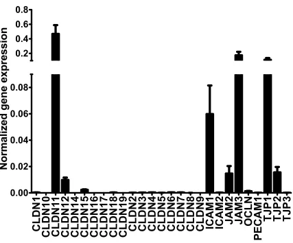
Characterisation of the tight junction expression profiles was initially approached in vitro through the microarray expression analysis of cultured human SC endothelial cell (SCEC) strains and TMC strains obtained from post mortem donor eyes. RNA was extracted from SCEC strains derived from four individual donors and the mean normalised expression (2-∆Ct) of genes encoding claudin and adhesion junctional
proteins was analysed, fig. 2.1. The resulting expression profile identified claudin-11, also known as oligodendrocyte specific protein, as one of the most highly expressed claudin family TJ protein in the cultured SCEC strains. Additionally, the TJ proteins, zonula ocludens-1 (ZO-1), also known as tight junction protein-1 (TJP1), an integral part of TJ complexes, and junctional adhesion molecule-3 (JAM3) were also identified as being expressed at high levels in these cells. Interestingly, the most significantly expressed TJ proteins that form the basis of TJ in vascular endothelial cell layers in human and murine brain and retinal endothelia, claudin-5 and occludin, were only detected at low levels in human SCEC in this array. These data indicate that claudin-11 is the dominant claudin in TJs of cultured SCEC, with ZO-1 being a major TJ associated protein in cultured SCEC. JAM3, while highly expressed, is unlikely to play a large role in endothelial permeability as, while it is a TJ component, it is not essential for TJ formation or maintenance, as is the case with claudin proteins and ZO-1.
Figure 2.2: Microarray expression profile of junctional proteins in human SCEC.
The expression levels of various tight junction and adhesion junction associated proteins were assessed using the Human Tight Junctions RT2 Profiler PCR array. Bar graphs represent 2−ΔCT relative expression levels of target transcripts from 4 separate SCEC strains, normalised to 5 different housekeeping genes, mean ± s.e.m.
Once the expression of these TJ components had been characterised at an mRNA level, it was desirable to further validate their role in the formation of endothelial barriers through confirmation of expression at a protein level. In order to do this, protein was extracted from primary cultured SCEC strains and used in western blot analysis, see fig. 2.3. These data agreed with the findings from the above microarray, showing that claudin-11 and ZO-1 were highly expressed in cultured human SCEC. We additionally showed here the presence of the TJ protein tricellulin, also known as MARVELD2, which was not included as a candidate in the original microarray. Tricellulin functions in the formation of TJs at tri-cellular endothelial junctions. We
CLDN1 CLDN10 CLDN1
1
CLDN12 CLDN14 CLDN15 CLDN16 CLDN17 CLDN18 CLDN19 CLDN2 CLDN3 CLDN4 CLDN5 CLDN6 CLDN7 CLDN8 CLDN9
IC
A
M1
IC
A
M2
JA
M2
JA
M3
OCLN
PEC
A
M1
T
JP1
T
JP2
T
JP3
0.00
0.02
0.04
0.06
0.08
0.2
0.4
0.6
0.8
also confirmed from this protein analysis that claudin-5 was indeed not expressed in SCECs.
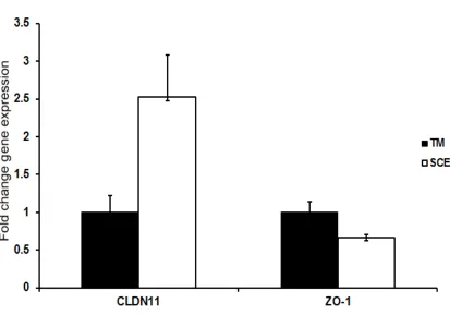
Figure 2.3: Comparison of claudin-11 and ZO-1 expression levels between TMC and SCEC.
The expression levels of claudin-11 and ZO-1 were determined using the Human Tight Junctions RT2 Profiler PCR array, with relative expression between cultured TMC and SCEC shown as 2−ΔΔCT, mean ± s.e.m. (N = 2).
Characterisation of expression of tight junction components in mouse and non-human primate outflow tissues
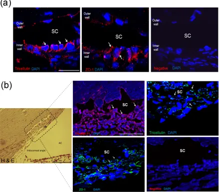
protein yields obtained from dissected material. Instead, immunohistochemistry (IHC) was used to analyse tissue sections from both mouse and primate for the presence and localisation of our proteins of interest. Figure 2.4 (a) shows the localisation of immunofluorescent signals corresponding to ZO-1 and tricellulin at the inner wall endothelial cells of SC in cryosectioned mouse eyes, with continuous junctional strands visible, indicating the presence of functional tight junctions. Staining for ZO-1 and tricellulin was also observed in the TM, but in this case was discontinuous, indicating the absence of a TJ controlled barrier to AH flow in the TM. In contrast to our results from human cells, we were unable to show the presence of claudin-11 or claudin-5 in the mouse SC inner wall endothelium, fig 2.5.
Figure 2.4: Analysis of protein expression of claudin-11, ZO-1, tricellulin, and claudin-5.
Figure 2.5: Immunohistochemical characterisation of TJ protein expression in murine and primate outflow pathways.
(a) Immunohistochemical staining of cryosections of murine conventional outflow pathway, with ZO-1 and tricellulin labeled by Cy3 (red), and nuclei labeled with DAPI (blue). Negative control staining was carried out with no primary antibody. SC = Schlemm’s canal lumen, scale bar = 50 µm. (b) Left: H&E staining of primate iridocorneal angle in paraffin section, with area enclosed in dotted box representing the region in inset immunohistochemical staining images. SC = Schlemm’s canal lumen, AC = anterior chamber, TM = trabecular meshwork, scale bar = 200 µm. Right:
[image:50.595.78.519.83.468.2]IHC was also used to characterise and localise the TJ components present in our non-human primate model, the African Green monkey (Chlorocebus sabeus), fig. 2.4 (b). This analysis showed the presence of continuous claudin-11 TJ strands at the endothelial cell boundaries of the inner wall cells of SC, indicating the role of these cells in the generation of outflow resistance through the maintenance of a TJ mediated endothelial barrier. In addition to this, the outer wall endothelial cells of SC were shown not to possess this strong claudin-11 staining. The same analysis was carried out for ZO-1 and tricellulin, and again an enrichment of expression was seen at the inner wall of SC, with continuous TJ strands visible in both cases. No continuous staining patterns were seen for any of our targets in the TM, indicating that TJ barrier formation in the TM is not likely to contribute greatly to the generation of outflow resistance. These data indicate that in vivo expression of TJ components in our non-human primate model matches well with the pattern seen in cultured primary human SCEC.
Figure 2.6: Immunohistochemical staining of murine conventional outflow tissue for claudin-11 and claudin-5.
Validation of tight junction siRNAs
From this initial analysis, claudin-11, ZO-1, and tricellulin were selected as potential targets for siRNA mediated downregulation as part of our therapeutic approach. In order to begin the process of determining whether modulation of the expression of these proteins could have an IOP lowering effect through increased paracellular permeability at SC inner wall, we first had to demonstrate the ability to downregulate their expression. These proteins were downregulated using RNA interference (RNAi) technology, with short interfering RNAs (siRNAs) that were specific to the mRNA transcript sequence of our selected target proteins.
Figure 2.7: Western blot and densitometric analysis of siRNA mediated TJ protein downregulation.
Figure 2.8: The effect of siRNA mediated downregulation of selected TJ components on cell viability.
Discussion
While TJs in general have been extensively studied in many different cell types, the TJ composition of SC cells had hitherto not been well characterised. As has been previously reported, ZO-1 was confirmed to be expressed in cultured human SC cells at an mRNA level by microarray and at a protein level by western blot (Underwood et al. 1999). Additionally, the expression of claudin-11 was shown by microarray and western blot of cultured cells also, while tricellulin expression was demonstrated by western blot alone, as it was not a candidate gene in the microarray. It was also demonstrated that the expression level of claudin-11 was 2.52 fold higher in cultured SC cells as compared to TM cells, while ZO-1 showed no significant difference in expression levels.
These data were reinforced by IHC staining showing continuous junctional strands of claudin-11, ZO-1, and tricellulin in paraffin sectioned primate outflow pathways, while ZO-1 and tricellulin alone were shown in cryosectioned murine outflow pathways. The staining pattern for these components was typified by enrichment at the inner wall of SC, where continuous strands were more frequently seen, with diffuse staining through the TM. This is consistent with models of outflow resistance generation in the conventional outflow pathway, where resistance to outflow in the TM is not generated through TJ mediated paracellular passage of AH, rather, it arises through heavy extracellular matrix deposition increasing the tortuosity of the pathway as AH approaches the JCT and SC inner wall (Overby et al. 2009). While some staining of TJ components was seen in the outer wall of SC, it was more diffuse and with less continuous strands than that seen in SC inner wall. This is as expected, as no fluid flow, and therefore no pressure drop, occurs across the SC outer wall, instead AH travels through the path of highest hydraulic conductivity, along the SC lumen to collector channels leading to the episcleral veins.