J. exp. Biol. US, 1-13 (1985)
Printed in Great Britain © The Company of Biologists Limited 1985
MUSCLE FUNCTION: A PERSONAL VIEW
B Y D . R. WILKIE
Department of Physiology, University College London, Gower Street, London WC1E 6BT
We are far more likely to arrive at our destination if we know what it is. For those of us who are interested in how muscles work, the destination is the progressive unification of our knowledge of the physics, chemistry and structure - at all levels - of muscular systems. The same integration is needed to understand other organs, but in my opinion the field of muscle research has progressed further than the others. Optimists that we are, our goal seems almost within our grasp. Perhaps it is just at the rainbow's end . . .
The need to relate muscular structure to its mechanical function has been apparent for centuries. Certainly it was clear to Galileo and Borelli (both of whom understood dimensional analysis), and to Leonardo da Vinci (who understood most things). To my mind, William Harvey is pre-eminent in this regard, because he added the stem discipline of experimentation - with all its cussedness - to control the somewhat easier task of speculation.
By the late 18th century the relationship between living function and the then new subject of chemistry was emerging from the work of geniuses like Priestley and especially Lavoisier. As early as 1807 the great Berzelius 'convinced himself that the amount of free lactic acid in a muscle is proportional to the extent to which it has been previously exercised.' (Quotation from Lehmann, 1851). This conclusion (with a few refinements) remains valid today.
The great achievements of the 19th century seem to me to be (not in order of importance):
(1) The perfection of our knowledge of gross anatomy.
(2) A great increase in our knowledge of chemistry and physics. This led to the dismissal of 'vital forces', i.e. the notion that living processes were in some essential way different from non-living ones. Liebig and Pasteur were among the giants who settled this issue.
(3) The demonstration of the continuity of life, i.e. the absence of 'spontaneous generation'. In this Pasteur also played a key part.
(4) Methods of light microscopy were brought almost to their modern state of perfection. However, we should never forget what amazing things Leeuwenhoek saw 200 years before, using a simple lens and a sharp mind. The observations made on living muscle during the 19th century were in fact more accurate than the ones that dominated the scene in my youth. This salutary warning story is brilliantly told in the (1957) review by Andrew Huxley.
1
D . R. WlLKIEMoving to the 20th century, my account is bound to be patchy and centred on my own interests. I beg your indulgence and, to quote from Shakespeare's prologue to Henry V, I hope that you will 'eke out my imperfection with your minds.'
[image:2.451.61.386.225.607.2]Every so often the synthesis between the chemistry and physics of muscle has seemed to be within our grasp. For example, during the 'lactic acid era', roughly 1920
Muscle function 3
to 1930, it was believed by many that the formation of lactic acid was the primary event that powers muscular contraction. This view disintegrated from the blow struck by Lundsgaard in 1931 by his demonstration that muscle contraction can take place in the complete absence of lactic acid formation. At a dinner to celebrate his retirement in 1969, Lundsgaard talked wryly about the great difficulties he had in reproducing his experiments in Meyerhof's laboratory, after his initial success in Copenhagen.
My own interest in muscle began abruptly on the Friday afternoon of August 22, 1941, during a practical class in a converted barn (the college had been severely damaged in October 1940). I became fascinated by a routine class experiment on free and afterloaded muscle twitches. I spent such time as a medical student's life would permit in doing more experiments and reading what I could find about muscle. An ironical consequence was that I did badly at the task set in my practical examination. I could not understand then any more than I can now how the work output in an isometric muscle twitch could be force X length -s- 6!
Unknown to me the stage was already set for the new revolution in which we live. The electron microscope was already waiting in the wings and, during the next two decades, our knowledge of the structure, chemistry and mechanics of muscle would be brought together in the sliding-filament theory of contraction. At the same time crossbridges between actin and myosin, powered by the free-energy change for ATP hydrolysis, were shown to be the most probable site of force generation. These theories seem to be surviving well, though you should never be overconfident about this strange machine which, unlike those made by man, transforms chemical energy into mechanical work at constant temperature. Muscle biology may still have a few tricks up its sleeve.
The elements that contribute to our current theories of muscle contraction come from diverse sources. A. V. Hill's classic paper (1938) had shown a seemingly clear and certainly intriguing relationship between the force-velocity curve and the heat production of muscle. Some years before, he and his co-workers had established many of the ideas still current in exercise physiology, such as that of 'oxygen debt'. I have always wondered whether A.V.'s interest in muscle and exercise physiology arose from his own fine athletic abilities. Fig. 1 shows a photograph that I took at University College in 1951. I have others that show him in more relaxed mood, in his garden in Highgate in 1959 with his wife, Margaret, family and friends. At gatherings like this he was in his element.
of calculator that everyone can now own for a few dollars 1
On the biochemical front, Lippman, Lohmann, Lundsgaard and many others had established by 1940 in 'test tube' studies that ATP is indeed the prime fuel for muscular contraction. The first step towards closing the gap between the chemical and the physical had been made in 1939 by V. A. Engelhardt and his wife, M. N. Lyubimova in Moscow when they showed that muscle protein had ATPase activity. To quote from the splendid book by Dorothy Needham (1971: consult it for detailed references)' . . . the idea of the enzymic activity of the muscle machinery itself was an entirely new one, and the Russian workers fully realised its implications'. They went on to examine the effect of ATP on muscle-protein threads. Perversely, the threads relaxed 1
Quite soon the next great step was made by Albert Szent-Gyorgyi, here shown (Fig. 3) on the beach at Tarpaulin Cove, Naushon Island, when the Woods Hole Laboratory was having its lobster picnic in 1967. We also see the back of Hugh Huxley's head and a charming picture of his wife Frances. In what seems to have been a flash of inspired imagination (who needs more?), Szent-Gyorgyi had devised the glycerinated rabbit psoas preparation that has been a steady and sturdy contributor to muscle research ever since. Under the action of ATP these fibres contract as strongly and as fast as living muscle does.
In retrospect it seems strange that it took so long for the 'biophysical' and the 'biochemical' approaches to link up. Partly it was a matter of mutual ignorance but
there was also an element of aversion: some of the key (and correct) biochemical experiments seemed very messy to the precise-minded biophysicists. The real coming-together of biophysics and biochemistry occurred in a new field, that of ultrastructure, when Hugh Huxley and Jean Hanson (1954 et seq.) showed how the long-demonstrated biochemical processes actually operate at ultra-microscopic level. In this volume, we shall hear further about the way in which Hugh Huxley progressed from the first low-angle, X-ray diffraction studies of muscle, then through a mastery of the electron microscope in the days when it was extremely difficult to use, to his latest studies of time-resolved changes in X-ray diffraction as observed using the powerful X-ray beams derived from a synchrotron.
An almost unknown and, for me, very sad story concerns the way in which Annemarie Weber and I could in principle have discovered in 1951 that muscle shortening results from sliding movement of actin relative to myosin filaments. At least we had assembled in my laboratory all the essential apparatus - polarizing microscope, glycerinated rabbit psoas, ATP and Mg solutions. But alas it never occurred to either of us to look critically at contraction down the microscope: we measured the force-velocity curve instead and because of our blinkers a golden opportunity was lost. Annemarie Weber is shown with A. V. Hill in Fig. 1.
In those days we were obliged to make much of our own apparatus, often out of 'Government surplus' bits and pieces. This was highly educational and at least spared us the present harassment over grant applications. Nevertheless I cannot help regretting the time spent in trying to build stable d.c. amplifiers (especially at Plymouth, where everything conducted electricity) and in making calculations. In some ways I do think the old-days were more suited to accurate experimentation. Walace Fenn once observed to me that in his youth there were few laboratory assistants and scientists performed all aspects of experiments themselves. On the other hand more time was available for these activities because social conditions allowed one to hire servants to look after the house. Today we have technicians in the laboratory and at home do our own washing up.
After a few years even I was forced to realise the importance of biochemistry as a result of thinking and writing about thermodynamics (Wilkie, 1960a). This showed among other things, that the measurements of heat and work that we had made up until then could not be interpreted without parallel chemical measurements. You will be spared details of the next fifteen years' work, whose chief conclusion (Gilbert, Kretzschmar & Wilkie, 1972; Curtin, Gilbert, Kretzschmar & Wilkie, 1974) was that although all the work could be accounted for by measured chemical changes, a large early element of the heat production was unaccounted for. That remains the situation today.
Muscle function
10
1
10
3
£.
3 O
01
0-01
Dog Man Horse
I r
I . . . . I
i i I • . i . l10 100
Weight (kg)
[image:7.451.67.333.122.569.2]1000
8
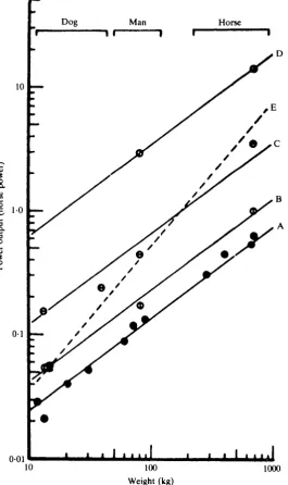
D . R. WlLKIEinvolve spending a couple of afternoons in the library. How mistaken! It must be a familiar experience that one seldom finds exactly what one wants and it took ages to assemble the information required (Wilkie, 19606).
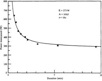
Physiologists had provided numberless measurements of oxygen consumption but the most complete measurements of mechanical power production were the work of a German engineer, Otto Ursinus (1936, 1937), who had made them in connection with an earlier burst of interest in man-powered flying machines. The human engine has the characteristics shown in Fig. 5. These results, obtained by direct measurement on a single subject, confirm the conclusions of the earlier literature survey. The mechanical power that can be produced varies, at first very steeply, with the duration for which it must be maintained. The physiological reason for this is that a fixed amount of work can be obtained from anaerobic sources (hydrolysis of phospho-creatine, formation of lactic acid) over a variable period of time. This is added to the steady power production from oxidative metabolism which can be maintained for long periods of time. Not surprisingly, we have found that sprinting athletes show a very high ability to produce anaerobic work, while endurance athletes are able to maintain a high, steady power output.
I
8UU 700 600 500 400 300 200 100 n- 1
—
—
i iE = 273 W A=16kJ T = 10s
^ * I T
-1
. . . . [image:8.451.49.398.302.577.2]10 Duration (min)
Muscle function 9
Clearly the region of interest in designing man-powered aircraft is from about 3 min onwards, where the very shallow negative slope means that a large increase of duration will be obtained from a small decrease in power requirement. On the basis of quite inadequate information we had to draw up regulations for the first Kremer competition that were to be difficult but not impossible. I think that our figure-of-eight course must have been about right since the prize was not won for 20 years.
The first straight flights took place in England in 1961 and were repeated and extended over a period of years mostly in England and Japan. However, the prize was not actually won until 1977 when a brilliant team from California, led by Dr Paul McCready, adopted a totally novel approach to the problem, ending up with a machine (the Gossamer Condor) that resembled in many ways the original design of the Wright brothers (see Grosser, 1981). Among many brilliant ideas was the one to fly at the very slow speed of 9 miles per hour, about half that of the other contestants. Since air resistance increases as the square of the speed the earlier aircraft had not been able to use the extensive system of bracing wires that made such an important contribution to the success of the Californian aircraft.
A successor to the original aircraft, the Gossamer Albatross, is shown in Fig. 6; this machine flew the English Channel. The team wore T-shirts with the message 'If it hasn't broken, it's too heavy'. Somehow for me this sums up the charm of the very many outstanding engineers and others with whom I was associated for more than 20 years. As Grosser (1981, p. viii) writes: 'Anyone who has lost faith in the kindness and altruism of humanity should try building a human-powered airplane'.
My most recent interest concerns the application of nuclear magnetic resonance spectroscopy to intact tissues which became possible in the early 1970s. I was fasci-nated by this technique. Radio had been one of my hobbies as a schoolboy, and now I could use radio-frequency signals to determine non-invasively the concentrations of metabolites in living muscle. To tell the truth, I was getting a bit weary after more than a decade of quick-freezing frog muscles and then laboriously analysing their extracts by chemical methods. Working at Oxford with our collaborator, David Gadian, Joan Dawson and I were fairly soon able to obtain useful results from oxygenated and anaerobic frog muscles (Dawson, Gadian&Wilkie, 1976, 1977, 1978,
1980).
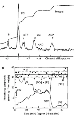
By 1980 technical advances allowed us to study human muscle as well. Fig. 7 shows Joan Dawson serving as the subject for the first physiological experiments on a human subject using NMR spectroscopy. These were undertaken in collaboration with personnel at Oxford Research Systems using their prototype Topical Magnetic Resonance Spectrometer. The spectrum of resting forearm muscle obtained that day (Oct. 8, 1980) is shown in Fig. 8A; Fig. 8B shows the changes in concentration of phosphocreatine (PCr) and inorganic phosphate (Pi) during and following 58 min of complete arterial occlusion.
Fig.
6.
The
Gcmamcr
Albatross,
built
by
a
team
lead
by
Paul
McCready
and
flown
am
the
Engliah
Channel,
June
12,
15'9,
by
Bryan
Allen
Grosser,
12
D . R. WlLKIE+5 - 5 - 1 0 Chemical shift (p.p.m)
B 40
I S
3 0
8 . 1
2 0 |
-M _
• ^s \A
ON
' » " J
[PCr]
/
i
OFF
— ; , •
-[PCr]
[Pi]
0 20 40 60 80 Time (min) (approx 2-5 min bins)
Fig. 8. (A) 31P nuclear magnetic resonance spectrum obtained from approximately 22 cm3 of human forearm muscle using a surface<coil in the 20-cm TMR 32 spectrometer at Oxford Research Systems on Oct. 8, 1980. The spectrum was accumulated in 26 min (800 pulses at 2-s intervals) and enhanced by convolution differencing. (B) The changes in concentration of phosphocreatine (PCr) and inorganic phosphate (Pi) during and following 58 min of complete arterial occlusion. Stars indicate the sum of [PCr] + [Pi] with solid and dashed lines representing mean ± s.D. After Cresshull et al. (1981).
important physiological result concerning the mechanism of activation of glycolysis emerged from this experiment as well. It has been widely believed that glycolysis is regulated by concentrations of phosphorus metabolites or by calculated quantities dependent upon them, such as 'phosphate potential' or 'adenylate charge'. These theories are presented as facts in well-known textbooks (Lehninger, 1975; Stryer, 1981).
[image:12.451.87.337.81.465.2]Muscle function 13
resting muscle, the intracellular pH (indicated by the position of Pi on the X-axis) goes slightly alkaline. Similar changes in phosphorus metabolites as a result of contraction rather than ischaemia are accompanied by a large acid shift in pH, indicating lactic acid formation through the glycolytic pathway. We concluded that whatever switches glycolysis on is closely associated with contraction and does not directly involve changes in phosphorus metabolite levels (see Dawson, 1983; Wilkie, 1983; Wilkie e* a/. 1984).
R E F E R E N C E S
CRESSHULL, I., DAWSON, M. J., EDWARDS, R. H. T . , GADIAN, D . G., GORDON, R. E., RADDA, G. K., SHAW,
D. & WILKIE, D . R. (1981). Human muscle analysed by 31P nuclear magnetic resonance in intact subjects.
jf. Physiol,, Land. 317, 18P.
CURTIN, N. A., GILBERT, C , KRETZSCHMAR, K. M. & W I L D E , D . R. (1974). The effect of the performance of
work on total energy output and metabolism during muscular contraction. J'. Physiol., Land. 238, 455—472. DAWSON, M. J. (1983). Phosphorus metabolites and the control of glycolysis studied by nuclear magnetic resonance. In Biochemistry of Exercise, (eds H. G. Knuttgen, J. A. Vogel & J. Pcortmans), pp. 116-125. International Series of Sports Sciences.
DAWSON, M. ] . , GADIAN, D . G. & WILKIE, D . R. (1976). Living muscle studied by 31P nuclear magnetic resonance. J . Physiol., Land. 258, 82-83P.
DAWSON, M. J., GADIAN, D. G. & WILXIE, D. R. (1977). Contraction and recovery of living muscle studied by
31
P nuclear magnetic resonance. J. Physiol., Land. 267, 703-735.
DAWSON, M. J., GADIAN, D. G. & WILKIE, D. R. (1978). Muscular fatigue investigated by phosphorus nuclear magnetic resonance. Nature, Land. 274, 861-866.
DAWSON, M. ] . , GADIAN, D. G. &WILKIE, D . R. (1980). Mechanical relaxation rate and metabolism studied in fatiguing muscle by phosphorus nuclear magnetic resonance. J. Physiol., Land. 299, 465—484.
GILBERT, C , KRETZSCHMAR, K. M. & WILKIE, D. R. (1972). Heat, work, and phosphocreatine splitting during muscular contraction. Cold Spring Harb. Symp. quant. Biol. 37, 613—618.
GROSSER, M. (1981). Gossamer Odyssey: the Triumph of Human-Powered Flight. London: Michael Joseph Ltd. HILL, A. V. (1938). The heat of shortening and the dynamic constants of muscle. Proc. R. Soc. B 126, 136-195. HUXLEY, A. F. (1957). Muscle structure and theories of contraction. Prog. Biophys. biophys. Chem. 7, 255-318. HUXLEY, H. E. & HANSON, J. (1954). Changes in the cross-striations of muscle during contraction and stretch
and their structural interpretation. Nature, Land. 173, 973-976.
LEHMANN, C. G. (1851). Physiological Chemistry (tr. by G. E. Day). London: Harrison & Son.
LEHNINGER, A. L. (1975). Biochemistry, Part 2, Chap. 16. 2nd Ed. p. 425. New York: Worth Publishers Inc. NEEDHAM, D . M. (1971). Machina Carnis. The Biochemistry of Muscular Contraction in its Historical
Development. Cambridge: Cambridge University Press.
STRYER, L. (1981). Biochemistry, 2nd ed. San Francisco: W. H. Freeman & Co. p. 299. URSINUS, O. (1936). Grfindung des Muskelflug. Instituts Frankfurt aM., Flugsport, pp.1-28. URSINUS, O. (1937). Versuche mit Energie-speichern. Instituts Frankfurt aM., Flugsport, pp. 33—40. WILKIE, D. R. (1950a). The relation between force and velocity in human muscle. J . Physiol. 110, 249-280. WILKIE, D. R. (19506). The circuit analogue of muscle. Electronic Engineering, October, 1950, pp. 435-438. WILKIE, D . R. (1959). The work output of animals: flight by birds and by manpower. Nature, Land. 183,
1515-1516.
WILKIE, D . R. (1960a). Thermodynamics and the interpretation of biological heat measurements. Prog.
Biophys. 10, 259-298.
WILKIE, D. R. (19606). Man as a source of mechanical power. Ergonomics 3, 1-8.
WILKIE, D. R. (1980). Equations describing power input by humans as a function of duration of exercise. In
Exercise Bioenergetics and Gas Exchange, (eds C. Cerretelli & B. J. Whipp), pp. 25—34. Amsterdam:
Elsevier/North Holland Biochemical Press.
WILKIE, D. R. (1981). Shortage of chemical fuel as a cause of fatigue: studies by nuclear magnetic resonance and bicycle ergometry. In Human Muscle Fatigue: Physiological Mechanisms, (eds. R. Porter&J. Whelan), (Ciba Found. Symp. No. 82). London: Pitman Medical.
WILKIE, D . R. (1983). The control of glycolysis in living muscle studied by nuclear magnetic resonance and other techniques. Biochem. Soc. Trans. 3, 244-246.
WILKIE, D . R., DAWSON, M. J., EDWARDS, R. H. T . , GORDON, R. E. & SHAW, D . (1984). 31P-NMR studies of