James M. Provenzale and Roger E. McLendon
PURPOSE: To determine the MR features of spinal angiolipomas and to compare these findings with their histologic appearance. METHODS: The MR examinations of three patients with surgically proved angiolipomas were reviewed for tumor location and extent, signal characteristics, and pattern of contrast enhancement, and were then compared with the histologic findings. RESULTS: Four tumors were found in the three patients, all located in the posterior epidural compartment, averaging about 2.5 vertebral bodies in length. On noncontrast T1-weighted images, all lesions were inhomogeneous and hypointense relative to epidural fat. Inhomogeneous enhancement was seen in three lesions on postcontrast T1-weighted images obtained with fat-saturation techniques. Angiolipomas were least conspicuous on T2-weighted images. A high vascular content correlated with the presence of large hypointense regions on T1-weighted images. CONCLUSION: Spinal angiolipomas are typically hyperintense on noncontrast T1-weighted images relative to other tumors. Angiolipomas that contain large hypointense foci on noncontrast T1-weighted images can be expected to have a high degree of vascularity.
Index terms: Lipoma; Spine, neoplasms
AJNR Am J Neuroradiol17:713–719, April 1996
Angiolipomas are uncommon, benign neo-plasms that consist primarily of adipose and vascular elements. They are usually found in the soft tissues of the extremities, trunk, or neck (1). Spinal angiolipomas are rare lesions, ac-counting for between 0.14% and 1.2% of spinal axis tumors (1). Previous reports of the mag-netic resonance (MR) imaging appearance of spinal angiolipomas have noted that these le-sions are typically hyperintense on T1-weighted images (because of their lipomatous content) and often inhomogeneous (1–5). We report the MR imaging and histologic findings in three cases of spinal angiolipoma.
Materials and Methods
A review of our neuropathologic records over a period of 3 years revealed three patients (all women; average age, 47 years) in whom histologic findings prompted a diagnosis of spinal angiolipoma. All three patients had
undergone preoperative MR imaging examinations. In ad-dition, in one patient, axial computed tomography (CT) was performed before and after administration of intrave-nous contrast material, and contiguous 3-mm sections were obtained. Pulse sequences used included noncon-trast T1-weighted (three patients), T2-weighted (three pa-tients), noncontrast T1-weighted with fat saturation (one patient), postcontrast T1-weighted with fat saturation (two patients), and postcontrast T1-weighted without fat satu-ration (one patient). T1-weighted images were obtained with parameters of 500 –749/11–18/2 (repetition time/ echo time/excitations). Precontrast and postcontrast T1-weighted images were obtained in all three cases. T2-weighted images were obtained using a fast spin-echo technique with parameters of 3000 – 4000/34 –38,102– 108 (effective)/2. Fat-suppression T1-weighted images acquired with a frequency-selective pulse followed by gra-dient dephasing were obtained in two patients.
MR images were examined to determine the number and rostrocaudal length of the lesions, the presence of hyperintense signal within the tumor on T1-weighted im-ages, presence of inhomogeneous signal within the tumor, and degree of contrast enhancement. The histologic find-ings of each tumor were reviewed for degree of vascularity and compared with the MR findings in each case.
Results
Four lesions were found in the three patients. Three of the lesions were proved histologically
Received July 7, 1995; accepted after revision October 30.
From the Departments of Radiology (J.M.P.) and Pathology (R.E.M.), Duke University Medical Center, Durham, NC.
Address reprint requests to James M. Provenzale, MD, Department of Radiology, Duke University Medical Center, Durham, NC 27710.
AJNR 17:713–719, Apr 1996 0195-6108/96/1704 –0713
qAmerican Society of Neuroradiology
at surgery and the fourth (a second lesion in patient 2) was presumed to be an angiolipoma on the basis of nearly identical signal character-istics as the first lesion. The lesions ranged from 1.6 to 4.0 vertebral bodies in length (average, 2.6 vertebral bodies). One lesion extended into adjacent neural foramina, resulting in foraminal widening.
Imaging Appearance
All lesions were predominantly hyperintense on noncontrast T1-weighted images but were slightly or moderately hypointense relative to epidural fat. All lesions were inhomogeneous on each of the pulse sequences, but this feature was most pronounced on noncontrast T1-weighted images, in which focal regions of hy-pointense signal relative to the overall signal of the tumors were seen (Figs 1A and 2A). These regions varied in size; they were small and had a mottled appearance (Fig 1A) in two cases, and occupied a relatively large volume of the tumor in one case (Fig 3A).
Postcontrast T1-weighted imaging with fat saturation was the best pulse sequence for dem-onstrating the tumor. All three lesions imaged using this sequence diffusely enhanced in an inhomogeneous manner and were brighter than epidural fat (Figs 1B and 2D). On proton den-sity–weighted images, angiolipomas appeared much brighter than cerebrospinal fluid but were nearly isointense with fat. The lesions were least conspicuous on T2-weighted images, on which they were only mildly hypointense relative to CSF and nearly isointense or slightly hypoin-tense compared with epidural fat (Fig 3C).
Noncontrast CT scans showed the mass in patient 1 to be slightly hyperdense relative to epidural fat (Fig 1C). Contrast-enhanced CT in the same patient showed the mass to enhance diffusely and to become relatively isodense with the thecal sac (Fig 1D).
Histologic Appearance
In patients 1 and 2 the lesions had the normal whitish yellow histologic appearance of benign epidural fat. A gross specimen was not available for the lesion from patient 3. Microscopically, the lesions from patients 1 and 2 showed sheets of mature adipocytes densely interspersed with numerous small vascular channels (Fig 1E). In addition, the specimen from patient 2 also
fo-cally exhibited dilated vascular channels en-meshed within dense fibrous connective tissue containing little fat. In contrast, the histologic appearance of the lesion from patient 3 was marked by intense vascularity with cavernous vascular channels that appeared to fold upon themselves in tortuous knots, coursing between islands of mature adipocytes (Fig 3D).
Radiologic-Pathologic Correlation
In the three lesions that were resected, there was good correlation between the degree of hy-perintense signal on noncontrast T1-weighted images and the degree of lipomatous content. The lesions in patients 1 and 2 were diffusely hyperintense and had the highest ratios of fat to vascular content. The lesion in patient 3 had relatively large foci of hypointense signal (rela-tive to the rest of the tumor) and had the highest degree of vascularity.
Discussion
Angiolipomas are benign tumors composed of varying proportions of mature fat cells and angiomatous proliferation of blood vessels (6, 7). They are considered a subgroup of lipomas and have been referred to by various terms, including vascular lipoma, hemangiolipoma, andfibromyolipoma(1). Mitoses and pleomor-phism are not features of these lesions (6). An-giolipomas may be composed predominantly of vascular and stromal elements, with only a small lipomatous component and blood vessels varying in size from capillaries to arteries (1, 7). Alternatively, they may be predominantly lipo-matous and contain only a few small angioma-tous regions. In the latter instance, they can be distinguished from lipoma by the presence of capillaries with a complex branching pattern and abnormal pericytic proliferation (7). Sparsely vascularized angiolipomas can be dis-tinguished from lipomas by the presence of mi-crothrombi and fibrous scarring in angiolipomas but not in lipomas. Angiolipomas can be distin-guished from liposarcomas by the typical pres-ence of mitotically active, pleomorphic lipo-blasts associated with an arcuate, or “chicken track,” vascular appearance in liposarcomas, features not found in angiolipomas (6). Further-more, angiolipomas often contain a greater number of mature, thick-walled vessels than do liposarcomas (6). Distinction from
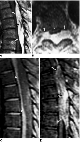
Fig 1. Patient 1: 38-year-old woman with lower back pain of 3 years’ duration.
A, Noncontrast T1-weighted sagittal MR image (500/11/2) of the lumbar spine shows a slightly inhomogeneous posterior epidural mass (white arrows), which is moderately hypointense relative to normal epidural fat (curved arrow) and that contains small foci that are even more hypointense (open arrow). The lesion was nearly isointense with epidural fat on T2-weighted images (not shown).
B, Postcontrast T1-weighted sagittal MR image (600/11/2) obtained with fat saturation technique shows dense, slightly inhomoge-neous enhancement of the mass (arrows).
C, Noncontrast axial CT scan shows anterior displacement of the thecal sac by a slightly inhomogeneous mass (arrow), which is mildly hyperdense relative to fat. The mass measured between259 and2112 Hounsfield units.
D, Contrast-enhanced axial CT scan shows moderate enhancement of the mass (arrows), which is now relatively isodense with the thecal sac. The mass now measured between224 and231 Hounsfield units.
Fig 2. Patient 2: 61-year-old woman with a 2-year history of increasing parapa-resis and two thoracic spinal lesions with similar imaging characteristics, one of which is illustrated.
A, Noncontrast T1-weighted sagittal MR image (500/11/2) shows an inhomo-geneous mass (white arrows) compress-ing the spinal cord. The mass is generally slightly hypointense relative to normal epi-dural fat but contains a region that is es-pecially hypointense (black arrow). This lesion was resected and, on microscopic examination, the degree of vascularity was relatively close to that of the lesion in pa-tient 1.
B, Noncontrast T1-weighted axial MR image (500/11/2) shows the mass dis-placing the thecal sac (arrow) anteriorly and extending through, and widening, the neural foramina bilaterally (arrowheads).
C, Noncontrast T1-weighted sagittal MR image (556/10/2) obtained with fat-saturation technique shows a diffuse, nearly homogeneous decrease in the sig-nal intensity of the lesion (arrows).
D, Postcontrast T1-weighted sagittal MR image (566/11/2) obtained with fat-saturation technique shows inhomoge-neous enhancement of the mass (white arrows). The most densely enhancing por-tion of the mass (black arrow) is near the site that was most hypointense on the non-contrast images (Fig 2A).
entiated low-grade liposarcomas lacking pleo-morphism can be difficult, if not impossible, and depends on the demonstration of lipoblasts in liposarcomas but not in angiolipomas (6). An-giomyolipomas, which generally occur in the kidney and are associated with tuberous sclero-sis, are distinguished from angiolipomas by the presence of smooth muscle proliferation within
[image:5.612.56.559.92.521.2]the former. Both encapsulated (ie, noninfiltrat-ing) and nonencapsulated (ie, infiltratnoninfiltrat-ing) forms of angiolipomas are recognized. Noninfiltrating angiolipomas typically involve the subcutane-ous tissues, are frequently multiple, are usually seen in young adults, present clinically as pain-ful nodules, and usually are easily cured by surgery (8). Infiltrating angiolipomas are rare, Fig 3. Patient 3: 42-year-old woman with midthoracic back pain of 2 years’ duration. A, Noncontrast T1-weighted sagittal MR image (566/18/2) of the midthoracic spine shows a very inhomogeneous mass that has a large component (long arrowheads) that is hypointense relative to fat, consistent with a more vascular nature compared with the lesion in Figure 1. Portions of the tumor that are isointense with fat (short arrows) are seen above and below the hypointense region.
B, Contrast-enhanced T1-weighted sagittal MR image (566/18/2) shows diffuse, slightly inhomogeneous enhancement of the mass (arrowheads). The regions that were hypointense relative to fat on Figure 3A are now relatively isointense with fat.
C, T2-weighted sagittal MR image (4000/108 effective/2) shows the mass (straight arrows) is hyperintense relative to spinal cord (arrowheads) and nearly isointense with normal fat (curved arrow).
usually involve the extremities, can infiltrate lo-cal structures, and have about a 50% recurrence rate (9).
Angiolipomas involving the central nervous system are extremely uncommon, with about 90% located within the spinal canal (10). In rare instances, angiolipomas have been found in-tracranially (11, 12). The vast majority of spinal angiolipomas are extradural (1, 13). Intramed-ullary angiolipomas are extremely rare (1). In the spinal canal, noninfiltrating forms of the tu-mor are much tu-more common (1, 14). Spinal angiolipomas usually extend over three to four vertebral bodies in length. Although angiolipo-mas are typically located in the posterior epi-dural space, infiltrating angiolipomas are gen-erally in the anterior epidural compartment (1). The typical clinical presentation of extradural angiolipoma is back pain, progressive parapa-resis, and lower extremity sensory changes, of-ten occurring over a period of a few years (1). The lesions are more common in females and generally present in the fifth decade, although intramedullary angiolipomas generally present at an earlier age (1, 13, 15). In unusual cases, a relapsing-remitting course has been noted (16). Symptom onset and exacerbations have been noted during pregnancy, possibly in conjunc-tion with an increase in tumor volume (resulting from the generalized rise in blood volume during pregnancy), impaired spinal venous drainage, or hormonal changes resulting in an increase in extravascular fluid volume (1, 17).
The CT appearance of spinal angiolipoma is that of a mass that is typically hypodense rela-tive to the spinal cord and has variable degrees of enhancement after contrast administration (1–3). On rare occasions, portions of angiolipo-mas within the spinal canal have been reported to be calcified (3). Infiltrating forms of the tumor can destroy adjacent bone and produce a tra-beculated appearance within the adjacent ver-tebral body, simulating hemangioma (1, 14). Vertebral bodies infiltrated by angiolipoma on CT scans have been reported to exhibit little or no contrast enhancement, which, according to some investigators, is a reliable way to distin-guish angiolipoma from vertebral hemangioma (14).
Because angiolipomas are mostly hyperin-tense on T1-weighted images and often nearly isointense with epidural fat, fat-suppression MR imaging is particularly well suited for the inves-tigation of these tumors. Contrast
administra-tion in conjuncadministra-tion with fat saturaadministra-tion in two of our patients served to make the lesions more conspicuous and to define better the borders of the tumor and aid in surgical planning. Almost all reported angiolipomas (and all lesions in our patients) have been predominantly hyperin-tense on T1-weighted images and inhomoge-neous owing to interspersed vascular elements (2, 3, 5). Rarely, angiolipomas have two dis-crete foci, one that is homogeneously hyperin-tense and another that is isoinhyperin-tense with spinal cord on noncontrast T1-weighted images (1, 2). The lesion in patient 3 differed from this form of angiolipoma because there were a large number of foci (rather than two foci) arrayed in some-what of a mosaic pattern (Fig 3A). The appear-ance of angiolipomas differs from that of lipo-sarcoma, another lipomatous (but malignant) lesion in a number of ways. Well-differentiated liposarcomas frequently have irregular, thick-ened septa on CT and MR studies and contain regions of hyperintense signal (compared with fat) on T2-weighted MR images (18), findings not seen in angiolipoma. Unlike angiolipomas, myxoid and pleomorphic liposarcomas gener-ally are relatively homogeneously isointense with muscle on T1-weighted images and hyper-intense relative to fat on T2-weighted images (18).
The MR appearance of angiolipomas corre-lated well with their observed histologic compo-sition. As expected, the lesions in patients 1 and 2 were composed primarily of fat and the lesion in patient 3 was highly vascular. Unlike some other vascular tumors (eg, glomus jugulare tu-mors), angiolipomas do not typically contain vascular flow voids on MR images (1, 2). This is probably because of the preponderance of cap-illaries and venous channels in angiolipomas, which distinguish them from those lesions with predominantly arteriolar circulation and from malformations with arteriovenous shunting, both of which produce fast flow, seen on MR images as flow-void phenomena. The presence of small vascular channels was sufficient to ren-der the angiolipomas inhomogeneous and hy-pointense relative to epidural fat, which, in con-junction with contrast enhancement, indicated that the lesions were probably some form of vascularized lipomatous tumor. Spinal angioli-pomas can, therefore, be distinguished from epidural lipomatosis, which is isointense with epidural fat and homogeneous and would not be expected to show contrast enhancement (19).
The treatment of spinal angiolipoma is surgi-cal extirpation. Total removal of epidural non-infiltrating lesions is possible in about 80% of cases (1, 14). As expected, infiltrating angioli-pomas are more difficult to remove, especially if they are anterior in location (1, 14). A staged procedure or only subtotal resection may be necessary. Because these lesions are slow growing and do not undergo malignant transfor-mation, partial resection often provides sub-stantial symptomatic relief (1).
Angiolipomas are rare but should be consid-ered when an inhomogeneous hyperintense mass on T1-weighted MR images is encoun-tered. Our findings indicate that the degree of central hypointensity on T1-weighted images is predictive of the degree of vascularity likely to be encountered at surgery. A preponderance of hypointense regions indicates a very vascular tumor, even though flow voids are not seen.
Acknowledgments
We thank Janet Garrett, MD, for contributing the MR image of patient 3 and Kathy Thompson for assistance in manuscript preparation.
References
1. Preul MC, Leblanc R, Tampieri D, Robitaille Y, Pokrupa R. Spinal angiolipomas: report of three cases.J Neurosurg1993;78:280 – 286
2. Weill A, del Carpio-O’Donovan R, Tampieri D, Melanson D, Ethier R. Spinal angiolipomas: CT and MR aspects.J Comput Assist Tomogr1991;15:83– 85
3. Matsushima K, Shinohara Y, Yamamoto M, Tanigaki T, Ikeda A, Satoh O. Spinal extradural angiolipoma: MR and CT diagnosis.J Comput Assist Tomogr1987;11:1104 –1106
4. Masalchi M, Arnetoli G, Dal Pozzo G, Canavero S, Pagni CA. Spinal epidural lipoma: MR findings. AJNR Am J Neuroradiol 1991;12:744 –745
5. Stranjalis G, Jamjoom A, Torrens MJ. MRI in the diagnosis of spinal extradural angiolipoma.Br J Neurosurg1992;6:481– 483 6. Lin JL, Lin F. Two entities in angiolipoma.Cancer1974;34:720 –
727
7. Dixon AY, McGregor DH, Lee SH. Angiolipomas: an ultrastructural and clinicopathological study.Hum Pathol1981;12:739 –747 8. Howard WR, Helwig EB. Angiolipoma.Arch Dermatol1960;82:
923–931
9. Dionne GP, Seemayer TA. Infiltrating lipomas and angiolipomas revisited.Cancer1974;33:732–738
10. Anson JA, Cybulski GR, Reyes M. Spinal extradural angiolipoma: report of two cases and review of the literature. Surg Neurol 1990;34:173–178
11. Wilkins PR, Hoddinott C, Hourihan MD, Davies KG, Sebugwawo S, Weeks RD. Intracranial angiolipoma.J Neurol Neurosurg Psychi-atry1987;50:1057–1059
12. Takeuchi J, Handa H, Keyaki A, Haibara H, Ozaki S. Intracranial angiolipoma.Surg Neurol1988;29:62– 66
13. Haddad FS, Abla A, Allam CK. Extradural spinal angiolipoma. Surg Neurol1986;26:473– 486
14. Kuroda S, Abe H, Akino M, et al. Infiltrating spinal angiolipoma causing myelopathy: case report. Neurosurgery 1990;27:315– 318
15. Palkovic S, Wassmann H, Bonse R, Kashab M. Angiolipomas of the spinal cord.Surg Neurol1988;29:243–245
16. Taylor J, Harries BJ, Schurr PH. Extrathecal haemangiolipomas of the spinal canal.Br J Surg1951;153:1–7
17. Cull DJ, Erdohazi M, Symon L. Extradural hemangiolipoma in the spinal canal: two cases presenting during pregnancy.Acta Neu-rochir1978;45:187–193
18. Jelinek JS, Kransdorf MJ, Shmookler BM, Aboulafia AJ, Malawer MM. Liposarcoma of the extremities: MR and CT findings in the histologic subtypes.Radiology1993;186:455– 459