Int J Clin Exp Med 2016;9(7):12804-12810 www.ijcem.com /ISSN:1940-5901/IJCEM0018562
Original Article
The use of the triangular closure technique for
defect coverage in pilonidal sinus treatment
Ethem Zobaci1, Metin Temel2, Ahmet Çınar Yasti3, Musa Zorlu3, Faruk Coskun3, Mete Dolapci3, İbrahim
Tayfun Şahiner3
1Department of Surgery, Corum Education and Research Hospital of Hitit University, Corum, Turkey; 2Department
of Plastic and Reconstructive Surgery, Mustafa Kemal University, School of Medicine, Hatay, Turkey; 3Department
of Surgery, Hitit University, Medical School, Corum, Turkey
Received October 26, 2015; Accepted May 17, 2016; Epub July 15, 2016; Published July 30, 2016
Abstract: In patients who require a more extensive surgical excision than normal, the defined choice of local flaps may be insufficient. The purpose of this study was to present the results of defect coverage with a new closure method in the surgical treatment of pilonidal sinus. The study included patients who presented at our clinic with chronic pilonidal sinus and were treated with Mutaf Triangular Closure Procedure. In this process, the wide sacral
defect that has been created is converted to a triangular shape. Two fasciocutaneous flaps are raised planned with
the unequal Z-plasty principle. One of the flaps is used for the closure of the defect area and the other for the donor site. The mean age of the 71 patients included was 23.7±6.9 years and comprised 57 males (80.3%). The average diameter of the defect after excision was found to be 89 mm. The duration of hospital stay was one day and the
median follow-up period was 16 months. The mean defect size was larger, and the mean work loss was longer at the recurrent group (P<0.001 and P<0.001, respectively), however, healing times were not different. No cases of flap necrosis, wound healing problems or recurrence were observed. The results of the current study have shown that the Mutaf Triangular Closure Procedure which can be easily applied and provides better aesthetic results can be successful in the treatment of pilonidal sinus with favorable results regarding time to turn to work.
Keywords: Pilonidal sinus, sacral defect, mutaf triangular procedure
Introduction
The word ‘pilonidal’ is derived from the Latin ‘pilus’ meaning ‘hair’ and ‘nidus’ meaning ‘nest’. Pilonidal sinus disease is a chronic infective disease seen in the sacrococcygeal region, which is often congenital. Previous hypotheses suggested that it originated from postcoccy
-geal cells and residual glands, but current views are that the disease is acquired [1, 2]. Although several surgical methods have been defined for the treatment of pilonidal sinus, as there are high recurrence rates no consensus has yet been reached on an ideal treatment
method.
The accepted treatment approach for pilonidal sinus is extensive removal of the affected soft tissue and coverage with local flaps of the defect that is created. Particularly in a primary disease where a wide area is affected and/or recurrent complicated patients who require a
more extensive surgical excision than normal, the defined choice of local flaps may be insu-fficient. Mutaf et al. introduced the Mutaf Triangular Closure Procedure for wide defects that are created after the removal of wide meningomyelocele lesions andsurgical treat
-ment of extensive sacrococcygeal hidradenitis suppurative [3, 4]. The aim of this study was to present the results of patients for whom defect coverage was achieved with the new Mutaf Triangular Closure technique in which surround
-ing tissue was better used compared to other local flap choices in the surgical treatment of pilonidal sinus. This study is the first publication of this technique in the closure of pilonidal sinus.
Material and method
pilonidal sinus, and were treated with the ‘Mutaf Triangular Closure Technique’ for repair of the extensive sacral defect which was formed after surgical removal. All procedures per
-formed in this study were in accordance with the ethical standards of the institutional re-search committee and with the 1964 Helsinki Declaration and its later amendments or com
-parable ethical norms. Patients gave informed consent prior to enrollment in the study. All patients, primary and recurrent, were operated by triangular closure technique.
Surgical technique
Within the Mutaf Triangular Closure technique, the pilonidal sinus tract is stained by being filled with methylene blue injection. Then the whole sinus tract is removed, keeping safe bor
-ders. The triangle thatis created, in the manner recommended in the technique, is changed into an appropriate form according to the shape
of the pilonidal sinus lesion and the newly-formed defect position. Finally, the defect cre
-ated by the excision is converted to a triangular shape. Two fasciocutaneous flaps are raised at 45° and 60° as previously described by Mutaf et al. [3], with the unequal Z-plasty principle adjacent to the long side of the created defect. One of the flaps is used for closure of the defect area and one for the donor site. Drawings of the flaps planned in different positions for use in various operations by the authors are shown
in Figure 1. Planning must be made taking into account the incisions to be made while prepar
-ing the flap, the defect location, the position and scar tissue that will form postoperatively and the suitability of the aesthetic lines of this postoperative scar.
To prevent the development of hematoma in the postoperative period, following placement of a vacuum drain, the operation is concluded with subcutaneous and cutaneous suturing of the flaps with 3-4/0 absorbable sutures and 4/0 polypropylene sutures.
Statistical analysis
For comparison of the clinical data SPSS (The Statistical Package for Social Sciences) 15.0 statistical package software was used. The
Chi-Figure 1. Flap modifications according to different defect positions. Illustration showing the design of Mutaf’s Trian
-gular Closure Technique. In this procedure, following extirpation of thelesion, the shape of the skin defect is surgi
[image:2.612.93.527.74.305.2]-cally converted to a triangle. Then, two triangular flaps are designed in an unequal z-plasty manner.
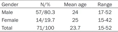
Table 1. Demographic Characteristics of Patients
Gender N/% Mean age Range
Male 57/80.3 24 17-52
Female 14/19.7 25 15-42
[image:2.612.90.287.392.446.2]Surgical treatment of pilonidal sinus
square test, ANOVA and Student T test were used where required to analyze the significance of the differences. P values of less than 0.05 were considered statistically significant at the 95% confidence interval.
Results
A total of 71 patients were included in the study, comprising 57 males (80.3%) and 14 females (19.7%) with a mean age of 23.7±6.9 years (range, 15-52 years) (Table 1). Of the patients, 52 were primary cases, and 19 were a recur
-rence. The operating techniques previously used on the recurrence cases were Limberg flap technique in 3, Karydakis flap technique in 6 and primary closure following lesion removal in 10 cases.
The size of the defect following excision was found to be mean 89 mm (range, 60-115 mm). In all cases, the tension-free closure was achieved using healthy and well-vascularised local skin. All the patients were mobilised on the postoperative first day. The duration of hospital stay was one day and the median fol
-low-up period was 15 months. The drain was removed on average on day 3. When the patients in the study were evaluated in respect of primary or recurrence, no difference was seen between the genders. The surgical param
-tion were found on the incision line that was treated with frequent dressing changes and ant biotherapy. No additional surgical intervention was made.
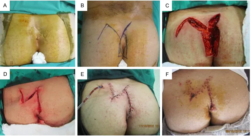
Illustrative case reports
Case 1:A 35-year old who had undergone two previous operations, the last one with the Limberg flap technique. Defect closure was applied with the triangular flap technique
(Figure 2).
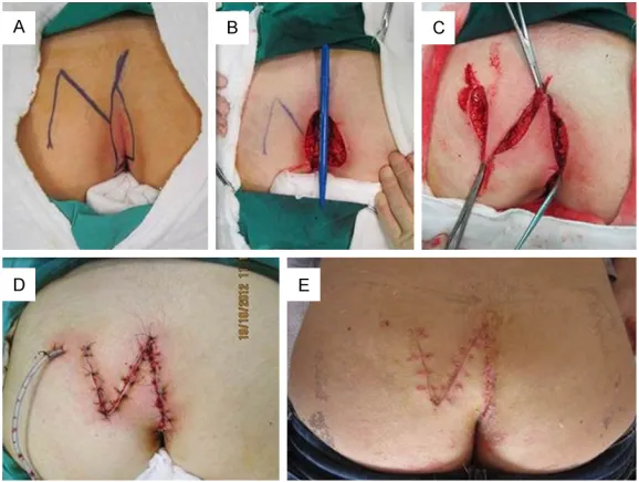
Case 2: A 27-year old female patient operated on with the triangular flap technique (Figure 3).
Discussion
Pilonidal sinus is a disease often seen in the sacrococcygeal region characterised by chronic repeated infection of hair follicles in the inter
-gluteal sulcus, which causes extreme discom
-fort to the patient. It was first described by Andersson in 1847 in a case found to have hair inside an ulcer and then in 1854, Warren pub
-lished the first series of similar cases. The word PILONIDAL was first used in 1880 by Hodges as a derivation from the Latin PILUS meaning ‘hair’ and NIDUS meaning ‘nest’ [5].
[image:3.612.93.345.86.166.2]Pilonidal sinus disease is seen 3-4 times more often in males than females. It is seen between
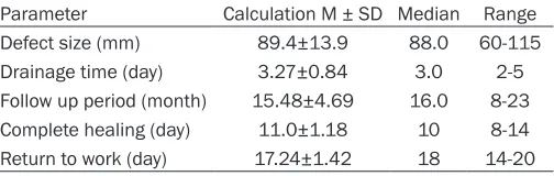
Table 2. Surgical parameters of the patients
Parameter Calculation M ± SD Median Range
Defect size (mm) 89.4±13.9 88.0 60-115
Drainage time (day) 3.27±0.84 3.0 2-5
Follow up period (month) 15.48±4.69 16.0 8-23
Complete healing (day) 11.0±1.18 10 8-14
Return to work (day) 17.24±1.42 18 14-20
[image:3.612.91.345.221.313.2]M ± SD: Mean ± standard deviation.
Table 3. Comparison of the primary and recurrent patients regarding mean defect sizes and surgical times
Parameter Diagnosis
Primary
diagnosis Recurrent P
N 52 19 <0.001
Defect size (mm) M ± SD 84.5±12.09 103.3±7.79 <0.001 Healing time (day) M ± SD 11.00±1.155 11.00±1.291 NS
Work loss (day) M ± SD 16.85±1.178 18.32±1.529 <0.001
M ± SD: Mean ± standard deviation, NS: not significant.
eters of all the patients are shown in
Table 2.
When the primary and recurrent cases were examined in terms of defect diameter, healing time and time to return to work, the mean diameter of the defect was seen to be greater and the time to work was sta
-tistically significantly longer in the recurrence group (P<0.001, P<0.001, respectively) (Table 3). In comparison of the median values of defect size, healing time and work loss, a statisti
-cally significant difference was deter
-mined between the groups in respect of defect size and time to return to work (Table 3).
No cases of flap necrosis or recur
-the ages of 15-35 years, with increasing rates in the 17-27 years age group and it is rarely seen over the age of 45 years [5]. There is as yet no consensus as to whether the aetiology is congenital or acquired. However, besides the factors facilitating the primary mechanism, such as excessive body hair, a deep gluteal cleft, damp and macerated areas associated with lengthy periods of sitting and poor hygiene conditions, this sinus also occurs in predis
-posed individuals [1, 2, 6, 7].
To date, various treatment methods have been used. As a conservative treatment, injections of sclerosing phenol, silver nitrate and alcohol have been applied to the pilonidal sinus cavity, but these have ceased to be used as they only provide a temporary solution. In conservative methods, return to daily life is delayed by a mean 40-50 days for the completion of wound healing. Rates of cure have been reported as approximately 70% [8] and said high rates of recurrence of 30% have resulted in the aban
-donment of conservative techniques. The basic principle of surgical treatment is the choice of a technique which provides excision of the lesion with safe borders and the reduction of potential recurrence to a minimum [9, 10].
To reduce the risk of recurrence, the frequently used choice of secondary wound healing may lead to restrictions on the patient’s daily life in the postoperative period at a higher rate than the problem created by the original disease and surgical morbidity may even exceed the dis
-ease itself. Also, in cases of secondary healing, the dressing changes and monitoring produce more work and increase the costs of the dis
-ease. Recurrence rates of 1-19% have been reported in cases left to secondary healing [11]. In the technique described by Bascom, according to the results of 161 treated patients, the follow-up period was 3.5-9 years, complete wound healing time was 3 weeks and recur
-rence was reported at the rate of 16%. In other treatment choices, the healing period ranged from 2-3 weeks in the primary closure of the defect and comparison with secondary healing, fewer dressings were required. How-ever, because of sulcus and tension that de-velops in the midline, recurrence rates are high
-er. Moreover, when applied in large defects, necrosis in the suture line and an extended healing period associated with that is a com
-monly encountered problem. In a series of 1129 patients operated on with pilonidal exci
[image:4.612.90.524.71.307.2]-sion and primary closure, Foss reported a recurrence rate of 16% [12].
Figure 2. A 35-year old who had undergone two previous operations, He had been operated with excision and primary closure of the defect and the last one with the Limberg flap technique. Defect closure was applied with
Surgical treatment of pilonidal sinus
Karydakis described asymmetrical closure that prevented the unwanted element of the suture line remaining over the midline [1]. Reasons for this choice are low reported recurrence rates besides advantages such as that it is a simple technique, the suture line remains lateral, and it provides early healing and early return to work. In a series of 7471 cases treated with this method and followed up over 2-20 years, the recurrence rate was reported as 0%-1% [1]. In another study that reported the recurrence rate as 7%, this rate was associated with fail
-ure of the technique and a shifting of the sut-ure line to the midline [1, 13, 14]. Kitchen [14] applied the Karydakis technique to 114 patients who had experienced failure of a previ
-ous surgical technique and recurrence was reported to be observed in 33 (29%) patients. When the recurrence rates in literature are examined, a mean recurrence rate of 4-7%, dif
-ficulties of application to large defects and the development of scar tissue are disadvantages of the Karydakis technique which is difficult to apply in recurrence cases. In the current study, which used the Mutaf Triangular Closure Technique, 19 patients were recurrence cases that had previously been operated on with dif
-ferent techniques. This easily applied surgical technique to large defects in the patients of the current study provided the advantages of no skatris tissue formation observed in either the
the presacral fascia with incompatible colour
-ing, and there is also the further problem of a longer hospital stay.
When the primary closure is not possible, the use of local flap methods is the only solution to protect against the handicaps of secondary surgical healing. Several different local flap techniques have been described in the litera
-ture, although none of these has removed the risk of recurrence. Consequently, there is as yet no consensus on the ideal choice of the local flap. With the use of a local flap, the defect area can be covered without tension, and the suture line is prevented from remaining in the midline. The decision for which flap to use must be made considering the shape of the sinus, whether or not it is complicated and how the remaining cavity will be filled. To cover the defect after pilonidal sinus excision, Z-plasty, V-Y-plasty, rhomboid flaps, rotation flaps, glute
-us maxim-us m-usculocutaneo-us flaps and recently, perforator based local flap techniques are often used. Using faciocutaneous flaps and myocutaneous flaps isindicated in the-case of extensive tissue defect with repeated infection and recurrence. These methods aim at wide excision of all the diseased tissue and closing the resultant defect with a well-vascu
-larized and bulky the sametissue. Also, it pro
[image:5.612.89.378.71.289.2]-vides a tension-freesuture line. On the other
Figure 3. A 27-year old female patient operated on with the triangular flap technique. A: Operation Plan. B: Defect created after sinus excision. C: Trans
-position of the flaps. D: Post-suturing image. E: Early postoperative appear -ance.
perioperative or postopera
-tive period, thereby resulting in no reduction in quality of
life.
Following sinus excision, cov
-ering the cavity with a skin graft is an effective method. Gu-yuron [15] applied this meth
-od to 58 patients, of whom 42 were recurrence cases and reported recurrence of the disease in 1 patient from a mean follow-up period of 5 years (range, 1-15 years). The reason that this technique is not currently preferred is that another body area is used for graft harvesting, and morbidi
hand, they are more complicated technique requiring greater surgical expertise and are related to higher rates of morbidity and wound dehiscence and a longer time of hospitalization [16].
The most popular of these is the Limberg flap, which has the advantages of widening the gluteal cleft and provides better cosmetic results. In a recent study by Çubukçu [17] of 129 patients operated on with Limberg flap, disease recurrence was reported at 5% in a 2-year follow-up period. Rhomboid flaps have been reported to have a recurrence rate of mean 0-5% [18-21]. Milito et al. [22] reported that after rhomboid flap had applied to 67 patients, 6 of whom were recurrence cases, no disease recurrence was observed throughout a 74-month follow-up period. When covering the defect which has been created, as the tissue for rhomboid flaps including the Limberg flap, is taken from a single anatomic area and a single flap is used in covering the defect, there is ten
-sion in the wound coverage as other healthy tis
-sues adjacent to the defect are not used and associated with this tension, seroma and wound separation may develop as the most sig
-nificant complication [16].
At the National Plastic Surgery Congress in 2011, Mutaf et al. presented this new tech
-nique, in which two flaps are prepared, one of these flaps is used for the defect closure while the other one is used for the closure of the donor site of the primary flap. Using two differ
-ent flaps, the Z-plasty manoeuvre provides extra tissue relaxation and closure without leading to tension in the suture lines, which are included in the coverage of large defects. By transferring a thick and well-vascularized neigh
-bouring tissue to the defect area, in this tech
-nique reduces the depth of the natal cleft, fills the dead space resulted from the wide excision of pilonidal sinus completely and allowed to transpose the resultant scar from midline. While in the Limberg and several other tech
-niques, the tissue required for defect coverage is obtained from only one area, in the Mutaf technique, the tissue to be used in the closure is moved from several directions and by equal distribution of the tension to all fields, the previ
-ously mentioned problems are overcome. Thus by reducing pressure, dog-ear formation is pre
-vented. In addition, the scar does not remain in the midline in this technique, the gluteal sulcus
is corrected by widening, and the formation of a dead cavity in the wound is prevented, thereby making it also possible to prevent recurrence. The dimensions of the flap can be changed depending on the diameter of the defect and therefore, the Mutaf technique can be recom
-mended for large defects where coverage is not possible with a Limberg flap and especially for recurrent cases.
In addition to other positive points, it is also noteworthy that in comparison with secondary healing and grafting techniques, the mobilisa
-tion period of the Mutaf technique provides an earlier return to work and normal daily life.
Conclusion
The principles in treatment of pilonidal sinus are complete excision of the sinus tract, ten
-sion-free and durable closure of the resultant defect with a well-vascularized tissue, obliterat
-ing the natal cleft, prevention of recurrence and also keeping the period of hospital care and time to return to work short. To obtain optimal results, the defect location, position, length of scar tissue which will remain and cosmetic results must be taken into consideration when planning the flap. For the application of this technique, a period of learning of flap surgery is required and it will also take time because of the difficulties experienced by surgeons in changing from previous habits.
The triangular closure technique was found to be a use fultechnique for the treatment of pilo
-nidalsinus with favorable results regarding time to turn to work.
Acknowledgements
None of the authors has a financial interest in any of the products, devices or drugs men
-tioned in this article. The authors thank Caroline Walker for assistance with the English language of the paper.
Disclosure of conflict of interest
None.
Surgical treatment of pilonidal sinus
References
[1] Karydakis GE. Easy and successful treatment of pilonidal sinus after explanation of its caus
-ative process. Aust N Z J Surg 1992; 62:
385-389.
[2] Bascom J. Pilonidal disease: origin from folli
-cles of hairs and results of follicle removal as treatment. Surgery 1980; 87: 567-572. [3] Mutaf M, Bekerecioglu M, Erkutlu I and Bulut
O. A new technique for closure of large menin
-gomyelocele defects. Ann Plast Surg 2007; 59:
538-543.
[4] Mutaf M, Gunal E, Berberoglu O and Gokce A. Surgical treatment of extensive sacrococcy
-geal hidradenitis suppurativa with triangular closure technique. Ann Plast Surg 2014; 73:
583-587.
[5] Akinci OF, Bozer M, Uzunkoy A, Duzgun SA and Coskun A. Incidence and aetiological factors in pilonidal sinus among Turkish soldiers. Eur J Surg 1999; 165: 339-342.
[6] Sondenaa K, Andersen E, Nesvik I and Soreide JA. Patient characteristics and symptoms in chronic pilonidal sinus disease. Int J Colorectal Dis 1995; 10: 39-42.
[7] Cubukcu A, Carkman S, Gonullu NN, Alponat A, Kayabasi B and Eyuboglu E. Lack of evidence that obesity is a cause of pilonidal sinus dis
-ease. Eur J Surg 2001; 167: 297-298.
[8] Rignault D, Pailler JL, Brillac J, Essioux H and Bon JC. Nonsurgical treatment of pilonidal fis
-tulas by the curettage-phenolization method. Gastroenterol Clin Biol 1977; 1: 941-944. [9] Matter I, Kunin J, Schein M and Eldar S. Total
excision versus non-resectional methods in the treatment of acute and chronic pilonidal disease. Br J Surg 1995; 82: 752-753. [10] Armstrong JH and Barcia PJ. Pilonidal sinus
disease. The conservative approach. Arch Surg 1994; 129: 914-917; discussion 917-919. [11] da Silva JH. Surgical treatment of pilonidal cyst
by incision and curettage. Rev Hosp Clin Fac
Med Sao Paulo 1974; 29: 199-203.
[12] Foss MV. Pilonidal sinus: excision and closure. Proc R Soc Med 1970; 63: 752.
[13] Sakr M, El-Hammadi H, Moussa M, Arafa S and Rasheed M. The effect of obesity on the re
-sults of Karydakis technique for the manage
-ment of chronic pilonidal sinus. Int J Colorectal Dis 2003; 18: 36-39.
[14] Kitchen PR. Pilonidal sinus: experience with the Karydakis flap. Br J Surg 1996; 83:
1452-1455.
[15] Guyuron B, Dinner MI and Dowden RV. Excision and grafting in treatment of recurrent piloni-dal sinus disease. Surg Gynecol Obstet 1983;
156: 201-204.
[16] Hull TL and Wu J. Pilonidal disease. Surg Clin North Am 2002; 82: 1169-1185.
[17] Cubukcu A, Gonullu NN, Paksoy M, Alponat A, Kuru M and Ozbay O. The role of obesity on the recurrence of pilonidal sinus disease in pa
-tients, who were treated by excision and Limberg flap transposition. Int J Colorectal Dis
2000; 15: 173-175.
[18] Quinodoz PD, Chilcott M, Grolleau JL, Chavoin JP and Costagliola M. Surgical treatment of sa
-crococcygeal pilonidal sinus disease by exci
-sion and skin flaps: the Toulouse experience. Eur J Surg 1999; 165: 1061-1065.
[19] Bozkurt MK and Tezel E. Management of pilo
-nidal sinus with the Limberg flap. Dis Colon
Rectum 1998; 41: 775-777.
[20] Ozgultekin R, Ersan Y, Ozcan M, Ozcelik F, Celik V, Cercel A and Sakaoglu M. Therapy of piloni
-dal sinus with the Limberg transposition flap. Chirurg 1995; 66: 192-195.
[21] Abu Galala KH, Salam IM, Abu Samaan KR, El Ashaal YI, Chandran VP, Sabastian M and Sim AJ. Treatment of pilonidal sinus by primary clo
-sure with a transposed rhomboid flap com
-pared with deep suturing: a prospective ran
-domised clinical trial. Eur J Surg 1999; 165:
468-472.
[22] Milito G, Cortese F and Casciani CU. Rhomboid flap procedure for pilonidal sinus: results from 67 cases. Int J Colorectal Dis 1998; 13: