REVIEW
Nucleophosmin: from structure
and function to disease development
Joseph K. Box, Nicolas Paquet
*, Mark N. Adams, Didier Boucher, Emma Bolderson, Kenneth J. O’Byrne
and Derek J. Richard
*Abstract
Nucleophosmin (NPM1) is a critical cellular protein that has been implicated in a number of pathways including
mRNA transport, chromatin remodeling, apoptosis and genome stability. NPM1 function is a critical requirement for
normal cellular biology as is underlined in cancer where NPM1 is commonly overexpressed, mutated, rearranged and
sporadically deleted. Consistent with a multifunctional role within the cell, NPM1 can function not only as a
proto-oncogene but also as a tumor suppressor. The aim of this review is to look at the less well-described role of NPM1 in
the DNA repair pathways as well as the role of NPM1 in the regulation of apoptosis and its mutation in cancers.
Keywords:
Nucleophosmin 1, DNA repair, Cancer, Apoptosis
© 2016 The Author(s). This article is distributed under the terms of the Creative Commons Attribution 4.0 International License (http://creativecommons.org/licenses/by/4.0/), which permits unrestricted use, distribution, and reproduction in any medium, provided you give appropriate credit to the original author(s) and the source, provide a link to the Creative Commons license, and indicate if changes were made. The Creative Commons Public Domain Dedication waiver (http://creativecommons.org/ publicdomain/zero/1.0/) applies to the data made available in this article, unless otherwise stated.
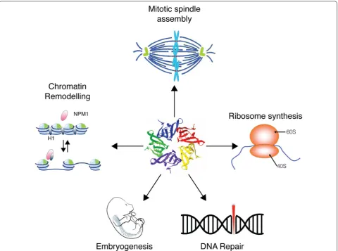
Background
Nucleophosmin (NPM1), also known as B23, No38 or
Numatrin, is an abundant nucleolar protein found in the
nuclei of proliferating cells. NPM1 has been documented
as participating in ribosome biogenesis, mRNA
process-ing, chromatin remodelprocess-ing, and embryogenesis (Fig.
1
).
While much is known about the function of NPM1 in
metabolic pathways, it is becoming clear that NPM1 also
has a critical function in maintaining genomic stability by
functioning in various DNA repair pathways and
regulat-ing apoptosis. In this review, we shall present an updated
overview of these roles, in particular the emerging data
supporting a role for NPM1 in DNA repair. Lastly, we
shall look at how NPM1 dysfunction contributes to
can-cer pathologies.
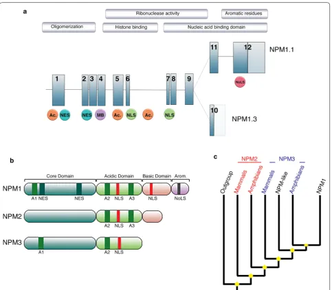
Gene organization and evolutionary history
Human
NPM1
is located on chromosome 5q35 and is
composed of 12 exons, encoding for at least two isoforms
(Fig.
2
a) [
1
]. NPM1.1 (or B23.1), corresponding to the
full-length transcript, results in a 294 amino acids protein
(35–40 kDa) abundantly expressed in all tissues.
Alterna-tively, NPM1.3 (also known as B23.2) results from the use
of a distinct 3
′
exon, and encodes for a protein expressed
at low levels in cells, lacking the last 35 amino acids of
the NPM1 C-terminus [
2
]. A third isoform NPM1.2 has
been suggested, but so far, no biological data support this
finding [
3
].
NPM1 belongs to a histone chaperones family, the
Nucleophosmin/nucleoplasmin (NPM) family, a group
that comprises multiple major functional members
(NPM1, NPM2, NPM3 and the invertebrate NPM-like),
and can be found amongst all Metazoan [
4
]. While this
family is well characterized functionally, little is known
about the evolution of these genes and proteins. At a
glance, all members of the NPM family exhibit
con-served structural motifs; a N-terminal core domain, an
acidic domain and a nuclear localization signal,
associ-ated with a less conserved, disorganized C-terminus
region (Fig.
2
b) [
5
]. Subsequently, crystallographic
stud-ies revealed a similar tertiary organization for NPM1 and
NPM2 with monomers organized into pentameric
donut-shaped complexes [
6
,
7
].
As simplified in Fig.
2
c, phylogenetic analysis revealed
the late emergence of the NPM1 monophyletic clade
while, in contrast, NPM2 and NPM3 lineages appear of
polyphyletic origin, with mammalians and amphibians
Open Access
sequences clearly differentiated [
4
]. Consistent with their
shared expression profile, localization and direct
physi-cal interaction, NPM1 and NPM3 are the most closely
related members of the family, suggesting functional
constraints between the two proteins [
4
]. Huang et al. [
8
]
further suggest that NPM3 may have evolved following
the loss of a nucleic acid binding domain of NPM1, and
functions as an element regulating NPM1 RNA binding.
Interestingly, codon usage within the NPM gene
fam-ily indicates a strong purifying selection, materialized
by a high rate of silent mutations which significantly
deviates from neutrality. The highly conserved
organi-zation of NPM proteins as pentamers further supports
the hypothesis of a strong negative selection operating
at the structural level. Interestingly, potential sites of
post-translational modifications are also selectively
con-strained, being conserved not only at the protein level
but also showing a preferred codon usage [
4
].
Remark-ably, these characteristics are shared with evolutionary
features observed in histones, suggesting intertwined
evolutionary history between the two families [
9
].
Despite being the most recent divergent NPM lineage,
the functions of NPM1 are diverse and include roles in
ribosome biogenesis [
10
,
11
], mRNA processing [
12
],
chromatin remodeling [
13
], embryogenesis [
14
],
regula-tion of apoptosis and maintenance of genome stability.
Characteristic structural features of nucleophosmin
NPM1 structural architecture is well characterized by
three distinct regions, onto which nucleolar and nuclear
localization motifs, nucleic acids binding domains,
oli-gomerization domains, histones binding regions, as well
as a putative metal binding domain, have been mapped
and described in detail [
15
–
17
] (Fig.
2
a, b).
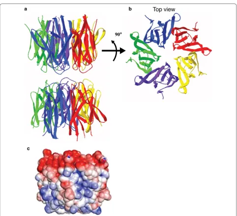
[image:2.595.59.540.87.444.2]The three dimensional structure of the human
NPM1-core has been determined by X-ray crystallography and
showed an organization into eight β-barrels forming a
jelly roll barrel. Further, NPM1 monomers associate as
donut-shaped homo-pentamers (Fig.
3
a, b). The
distri-bution of charges in this region is extremely
asymmet-ric with negatively charged residues clustered on one
side of the oligomer. Two pentamers of NPM1 interact
[image:3.595.58.542.85.508.2]the pentamer into unstable, unfolded monomers. This
structural polymorphism participates in the regulation
of NPM1 localization and function [
6
]. As such,
oli-gomerization of NPM1 has been linked to its nucleolar
localization and role in cellular proliferation, while the
monomeric form of NPM1 is associated with its role in
the DNA damage response and induction of apoptosis
[
19
].
The NPM1 central region appears unstructured and
is marked by the presence of highly acidic regions
composed of strings of aspartic and glutamic acids
(A1, A2 and A3). They provide long tracks of negatively
charged residues, known to be involved in the binding to
Histones H1, H3, H4, H2A and H2B, potentially by
mim-icking the charges of DNA and RNA [
20
,
21
]. It also
con-tains a nuclear localization signal.
[image:4.595.57.540.81.521.2]to nucleic acids and ATP [
16
,
22
]. These aromatic
resi-dues constitute an atypical nucleolar localization
sig-nal (NoLS), and their mutation are responsible for the
unfolding and the aberrant NPM1 localization typical in
acute myeloid leukemia (AML) cases.
Function of NPM1 in apoptosis
Although the best described function of NPM1 is in
ribo-some biogenesis, NPM1 also displays a critical role in
regulating apoptosis. NPM1 expression levels have been
implicated in controlling the cellular apoptotic response.
In a variety of cell based models, several studies have
demonstrated that down-regulation of NPM1 sensitizes
cells to apoptosis, while increased levels of the protein
protects against apoptosis [
23
–
25
]. In a disease setting,
the balance between NPM1 expression and cell fate is
demonstrated in hypoxia-driven cancers. For example,
suppression of hypoxia-induced NPM1 expression
pro-motes apoptosis whereas overexpression protects from
hypoxia-mediated cell death [
26
]. From a cancer
perspec-tive, elevated levels of NPM1 might promote malignant
transformation by enabling cell survival.
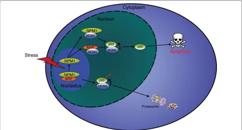
Several proteins have been identified that impact
cell survival by interacting with and regulating NPM1
protein levels. In the nucleolus, the tumor suppressor
p14
ARFinteracts with NPM1 to promote degradation of
the protein and induce cell death (Fig.
4
) [
27
]. However,
the interplay between NPM1 and p14
ARFis more
com-plex, with NPM1 also acting as major cellular reservoir
of p14
ARF(Fig.
4
). After various stimuli NPM1 releases
p14
ARFallowing binding to MDM2 and preventing the
proteasomal degradation of p53 (Fig.
4
) [
28
].
Consist-ently, depletion of NPM1 with siRNA results in increased
apoptosis due to a greater amount of free p14
ARF[
28
]. On
the contrary, up-regulation of NPM1 appears to
antago-nize p14
ARFand increases its nucleolar retention [
29
].
These observations provide evidence that NPM1
regu-lates cell fate in a p53-dependent manner by directing
p14
ARFto nucleoli and preventing inhibition of MDM2
(Fig.
4
) [
27
,
30
,
31
]. Other studies have also linked NPM1
with the tumor suppressor activity of p53. For example,
NPM1 is also able to interact directly with MDM2,
inde-pendently of p14
ARF, and act as a p53:MDM2 inhibitor to
protect p53 from degradation (Fig.
4
) [
32
,
33
].
Interest-ingly, NPM1 is also reported to directly associate with
p53 [
34
]. However, this interaction remains contentious
as an inability for NPM1 to interact with p53 has also
been reported [
27
]. It may be that an interaction between
NPM1 and p53 might only occur in certain cellular
con-texts. Nonetheless, this observation requires clarification.
Fig. 4 Regulation of apoptosis by NPM1. In unstressed cells, p14ARF and NPM1 form a dimer in the nucleoli, allowing MDM2 to target p53 for
proteasomal degradation. Following a stress, such as DNA damages, p14ARF and NPM1 dissociate and relocate to the nucleus were they sequester
[image:5.595.57.539.417.677.2]In addition to p14
ARF, MDM2 and possibly p53, NPM1
has been reported to bind PKB/Akt in order to modulate
cell survival. In the nucleus, Akt binds NPM1 in response
to growth factor stimulation to protect NPM1 against
caspase-3-mediated proteolytic degradation and promote
cell survival [
35
]. Similarly, NPM1 stability is enhanced
by interacting with erythroid differentiation-associated
gene (EDAG), promoting acute myeloid leukemia (AML)
cell survival [
36
]. In another instance, the interaction of
GAGE with NPM1 also enables NPM1 stability to
pro-mote resistance to interferon-γ-induced apoptosis [
37
].
NPM1 has also been portrayed as regulating cell fate
by modulating both the intrinsic and extrinsic apoptosis
pathways. During the intrinsic apoptotic response, p53
is required in the mitochondrial to enable cytochrome C
release. Overexpression of NPM1 prevents the
transloca-tion of p53 from the nucleus to the mitochondria [
38
],
suggesting that NPM1 may protect cells from
apopto-sis by reducing the mitochondrial level of p53. In acute
promyelocytic leukemia cells expressing the
NPM1-retinoic acid receptor α (NPM1-RAR) fusion protein,
NPM1-RAR blocked TNF-induced extrinsic apoptosis
by inhibiting signaling to activate caspase-3 and -8 [
39
].
Similarly, mutant forms of NPM1 are indicated to impede
apoptosis by directly inhibiting the proteolytic function
of caspase-6 and -8 in the cytoplasm [
40
]. Interestingly,
in anaplastic large-cell lymphoma cells, the
cytoplas-mic fraction of the NPM1-ALK fusion protein is solely
responsible for inducing apoptosis by engaging the
DNA-damage response [
41
]. These studies suggest, at least for
the mutant or NPM1 fusion proteins, that the
cytoplas-mic fraction of NPM1 may be required to regulate the
apoptotic pathways.
Role of NPM1 in the DNA repair response
Loss of NPM1 function has been shown to be associated
with increased genome instability [
42
]. Several studies
have demonstrated the critical role of NPM1 in the
main-tenance of genome stability through its interaction with
unduplicated centrosomes [
42
]. The phosphorylation of
NPM1 by CDK2/Cyclin E promotes the release of NPM1
from the centrosome during duplication; this represents
an essential step for duplication to occur. However,
dur-ing mitosis NPM1 re-associates with the centrosomes at
the spindle bodies and appears to be controlling
centro-some duplication [
43
]. Indeed, depletion of NPM1 has
been shown to promote genome instability (unaligned
chromosomes, micronuclei) [
44
,
45
]. In mice, the
chro-mosomal instability associated with NPM1 depletion
partially explains the embryonic lethality [
14
].
However, it has only recently become clear that NPM1
is likely to have a direct role in the repair of DNA lesions.
Multiple DNA repair pathways promote the repair of
different DNA lesions and NPM1 has been implicated in
several of these repair pathways (Fig.
5
).
DNA double-strand breaks (DSB) are the most
destruc-tive and genotoxic lesions encountered by cells and as
such complex cascades have evolved to sense, signal and
repair these breaks. DNA DSBs can then be repaired by
two pathways, homologous recombination (HR) and
non-homologous end-joining (NHEJ) (reviewed in [
46
]).
HR utilizes a sister chromatid as a template for repair and
therefore can only be performed in the late S-phase and
G2 phases of the cell cycle. In contrast, NHEJ can occur
in any phase of the cell cycle and involves a less complex
method of ligating the two DNA ends together. As
over-hanging DNA may be resected during this method, NHEJ
is generally known as the more error-prone mechanism
of DNA DSB repair. In the HR process, DSBs are detected
and signaling pathways are initiated by the ATM/ATR
kinases, promoting the recruitment of repair proteins,
including nucleases that resect DNA with a 5
′
–3
′
polar-ity. This resection generates stretches of single stranded
DNA, which invade into a sister chromatid, allowing it to
act as a template for polymerase-mediated extension of
the invading strand. Following this extension and
re-liga-tion of DNA strands this reacre-liga-tion then yields two intact
and identical DNA molecules [
47
].
altering its function. YTR107 inhibits DNA repair and
radiosensitizes cells in an NPM1-dependent process [
51
].
In light of the above it is clear that NPM1 has a role in
DNA DSB repair, however much is still to be elucidated
about the exact mechanism of its function in these
path-ways (Fig.
5
).
Following exposure of cells to agents such as UV, DNA
lesions may cause replication forks to stall as the
repli-cative polymerases are unable to bypass the UV-induced
bulky lesions. In order for these stalled replication forks
to be restarted, they can be repaired by a form of HR or
be bypassed in a mechanism known as translesion
syn-thesis (TLS, reviewed in [
52
], Fig.
5
). A recent study
iden-tified NPM1 as a key player of the TLS pathway [
53
]. This
process enables switching of the DNA polymerase to a
low fidelity DNA polymerase that can replicate the DNA
across the lesion. NPM1 regulates TLS by binding to
and protecting DNA Polymerase Eta (POLH, polη) from
proteosomal degradation promoting its role in TLS. The
mutated NPM1 (NPM1c+, found in 30 % of AML cases)
was found to result in increased degradation of polη,
per-haps explaining the improved prognosis in AML patients
with NPM1 mutations [
53
].
In addition to its role in DNA double-strand break
repair and translesion synthesis, NPM1 has also been
shown to respond to DNA lesions induced by UV light.
The alterations to nucleotides caused by UV irradiation
are repaired by the nucleotide excision repair pathway, a
process that is dependent upon the PCNA homo-trimer
(reviewed in [
54
]). The levels of NPM1 protein were
shown to increase following cellular exposure to UV
[
55
]. Exogenous overexpression of NPM1 was also found
to increase cellular survival and DNA repair capacity
following UV irradiation. Supporting a role for NPM1 in
nucleotide excision repair (NER), it was found to
tran-scriptionally regulate the crucial NER protein PCNA
[
55
]. Following UV irradiation, dephosphorylation of
NPM1 on Threonine 199, 234 and 237 residues occurs
in a PP1β-dependent manner. Dephosphorylation of
these sites on NPM1 enhances the interaction between
NPM1 and the retinoblastoma tumor suppressor protein
(pRB), which then allows the release of E2F1 from pRB.
E2F1 subsequently functions to transcriptionally activate
several downstream DNA repair genes, including XPC,
DDB2 and RPA14, facilitating DNA repair [
56
].
Oxidative damage to DNA is caused predominantly by
normal cellular metabolism and is repaired by the base
excision repair pathway (BER reviewed in [
57
]). NPM1
has been shown to modulate the BER pathway through
control of the apurinic/apyrimidinic endonuclease 1
(APE1) protein levels and modulation of the AP-site
inci-sion activity of APE1, which is required for base
exci-sion repair (Fig.
5
) [
58
–
60
]. Several nucleolar proteins
involved in BER were also mislocalised in
NPM1-defi-cient cells, including APE1, Fen1 and LigI [
61
]. NPM1
was also found to belong to a complex containing several
BER proteins, including APE1, Fen1, Polβ and LigI [
62
,
63
].
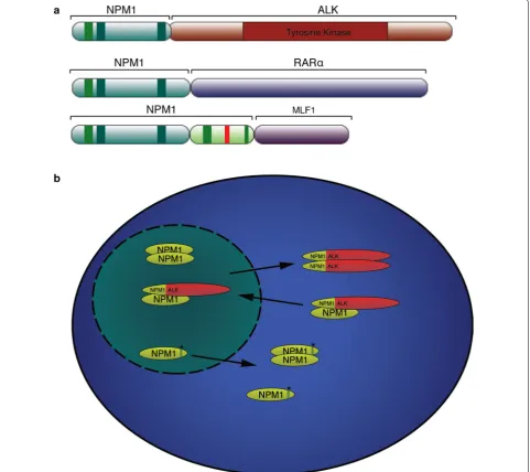
NPM1 and cancer
[image:7.595.56.541.87.247.2]express a fusion between NPM1 and the catalytic domain
of anaplastic lymphoma receptor tyrosine kinase (ALK)
[
64
]. In addition, 35 % of all AML patients (50–60 % in
adults with normal karyotype) show NPM1
rearrange-ments or mutations [
65
], leading the World Health
Organization to introduce mutated NPM1 as an AML
entity [
66
]. All NPM1 mutations reported occur with
the C-terminus of the protein altering either the
fold-ing of this region or the Nucleolar localization signal
itself. Named NPM1 Cytoplasm positive or NPM1c+,
these mutations result in cytoplasmic localization of the
protein, and act as dominant negative by retaining WT
NPM1 in the cytoplasm (Fig.
6
b) [
67
].
Patients with NPM1 mutations proved to have a
bet-ter outcome with increased complete remission and
improved overall survival [
68
]. However, within this group
of patients, the antigen expression pattern of HLADR(+)
CD34(+) CD7(+) is associated with poor prognosis [
69
].
NPM1c+ has been shown to result in microRNAs
dereg-ulation [
70
], and a recent in vitro study established that
NPM1c+ could enhance leukemia cells adhesion,
migra-tory and invasive potential through MEK/ERK activation
[image:8.595.58.540.237.665.2][
71
]. Notably, NPM1 mutation in a knock-in mouse
model resulted in AML initiation [
72
,
73
], and studies on
MEFs have shown that NPM1 mutation results in p14
ARFdestabilization [
74
] and c-Myc stabilization [
75
].
In anaplastic large-cell lymphoma (ALCL), NPM1
fusion with anaplastic lymphoma receptor tyrosine
kinase (ALK) can be found in 85 % of the ALK+ ALCL,
this results in the expression of a chimeric oncogenic
protein formed by the C-terminus of NPM1 and the
kinase domain of ALK (Fig.
6
a) [
64
,
76
]. In these cells
both NPM1 and ALK exhibits aberrant localization.
Fur-ther, dimerization of NPM1-ALK leads to a constitutive
activation of the ALK kinase (Fig.
6
b) [
76
].
Chimeric proteins between NPM1 and the retinoic acid
receptor-α gene (RARα) or between NPM1 and the
mye-lodysplasia/myeloid leukemia factor 1 (MLF1) have also
been reported in rare cases of leukemia (Fig.
6
a) [
77
,
78
].
Besides leukemia, involvement of NPM1 has also been
reported in several solid cancers. Overall, NPM1
overex-pression is linked to high grade tumors and poor
progno-sis, as observed in brain glioblastoma [
79
], oral squamous
cell carcinoma [
80
], non-small cells lung cancer (NSCLC)
[
51
], hepatocellular carcinomas (HCC) [
81
], colon
can-cer [
82
–
84
], ovarian cancer [
62
,
85
], and endometrial
carcinoma [
86
]. A higher level of NPM1 has also been
observed in prostate cancer, when compared to normal
tissue [
87
,
88
], promoting cells growth and invasiveness
[
89
]. In bladder cancer, high levels of NPM1are not
asso-ciated with tumor grade, but with cancer progression,
recurrence, and poor prognosis [
90
]. NPM1
overexpres-sion has also been reported in thyroid cancer [
91
], with
thyroid cancer cell lines showing NPM1
mislocaliza-tion in the absence of the mutamislocaliza-tions observed in AML
patients [
92
].
However, low levels of NPM1 have also been observed
in some cancers, such as gastric cancers (mRNA and
protein) compared to normal tissue [
93
]. In breast
can-cer, low NPM1 levels are also associated with poor
out-come, independently of the molecular subtype, with
granular staining of NPM1 correlating with poor
prog-nosis [
94
].
Since NPM1 is overexpressed in many types of cancer
and because of its role in genome stability, it could be a
potential target for new cancer therapy strategies.
Differ-ent molecules have been trialed to induce cell death by
destabilizing NPM1 [
95
–
98
] or inhibiting its interaction
with other DNA repair proteins [
99
]. Recent studies have
focused on combining NPM1 inhibition with a DNA
damaging agent, for instance ionizing radiation [
51
,
100
],
or a cytotoxic drug [
101
]. Other strategies are aiming
specifically at NPM1c+ itself [
102
], or in combination
with damage induction by increasing oxidative stress in
cells [
103
–
106
].
Frontiers
As summarized here, NPM1 plays multiple roles within
human cells while the best documented is in RNA
trans-port and ribosome biogenesis, it is clear that NPM1 also
plays a critical role in the regulation of apoptosis and in
the maintenance of genomic homeostasis.
Genome instability is one of the underlying causes of
cellular transformation and cancer development. Once a
cancer does form, genome instability becomes a common
feature seen as a universal hallmark of all cancers [
107
].
This instability provides the cancer the ability to evolve
and adapt to the environment in which it is located;
fur-ther, defects in the apoptosis pathways allow the cancer
cell to cope and survive with levels of genetic instability
that would normally induce cell death.
Given the critical role NPM1 plays in genome stability
and apoptosis it is hardly surprising that NPM1
dysfunc-tion is a frequent feature in cancers. Indeed, as evidence
of NPM1 function in DNA repair pathways increases, it
explains at least in part the genetic instability associated
with cancers such as AML. Further, NPM1
deregula-tion or mutaderegula-tion will suppress the ability of that cell to
respond to apoptotic stimuli, allowing for the tolerance
of the genetic instability. Consistent with a role in genetic
instability and specifically the repair of double-strand
DNA breaks we see that AML patients with a mutated
NPM1 have a >2 fold higher odds of achieving
com-plete remission as compared to patients with a wild type
NPM1 [
108
]. NPM1 in cancer is most strikingly
high-lighted in acute myeloid leukemia where approximately
30 % of patients will have a mutation or fusion event
implicating NPM1.
Despite the importance of NPM1 in genome stability it
is clear that we do not fully understand how NPM1
func-tions in the repair of DNA damage. We know that NPM1
moves to sites of double-strand DNA breaks within the
genome, but we do not yet understand how NPM1
func-tions at those break sites. We do not know if NPM1 binds
to nucleic acids at those sites or if it is involved in
remod-eling chromatin. It is clear, however, that
understand-ing how NPM1 participates in DNA repair and genome
stability will help to delineate the role of NPM1 in both
normal and cancerous cells. This will perhaps provide
insights into why NPM1 dysfunction is a marker of drug
and radiation sensitivities.
Abbreviations
2 homolog; MEF: mouse embryonic fibroblast; MLF1: myelodysplasia/myeloid leukemia factor 1; mRNA: messenger RNA; NER: nucleotide excision repair; NHEJ: non-homologous end-joining; NoLS: nucleolar localisation signal; NPM1: nucleophosmin1; PCNA: proliferating cell nuclear antigen; PKB: protein kinase B; RAR: retinoic acid receptor α; RNA: ribonucleic acid; RNF: ring finger protein1; RPA: replication protein A; TLS: translesion DNA synthesis; TNF: tumor necrosis factor; UV: ultra violet; XPC: xeroderma pigmentosum, complementa-tion group C.
Authors’ contributions
JKB, NP, MNA, DB, EB, KJO and DJR wrote the manuscript. All authors read and approved the final manuscript.
Competing interests
The authors declare that they have no competing interests.
Funding information
This work was supported by a NHMRC project Grant (1066550), an ARC project Grant (D.J.R, DP 120103099) and by a Queensland Health Senior Clinical Research Fellowship (K.J.O.). M.A. holds a NHMRC Early Career Fellowship (1091589). E.B. is supported by an Advance Queensland Research Fellowship.
Received: 8 May 2016 Accepted: 16 August 2016
References
1. Umekawa H, Chang JH, Correia JJ, Wang D, Wingfield PT, Olson MO. Nucleo-lar protein B23: bacterial expression, purification, oligomerization and secondary structures of two isoforms. Cell Mol Biol Res. 1993;39(7):635–45. 2. Dalenc F, Drouet J, Ader I, Delmas C, Rochaix P, Favre G, Cohen-Jonathan
E, Toulas C. Increased expression of a COOH-truncated nucleophosmin resulting from alternative splicing is associated with cellular resistance to ionizing radiation in HeLa cells. Int J Cancer. 2002;100(6):662–8. 3. Lim MJ, Wang XW. Nucleophosmin and human cancer. Cancer Detect
Prev. 2006;30(6):481–90.
4. Eirin-Lopez JM, Frehlick LJ, Ausio J. Long-term evolution and functional diversification in the members of the nucleophosmin/nucleoplasmin family of nuclear chaperones. Genetics. 2006;173(4):1835–50. 5. Frehlick LJ, Eirin-Lopez JM, Ausio J. New insights into the
nucle-ophosmin/nucleoplasmin family of nuclear chaperones. BioEssays. 2007;29(1):49–59.
6. Mitrea DM, Grace CR, Buljan M, Yun MK, Pytel NJ, Satumba J, Nourse A, Park CG, Madan Babu M, White SW, et al. Structural polymorphism in the N-terminal oligomerization domain of NPM1. Proc Natl Acad Sci USA. 2014;111(12):4466–71.
7. Platonova O, Akey IV, Head JF, Akey CW. Crystal structure and function of human nucleoplasmin (npm2): a histone chaperone in oocytes and embryos. Biochemistry. 2011;50(37):8078–89.
8. Huang N, Negi S, Szebeni A, Olson MO. Protein NPM3 interacts with the multifunctional nucleolar protein B23/nucleophosmin and inhibits ribosome biogenesis. J Biol Chem. 2005;280(7):5496–502.
9. Eirin-Lopez JM, Gonzalez-Romero R, Dryhurst D, Mendez J, Ausio J. Long-term evolution of histone families: old notions and new insights into their mechanisms of diversification across eukaryotes. In: Pontarotti P, editor. Evolutionary biology: concept, modeling and application. Berlin: Springer-Verlag; 2009. p. 139–62.
10. Yu Y, Maggi LB Jr, Brady SN, Apicelli AJ, Dai MS, Lu H, Weber JD. Nucle-ophosmin is essential for ribosomal protein L5 nuclear export. Mol Cell Biol. 2006;26(10):3798–809.
11. Savkur RS, Olson MO. Preferential cleavage in pre-ribosomal RNA byprotein B23 endoribonuclease. Nucleic Acids Res. 1998;26(19):4508–15.
12. Murano K, Okuwaki M, Hisaoka M, Nagata K. Transcription regula-tion of the rRNA gene by a multifuncregula-tional nucleolar protein, B23/ nucleophosmin, through its histone chaperone activity. Mol Cell Biol. 2008;28(10):3114–26.
13. Okuwaki M, Matsumoto K, Tsujimoto M, Nagata K. Function of nucle-ophosmin/B23, a nucleolar acidic protein, as a histone chaperone. FEBS Lett. 2001;506(3):272–6.
14. Grisendi S, Bernardi R, Rossi M, Cheng K, Khandker L, Manova K, Pandolfi PP. Role of nucleophosmin in embryonic development and tumorigen-esis. Nature. 2005;437(7055):147–53.
15. Okuwaki M. The structure and functions of NPM1/nucleophosmin/B23, a multifunctional nucleolar acidic protein. J Biochem. 2008;143(4):441–8. 16. Hingorani K, Szebeni A, Olson MO. Mapping the functional domains of
nucleolar protein B23. J Biol Chem. 2000;275(32):24451–7. 17. Chan WY, Liu QR, Borjigin J, Busch H, Rennert OM, Tease LA, Chan
PK. Characterization of the cDNA encoding human nucleophosmin and studies of its role in normal and abnormal growth. Biochemistry. 1989;28(3):1033–9.
18. Lee HH, Kim HS, Kang JY, Lee BI, Ha JY, Yoon HJ, Lim SO, Jung G, Suh SW. Crystal structure of human nucleophosmin-core reveals plasticity of the pentamer–pentamer interface. Proteins. 2007;69(3):672–8.
19. Koike A, Nishikawa H, Wu W, Okada Y, Venkitaraman AR, Ohta T. Recruit-ment of phosphorylated NPM1 to sites of DNA damage through RNF8-dependent ubiquitin conjugates. Cancer Res. 2010;70(17):6746–56. 20. Gadad SS, Senapati P, Syed SH, Rajan RE, Shandilya J, Swaminathan V,
Chatterjee S, Colombo E, Dimitrov S, Pelicci PG, et al. The multifunc-tional protein nucleophosmin (NPM1) is a human linker histone H1 chaperone. Biochemistry. 2011;50(14):2780–9.
21. Swaminathan V, Kishore AH, Febitha KK, Kundu TK. Human histone chaperone nucleophosmin enhances acetylation-dependent chroma-tin transcription. Mol Cell Biol. 2005;25(17):7534–45.
22. Choi JW, Lee SB, Kim CK, Lee KH, Cho SW, Ahn JY. Lysine 263 residue of NPM/B23 is essential for regulating ATP binding and B23 stability. FEBS Lett. 2008;582(7):1073–80.
23. Ahn JY, Liu X, Cheng D, Peng J, Chan PK, Wade PA, Ye K. Nucleophos-min/B23, a nuclear PI(3,4,5)P(3) receptor, mediates the antiapoptotic actions of NGF by inhibiting CAD. Mol Cell. 2005;18(4):435–45. 24. Wu MH, Chang JH, Yung BY. Resistance to UV-induced cell-killing in
nucleophosmin/B23 over-expressed NIH 3T3 fibroblasts: enhancement of DNA repair and up-regulation of PCNA in association with nucle-ophosmin/B23 over-expression. Carcinogenesis. 2002;23(1):93–100. 25. Wu MH, Chang JH, Chou CC, Yung BY. Involvement of
nucleophos-min/B23 in the response of HeLa cells to UV irradiation. Int J Cancer. 2002;97(3):297–305.
26. Li J, Zhang X, Sejas DP, Bagby GC, Pang Q. Hypoxia-induced nucle-ophosmin protects cell death through inhibition of p53. J Biol Chem. 2004;279(40):41275–9.
27. Itahana KBK, Jin A, Itahana Y, Hawke D, Kobayashi R, Zhang Y. Tumor suppressor ARF degrades B23, a nucleolar protein involved in ribosome biogenesis and cell proliferation. Mol Cell. 2003;12(5):1151–64. 28. Qin FX, Shao HY, Chen XC, Tan S, Zhang HJ, Milao ZY, Wang L, Hui-Chen,
Zhang L. Knockdown of NPM1 by RNA interference inhibits cells proliferation and induces apoptosis in leukemic cell line. Int J Med Sci. 2011;8(4):287–94.
29. Korgaonkar C, Hagen J, Tompkins V, Frazier AA, Allamargot C, Quelle FW, Quelle DE. Nucleophosmin (B23) targets ARF to nucleoli and inhibits its function. Mol Cell Biol. 2005;25(4):1258–71.
30. Korgaonkar CHJ, Tompkins V, Frazier AA, Allamargot C, Quelle FW, Quelle DE. Nucleophosmin (B23) targets ARF to nucleoli and inhibits its function. Mol Biol Cell. 2005;25(4):1258–71.
31. Ye K. Nucleophosmin/B23, a multifunctional protein that can regulate apoptosis. Cancer Biol Ther. 2005;4(9):918–23.
32. Kurki SPK, Latonen L, Kiviharju TM, Ojala PM, Meek D, Laiho M. Nucleolar protein NPM interacts with HDM2 and protects tumor suppres-sor protein p53 from HDM2-mediated degradation. Cancer Cell. 2004;5(5):465–75.
33. Jin AIK, O’Keefe K, Zhang Y. Inhibition of HDM2 and activation of p53 by ribosomal protein L23. Mol Cell Biol. 2004;24(17):7669–80.
34. Colombo E, Marine JC, Danovi D, Falini B, Pelicci PG. Nucleophosmin regulates the stability and transcriptional activity of p53. Nat Cell Biol. 2002;4(7):529–33.
36. Zhang MJ, Ding YL, Xu CW, Yang Y, Lian WX, Zhan YQ, Li W, Xu WX, Yu M, Ge CH, et al. Erythroid differentiation-associated gene interacts with NPM1 (nucleophosmin/B23) and increases its protein stability, resisting cell apoptosis. FEBS J. 2012;279(16):2848–62.
37. Kular RK, Yehiely F, Kotlo KU, Cilensek ZM, Bedi R, Deiss LP. GAGE, an antiapoptotic protein binds and modulates the expression of nucle-ophosmin/B23 and interferon regulatory factor 1. J Interferon Cytokine Res. 2009;29(10):645–55.
38. Dhar SKSCD. Nucleophosmin blocks mitochondrial localization of p53 and apoptosis. J Biol Chem. 2009;284(24):16409–18.
39. Chattopadhyay A, Hood BL, Conrads TP, Redner RL. Extrinsic apoptosis is impeded by direct binding of the APL fusion protein NPM-RAR to TRADD. Mol Cancer Res. 2014;12(9):1283–91.
40. Leong SM, Tan BX, Bte Ahmad B, Yan T, Chee LY, Ang ST, Tay KG, Koh LP, Yeoh AE, Mok YK, Lim TM. Mutant nucleophosmin deregulates cell death and myeloid differentiation through excessive caspase-6 and -8 inhibition. Blood. 2010;116(17):3286–96.
41. Ceccon M, Merlo ME, Mologni L, Poggio T, Varesio LM, Menotti M, Bom-belli S, Rigolio R, Manazza AD, Di Giacomo F, et al. Excess of NPM-ALK oncogenic signaling promotes cellular apoptosis and drug depend-ency. Oncogene. 2016;35(29):3854–65.
42. Wang W, Budhu A, Forgues M, Wang XW. Temporal and spatial control of nucleophosmin by the Ran-Crm1 complex in centrosome duplica-tion. Nat Cell Biol. 2005;7(8):823–30.
43. Zatsepina OV, Rousselet A, Chan PK, Olson MO, Jordan EG, Bornens M. The nucleolar phosphoprotein B23 redistributes in part to the spindle poles during mitosis. J Cell Sci. 1999;112(Pt 4):455–66.
44. Amin MA, Matsunaga S, Uchiyama S, Fukui K. Nucleophosmin is required for chromosome congression, proper mitotic spindle forma-tion, and kinetochore-microtubule attachment in HeLa cells. FEBS Lett. 2008;582(27):3839–44.
45. Amin MA, Matsunaga S, Uchiyama S, Fukui K. Depletion of nucleophos-min leads to distortion of nucleolar and nuclear structures in HeLa cells. Biochem J. 2008;415(3):345–51.
46. Ceccaldi R, Rondinelli B, D’Andrea AD. Repair pathway choices and conse-quences at the double-strand break. Trends Cell Biol. 2016;26(1):52–64. 47. San Filippo J, Sung P, Klein H. Mechanism of eukaryotic homologous
recombination. Annu Rev Biochem. 2008;77:229–57.
48. Lee SY, Park JH, Kim S, Park EJ, Yun Y, Kwon J. A proteomics approach for the identification of nucleophosmin and heterogeneous nuclear ribonucleoprotein C1/C2 as chromatin-binding proteins in response to DNA double-strand breaks. Biochem J. 2005;388(Pt 1):7–15.
49. Okuda M, Horn HF, Tarapore P, Tokuyama Y, Smulian AG, Chan PK, Knudsen ES, Hofmann IA, Snyder JD, Bove KE, et al. Nucleophosmin/ B23 is a target of CDK2/cyclin E in centrosome duplication. Cell. 2000;103(1):127–40.
50. Koike A, Nishikawa H, Wu W, Okada Y, Venkitaraman AR, Ohta T. Recruit-ment of phosphorylated NPM1 to sites of DNA damage through RNF8-dependent ubiquitin conjugates. Cancer Res. 2010;70:6746–56. 51. Sekhar KR, Benamar M, Venkateswaran A, Sasi S, Penthala NR, Crooks
PA, Hann SR, Geng L, Balusu R, Abbas T, et al. Targeting nucleophosmin 1 represents a rational strategy for radiation sensitization. Int J Radiat Oncol Biol Phys. 2014;89:1106–14.
52. Jansen JG, Tsaalbi-Shtylik A, de Wind N. Roles of mutagenic translesion synthesis in mammalian genome stability, health and disease. DNA Repair. 2015;29:56–64.
53. Ziv O, Zeisel A, Mirlas-Neisberg N, Swain U, Nevo R, Ben-Chetrit N, Martelli MP, Rossi R, Schiesser S, Canman CE, et al. Identification of novel DNA-damage tolerance genes reveals regulation of translesion DNA synthesis by nucleophosmin. Nat Commun. 2014;5:5437.
54. Dijk M, Typas D, Mullenders L, Pines A. Insight in the multilevel regula-tion of NER. Exp Cell Res. 2014;329(1):116–23.
55. Wu MH, Yung BYM. UV stimulation of nucleophosmin/B23 expression is an immediate-early gene response induced by damaged DNA. J Biol Chem. 2002;277:48234–40.
56. Lin CY, Tan BC, Liu H, Shih CJ, Chien KY, Lin CL, Yung BY. Dephosphoryla-tion of nucleophosmin by PP1B facilitates pRB binding and consequent E2F1-dependent DNA repair. Mol Biol Cell. 2010;21(24):4409–17. 57. Krokan HE, Bjoras M. Base excision repair. Cold Spring Harb Perspect
Biol. 2013;5(4):a012583.
58. Vascotto C, Fantini D, Romanello M, Cesaratto L, Deganuto M, Leonardi A, Radicella JP, Kelley MR, D’Ambrosio C, Scaloni A, et al. APE1/Ref-1 interacts with NPM1 within nucleoli and plays a role in the rRNA quality control process. Mol Cell Biol. 2009;29:1834–54.
59. Vascotto C, Lirussi L, Poletto M, Tiribelli M, Damiani D, Fabbro D, Damante G, Demple B, Colombo E, Tell G. Functional regulation of the apurinic/apyrimidinic endonuclease 1 by nucleophosmin: impact on tumor biology. Oncogene. 2013;33:1–12.
60. Tell G, Fantini D, Quadrifoglio F. Understanding different functions of mammalian AP endonuclease (APE1) as a promising tool for cancer treatment. Cell Mol Life Sci. 2010;67(21):3589–608.
61. Poletto M, Lirussi L, Wilson DM 3rd, Tell G. Nucleophosmin modulates stability, activity, and nucleolar accumulation of base excision repair proteins. Mol Biol Cell. 2014;25(10):1641–52.
62. Londero AP, Orsaria M, Tell G, Marzinotto S, Capodicasa V, Poletto M, Vascotto C, Sacco C, Mariuzzi L. Expression and prognostic significance of APE1/Ref-1 and NPM1 proteins in high-grade ovarian serous cancer. Am J Clin Pathol. 2014;141:404–14.
63. Poletto M, Malfatti MC, Dorjsuren D, Scognamiglio PL, Marasco D, Vascotto C, Jadhav A, Maloney DJ, Wilson DM 3rd, Simeonov A, et al. Inhibitors of the apurinic/apyrimidinic endonuclease 1 (APE1)/nucle-ophosmin (NPM1) interaction that display anti-tumor properties. Mol Carcinog. 2016;55(5):688–704.
64. Morris SW, Kirstein MN, Valentine MB, Dittmer KG, Shapiro DN, Saltman DL, Look AT. Fusion of a kinase gene, ALK, to a nucleo-lar protein gene, NPM, in non-Hodgkin’s lymphoma. Science. 1994;263(5151):1281–4.
65. Falini B, Mecucci C, Tiacci E, Alcalay M, Rosati R, Pasqualucci L, La Starza R, Diverio D, Colombo E, Santucci A, et al. Cytoplasmic nucleophosmin in acute myelogenous leukemia with a normal karyotype. N Engl J Med. 2005;352(3):254–66.
66. Vardiman JW, Thiele J, Arber DA, Brunning RD, Borowitz MJ, Porwit A, Harris NL, Le Beau MM, Hellstrom-Lindberg E, Tefferi A, et al. The 2008 revision of the World Health Organization (WHO) classification of myeloid neoplasms and acute leukemia: rationale and important changes. Blood. 2009;114(5):937–51.
67. Falini B, Nicoletti I, Martelli MF, Mecucci C. Acute myeloid leukemia carrying cytoplasmic/mutated nucleophosmin (NPMc+ AML): biologic and clinical features. Blood. 2007;109(3):874–85.
68. Jain P, Kantarjian H, Patel K, Faderl S, Garcia-Manero G, Benjamini O, Borthakur G, Pemmaraju N, Kadia T, Daver N, et al. Mutated NPM1 in patients with acute myeloid leukemia in remission and relapse. Leuk Lymphoma. 2014;55:1337–44.
69. Hirsch P, Qassa G, Marzac C, Tang R, Perrot J-Y, Isnard F, Mohty M, Marie JP, Legrand O. Acute myeloid leukemia in patients older than 75: prognostic impact of FLT3-ITD and NPM1 mutations. Leuk Lymphoma. 2015;56:147–50.
70. Russ AC, Sander S, Lück SC, Lang KM, Bauer M, Rücker FG, Kestler HA, Schlenk RF, Döhner H, Holzmann K, et al. Integrative nucleophosmin mutation-associated microRNA and gene expression pattern analysis identifies novel microRNA—target gene interactions in acute myeloid leukemia. Haematologica. 2011;96:1783–91.
71. Xian J, Shao H, Chen X, Zhang S, Quan J, Zou Q, Jin H, Zhang L. Nucleophosmin mutants promote adhesion, migration and invasion of human leukemia THP-1 cells through MMPs up-regulation via Ras/ERK MAPK signaling. Int J Biol Sci. 2016;12:144–55.
72. Cheng K, Sportoletti P, Ito K, Clohessy JG, Teruya-Feldstein J, Kutok JL, Pandolfi PP. The cytoplasmic NPM mutant induces myeloproliferation in a transgenic mouse model. Blood. 2010;115:3341–5.
73. Vassiliou GS, Cooper JL, Rad R, Li J, Rice S, Uren A, Rad L, Ellis P, Andrews R, Banerjee R, et al. Mutant nucleophosmin and cooperating path-ways drive leukemia initiation and progression in mice. Nat Genet. 2011;43:470–5.
74. Colombo E, Martinelli P, Zamponi R, Shing DC, Bonetti P, Luzi L, Volorio S, Bernard L, Pruneri G, Alcalay M, et al. Delocalization and destabiliza-tion of the Arf tumor suppressor by the leukemia-associated NPM mutant. Cancer Res. 2006;66(6):3044–50.
• We accept pre-submission inquiries
• Our selector tool helps you to find the most relevant journal • We provide round the clock customer support
• Convenient online submission • Thorough peer review
• Inclusion in PubMed and all major indexing services • Maximum visibility for your research
Submit your manuscript at www.biomedcentral.com/submit
Submit your next manuscript to BioMed Central
and we will help you at every step:
76. Duyster J, Bai RY, Morris SW. Translocations involving anaplastic lym-phoma kinase (ALK). Oncogene. 2001;20(40):5623–37.
77. Redner RL, Rush EA, Faas S, Rudert WA, Corey SJ. The t(5;17) variant of acute promyelocytic leukemia expresses a nucleophosmin-retinoic acid receptor fusion. Blood. 1996;87(3):882–6.
78. Yoneda-Kato N, Look AT, Kirstein MN, Valentine MB, Raimondi SC, Cohen KJ, Carroll AJ, Morris SW. The t(3;5)(q25.1;q34) of myelodysplastic syndrome and acute myeloid leukemia produces a novel fusion gene, NPM-MLF1. Oncogene. 1996;12(2):265–75.
79. Holmberg Olausson K, Elsir T, Moazemi Goudarzi K, Nistér M, Lindström MS. NPM1 histone chaperone is upregulated in glioblastoma to pro-mote cell survival and maintain nucleolar shape. Sci Rep. 2015;5:16495. 80. Coutinho-Camillo CM, Lourenco SV, Nishimoto IN, Kowalski LP,
Soares FA. Nucleophosmin, p53, and Ki-67 expression patterns on an oral squamous cell carcinoma tissue microarray. Hum Pathol. 2010;41(8):1079–86.
81. Liu X, Liu D, Qian D, Dai J, An Y, Jiang S, Stanley B, Yang J, Wang B, Liu X, et al. Nucleophosmin (NPM1/B23) interacts with activating transcrip-tion factor 5 (ATF5) protein and promotes proteasome- and caspase-dependent ATF5 degradation in hepatocellular carcinoma cells. J Biol Chem. 2012;287:19599–609.
82. Kim K-H, Yoo BC, Kim WK, Hong JP, Kim K, Song EY, Lee JY, Cho JY, Ku J-L. CD133 and CD133-regulated nucleophosmin linked to 5-fluorouracil susceptibility in human colon cancer cell line SW620. Electrophoresis. 2014;35:522–32.
83. Wong JCT, Hasan MR, Rahman M, Yu AC, Chan SK, Schaeffer DF, Ken-necke HF, Lim HJ, Owen D, Tai IT. Nucleophosmin 1, upregulated in adenomas and cancers of the colon, inhibits p53-mediated cellular senescence. Int J Cancer. 2013;133:1567–77.
84. Liu Y, Zhang F, Zhang X-F, Qi L-S, Yang L, Guo H, Zhang N. Expression of nucleophosmin/NPM1 correlates with migration and invasiveness of colon cancer cells. J Biomed Sci. 2012;19:53.
85. Kalra RS, Bapat SA. Enhanced levels of double-strand DNA break repair proteins protect ovarian cancer cells against genotoxic stress-induced apoptosis. J Ovarian Res. 2013;6:66.
86. Zhou Y, Shen J, Xia L, Wang Y. Estrogen mediated expression of nucle-ophosmin 1 in human endometrial carcinoma clinical stages through estrogen receptor-α signaling. Cancer cell Int. 2014;14:540.
87. Leotoing L, Meunier L, Manin M, Mauduit C, Decaussin M, Verrijdt G, Claessens F, Benahmed M, Veyssiere G, Morel L, et al. Influence of nucleophosmin/B23 on DNA binding and transcriptional activ-ity of the androgen receptor in prostate cancer cell. Oncogene. 2008;27(20):2858–67.
88. Subong EN, Shue MJ, Epstein JI, Briggman JV, Chan PK, Partin AW. Monoclonal antibody to prostate cancer nuclear matrix protein (PRO:4-216) recognizes nucleophosmin/B23. Prostate. 1999;39(4):298–304. 89. Loubeau G, Boudra R, Maquaire S, Lours-Calet C, Beaudoin C, Verrelle P,
Morel L. NPM1 silencing reduces tumour growth and MAPK signalling in prostate cancer cells. PLoS One. 2014;9:e96293.
90. Tsui KH, Cheng AJ, Chang P, Pan TL, Yung BY. Association of nucle-ophosmin/B23 mRNA expression with clinical outcome in patients with bladder carcinoma. Urology. 2004;64(4):839–44.
91. Pianta A, Puppin C, Franzoni A, Fabbro D, Di Loreto C, Bulotta S, Deganuto M, Paron I, Tell G, Puxeddu E, et al. Nucleophosmin is overexpressed in thyroid tumors. Biochem Biophys Res Commun. 2010;397(3):499–504.
92. Pianta A, Puppin C, Passon N, Franzoni A, Romanello M, Tell G, Di Loreto C, Bulotta S, Russo D, Damante G. Nucleophosmin delocalization in thyroid tumour cells. Endocr Pathol. 2011;22:18–23.
93. Leal MF, Mazzotti TKF, Calcagno DQ, Cirilo PDR, Martinez MC, Demachki S, Assumpção PP, Chammas R, Burbano RR, Smith MC. Deregulated expression of nucleophosmin 1 in gastric cancer and its clinicopatho-logical implications. BMC Gastroenterol. 2014;14:9.
94. Karhemo P-R, Rivinoja A, Lundin J, Hyvönen M, Chernenko A, Lammi J, Sihto H, Lundin M, Heikkilä P, Joensuu H, et al. An extensive tumor array analysis supports tumor suppressive role for nucleophosmin in breast cancer. Am J Pathol. 2011;179:1004–14.
95. Jian Y, Gao Z, Sun J, Shen Q, Feng F, Jing Y, Yang C. RNA aptamers inter-fering with nucleophosmin oligomerization induce apoptosis of cancer cells. Oncogene. 2009;28:4201–11.
96. Perera Y, Farina HG, Gil J, Rodriguez A, Benavent F, Castellanos L, Gómez RE, Acevedo BE, Alonso DF, Perea SE. Anticancer peptide CIGB-300 binds to nucleophosmin/B23, impairs its CK2-mediated phosphoryla-tion, and leads to apoptosis through its nucleolar disassembly activity. Mol Cancer Ther. 2009;8:1189–96.
97. Qi W, Shakalya K, Stejskal A, Goldman A, Beeck S, Cooke L, Mahade-van D. NSC348884, a nucleophosmin inhibitor disrupts oligomer formation and induces apoptosis in human cancer cells. Oncogene. 2008;27:4210–20.
98. Wulff JE, Siegrist R, Myers AG. The natural product avrainvilla-mide binds to the oncoprotein nucleophosmin. J Am Chem Soc. 2007;129:14444–51.
99. Poletto M, Malfatti MC, Dorjsuren D, Scognamiglio PL, Marasco D, Vas-cotto C, Jadhav A, Maloney DJ, Wilson DM, Simeonov A, et al. Inhibitors of the apurinic/apyrimidinic endonuclease 1 (APE1)/nucleophosmin (NPM1) interaction that display anti-tumor properties. Mol Carcinog 2015;1:n/a–n/a.
100. Penthala NR, Ketkar A, Sekhar KR, Freeman ML, Eoff RL, Balusu R, Crooks PA. 1-Benzyl-2-methyl-3-indolylmethylene barbituric acid derivatives: anti-cancer agents that target nucleophosmin 1 (NPM1). Bioorg Med Chem. 2015;23:7226–33.
101. Destouches D, Sader M, Terry S, Marchand C, Maille P, Soyeux P, Carpentier G, Semprez F, Ceraline J, Allory Y, et al. Implication of NPM1 phosphorylation and preclinical evaluation of the nucleoprotein antagonist N6L in prostate cancer. Oncotarget. 2016. doi:10.18632/ oncotarget.8043.
102. Luskin MR, Lee JW, Fernandez HF, Abdel-Wahab O, Bennett JM, Ket-terling RP, Lazarus HM, Levine RL, Litzow MR, Paietta EM, et al. Benefit of high-dose daunorubicin in AML induction extends across cytogenetic and molecular groups. Blood. 2016;127(2):1551–8.
103. El Hajj H, Dassouki Z, Berthier C, Raffoux E, Ades L, Legrand O, Hleihel R, Sahin U, Tawil N, Salameh A, et al. Retinoic acid and arsenic trioxide trig-ger degradation of mutated NPM1, resulting in apoptosis of AML cells. Blood. 2015;125:3447–54.
104. Martelli MP, Gionfriddo I, Mezzasoma F, Milano F, Pierangeli S, Mulas F, Pacini R, Tabarrini A, Pettirossi V, Rossi R, et al. Arsenic trioxide and all-trans retinoic acid target NPM1 mutant oncoprotein levels and induce apoptosis in NPM1-mutated AML cells. Blood. 2015;125:3455–65. 105. Garcia JS, Huang M, Medeiros BC, Mitchell BS. Selective toxicity of
investigational ixazomib for human leukemia cells expressing mutant cytoplasmic NPM1: role of reactive oxygen species. Clin Cancer Res. 2016;22(8):1978–88.
106. Huang M, Thomas D, Li MX, Feng W, Chan SM, Majeti R, Mitchell BS. Role of cysteine 288 in nucleophosmin cytoplasmic mutations: sensitiza-tion to toxicity induced by arsenic trioxide and bortezomib. Leukemia. 2013;27:1970–80.
107. Negrini S, Gorgoulis VG, Halazonetis TD. Genomic instability—an evolv-ing hallmark of cancer. Nat Rev Mol Cell Biol. 2010;11(3):220–8. 108. Liu Y, He P, Liu F, Shi L, Zhu H, Zhao J, Wang Y, Cheng X, Zhang M.