A Plasmid vector encoding functional human
keratinocyte growth factor gene in vitro
—
Functional human KGF gene expression in vitro
Lin Qiu, Chunbao Guo*Laboratory of Surgery, Children’s Hospital of Chongqing Medical University, Chongqing, China; *Corresponding Author: [email protected]
Received 9 February 2010; revised 7 March 2010; accepted 2 April, 2010
ABSTRACT
In this study, we cloned human KGF (hKGF) genes using RT-PCR techniques and developed a eukaryotic expression plasmid vector capable of directing the expression of functional hKGF. Monolayer culture of human embryo lung fibro-blast (HLF) was used for isolation of total RNA. Then the total RNA was purified and reverse- transcribed into cDNA using an oligo (dT) primer. A full PCR fragment for hKGF was generated and cloned. Restriction digestion and nucleo-tide sequence analysis validated the complete hKGF transcription. The hKGF cDNA fragment was inserted into pEGFP-C2 vector by means of recombinant DNA technology and verified by restriction analysis and sequencing. We have constructed pEGFP-C2-hKGF encoding the green fluorescent protein (GFP). Furthermore, hKGF had the effect on AEC II proliferation. These results suggest that the potential appli-cation of a hKGF plasmid of gene expression should be useful for sustained AEC proliferation, and its in vivo efficacy needs to be validated. Keywords:Human Embryo Lung Fibroblast; Gene Clone; Reverse Transcriptage Polymerase Chain Reaction; Eukaryotic Expression Vector
1. INTRODUCTION
It has become clear that type II alveolar epithelial cell (AEC II) proliferation is not only important in physio-logical processes of fetal and neonatal lung development in association with somatic growth, mechanical stretch-ing, physical, chemical and biological stimulation, but also plays a pivotal role in pathological conditions, such as lung inflammation, injury and reparation due to hy-peroxia and hypoxia, barotrauma and volotrauma, radia-tion, and other natural or iatrogenic hazards [1,2,3].
Growth factors that are mitogenic for adult rat,rabbit, or human AEC II in vitro include epidermal growthfactor, transforming growth factor-α (TGF-α), insulin, and acidicand basic fibroblast growth factor (FGF).
Keratinocyte growth factor (KGF; also known as FGF-7), a memberof the FGF family, is a potent mito-gen specific for epithelialcells [4]. Unlike other mem-bers of this family, KGFis produced by cells of mesen-chymal origin of several stromalfibroblast lines derived from embryonic, neonatal, and adult sources [5,6]. In these tissues, KGF functions in mesenchymalinduction of epithelial growth as a stromal-derived paracrine regu-lator of epithelial proliferation. It is important in fetal pulmonary growth and differentiation [7]. A protective role of KGF to block apoptosis and to prevent oxidative damage has also been suggested for AECs, hepatocytes, and keratinocytes [1,2,8,9]. KGF induces AEC II prolif-erationin vitro and in vivo [3,10,11]. Furthermore, lungs of transgenic mice expressing a dominant negative for the KGF receptor (FGF receptor 2-IIIb)under control of the surfactant protein C promoter have grosslyabnormal lung development, with only two primordial epithelial tubes and no branching morphogenesis [12]. Transgenic mice overexpressingKGF exhibit lethal papillary cysta-denoma, with marked enlargementof the bronchial air spaces [13].
expres-Copyright © 2010 SciRes. Openly accessible athttp://www.scirp.org/journal/jbpc/
sion vector is one of the most popular and promising tools for gene delivery [15]. But how to construct a proper recombinant vector carrying the interested gene has become the bottle neck which restricts the applica-tion of the vector in clinical gene therapy. In the present study we sought to construct a eukaryotic expression vector containing hKGF gene, which was capable of introducing exogenous gene in vitro, so as to enable fur-ther experimental foundation for ALI study.
2. MATERIALS AND METHODS
2.1. Bacterial Strains, Plasmids, and Cultivation
For preparation of total DNA, human embryo lung fi-broblast was cultivated. E. coli DH5α and an expression vector pMD18-T Vector (Takara Bio, Dalian, China) were used for cloning and expression studies. E. coli
DH5αand an expression vector pEGFP-C2 used for eu-karyotic expression. E. coli strain were cultivated in Lu-ria-Bertani (LB) medium supplemented with antibiotics (100 μg/ml ampicillin and/or 35 μg/ml chloramphenicol) when they were required. The recombinant plasmid pEGFP-C2-hKGF ncludes hKGF genes from human embryo lung fibroblast in pEGFP-C2. The nucleotide sequence of hKGF is available from GenBank under Accession No. M60828.
2.2. Human Embryo Lung FibroblastCell Culture
Human embryo lung fibroblast was cultivated were ob-tained from cell Bank and seed into 12-well plates and cultured in growth medium DMEM supplemented with 10% heat-inactivated fetal bovine serum (FBS), 100 U/mL penicillin, and 100 μg/mL streptomycin in a hu-midified atmosphere containing 5% CO2 and 95% air at
37℃. After six days of differentiation, human embryo lung fibroblast were grown in monolayers until they reached a confluency of 70~80%.
2.3. RNA Isolation and CDNA Synthesis
Total RNA was isolated from monolayers of human em-bryo lung fibroblast using the guanidine isothiocyanate based TRIzol solution (GIBCO-BRL, Burlington, ON, Canada) according to the manufacturer's specifications. The RNA samples were resuspended in 100% forma-mide and quantified spectrophotometrically at 260 nm. All RNA isolates had an OD260:OD280 between 1.8 and
2.0, indicating clean RNA isolates. The RNA quality was also checked by 1.0% agarose gel electrophoresis, stained with 1 ug/ml ethidium bromide. Oligo-(dT)18
(Invitrogen, Burlington, ON) was used as primer in the first step of cDNA synthesis. Total RNA (5 μg) was combined with 0.5 μg oligo-dT, 200 μM dNTPs and H2O
and preheated at 65℃ for 2 min to denature secondary structures. The mixture was then cooled rapidly to 20℃
and then 10 μl 5 × RT Buffer, 10 mM DTT and 200 U MMLV Reverse Transcriptase (Sigma-Aldrich, Oakville, ON, Canada) was added for a total volume of 50 μl. The reverse transcription mix was incubated at 37℃ for 90 min. then stopped by heating at 95℃ for 5 min. The cDNA stock was stored at –20℃. The yield of cDNA was measured according to the PCR signal generated from the internal standard house-keeping gene β-actin amplified from 18 to 24 cycles starting with 0.1 μl of the cDNA solution. The volume of each cDNA pool was adjusted to give the same exponential phase PCR signal strength for β-actin after 20 cycles.
2.4. Amplification of the HKGF Gene
The hKGF gene (GenBank Accession No. M60828) was amplified by PCR from the cDNA solution as described above, using two oligonucleotide primers P1 (5’-CCT AGA TCT GCC ACC ATG CAC AAA TGG ATA CTG AC-3’, the BglII restriction site is underlined and the ATG initiation codon is indicated by boldface type) and P2 (5’-CCT CTC GAG TTA AGT TAT TGC CAT AGG AAG-3’, the XhoI restriction site is underlined and the TTA termination codon is indicated by boldface type), with the aid of the computer program Primer Premier 5.0. The sense primer is homologous to nucleotides 1 to 20 in the hKGFopen reading frame and contains a consensus (GCCACC) Kozak sequence immediately upstream of the initiation codon. In addition, a BglIIsite was inserted at the 5’-terminus to facilitate cloning. The antisense primer is homologous to nucleotides 565 to 585 and con-tains an XhoI recognition sequence at its 5’ terminus. These primersallowed amplification of a 609-bp cDNA fragment16. The PCR process involved at 95℃ for 2 min,
and then 35 cycles of 95℃ for 30 s, 60℃ for 1 min, 72
℃ for 1 min. at the last cycle, PCR products were ex-tended by keeping the temperature at 72℃ for 10 min. Then 10 µl of each PCR product was electrophoresed on 1% agarose gel containing 0.5 µg/mL EB, and PCR products were then purified from agarose gel according to the protocol of DNA purification kit. The resulting PCR fragment was blunted by T4 DNA polymerase, phosphorylated by T4 polynucleotide linase, and ligated to the ScaI-digested and phosphorylated pMD18-T Vec-tor by T4 DNA ligase. The resulting recombinant plas-mid was mixed with E. coli DH5α cells (1:19), on ice for 30 min, 42℃ 2 min, then added 2 ml LB, ice for 2 min, 37℃ 80 min, and added to X-gal 40 μl, IPTG 20 μl, 37℃
overnight. Transformed cells were spread onto LB me-dium agar plates for blue-white blot screening. The posi-tive clones were selected from the transfected E. coli
car-rying hKGF gene from E. coli DH5α cells. The recom-binant plasmid was isolated from E. coli DH5α cells using Plasmid Extract Kit. The insert was digested and sequenced to ensure that the correct construction had been obtained.
2.5. Construction of an Expression Vector
The amplified recombinant plasmid was digested with endonucleases BglII and XhoI then followed with elec-trophoresis. The amplified hKGF fragment was retrieved using PCR Fragment Recovery Kit (Takara, Bio, Dalian, China) according to manufacturer’s guide. Recovery hKGF cDNA was ligated to pEGFP-C2 digested by
BglII and XhoI. Briefly, 1 μl pEGFP-C2, 1 μl cDNA fragment and 5 μl Universal Buffer were mixed at 4℃
overnight. The pEGFP-C2 plasmid inserted with correct hKGF gene was simultaneously introduced into E. coli
DH5α cells. The recombinant plasmid was isolated, di-gested and sequenced to get correct construction plasmid of pEGFP-C2-hKGF.
2.6. S-D Rat AECII Cells Isolation
Alveolar epithelial cells were isolated from patho-gen-free male Sprague-Dawley rats (180-200 g) as pre-viously described [10]. The cell pellet(70% purity, > 95% viability, 8-10 × 106 cells/rat) was plated at a
den-sity of 7-10 × 105 cells/cm2 in six-well culture dishes.
Culture medium consisted of DMEMcontaining 25 mM D-glucose, 10 mM HEPES, 23.8 mM NaHCO3, 2 mM
L-glutamine, 10% FCS, 50 U/ml penicillin, 50 µg/ml streptomycin,and 10 µg/ml gentamicin incubated in a 5% CO2-95% air atmosphere. Culture medium was
changed 24 h after isolation and then on alternatedays.
2.7. Cell Transfection, Cell Count
Cells were seeded in a 6-well plate with 2 × 10⁵ cells per
well 24 h prior to transfection when they were cultured to a confluency of about 90%. Cell transfection was performed according to the manufacturers instructions. Briefly, a transfection mixture was prepared by adding 6
g of plasmid DNA and 20 l lipofectame (GIBCO BRL) to 500 l serum-free RPMI1640. After incubated at room temperature for 20 min, the transfection mixture was added to the cells to be cultivated for 4 h at 37℃
when the media containing the transfection mixture was exchanged for growth medium. After transfection cells were observed and photographed by fluorescence mi-croscope and counted at 24, 48, 72, and 96 h.
2.8. Expression Analysis by Immunohistochemistry
The gene transferred AECII cells were washed and fixed with 4% paraformaldehyde then incubated with the 1:200 dilution of rabbit anti human primary antibody to KGF (Santa Cruz Biotechnology, Santa Cruz, CA) or
control IgG (1:1000) overnight at 4 . The tissue se℃ c-tions were washed in PBS, then incubated with a 1:300 dilution of biotinylated secondary sheep anti-rabbit or goat anti-rabbit IgG. After washing with PBS, tissue sec-tions were incubated with an avidin-biotin complex and developed in 0.075% (w:v) 3, 3 diaminobenzidine (DAB). After lightly counterstaining with haematoxylin, the sections were dehydrated. The intensity of immu-nostaining around the stent struts was scored as follows: 0, no staining; 1, minor staining only; 2, moderate stain-ing; and 3, heavy staining. Scoring was performed on every third strut in each vessel beginning with the strut closest to the top of the slide by an investigator blinded to the treatment allocation.
2.9. Akt Kinase Assay
Akt kinase assay was performed using Akt Kinase Assay kit (Cell Signaling Technology, Beverly, MA), following the manufacturer's instructions. In brief, soluble extract of lung tissue was prepared by using Triton X-100- containing extraction buffer and used for immunopre-cipitation of Akt with anti-Akt antibody conjugated to agarose beads. The immunoprecipitated Akt was incu-bated with glycogen synthase kinase (GSK)-3 protein in kinase assay buffer containing ATP. After centrifugation, an aliquot of the supernatant was removed and tested for the presence of phospho-GSK-3 by western blotting us-ing anti–phospho-GSK-3α/ß (Ser 21/9) antibody. Equal amounts of soluble extracts were immunoprecipitated with anti-Akt Ab, and The Akt bound to the agarose beads was released by boiling in SDS-sample buffer and subjected to western blot analysis.
3. RESULTS
3.1. The HKGF Gene Fragment Amplified by PCR from Human Embryo Lung
Fibroblast
To clone the full sequence gene encoding human KGF protein for functional expression, standardized RT-PCR assay was performedfor cloning the gene from the cDNA of human embryo lung fibroblast. Total cellular RNA was purified and RT-PCR was performed using primers based on the published hKGF cDNA sequence, as described in methods. Conditions and cycle number were optimized for primer. This yielded target frag-ments of hKGF gene with expected 609 bp in length that was subsequently TA-cloned into a pMD18-T vec-tor (Figure 1).
3.2. Enzyme Digestion Analysis of the Recombinant Plasmid
Copyright © 2010 SciRes. Openly accessible athttp://www.scirp.org/journal/jbpc/
Figure 1. Generation of hKGF cDNA amplified from human embryo lung fibroblast. M: marker (500bp DNA ladder), 1: Lane 1 shows the PCR product (arrow shown), corresponding to the hKGF cDNA (609 bp). Samples were run on a 3% agarose gel with a DNA ladder.
plasmid inserted hKGF cDNA were both doubly di-gested with PST1/ECOR1 (Figure 2). A 601 bp frag-ment was released from pMD18-T. This feature of ex-ogenous gene fragment introduced argues in favor of a successful clone.
3.3. Nucleotide Sequence Analysis
Further analysis of the nucleotide sequences of hKGF
gene in pMD18-T revealed that the sequences of the cloned gene was completely coincidence with the pub-lished hKGF sequences [16].
3.4. Insertion of HKGF Gene into PEGFP-C2 Plasmid
To construct eukaryotic expression vector, PCR products were subcloned into a pEGFP-C2 vector. The construc-tion strategies of recombinant exogenous hKGF gene and eukaryotic expression plasmid pEGFP-C2-hKGF are described elsewhere. Recombinant gene hKGF was 609 bp with PST1/ECOR1 restriction site on each side, the size of pEGFP-C2-hKGF was 5.3 kb.
3.5. Identification of Eukaryotic Expression Vector AEGFP-C2-HKGF
The eukaryotic expression vector pEGFP-C2-hKGF was identified by restriction endonucleases cut with PST1/
ECOR1 and electrophoresis generating a 601 bp fragment according to the result from GeneBank (Figure 3). By nucleotide sequencing, coinciding completely with the sequence from GeneBank. There were no endonuclease cut sites of PST1/ECOR1 by zymogram analysis.
[image:4.595.127.225.79.274.2]Figure 2. The recombinant plasmid was dou-bly-digested with PST1/ECOR1. M: marker (1 kb DNA ladder), 1: the product of PCR doubly di-gested with PST1/ECOR1, yielding expected 601 bp and 2692bp cDNA fragments.
Figure 3. Expressing vector pEGFP-C2-hKGF was digested by PST1/ECOR1. M: Marker (1 kb DNA ladder); 1. 601 bp and 4.7 kb DNA fragments were appeared as expected.
3.6. Expression of the Target HKGF Protein by Immunohistochemistry
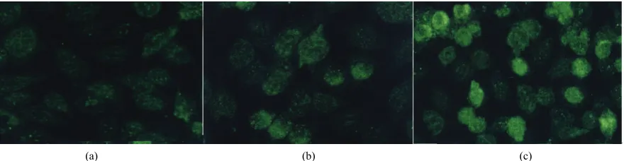
[image:4.595.376.468.343.537.2]peak at 72 h (mean score 2.1 ± 0.8) (Figure 3(b)). But, there was no statisticalsignificant between 48 h and 72 h groups (Figure 4, P > 0.05). KGF expression was unde-tectable at 24 h and 48 h control groups. The results of fluorescence expression were similar to that of immuno-histochemistry (Figure 5).
KGF in our study was designed for two purposes. The first was to study the underling mechanism of KGF such as producingextensive type II cell hyperplasia. It is much more difficult to find this extensive type II cell hyperplasia in rodent lungs with a variety of injuries, includingbleomycin. Therefore in rodents, it has been difficult to investigatethe biologic properties of hyper-plastic type II cells and theirability to alter the inflam-matory and fibroproliferative response.Type II cells can augment fibroticreaction by producing PDGF, TGF-α, and TGF-β [17-20]. However,type II cells also inhibit fibroblast proliferation [21]. Exogenous KGF should make it possible to study the biologicproperties of hy-perplastic type II cells. The second purpose was to have a means of delivering KGF for a sustained period in a local areafor the treatment of lung injury. Currently, because of the inflammationinduced by adenoviral vec-tors, therapy of acute lung injury may not be possible. Another approach is to decrease the host response by using plasmid vector.
3.7. Effect of HKGF on AECII Cell Growth
Cell growth was measured by cell count. The cell num-bers were 2.8 × 10⁶, 3.6 × 10⁶, and 5.0 × 10⁶ in the
ex-perimental group and 2.6 × 10⁶, 3.2 × 10⁶, and 3.9 × 10⁶
in the control group at 24, 48, and 72 h respectively. The growth of AECII was stimulated by KGF in a time- dependent manner (Figure 6, P < 0.05).
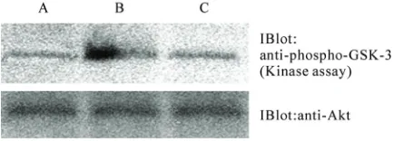
3.8. KGF-Induced Activation of Akt in Vivo
We next have investigated whether high KGF expression follow up a dramatic increase in Akt activation as evalu-ated by Akt kinase assay. We have shown that expression of constitutively active Akt appeared strong transfected by eukaryotic expression vector pEGFP-C2-hKGF (Figure 7). Thus, one possible explanation for KGF-mediated effect is associated with activation of the Akt signaling axis.
Over the past 10 yr, it has become increasingly clear that KGF play important roles in both the normal and the injured lung and ultimately may have therapeutic poten-tial asa targeted therapy to facilitate lung epithelial re-pair in lung disease. KGF has been shown to increase the expression of SP-A and SP-D in cultured alveolar type II cells in vitro [22,23]. Administration of KGF has been
4. DISCUSSION
This Eukaryotic expression plasmid vector expressing
[image:5.595.75.518.404.514.2](a) (b) (c)
Figure 4. Immunolocalization of KGF in S-D AECII cells. Positively stained cells appear brown color. Positive immuno-reactivity to KGF is present in surrounding cytomembrane and is weak in the cytoplasm. The control group data did not show. Note the strong cytomembrane staining in S-D AECII cells 72 h transfection with hKGF vector (c), whereas moder-ate in S-D AECII cells 48 h transfection with hKGF vector (b) and weak in S-D AECII cells 24 h transfection with hKGF vector (a). Original magnification × 200.
(a) (b) (c)
[image:5.595.80.520.581.695.2]Copyright © 2010 SciRes. Openly accessible athttp://www.scirp.org/journal/jbpc/
Figure 6. Cell count of AECII cells transfected by eukaryotic expression vector pEGFP-C2-hKGF in different time (× 106).
Each point is mean ± SE from 6 observations from a represen-tative experiment. Where no error bars are shown, they are included in the point. Similar results were obtained in 2 further experiments.
Figure 7. Activity of Akt kinase in the AECII by eukaryotic expression vector pEGFP-C2-hKGF. Akt kinase activity of AECII transfected with eukaryotic expression vector pEGFP- C2-hKGF for 72 h (b) was detected in a high level as com-pared with normal AECII (a) and vector control (c). The blot was stripped and probed with anti-Akt Ab as described in Ma-terials and Methods and shown equal amounts of Akt in im-munoprecipitates. Three observations were included per condi-tion and data shown are representative of two independent experiments.
shown to protect the lung from a variety of insults in-cluding oxygen, radiationand chemotherapy. In bleomy-cin-induced lung injury, KGF was shown todecrease lung edema.
However, the KGF has had to be given before the in-jury in order to be effective. A more sustained delivery of KGF or expression of KGF might allow for successful post-treatmenttherapy. Gene transfer into specific tissues or cell types is a key technique in the development of gene delivery strategies study, gene function research and gene therapy. Adenoviral vectors can efficiently transfer foreign genes into lung tissue in vivo [24,25]. However, adenovirus-mediatedgene expression is tran-sient and may be associated with a significant inflam-matory response, depending on the viral dose [26]. Un-fortunately, this inflammatory response limits the use of
these vectors in treating acute lung injury.The host re-sponse is mainly to the virus.
Eukaryotic expression plasmid vector capable of di-recting the expression of exogenous gene has been found to be potentially valuable for the study of specific gene function and for gene therapy [27-29]. We described the establishment of a eukaryotic expression system, which allows us to express hKGF in S-D rat AECII. The versa-tilityof this system is evident in our ability to express hKGF. Because ourobjective in this study was to study the effects of KGF, it wasimportant to develop this sys-tem. The results of immunohistochemistry performed in this study demonstrate that the constructed eukaryotic expression system pEGFP-C2-hKGF efficiently pro-duces hKGF gene. And cell count result suggested that hKGF could induce S-D rat AECII cell proliferation.
As plasmid vector gene transfer is one of the most re-liable and convenient methods for introducing genes into almost all types of mammalian cells and for expressing the genes at high levels since many cells receive multi-ple copies of the recombinant genome [30-32]. Gene delivery using eukaryotic expression plasmid vector that propagates in cultured cells might be an effective way for research [33,34]. In order to increase the effect of putative gene therapy, in this study, primers containing specific enzyme-cutting sites were designed to amplify the 0.6 kb hKGF from human genome, and the 0.6 kb sequence was cloned into the eukaryotic plasmid. The
hKGF gene, cloned from human embryo lung fibroblast, showed high homologies of nucleotide and putative amino acid sequences compared with the published cor-responding sequences [35,16].
It was shown that there are several different signal transduction pathways to be stimulated by growth fac-tors (36) and through Akt signaling to transmit induced apoptosis signaling to link life and death decisions (37). our data support the idea that high levels of KGF posi-tively regulate Akt during lung development. KGF- induced Akt activation may play an important role in inhibiting lung alveolar cell death thereby preserving the lung architecture and function during oxidative stress (38). At present, we have no simple explanation for how KGF modifies the response of Akt signal. Fu-ture insight into the relationships between KGF and Akt in vivo will come from functional and biochemical stud-ies on the pathway of the two ligands, and from the study of both KGF-deficient and KGF, Akt double null mutant mice.
[image:6.595.57.274.339.417.2]5. ACKNOWLEDGEMENTS
We thank Prof. Wang Fu-Liang for providing technical assistance and insightful discussions during the preparationof the manuscript. This work was supported by National Scentific Foundation of China (No: 30030781).
REFERENCES
[1] Haddad, I. Y., Milla, C., Yang, S., Panoskaltsis-Mortari, A., Hawgood, S., Lacey, D. L. and Blazar, B. R. (2003) Surfactant protein A is a required mediator of keratino-cyte growth factor after experimental marrow transplan-tation. American Journal of Physiology. Lung Cell Mo-lecular Physiology, 285,L602-L610.
[2] Yi, E. S., Williams, S. T., Lee, H., Malicki, D. M., Chin, E. M, Yin, S., Tarpley, J. and Ulich, T. R. (1996) Keratinocyte growth factor ameliorates radiation- and bleomycin- induced lung injury and mortality. American Journal of Pathology, 149,1963-1970.
[3] Keijzer, R., van Tuyl, M., Meijers, C., Post, M., Tibboel, D., Grosveld, F. and Koutsourakis, M. (2001) The tran-scription factor GATA6 is essential for branching morpho-genesis and epithelial cell differentiation during fetal pul-monary development. Development, 128, 503-511.
[4] Ware, L. B. and Matthay, M. A. (2002) Keratinocyte and hepatocyte growth factors in the lung: Roles in lung de-velopment, inflammation, and repair. American Journal of Physiology. Lung Cell Molecular Physiology, 282, L924-L940.
[5] Ray, P., Devaux, Y., Stolz, D. B., Yarlagadda, M., Wat-kins, S. C., Lu, Y., Chen, L., Yang, X. F. and Ray, A. (2003) Inducible expression of keratinocyte growth fac-tor (KGF) in mice inhibits lung epithelial cell death in-duced by hyperoxia. Proceedings of the National Acad-emy of Science, 100, 6098-6103.
[6] Atabai, K., Ishigaki, M., Geiser, T., Ueki, I., Matthay, M. A. and Ware, L. B. (2002) Keratinocyte growth factor can enhance alveolar epithelial repair by nonmitogenic mechanisms. American Journal of Physiology. Lung Cell Molecular Physiology, 283, L163-L169.
[7] Hohlfeld, J. M., Hoymann, H. G.., Tschernig, T., Fehren-bach, A., Krug, N. and FehrenFehren-bach, H. (2004) Keratino-cyte growth factor transiently alters pulmonary function in rats. Journal of Applied Physiology, 96, 704-710. [8] Buckley, S., Barsky, L., Driscoll, B., Weinberg, K.,
Anderson, K. D. and Warburton, D. (1998) Apoptosis and DNA damage in type 2 alveolar epithelial cells cultured from hyperoxic rats. American Journal of Physiology. Lung Cell Molecular Physiology, 274, L714-L720. [9] Senaldi, G., Shaklee, C. L., Simon, B., Rowan, C. G.,
Lacey, D. L. and Hartung, T. (1998) Keratinocyte growth factor protects murine hepatocytes from tumor necrosis factor-induced apoptosis in vivo and in vitro. Hepatology,
27, 1584-1591.
[10] Portnoy, J., Curran-Everett, D., Mason, R. J. (2004) Keratinocyte growth factor stimulates alveolar type II cell proliferation through the extracellular signal-regulated kinase and phosphatidylinositol 3-OH kinase pathways.
American Journal of Respiratory Cell and Molecular Bi-ology, 30, 901-907.
[11] Fehrenbach, A., Bube, C., Hohlfeld, J. M., Stevens, P., Tschernig, T., Hoymann, H. G., Krug, N. and Fehrenbach, H. (2003) Surfactant homeostasis is maintained in vivo during keratinocyte growth factor-induced rat lung type II cell hyperplasia. American Journal of Respiratory and Critical Care Medicine, 167, 1264-1270.
[12] Chedid, M., Rubin, J. S., Csaky, K. G. and Aaronson, S. A. (1994) Regulation of keratinocyte growth factor gene expression by interleukin 1. Journal of Biological Chem-istry,269, 10753-10757.
[13] Simonet, W. S., DeRose, M. L., Bucay, N., Nguyen, H. Q., Wert, S. E., Zhou, L., Ulich, T. R., Thomason, A., Danilenko, D. M. and Whitsett, J. A. (1995) Pulmonary malformation in transgenic mice expressing human keratinocyte growth factor in the lung. Proceedings of the National Academy of Science, 92, 12461-12465.
[14] Spilde, T. L., Bhatia, A. M., Marosky, J. K., Preuett, B., Kobayashi, H., Hembree, M. J., Prasadan, K., Daume, E., Snyder, C. L. and Gittes, G. K. (2003) Fibroblast growth factor signaling in the developing tracheoesophageal fis-tula. Journal of Pediatric Surgery, 38, 474-477. [15] Fu, Y. G., Qu, Y. J., Wu, K. C., Zhai, H. H., Liu, Z. G. and
Fan, D.M. (2003) Apoptosis-inducing effect of recombi-nant Caspase-3 expressed by constructed eukaryotic vector on gastric cancer cell line SGC7901. World Jour-nal of Gastroenterology, 9, 1935-1939.
[16] Morikawa, O., Walker, T. A., Nielsen, L. D., Pan, T., Cook, J. L. and Mason, R. J. (2000) Effect of adenovec-tor-mediated gene transfer of keratinocyte growth factor on the proliferation of alveolar type II cells in vitro and in vivo. American Journal of Respiratory Cell and Mo-lecular Biology, 23, 626-635.
[17] Paine, R. 3rd., Rolfe, M. W., Standiford, T. J., Burdick, M. D., Rollins, B. J. and Strieter, R. M. (1993) MCP-1 expression by rat type II alveolar epithelial cells in pri-mary culture. Journal of Immunology, 150, 4561-4570. [18] Li, C., Zhu, N. L., Tan, R. C., Ballard, P. L., Derynck, R.
and Minoo, P. (2002) Transforming growth factor-beta inhibits pulmonary surfactant protein B gene transcrip-tion through SMAD3 interactranscrip-tions with NKX2.1 and HNF-3 transcription factors. Journal of Biological Chem-istry, 277, 38399-38408.
[19] Beers, M.F., Solarin, K.O., Guttentag, S.H., Rosenbloom, J., Kormilli, A., Gonzales, L.W. and Ballard, P. L. (1998) TGF-beta1 inhibits surfactant component expression and epithelial cell maturation in cultured human fetal lung.
American Journal of Physiology. Lung Cell Molecular Physiology, 275, L950-L960.
[20] Zhao, Y., Young, S. L., McIntosh, J. C., Steele, M. P. and Silbajoris, R. (2000) Ontogeny and localization of TGF- beta type I receptor expression during lung development.
American Journal of Physiology. Lung Cell Molecular Physiology, 278, L1231-L1239.
[21] Mason, R. J., Lewis, M. C., Edeen, K. E., McCormick- Shannon, K., Nielsen, L. D. and Shannon, J. M. (2002) Maintenance of surfactant protein A and D secretion by rat alveolar type II cells in vitro. American Journal of Physiology. Lung Cell Molecular Physiology, 282, L249- L258.
Copyright © 2010 SciRes. Openly accessible athttp://www.scirp.org/journal/jbpc/ II cells. American Journal of Respiratory Cell and
Mo-lecular Biology,18, 168-178.
[23] Chelly, N., Mouhieddine-Gueddiche, O. B., Barlier-Mur, A. M., Chailley-Heu, B., Bourbon, J. R. (1999) Kerati-nocyte growth factor enhances maturation of fetal rat lung type II cells. American Journal of Respiratory Cell and Molecular Biology, 20, 423-432.
[24] Xing, Z., Braciak, T., Jordana, M., Croitoru, K., Graham, F. L. and Gauldie, J. (1994) Adenovirus-mediated cyto-kine gene transfer at tissue sites. Overexpression of IL-6 induces lymphocytic hyperplasia in the lung. Journal of Immunology, 153, 4059-4069.
[25] Yei, S., Mittereder, N., Wert, S., Whitsett, J. A., Wilmott, R. W. and Trapnell, B. C. (1994) In vivo evaluation of the safety of adenovirus-mediated transfer of the human cystic fibrosis transmembrane conductance regulator cDNA to the lung. Human Gene Therapy, 5, 731-744. [26] Roy, S., Gao, G., Lu, Y., Zhou, X., Lock, M., Calcedo, R.
and Wilson, J. M. (2004) Characterization of a family of chimpanzee adenoviruses and development of molecular clones for gene transfer vectors. Human Gene Therapy,
15, 519-530.
[27] Ebelt, H. and Braun, T. (2003) Optimized, highly effi-cient transfer of foreign genes into newborn mouse hearts in vivo. Biochemical and Biophysical Research Commu-nications, 310, 1111-1116.
[28] Bosma, P. T., van Eert, S. J., Jaspers, N. G., Stoter, G. and Nooter, K. (2003) Functional cloning of drug resistance genes from retroviral cDNA libraries. Biochemical and Biophysical Research Communications, 309, 605-611. [29] Yan, J., Liang, S. H., Mao, Y. F., Li, L. W., Li, S. P. (2003)
Construction of expression systems for flaA and flaB genes of Helicobacter pylori and determination of im-munoreactivity and antigenicity of recombinant proteins.
World Journal of Gastroenterology, 9, 2240-2250. [30] Ebensen, T., Paukner, S., Link, C., Kudela, P., de Domenico,
C., Lubitz, W. and Guzman, C. A. (2004) Bacterial ghosts are an efficient delivery system for DNA vaccines. Journal of Immunology, 172, 6858-6865.
[31] Piao, Y. F., Tang, T. Y., Niu, J. Q. and Wang, F. (2004) Construction of eukaryotic expression plasmids of hepa-titis B surface antigen and helper T lymphocyte epitope.
Hepatobiliary & Pancreatic Diseases International Jour-nal, 3, 219-222.
[32] Schuck, S., Manninen, A., Honsho, M., Fullekrug, J. and Simons, K. (2004) Generation of single and double knock-downs in polarized epithelial cells by retrovirus-mediated RNA interference. Proceedings of the National Academy of Science, 101, 4912-4917.
[33] Belin, D., Guzman, L.M., Bost, S., Konakova, M., Silva, F. and Beckwith, J. (2004) Functional activity of eu-karyotic signal sequences in Escherichia coli: The oval-bumin family of serine protease inhibitors. Journal of Biological Chemistry, 335, 437-453.
[34] Baldwin, S. L., Powell, T. D., Wonderling, R. S., Keiser, K. C., Morales, T., Hunter, S., McDermott, M., Radecki, S. V. and Milhausen, M. J. (2003) Transient and stable transfection of Chinese hamster ovary cells with the re-combinant feline erythropoietin gene and expression, pu-rification, and biological activity of feline erythropoietin protein. American Journal of Veterinary Research, 64, 1465-1471.
[35] Finch, P. W., Rubin, J. S., Miki, T., Ron, D. and Aaronson, S. A. (1989) Human KGF is FGF-related with properties of a paracrine effector of epithelial cell growth. Science,
245, 752-755.
[36] Fujio, Y. and Walsh, K. (1999) Akt mediates cytoprotec-tion of endothelial cells by vascular endothelial growth factor in an anchorage-dependent manner. Journal of Bio-logical Chemistry, 274, 16349-16354.
[37] Kauffmann-Zeh, A., Rodriguez-Viciana, P., Ulrich, E., Gilbert, C., Coffer, P., Downward, J. and Evan, G. (1997) Suppression of c-Myc-induced apoptosis by Ras signal-ling through PI(3)K and PKB. Nature, 385, 544-548. [38] Lu, Y., Parkyn, L., Otterbein, L. E., Kureishi, Y., Walsh,