Original Article
Study on expression of IL-6/JAK/STAT3 signaling
pathway in category IIIB prostatitis
Guangyu Li1, Haiyan Lan2, Jihong Liang1, Jing Xian3, Dan Fang3, Yuncong Mo4, Cheng Zheng1, Yingjin Dong1, Yuanfa Li1
Departments of 1Andrology, 3Endocrinology, 4Nuclear Medicine,The First Affiliated Hospital of Guangxi Medical University, Nanning City, Guangxi Zhuang Autonomous Region, China; 2Department of Medical Oncology, The People’s Hospital of Guangxi Zhuang Autonomous Region, Nanning City, Guangxi Zhuang Autonomous Region,
China
Received November 20, 2017; Accepted January 19, 2018; Epub April 15, 2018; Published April 30, 2018
Abstract: Objective: To investigate the expression levels of interleukin (IL)-6 and downstream JAK/STAT3 signaling pathway in category IIIB prostatitis. Methods: A total of 44 patients diagnosed with category IIIB prostatitis in our hospital were selected as study group. During the same period, forty healthy people underwent physical examina-tion in our hospital were enrolled in control group. The differences of IL-6, IL-8 and IL-2 in serum and prostatic fluid in the two groups were compared. In addition, 80 Sprague-Dawley rats were randomly divided into four groups: control group (normal saline was injected every other day), prostatitis group (animal models of category IIIB prostatitis), prostatitis + IL-6 group (IL-6 was intraperitoneally injected to rats with prostatitis every other day for one month), prostatitis + blocker group (IL-6 downstream JAK2/STAT3 signaling pathway blocker AG490 was given to rats with prostatitis every other day for one month), with 20 rats in each group. The levels of IL-6, IL-8 and IL-2 in serum of the four groups of rats were detected. And the differences of expression levels of IL-6 and downstream signaling molecules (JAK and STAT3) in prostate tissues as well as the inflammatory degree of prostate pathological tissues of rats in the four groups were compared. Results: Compared with control group, the IL-6, IL-8 and IL-2 levels in pros-tatic fluid of patients in study group were obviously higher (all P<0.01), but the differences of those levels in serum were no significant (all P>0.05). The IL-6, IL-8 and IL-2 levels in serum of rats in the four groups had no significant difference (all P>0.05). However, in the prostate tissues of the four groups of rats, the numbers of IL-6 positive cells were ranked as prostatitis + IL-6 group > prostatitis group > control group > prostatitis + blocker group (all P<0.05), and the levels of JAK and STAT3 in the four groups were also with the same sequence (P<0.05), also, the inflamma-tory degrees were with the same trend. Conclusion: IL-6 and its downstream JAK/STAT3 signaling molecules present high expression in category IIIB prostatitis, and they may be involved in sterile inflammation.
Keywords: Category IIIB prostatitis, IL-6, JAK/STAT3 signaling pathway
Introduction
Category III chronic prostatitis/chronic pelvic pain syndrome is the most common chronic prostatitis, and category IIIB prostatitis is one of that without existing evidence of white blood cell in prostatic fluid [1]. Although lots of stud-ies on category IIIB prostatitis have been car-ried out, its etiology and pathogenesis remain unknown. In recent years, study showed that immune reaction played an important part in the development of category IIIB prostatitis, in which IL-6 had the most concern, but specific signaling pathway of IL-6 in pathogenesis and
Materials and methods
Subjects
This study was approved by Ethics Committee in our hospital and the informed consents were obtained from all subjects.
Forty-four patients diagnosed with category IIIB prostatitis in our hospital were selected as study group, aged 55-69 years, mean age: 61.6±7.2 years. Inclusion criteria: Patients who met the diagnostic criteria of category IIIB pros-tatitis in Diagnosis and Treatment of Chronic Prostatitis set by Chinese Urological Associa- tion; patients who signed informed consents in written form. Exclusion criteria: Patients who suffered from prostate cancer; patients with severe dysfunction in heart, lung, liver, kidney or other important organs; patients who were receiving oral medication for the treatment of chronic prostatitis. In the same period, forty healthy people underwent physical examina-tion in our hospital were enrolled in control group, aged 53-72 years, mean age: 63.5±9.7 years.
Animal model and grouping
A total of 80 male Sprague-Dawley rats (weight: 250 to 300 g), which were purchased from the laboratory animal center of our hospital, were randomly divided into four groups with 20 rats in each group: control group, prostatitis group, prostatitis + IL-6 group and prostatitis + block-er group. In prostatitis group, category IIIB pros-tatitis animal models were established, and specific methods were described in previous experimental research [4]. Right and left ven-tral lobes of rat prostate were injected 3% car-rageenan 50 μl, and experimental animal mod-els were successfully established after a week. After that, intraperitoneal injection of IL-6 was given for rats in prostatitis + IL-6 group every other day for one month. In prostatitis + blocker group, JAK2/STAT3 signaling pathway blocker AG490 was given to rats with prostatitis every other day for one month. And in control group and prostatitis group, the rats were injected equal amounts of normal saline every other day for a month.
Main reagents
Enzyme Linked Immunosorbent Assay (ELISA) kits of IL-6, IL-8 and IL-2 were purchased from Beijing Biofriendship Co., Ltd. And the sources
of other reagents were as follows: AG490 from Calbiochem company, Germany; IL-6 primary antibody from Pepro Tech company, America; JAK and STAT3 primary antibodies from Cell Signal company, America; β-actin primary anti-body from Santa Cruz Biotechnology company, America); all the secondary antibodies were from Beijing Zhongshan Golden Bridge Biote- chnology Co. Ltd.; cell lysis buffer (included 1 mM DTT, 5 mM EDTA and 1 × protease inhibitor cocktail) and protein loading buffer were from Sigma-Aldrich company, America.
Detection of cytokines levels
Detection of cytokines levels in human serum
and prostatic fluid: Fasting serum of the two groups of patients was collected in the morning respectively, and prostatic fluid was collected through prostatic massage. The concentrations of cytokine IL-6, IL-8 and IL-2 in serum and pros-tatic fluid were determined by human ELISA kits respectively, and the differences were com- pared.
Detection of cytokines levels in rat serum: Anesthesia was carried out for four groups of rats with 10% chloral hydrate. Then the rats were killed after serum samples were collected by carotid artery bloodletting, and their serum samples and prostate samples were kept. The concentrations of cytokine IL-6, IL-8 and IL-2 in serum of rats were detected by rat ELISA kits.
Detection of IL-6 positive cells in prostatic
tis-sues of rats: Immunohistochemical S-P method was used to detect the protein expression level of cytokine IL-6 in prostatic tissue. Parts of rat prostatic tissues were sliced and successively incubated with rat IL-6 primary antibody, biotin-labeled goat anti-rat IgG secondary antibody and horseradish peroxidase conjugated strep-tavidin in room temperature for color develop-ment. Ten slices were randomly selected from each group, and three visual fields were ran-domly selected in each slice for high-power lens to perform data measurement and analy-sis, and the number of positive cells in every visual field was calculated.
Analysis of inflammation degree of prostatic
tissues in rats
optical microscope at 400 times magnification to perform data measurement and analysis, and the mean value was calculated after inflam-matory cells were counted. The differences of prostate weight and inflammatory cell numbers of the four groups of rats were compared. Expression level of JAK and STAT3 signaling
pathway
Western blot method was used to detect molec-ular levels of JAK and STAT3 in rat prostatic tis-sues, and specific methods were as follows: parts of rat prostatic tissues were taken from the rats in four groups respectively, and 0.5 ml cell lysis buffer was added in the homogenized tissue, then they were boiled for five minutes to extract cell protein (20 μg protein was added for each well). Loading buffer was added in pre-pared samples, then they were mixed and boiled for 5 minutes for protein denaturation. After that, equal amount of mixed sample was added in each well of 7.5% polyacrylamide gel for electrophoresis. The proteins were trans-ferred to polyvinylidene fluoride membrane, then 3% skimmed milk dissolved in phosphate buffer saline (freshly prepared) was used for blocking. Next, JAK mouse monoclonal anti-body, STAT3 mouse monoclonal antibody (1:1,000) and anti-β-actin antibody (1:500) were added, then they were incubated
over-night at 4°C. After that, the proteins were washed for three times with phosphate buffer saline. Then, they were incubated after adding the horseradish peroxidase labeled secondary antibody (goat anti-rat IgG). At last, chemiluminescence and X- ray exposure were used. Image processing system (NIH image) was used for the analysis of optical density.
Statistical analysis
SPSS17.0 statistical soft-ware package was used for statistical analysis. Norma- lity test was used for contin-uous data before com- parison and analysis, and Levene test was used for homogeneity test of
vari-Table 1. Comparison of cytokine levels in serum and prostatic fluid between two groups (pg/ml)
Group Study group Control group t P
Case 44 40
IL-6 Serum 14.5±4.1 13.1±3.2 1.732 0.087
Prostatic fluid 50.4±7.6 43.1±5.9 4.882 0.000
IL-8 Serum 296.6±35.3 285.4±29.6 1.567 0.121
Prostatic fluid 930.2±47.5 898.9±40.7 3.227 0.002
IL-2 Serum 89.4±8.9 86.3±8.2 1.655 0.102
[image:3.612.91.381.99.203.2]Prostatic fluid 342.3±26.8 319.5±24.3 4.070 0.000
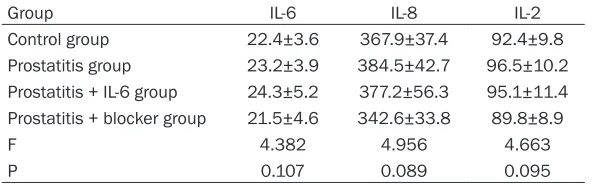
Table 2. Comparison of cytokine levels in serum of four groups of experimental rats (n=20/group, pg/ml)
Group IL-6 IL-8 IL-2
Control group 22.4±3.6 367.9±37.4 92.4±9.8
Prostatitis group 23.2±3.9 384.5±42.7 96.5±10.2 Prostatitis + IL-6 group 24.3±5.2 377.2±56.3 95.1±11.4 Prostatitis + blocker group 21.5±4.6 342.6±33.8 89.8±8.9
F 4.382 4.956 4.663
P 0.107 0.089 0.095
ance. The measurement data were expressed as mean±standard deviation (_x±sd), and com- parison of cases between study group and control group was performed with two independent samples t-test; one-way ANOVA and Newman-Student-Keuls test were used for the analysis of the inter-group differences among four animal groups. Enumeration data were expressed as rate, and chi-square test and chi-square partition test were used for inter-group comparison. The differences were statistically significant when P<0.05.
Results
Comparison of cytokine levels in serum and
prostatic fluid between two human groups
Compared with control group, patients in study group had apparently higher levels of IL-6, IL-8 and IL-2 in their prostatic fluid (all P<0.01), while differences of those levels in serum were not significant (all P>0.05). See Table 1.
Comparison of cytokine levels in serum of
experimental rats
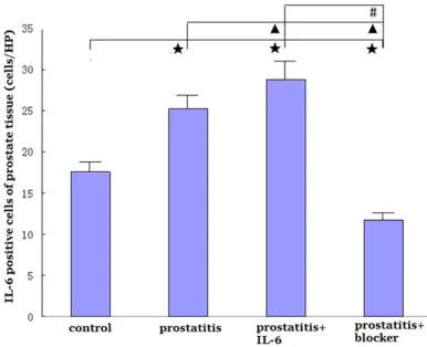
[image:3.612.89.385.250.342.2]Comparison of IL-6 levels in prostate tissues of rats
In the four groups, the amounts of IL-6 positive cells in rat prostate tissues were as follows: control group (17.6±4.7/HP), prostatitis group (25.2±4.3/HP), prostatitis + IL-6 (28.8±5.2/ HP), prostatitis + blocker group (11.7±4.1/HP); namely, the order of numbers of IL-6 positive cells in these groups was: prostatitis + IL-6 > prostatitis group > control group > prostatitis + blocker group (all P<0.05). See Figure 1.
Analysis of pathological prostatic tissue of rats
In prostatitis group, structures of rat prostate tissue were damaged with inhomogeneous lym-phoid hyperplasia. Catheters were dilatated or damaged; parts of basement membrane were destroyed with visible diffuse infiltration of lymph, monocyte and other chronic inflamma-tory cells existing around the mesenchyme and glands. In prostatitis + IL-6 group, structures of rat prostate tissue were damaged severely with gathering inflammatory cells; plenty of glandu-lar epithelial tissues were damaged, and lym-phoid nodule or folliculus were formed also with a full view of inflammatory cells. As for prostati-tis + blocker group, the inflammatory cells in rat prostatic tissues were scattered and numbered 1-10/HP. In control group, the rat prostate tis-sues had a relatively complete structure with neither inflammatory cell infiltration in mesen-chyme or gland, nor edema in glandular cavity. See Figure 2.
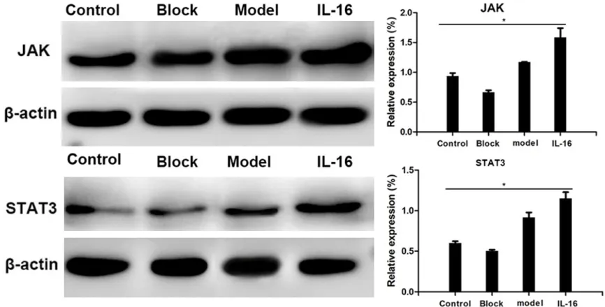
Comparison of JAK and STAT3 levels in rat
prostate tissues
The levels of JAK and STAT3 in prostate tissues of the four groups of rats were ranked as pros-tatitis + IL-6 group > prospros-tatitis group > control group > prostatitis + blocker group (P<0.05). See Figure 3.
Discussion
The immune inflammatory reaction has been proved vital to the pathogenic process of cate-gory IIIB prostatitis. As the previous studies demonstrated, after category IIIB prostatitis model rats received high level 5-α dihydrotes-tosterone, the cell inflammatory reaction of their prostate tissues could be significantly inhibited; therefore, many investigators be- lieved that category IIIB prostatitis was essen-tially a cytotoxic immune response, but its spe-cific pathogenesis was not yet fully defined [4-6]. The extremely complex body immune inflammatory reaction included the participa-tion of cytokines network and downstream sig-nal transduction, at the same time, there was a study proved that multiple cytokines networks and their interaction in the inflammatory reac-tion determined the outcome of overall body immune inflammation [7]. Among the cyto-kines, IL-6 might play a pivotal role in the patho-logical process of category IIIB prostatitis [8]. There were studies revealed that in patients with prostatitis, the levels of IL-6 in prostate fluid, sperm, and biopsy tissues were signifi-cantly related to their symptoms [9, 10]. Another study showed that after the treatment, the level of IL-6 in seminal plasma of patients with category IIIB prostatitis was significantly lower than that of before treatment, and it was positively correlated to clinical efficacy [11].
[image:4.612.90.283.72.229.2]This study found that the levels of IL-6, IL-8 and IL-2 in prostate fluid of patients with category IIIB prostatitis were obviously higher than those of normal subjects, which was consistent with the results of previous studies [12-14]. However, when the patients were compared with the nor-mal subjects in control group, there was no sig-nificant difference in IL-6, IL-8 and IL-2 in peripheral blood, which suggested that the immune inflammatory reaction of category IIIB prostatitis was localized in the prostate rather than the whole body [13-15]. In animal model experiment, there was no significant difference
Figure 1. Comparison of numbers of IL-6 positive cells in prostate tissues of four groups of rats. Com-pared with control group, ★P<0.05; compared with prostatitis group, ▲P<0.05; compared with
in rat serum IL-6, IL-8 and IL-2 levels. Nevertheless, according to the immunohisto-chemistry analysis of prostate tissues, we found that the IL-6 levels of rats in prostatitis group and prostatitis + IL-6 group were higher than that in prostatitis + blocker group, which suggested that IL-6 played a role in prostate chronic immune inflammatory response and also implied that the pathological process of category IIIB prostatitis might incline to be a kind of chronic autoimmune disease.
After the combination of IL-6 and cell mem-brane receptor complex in vivo, JAK kinase was activated, and therewith STAT was also activat-ed by tyrosine phosphorylation, and then sig-nals were transduced to intranuclear to act the biological roles in related regulation [15]; at the same time, JAK/STAT signaling pathway was appearing a positive feedback regulation to the
cellular expression of IL-6 [16-19]. Previous studies have shown that IL-6 promotes the expression of anti-apoptotic genes such as bcl-xl, c-Jun and Fas by activating the JAK/STAT sig-naling pathway, which is closely related to angiogenesis, cell proliferation and apoptosis [20-22]. But it hasn’t been reported by any studies that whether IL-6 can promote the pathogenesis and progression of category IIIB prostatitis by activating the JAK/STAT signaling pathway or not. Therefore, this study discussed this question by analyzing the clinical data of patients with category IIIB prostatitis in our hospital and establishing rat models of catego-ry IIIB prostatitis.
[image:5.612.90.526.72.192.2]Therefore, this study suggests that IL-6 may participate in the pathological process of cate-gory IIIB prostatitis through JAK/STAT signaling pathway, but this mechanism still needs further
Figure 2. Inflammatory infiltration of rat prostate tissues in four groups (*400). (A) prostatitis group, (B) prostatitis + IL-6 group, (C) prostatitis + blocker group, (D) control group.
[image:5.612.88.521.242.462.2]experiment to confirm; meanwhile, it may pro-vide a new potential therapeutic target for the treatment of category IIIB prostatitis, and it is worthy of further researches in subsequent studies.
Acknowledgements
This work was supported by the Natural Science Foundation of Guangxi Zhuang Autonomous Region (2017GXNSFAA198062) and the Basic Ability Improvement Project of Young Tea- chers in Guangxi Colleges and Universities (2017KY0112).
Disclosure of conflict of interest
None.
Address correspondence to: Guangyu Li and Jihong Liang, Department of Andrology, The First Affiliated Hospital of Guangxi Medical University, No.6 Shuangyong Road, Nanning City 530021, Guangxi Zhuang Autonomous Region, China. Tel: +86-13768275647; E-mail: [email protected] (GYL); Tel: +86-0771-5356155; E-mail: [email protected] (JHL)
References
[1] Lan T, Wang Y, Chen Y, Qin W, Zhang J, Wang Z, Zhang W, Zhang X, Yuan J and Wang H. Influ-ence of environmental factors on prevalInflu-ence, symptoms, and pathologic process of chronic prostatitis/chronic pelvic pain syndrome in northwest china. Urology 2011; 78: 1142-1149.
[2] Culig Z and Puhr M. Interleukin-6: a multifunc-tional targetable cytokine in human prostate cancer. Mol Cell Endocrinol 2012; 360: 52-58. [3] Jo HA, Kim JY, Yang SH, Han SS, Joo KW, Kim
YS and Kim DK. The role of local IL6/JAK2/ STAT3 signaling in high glucose-induced podo-cyte hypertrophy. Kidney Res Clin Pract 2016; 35: 212-218.
[4] Wang XJ, Zhong S, Zhang CM and Shen ZJ. Es-tablishment and histopathological characteris-tics of rat model of chronic nonbacterial pros-tatitis. Chinese Journal of Urology 2012; 33: 282-287.
[5] Lou L, Zhou J, Liu Y, Wei YI, Zhao J, Deng J, Dong B, Zhu L, Wu A, Yang Y and Chai L. Chlo-rogenic acid induces apoptosis to inhibit in-flammatory proliferation of IL-6-induced fibro-blast-like synoviocytes through modulating the activation of JAK/STAT and NF-kappaB signal-ing pathways. Exp Ther Med 2016; 11: 2054-2060.
[6] Krieger JN, Nyberg L Jr, Nickel JC. NIH consen-sus definition and classification of prostatitis. JAMA 1999; 282: 236-237.
[7] Hu Y, Niu X, Wang G, Huang J, Liu M and Peng B. Chronic prostatitis/chronic pelvic pain syn-drome impairs erectile function through in-creased endothelial dysfunction, oxidative stress, apoptosis, and corporal fibrosis in a rat model. Andrology 2016; 4: 1209-1216. [8] Aghazarian A, Plas E, Stancik I, Pfluger H and
Lackner J. New method for differentiating chronic prostatitis/chronic pelvic pain syn-drome IIIA from IIIB involving seminal macro-phages and monocytes. Urology 2011; 78: 918-923.
[9] Nickel JC, Nyberg LM and Hennenfent M. Re-search guidelines for chronic prostatitis: con-sensus report from the first national institutes of health international prostatitis collaborative network. Urology 1999; 54: 229-233.
[10] Milicevic N, Mrcela M, Galic J and Marjanovic K. Expression of proinflammatory cytokine in-terleukin-6 in tissue samples of human pros-tate obtained by needle biopsy. Pathol Res Pract 2015; 211: 865-870.
[11] Castiglione R, Salemi M, Vicari LO and Vicari E. Relationship of semen hyperviscosity with IL-6, TNF-alpha, IL-10 and ROS production in semi-nal plasma of infertile patients with prostatitis and prostato-vesiculitis. Andrologia 2014; 46: 1148-1155.
[12] Korrovits P, Ausmees K, Mandar R and Punab M. Seminal interleukin-6 and serum prostate-specific antigen as possible predictive bio-markers in asymptomatic inflammatory prosta-titis. Urology 2011; 78: 442-446.
[13] Cai T, Verze P, La Rocca R, Palmieri A, Tiscione D, Luciani LG, Mazzoli S, Mirone V and Malos-sini G. The clinical efficacy of pollen extract and vitamins on chronic prostatitis/chronic pelvic pain syndrome is linked to a decrease in the pro-inflammatory cytokine interleukin-8. World J Mens Health 2017; 35: 120-128. [14] Engelhardt PF, Seklehner S, Brustmann H,
Lu-suardi L and Riedl CR. Immunohistochemical expression of interleukin-2 receptor and inter-leukin-6 in patients with prostate cancer and benign prostatic hyperplasia: association with asymptomatic inflammatory prostatitis NIH category IV. Scand J Urol 2015; 49: 120-126. [15] Shirzad M, Heidarian E, Beshkar P and
Ghola-mi-Arjenaki M. Biological effects of hesperetin on interleukin-6/phosphorylated signal trans-ducer and activator of transcription 3 pathway signaling in prostate cancer PC3 cells. Phar-macognosy Res 2017; 9: 188-194.
induction in prostate epithelial cells stimulated with trichomonas vaginalis. Parasite Immunol 2016; 38: 678-687.
[17] Milicevic N, Mrcela M, Lukic I, Mandic S, Hor-vat V and Galic J. Comparison between clinical significance of serum proinflammatory protein interleukin-6 and classic tumor markers total PSA, free PSA and free/total PSA prior to pros-tate biopsy. Coll Antropol 2014; 38: 147-150. [18] Culig Z. Proinflammatory cytokine interleukin-6
in prostate carcinogenesis. Am J Clin Exp Urol 2014; 2: 231-238.
[19] Nguyen DP, Li J and Tewari AK. Inflammation and prostate cancer: the role of interleukin 6 (IL-6). BJU Int 2014; 113: 986-992.
[20] Chen X, Wang W, Man H, Li P and Shan B. In-creased B7-H4 expression during esophageal squamous cell carcinogenesis is associated with IL-6/STAT3 signaling pathway activation in mice. Oncol Lett 2017; 13: 2207-2215.
[21] Albino D, Civenni G, Rossi S, Mitra A, Catapano CV and Carbone GM. The ETS factor ESE3/EHF represses IL-6 preventing STAT3 activation and expansion of the prostate cancer stem-like compartment. Oncotarget 2016; 7: 76756-76768.