ISSN Online: 2165-3410 ISSN Print: 2165-3402
DOI: 10.4236/aim.2018.87036 Jul. 27, 2018 519 Advances in Microbiology
Development and Validation of Multiplex
One-Step Real-Time TaqManqRT-PCR Assays
for Detection and Quantification of Arboviral
Encephalitis Viruses
Donggen Zhou
*, Jie Luo
Ningbo International Travel Healthcare Center, Ningbo, China
Abstract
Arboviral encephalitis is a group of animal and human illness that is mostly caused by several distinct families of viruses including orthobunya virus, phlebovirus, flaviviruses, and the alphaviruses. Although specific signs and symptoms vary by the type of central nervous system (CNS), initial signs and symptoms are very similar. Therefore rapid immunologic and molecular tools for differential diagnosis of arboviral encephalitis viruses are important for effective case management and control of the spread of encephalitis. The qRT-PCR assay, especially multiplex PCR, has the potential to produce con-siderable savings in time and resources in the laboratory detection. Mean-while, the use of IC can prevent false negatives effectively by monitoring the processes of nucleic acid extraction and amplification. This report describes the development of a panel of internally controlled multiplex one-step real-time RT-PCR assays in which two virus specific-probe sets were used in the same reaction for the detection of 15 species arboviral encephalitis virus-es: the comparative sensitivity of multiplex one-step qRT-PCR assays to sin-gle plex one-step qRT-PCR assays as well as one-step RT-PCR assays for tection of each viral species. And total of 150 human serum samples were de-tected to evaluate the multiplex one-step qRT-PCR assays. These multiplex one-step real-time RT-PCR assays with IC were evaluated in terms of sensi-tivity, linearity, precision, specificity, and also field samples including serum and vector. These assays can detect and differentiate arboviral encephalitis viruses by high throughput, sensitive, and specific way. It is useful for clinical management and outbreak control of arboviral encephalitis viruses and vec-tor surveillance.
How to cite this paper: Zhou, D.G. and Luo, J. (2018) Development and Validation of Multiplex One-Step Real-Time Taq-ManqRT-PCR Assays for Detection and Quantification of Arboviral Encephalitis Vi-ruses. Advances in Microbiology, 8, 519-557.
https://doi.org/10.4236/aim.2018.87036
Received: June 26, 2018 Accepted: July 24, 2018 Published: July 27, 2018
Copyright © 2018 by authors and Scientific Research Publishing Inc. This work is licensed under the Creative Commons Attribution International License (CC BY 4.0).
http://creativecommons.org/licenses/by/4.0/
D. G. Zhou, J. Lu
DOI: 10.4236/aim.2018.87036 520 Advances in Microbiology
Keywords
Multiplex One-Step Real-Time TaqManqRT-PCR Assays, Arboviral Encephalitis Viruses, Internal Control
1. Introduction
Arboviral encephalitis is caused by infection with an arbovirus, which transmit-ted by a mosquito, tick or another arthropod. The commonest cause of human disease is flaviviruses, alphaviruses, orthobunyavirus and the phlebovirus [1][2] [3]. Eastern equine Encephalitis virus (EEEV) [4][5], Western Equine Encepha-litis virus (WEEV) [6][7] and Venezuelan Encephalitis virus (VEEV) [8] belong to the alphaviruses, Japanese Encephalitis virus (JEV) [9], St. Louis Encephalitis virus (SLEV) [6][10][11], West Nile virus (WNV) [5][6][12] and Tick-borne Encephalitis virus (TBEV) [13] [14] [15] [16] are from the flaviviruses, while California Encephalitis virus (CEV) and La Crosse virus (LACV) [17] are mem-bers of the orthobunyavirus, and Rift Valley Fever virus (RVFV) [18] [19] and Toscana virus (TOSV) [20] are members of the phlebovirus. Many types of ar-boviral encephalitis occur throughout the world. They include Japanese Ence-phalitis (JE), Rift Valley Fever (RVF), Tick-borne EnceEnce-phalitis (TBE), Murray Valley Encephalitis (MVE) [21] [22] and, most notorious of all, the West Nile virus (WNV) [23][24] which causes West Nile encephalitis, also known as West Nile fever.
Recently, increasing evidence has shown that certain arboviruses such as dengue virus and chikungunya virus may occasionally cause encephalitis in ad-dition to their conventional symptoms, which usually involves headaches, mus-cle and joint pain, and rashes [25] [26] [27] [28]. Most of these diseases are problems only for those individuals traveling to countries where the viruses are endemic, having similar symptoms. Therefore, it is too difficult to distinguish the various etiologic agents based on clinical signs and symptoms, which makes the accurate and timely laboratory detection of viruses important in early diag-nosis of arboviral encephalitis.
In view of its identifying the selected target gene of RNA viruses rapidly and specifically, probe-based real-time quantitative reverse transcription-polymerase chain reaction (qRT-PCR) assay is widely used for virus detection. Some me-thods for detection of arboviral encephalitis viruses have been published, which provides useful references for people working on them. However, most of these qRT-PCR assays may cover only one or parts of virus strains.
DOI: 10.4236/aim.2018.87036 521 Advances in Microbiology inhibitors present a significant source of false-negative results, which may be particularly important in the performance of nucleic acid detection [29].
Therefore, a panel of reliable comprehensive duplex one-step real-time qRT-PCR assays covering all important pathogens, suitable for multiplex screening or spe-cific quantitative identification with fast turn-around time and identical cycling parameters is still urgently needed, so that the unknown samples can be tested simultaneously and effectively.
Here, we established a panel of species-specific internally controlled one-step real-time qRT-PCR assays for multiplex detection of 15 viruses, which covered nearly all the important viral pathogens that cause arboviral encephalitis, in-cluding Eastern equine encephalitis virus (EEEV), Western Equine Encephalitis virus (WEEV), Venequilan Equine Encephalitis virus (VEEV), Japanese Ence-phalitis virus (JEV), Saint Louis EnceEnce-phalitis virus (SLEV), Murray Valley En-cephalitis virus (MVEV), West Nile virus (WNV), Powassan virus (POWV), California Encephalitis virus (CEV), La Crosse virus (LCV), Tick-borne Ence-phalitis virus (TBEV), Rift Valley Fever virus (RVFV), Toscana virus (TOSV), Dengue virus (DENV), Chikungunya virus (CHIKV) and internal control (IC). All assays were optimized at a same thermal cycling condition, and evaluated under single plex, duplex qRT-PCR assays or RT-PCR assays for detection of the in vitro-transcribed Viral RNAs, which were proved to be reliable molecular tools of early diagnosis. And total of 150 RNA samples from human serum were examined using the multiplex one-step real-time qRT-PCR assays. The duplex one-step real-time qRT-PCR assays were verified that the assays were sensitive, specific and reliable methods for detection of arboviral encephalitisis viruses. And they are useful for clinical management and outbreak control of arboviral encephalitisis viruses and vector surveillance.
2. Materials and Methods
2.1. Primers and Probes Design
In this study, 15 species viruses were detected, all of genomic sequences were all retrieved from the GenBank database of NCBI
(http://www.ncbi.nlm.nih.gov/nuccore/). The multiple alignments and
identifi-cation of conserved regions of genomic sequences were carried out respectively by Perl script, which using Clustal W alignment program and sequence analysis algorithm. Primers and probe for each virus were designed using a Primer Premier software (version 3.0), and optimized using Oligo software (version 6.0) by analysis of potentials for dimerization, cross-linking and secondary struc-tures. The specificity of primer and probe sequences was further confirmed us-ing primer-BLAST (NCBI). The probes were differently labeled with the fluo-rescent dyes, FAM, HEX. All oligonucleotides were synthesized by Invitrogen Technology Co., Ltd.
2.2. IC Design
ac-D. G. Zhou, J. Lu
DOI: 10.4236/aim.2018.87036 522 Advances in Microbiology
cording to the sequence of the tobacco mosaic virus (isolate Guangyuan, com-plete genome, http://www.ncbi.nlm.nih.gov/nuccore/HE818460.1). The details of its primers and probes are listed in Table 1. In order to check from RNA ex-traction to amplification, IC DNA sequence was inserted into pET28a (+) – MS2 vector and then be constructed to an IC sequence RNA contained armored virus.
2.3. Viruses and Sample Preparation
[image:4.595.64.539.229.733.2]Viral isolates propagated in C6/36 or Vero cells, including DENV 1-4 types,
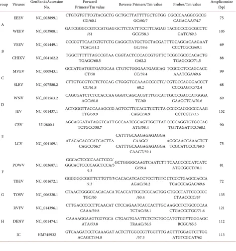
Table 1. Primers, probes, and amplicon sizes of the one-step real-time qRT-PCR assays.
Group Viruses GenBank\Accession No. Primers/Tm value Forward Reverse Primers/Tm value Probes/Tm value Ampliconsize (bp)
A
EEEV NC_003899.1 CTGTGTGTTCGTACGCTGCG/60.1 GCTGCTTATTTTGCTGTGGGC/60/7 CGCCCAAGGCGCCGCAGACAA/74.7 75
WEEV NC_003908.1 GATCGGGCCGTCCATGAG/61 GCTTCTATTTCCTTCAGAGGCG/58.3 TACGCCCCGCGCCTCGATC/69.3 105
B
VEEV NC_001449.1 CCCCGTTCAATGTGTCTGTCAC/61.2 CAGGCTATGCTGCTACGATGC/59.6 TTGCAGCACAAGAATCCCTCGCG/69.1 69
CHIKV NC_004162.2 TGGCTTTTTTAGCCGTAATGAGC/60.5 CGGTACTCCCACCGTGTTCG/62.2 TCGGTGCCCACACTGTGAGCGC/71.5 88
C
MVEV NC_000943.1 GCCATGATGGTGATGCAACT/58 CTGTCTGGGAATGAGCAGCC/59.4 TCGCCCTCCAGCACCAAATCGA/69.6 99
SLEV NC_007580.2 CTTGTGCGTCCTCTCCAGCC/61.8 CTGGGTGCAAAGCCCCTC/60.2 CGTGCCAGGGACCCTCCCGAGTC/72.4 68
D
WNV NC_001563.2 CAGCGATCTCTCCACCAAAGC/60.8 GGGTCAGCACGTTTGTCATTG/60 TGCCCGACCATGGGAGAAGCTCA/70.6 69
JEV NC_001437.1 ACTGGGTTACCAAAGCCGTTG/59.9 AGTCCTTCCACCTCCTCTACAGC/58.9 CCCCCACGGCCCAAGCCTCGT/73.5 152
E
CEV U12800.1 AGCAGGATATAGGTCATTTCTGCC/58.7 GCCAATCGCAGTTGCTTATATG/58.4 CCCCAGGTGTGCCACTGTTAGATTCC/68.1 90
LCV NC_004109.1 ATACACACCCATCACTTACAGCC/56.7
CATTTGCAAGAGAGAGGA CAAGC/ CATTTGCAAGAGAGAGGA
CAAGT/59.1
AGGCAACCAAACTCT
TCGCATCCCC/69.5 75
F
POWV NC_003687.1 GGCACTCCCCAGCTCCA/5GGCACTCCCCAACTCCG/ 9.3
GCTGGGGCAAGTCAATCTT
G/59.4 TCAACCCCCATCATCATGCGCCT/70.1 81
TBEV NC_001672.1 GGGGGGCGGTTCTTGTT/59.3 CACACATCACCTCCTTGTCAGAC/58.2 CTCCCTGAGCCACCATCACCCAGAC/69.6 72
G TOSV NC_006320.1 CTAACTGGGCCACACACATGC/60 TCACCATTGCTCGCACTGG/60.4 CTGCCTATTCCCCCCCTAACCCC/67 135
RVFV NC_014396.1 CTTGACCCCCTTCAACATCAAA/59.8 CTCCAGAATCACCACTTGCTCTAC/58.1 AAGCCTCTGCCCCAACTGACCCTGC/71.6 121
H DENV NC_001474.1 CAAAAGGAAGTCGYGCAATA/53.8 CTGAGTGAATTCTCTCTGCTRAAC/56.5 CATGTGGYTGGGAGCRCGC/65.5 112
DOI: 10.4236/aim.2018.87036 523 Advances in Microbiology CHIKV, TBEV, RVFV and WNV were provided by Wuhan institute of virology, CAS. Human serum samples from healthy persons (n = 150) were assembled from samples library of Ningbo International travel healthcare center. The hu-man serum from JEV patients (N = 20), TBEV patients (N = 13) and DENV pa-tients (N = 29) in the acute phase were from Ningbo center for disease preven-tion and control, other serum from DENV patients (N = 16) and CHIKV pa-tients (N = 8) were from laboratory of Ningbo International travel healthcare center, which were all confirmed by single plex real-time qRT-PCR assays, and other specific detection methods (virus isolation or IgG detection). These healthy human serums were used as negative control in all the tests, whereas the other viral isolates were implied as positive control in the detection assays for different viruses. Vector tissue samples were collected by our laboratory during vector surveillance in 2015, including mosquito pools (N = 112) and tick (N = 38).
2.4. RNA Extraction
RNA samples used for detection and quantification were prepared using QIAampViral RNA Mini Kit (Qiagen). A total 140 ul of samples which from serum,strain and culture supernatant of virus-infected cells were used for detection and quantification. RNA extraction was performed according to the manufacturer’s instructions for use of the RNA extraction procedure selected, and finally eluted in 60 µL sterilized RNase free water. Viral RNA samples were stored at −80˚C before use, and samples are aliquoted into sample sizes adequate for future use in the lab in order to avoid repeated freeze-thawing.
D. G. Zhou, J. Lu
DOI: 10.4236/aim.2018.87036 524 Advances in Microbiology
2.6. Development of Multiplex One-Step Real-Time qRT-PCR
Assays
To reduce the assay cost and improve condition for the multiple reaction. Mul-tiplex assays were assembled by grouping the primers and probes according to the hosts/Vectors or viral families for the duplex reaction. Originally, the linear dynamic range of detection for reaction containing one primer-probe set (sin-gleplx) and multiple prime-probe sets for multiple targets (duplex) was deter-mined using One-step real-time qRT-PCR in duplicates with 10-fold serial dilu-tion of a single species of target RNA. Singlex or Multiplex One-step real-time qRT-PCR reactions were performed using AgPath-IDTM one-step RT-PCR Kit (Applied Bio systems), and performed according to the protocol. It was per-formed in a total reaction volume of 25 ul consisting of 12.5 µL of 2 × RT-PCR buffer, 400 nM of each primer, 120 nM of each probe, 1 µL of Enzyme Mix and 5 µL of viral RNA transcripts or RNA samples. DEPC water was used as negative control. The qRT-PCR standard curve ranging from 1.0 × 10³ to 1.0 × 10⁷ cop-ies/µL, was generated from a 10-fold serial dilution of RNA transcripts. Real-time qRT-PCR cycling was performed on ABI 7500 fast system as follows: 45˚C for 10 min, 95˚C for 15 min, then 40 cycles of 15 s at 95˚C for denaturation and 60 s at 60˚C for annealing and extension incubations. Raw data was analyzed with 7500 Software v2.0.6 to determine the amount of viral RNA base on the threshold cycle value (Ct). Multiplex one-step real-time qRT-PCR assays and single one-step real-time qRT-PCR assays were compared for each of the species virus-es.
2.7. One-Step RT-PCR Assays
As the standard for comparison, One-step RT-PCR arrays were conduct accord-ing to previously reported method. One-step RT-PCR reactions were performed using Ag Path-IDTM one-step RT-PCR Kit (Applied Bio systems), and performed according to the protocol. Briefly, the primers for RT-PCR of each assay are the same as those for qRT-PCR. Also, the templates for RT-PCR are the same as those for qRT-PCR, including the reaction system. The amplified product was analyzed by electrophoresis using 2% agarose gel. The gel was stained with ethi-dium bromide and the amplified product was visualized under UV light. Mul-tiplex One-step real-time qRT-PCR assays and One-step RT-PCR assays were compared for sensitivity for 15 species viruses.
2.8. Specificity, Sensitivity and Reproducibility
DOI: 10.4236/aim.2018.87036 525 Advances in Microbiology To evaluate sensitivity of single plex, multiplex one-step real-time qRT-PCR assays, single plex one-step real-time qRT-PCR assays and one-step RT-PCR as-says, each group of 10-fold serial dilutions of RNA transcripts, ranging from 1.0 × 10³ to 1.0 × 10⁷ copies/µL, were used as standard preparations to assess the de-tection limits of viral RNA copy load. Duplicates of the assay within or between runs were performed to assess the reproducibility, and the intra-assay and in-ter-assay variations over the linear range of the assays were statistically calcu-lated.
2.9. Statistical Analysis
Regression, reproducibility and the coefficient of variation (CV)of the mean Ct value for each standard concentration within and between individual PCR runs were statically calculated by SPSS 15 to evaluate linearity and determine the quantitative performance of each assay.
Calculation method:Ct (threshold cycle) is the intersection between an am-plification curve and a threshold line. It is a relative measure of the concentra-tion of target in the PCR reacconcentra-tion.
Equation for Ct value: lg 0X = − ×Ct lg 1
(
+Ex)
+lgMLinear equation:
1 slope
Efficiency 10 1 100%
−
= − ×
3. Result
3.1. Primer-Probe Selection and Design
Genomic sequences of all representative strains of each viral species were down-loaded from the GenBank database (Supplementary Table S1). The internal control (IC) chooses synthetic construct sequence as the target gene having no homology with these arboviral encephalitis viruses. In total, 16 primer-probe pairs were designed. All primers-probes were grouped into eight groups for the duplex reaction based on the related diseases or virus families. Using the devel-oped reaction system, we tested each primer/probe set in the single plex assays, and then combined them into duplex reactions for multiplex one-step real-time qRT-PCR assays according to Table 1.
3.2. Generation of Viral RNAs
For further assessment of specificity and sensitivity for the developed Multip-lexone-step real-time qRT-PCR assays, one-step real-time qRT-PCR assays, and one-step RT-PCR assays against the Viral RNAs as the closets virus with DENV, CHIKV, TBEV, RVFV and WNV, Viral RNAs were used and generated via in
vitro transcription of single-stranded DNA fragments containing cDNA derived
D. G. Zhou, J. Lu
DOI: 10.4236/aim.2018.87036 526 Advances in Microbiology
indicating that the RNA products were highly pure. The concentration of RNA transcripts was quantified and the copynumbers were calculated respectively according to the concentration and size of each single-stranded RNA fragment (Supplementary Table S2). The invitro-transcribed Viral RNAs were used in the Multiplexone-step real-time qRT-PCR assays, one-step real-time qRT-PCR assays, and one-step RT-PCR assays for specificity and sensitivity evaluation.
3.3. Sensitivity and Reproducibility of Multiplex One-Step
qRT-PCR Assays
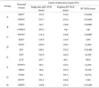
The sensitivity of multiplex one-step qRT-PCR assays, single plex one-step qRT-PCR assays and one-step RT-PCR assays for detection of each viral species. As shown in Supplementary Table S2, The in vitro-transcribed Viral RNAs were subjected to the sensitivity test. The RNA transcripts as RNA standards were se-rially diluted 10-folds from 1.0 × 10³ to 1.0 × 10⁷ copies/µL. Sensitivity of mul-tiplex one-step qRT-PCR assays, single plex one-step qRT-PCR assays and one-step RT-PCR assays was amplified as RNA samples. The amplification effi-ciencies of the single plex assays for the 15 arboviral encephalitis viruses were all above 90%. The standard curves showed a high correlation coefficient, R2 > 0.99, for all the viruses detections (data not shown). The potential limits of detection (LODs) of these assays were determined to be at a range from 85.7 to 155.7 RNA copies/PCR (Table 2). The synthesized RNA standards were used for the mul-tiplex assays testing, and standard curves of detections for each virus RNA tran-scripts were also constructed and showed high correlation coefficient, R2 > 0.98 (Figure 1). In most multiplex assays (13 out of 15 virus detections), the LODs were at a range from 94 to 150 RNA copies/PCR, which was similar to that in the single plex assays (Table 2). Besides, WEEV and JEV detection assays showed a little lower sensitivity with the LODs of 215.6 and 174.3 copies/PCR, respective-ly. As shown in Table 2, duplex qRT-PCR was 1000-fold more sensitive than one-step RT-PCR for the amplification of EEEV, WEEV, VEEV, RVFV, JEV and TOSV, and was 100-fold more sensitive than one-step RT-PCR for that of WNV, CEV, LCV, POWV, TBEV and DENV, and was 10-fold more sensitive than one-step RT-PCR for that of MVEV and CHIKV (Figure 1). The analysis of the LOD indicated that the strategy of multiplex detection ensures the sensitivity of the assay system.
DOI: 10.4236/aim.2018.87036 527 Advances in Microbiology
Table 2. Detection limits of multiplex one-step real time qRT-PCR assays.
Group Detected viruses Single plex qRT-PCR Limits of detection (copies/UL)
assays Duplexq RT-PCR assays RT-PCR assays
A EEEV 133.5 114.4 114,400
WEEV 155.7 215.6 215,600
B VEEV 94.3 134.8 134,800
CHIKV 103.5 146 146
C MVEV 114.2 124.8 124,800
SLEV 116.9 110.4 1104
D WNV 105.6 116.5 11,650
JEV 100.1 174.3 174,300
E CEV 143.9 143.5 14,350
LCV 85.7 94.1 9410
F POWV 86.3 124.3 12,430
TBEV 90.4 103.5 10,350
G TOSV 78.1 107.1 10,710
RVFV 101.3 110.6 1106
H DENV 110.8 131.2 131,200
3.4. Application of Multiplex One-Step qRT-PCR Assays
D. G. Zhou, J. Lu
D. G. Zhou, J. Lu
D. G. Zhou, J. Lu
D. G. Zhou, J. Lu
D. G. Zhou, J. Lu
D. G. Zhou, J. Lu
D. G. Zhou, J. Lu
D. G. Zhou, J. Lu
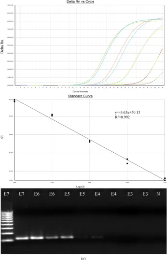
[image:24.595.124.471.75.614.2]DOI: 10.4236/aim.2018.87036 542 Advances in Microbiology
Figure 1. Amplification plots and standard curves of multiplex one-step qRT-PCR assays and comparison with RT-PCR assays.
The multiplex one-step qRT-PCR assays and RT-PCR assays were tested using synthesized in vitro target viral RNA transcripts ranging from 1.2 × 10³ to 1.2 × 10⁷ copies/mL. A PCR baseline subtractive curve fit view of the data is shown with relative
fluores-cence units (RFUs) plotted against cycle numbers. Standard curves generated from the Ct values obtained against known concen-trations, the coefficient of determination (R2) and slope of the regression curve for each assay are indicated. M:100 bp ladder; E3-E7: E3,1.0 × 103, E4, 1.0 × 10⁴, E5,1.0 × 105, E6,1.0 × 106, E7, 1.0 × 10⁷; A-O group: A group, EEEV, B group, WEEV, C group,
DOI: 10.4236/aim.2018.87036 543 Advances in Microbiology A total of 150 RNA samples which contained 8 CHIKV patients, 20 JEV pa-tients and 13 TBEV papa-tients were tested using the multiplex one-step real-time qRT-PCR assays, the assay sensitivity was 100% with all the tested samples. The result showed 8 positive (8/8) in Group B, 20 positive (20/20) in Group D, 13 positive (13/13) in Group F, and the healthy human sera were negative (Table 5).
Figure 2. Coefficients of variation of Ct values in the multiplex one-step qRT-PCR assays.
The multiplex one-step real-time RTPCR assays were performed in three independent experiments of replicates. The Coefficients of variation (CV) of Ct values were calculated in both intra-assays (A) and inter-assays (B), and showed all less than 5%.
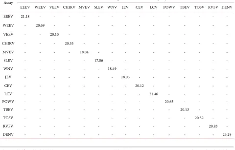
Table 3. Specificity analysis using in vitro transcribed viral RNAs.
Assay In vitro transcribed target viral RNA (1 × 10
6 copies/µL)
EEEV WEEV VEEV CHIKV MVEV SLEV WNV JEV CEV LCV POWV TBEV TOSV RVFV DENV
EEEV 21.18 - - - -
WEEV - 20.69 - - - -
VEEV - - 20.10 - - - -
CHIKV - - - 20.53 - - - -
MVEV - - - - 18.04 - - - -
SLEV - - - 17.86 - - - -
WNV - - - 18.49 - - - -
JEV - - - 18.05 - - - -
CEV - - - 20.12 - - - -
LCV - - - 21.46 - - - - -
POWV - - - 20.65 - - - -
TBEV - - - 20.13 - - -
TOSV - - - 20.52 - -
RVFV - - - 20.83 -
DENV - - - 23.29
0 1 2 3 4 5 6
2 3 4 5 6 7 8
0 1 2 3 4 5 6
2 3 4 5 6 7 8
[image:25.595.59.540.437.744.2]D. G. Zhou, J. Lu
[image:26.595.208.538.90.394.2]DOI: 10.4236/aim.2018.87036 544 Advances in Microbiology
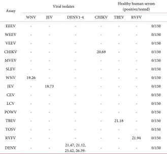
Table 4. Specificity analysis using viral isolates and healthy human sera.
Assay Viral isolates
Healthy human serum (positive/tested)
WNV JEV DENV1-4 CHIKV TBEV RVFV
EEEV - - - 0/150
WEEV - - - 0/150
VEEV - - - 0/150
CHIKV - - - 20.69 - - 0/150
MVEV - - - 0/150
SLEV - - - 0/150
WNV 19.26 - - - 0/150
JEV - 18.73 - - - - 0/150
CEV - - - 0/150
LCV - - - 0/150
POWV - - - 0/150
TBEV - - - - 21.18 - 0/150
TOSV - - - 0/150
RVFV - - - 21.94 0/150
DENV - - 25.42, 26.39- 21.47, 21.12, - - - 0/150
Table 5. Evaluation of the multiplex real-time qRT-PCR assays using clinical specimens.
Group Detected Viruses (positive/tested) Patients sera (positive/tested) Vectors tissues Healthy human sera (positive/tested)
B CHIKV 8/8 - 0/150
VEEV - - 0/150
D WNV - - 0/150
JEV 20/20 6/112 0/150
F POWV - - 0/150
TBEV 13/13 2/38 0/150
H DENV 45/45 2/112 0/150
IC - - -
[image:26.595.208.539.426.588.2]DOI: 10.4236/aim.2018.87036 545 Advances in Microbiology
4. Discussion
There are many central nervous system diseases and conditions, including infec-tions of the central nervous system such as encephalitis. Arboviral Encephalitis Viruses are member of animal viruses, including flaviviruses, phlebovirus, or-thobunyavirus, and the alphaviruses. And mostly Arboviral Encephalitis Viruses may cause encephalitis in a minority of infected humans.
D. G. Zhou, J. Lu
DOI: 10.4236/aim.2018.87036 546 Advances in Microbiology
and inter-assays, suggesting that the multiplex assays were of good reproducibil-ity. To verify the Multiplex One-Step Real-time TaqManqRT-PCR Assays were detecting infectious virus, total of 150 human serum RNA samples were ex-amined. The result showed that the assay sensitivity was 100% with all the tested samples and there were no false positive results observed in the unrelated patient sera and healthy humans era. Also, in this study, IC was used to monitor all as-say results. All the negative results were validated through observation of the amplifications of IC to avoid the false negative result. In this way, IC increased the assay’s sensitivity. The assay sensitivity and specificity for diagnosis of JEV, TBEV, DENV, CHIKV virus infection inpatient sera were reliable and desirable. The specificity and reproducibility of the assays were demonstrated and the sen-sitivity of the systems was acceptable. Furthermore, evaluation with clinical samples of patients and vectors showed the reliable specificities and sensitivities for laboratory detection of the infections with these viruses and provided poten-tial use for clinical diagnosis and vector surveillance.
5. Conclusions
In conclusion, the comprehensive multiplex one-step real-time TaqManqRT-PCR assays for rapid detection of 15 viruses was established and evaluated in this study. The developed multiplex one-step real-time qRT-PCR assay was tested using different simulate samples and showed excellent parameters in the fol-lowed statistical analysis. Therefore, this assay proved to be specific, sensitive and, apparently, convenient for rapid and simultaneous identification in labora-tory, and could be certainly extended to routine diagnosis and epidemiological detection of arboviral encephalitis infections.
The arboviral encephalitis virus panel with IC developed in this study was found to be highly specific and sensitive in the detection of 15 encephalitis vi-ruses from clinical specimens and vector tissues. The use of IC prevented false negative readings and improved accuracy of the assay. The panel can be a great aid to clinical management, vector surveillance and outbreak response of CNS in the future.
Acknowledgements
The authors thank Dr. Bo Zhang, Dr. Hongping Wei and staff of Wuhan Insti-tute of Virology for their technical assistance and for virus strains. We thank Dr. Yuping Luo for her linguistic advice.
Ethical Consideration
DOI: 10.4236/aim.2018.87036 547 Advances in Microbiology the donors.
Competing Interests
The authors have declared no competing financial interests exist.
Funding
This work was supported by Project of Zhejiang Provincial Natural Science Foundation (LY16H260004) and Project of Ningbo Entry-exit Inspection and Quarantine Bureau Science and Technology Program (Y2015-15).
References
[1] Anderson, P.D. and Bokor, G. (2012) Bioterrorism: Pathogens as Weapons. Journal of Pharmacy Practice, 25, 521-529. https://doi.org/10.1177/0897190012456366
[2] Mackay, I.M., Arden, K.E. and Nitsche, A. (2002) Real-Time PCR in Virology.
Nucleic Acids Research, 30, 1292-1305. https://doi.org/10.1093/nar/30.6.1292
[3] Espy, M.J., Uhl, J.R., Sloan, L.M., Buckwalter, S.P., Jones, M.F., Vetter, E.A., Yao, J.D., Wengenack, N.L., Rosenblatt, J.E., Cockerill 3rd, F.R. and Smith, T.F. (2006) Real-Time PCR in Clinical Microbiology: Applications for Routine Laboratory Testing. Clinical Microbiology Reviews, 19, 165-256.
https://doi.org/10.1128/CMR.19.1.165-256.2006
[4] Armstrong, P.M. and Andreadis, T.G. (2013) Eastern Equine Encephalitis Vi-rus—Old Enemy, New Threat. New England Journal of Medicine, 368, 1670-1673.
https://doi.org/10.1056/NEJMp1213696
[5] Zink, S.D., Jones, S.A., Maffei, J.G. and Kramer, LD. (2013) Quadraplex qRT-PCR Assay for the Simultaneous Detection of Eastern Equine Encephalitis Virus and West Nilevirus. Diagnostic Microbiology and Infectious Disease, 77, 129-132.
https://doi.org/10.1016/j.diagmicrobio.2013.06.019
[6] Brault, A.C., Fang, Y. and Reisen, W.K. (2015) Multiplex qRT-PCR for the Detec-tion of Western Equine Encephalomyelitis, St. Louis Encephalitis, and West Nile Viral RNA in Mosquito Pools (Diptera: Culicidae). Journal of Medical Entomology, 52, 491-499. https://doi.org/10.1093/jme/tjv021
[7] Bergren, N.A., Auguste, A.J., Forrester, N.L., Negi, S.S., Braun, W.A. and Weaver, S.C. (2014) Western Equine Encephalitis Virus: Evolutionary Analysis of a Declin-ing Alphavirus Based on Complete Genome Sequences. Journal of Virology, 88, 9260-9267. https://doi.org/10.1128/JVI.01463-14
[8] Gutiérrez, S., Thébaud, G., Smith, D.R., Kenney, J.L. and Weaver, S.C. (2015) De-mographics of Natural Oral Infection of Mosquitos by Venezuelan Equine Ence-phalitis Virus.
[9] Moureau, G., Temmam, S., Gonzalez, J.P., Charrel, R.N., Grard, G. and de Lambal-lerie, X. (2007) A Real-Time RT-PCR Method for the Universal Detection and Identification of Flavi Viruses. Vector-Borne and Zoonotic Diseases, 7, 467-477.
https://doi.org/10.1089/vbz.2007.0206
[10] Oyer, R.J., David Beckham, J. and Tyler, K.L. (2014) West Nile and St. Louis Ence-phalitis Viruses. HandbClin Neurol., 123, 433-447.
https://doi.org/10.1016/B978-0-444-53488-0.00020-1
Jung-D. G. Zhou, J. Lu
DOI: 10.4236/aim.2018.87036 548 Advances in Microbiology
len, S. (2013) Provenance and Geographic Spread of St. Louis Encephalitis Virus.
MBio, 4, e00322-13. https://doi.org/10.1128/mBio.00322-13
[12] Kilpatrick, A.M. and Pape, W.J. (2013) Predicting Human West Nile Virus Infec-tions with Mosquito Surveillance Data. American Journal of Epidemiology, 178, 829-835. https://doi.org/10.1093/aje/kwt046
[13] Kovalev, S.Y. and Mukhacheva, T.A. (2014) Tick-Borne Encephalitis Virus Subtypes Emerged through Rapid Vector Switches Rather than Gradual Evolution. Ecology and Evolution, 4, 4307-4316. https://doi.org/10.1002/ece3.1301
[14] Veje, M., Studahl, M., Norberg, P., Roth, A., Möbius, U., Brink, M. and Bergström, T. (2014) Detection of Tick-Borne Encephalitis Virus RNA in Urine. Journal of Clinical Microbiology, 52, 4111-4112. https://doi.org/10.1128/JCM.02428-14
[15] Achazi, K., Nitsche, A., Patel, P., Radonić, A., DonosoMantke, O. and Niedrig, M. (2011) Detection and Differentiation of Tick-Borne Encephalitis Virus Subtypes by a Reverse Transcription Quantitative Real-Time PCR and Pyrosequencing. Journal of Virological Methods, 171, 34-39. https://doi.org/10.1016/j.jviromet.2010.09.026
[16] Ananthan, D., Shah, S., Haseer-Koya, H. and Patel, A. (2014) Powasson Virus Causing Tick-Borne Encephalitis: A Diagnostic Dilemma. QJM, 107, 909-910.
https://doi.org/10.1093/qjmed/hcu082
[17] Reese, S.M., Blitvich, B.J., Blair, C.D., Geske, D., Beaty, B.J. and Black, W.C. (2008) Potential for La Crosse Virus Segment Reassortment in Nature. Virology Journal, 30, 16.https://doi.org/10.1186/1743-422X-5-164
[18] Maquart, M., Temmam, S., Héraud, J.M., Leparc-Goffart, I., Cêtre-Sossah, C., Del-lagi, K., Cardinale, E. and Pascalis, H. (2014) Development of Real-Time RT-PCR for the Detection of Low Concentrations of Rift Valley Fever Virus. Journal of Vi-rological Methods, 195, 92-99.https://doi.org/10.1016/j.jviromet.2013.10.001
[19] Mwaengo, D., Lorenzo, G., Iglesias, J., Warigia, M., Sang, R., Bishop, R.P. and Brun, A. (2012) Detection and Identification of Rift Valley Fever Virus in Mosquito Vec-tors by Quantitative Real-Time PCR. Virus Research, 169, 137-143.
https://doi.org/10.1016/j.virusres.2012.07.019
[20] Charrel, R.N., Bichaud, L. and de Lamballerie, X. (2012) Emergence of Toscana Vi-rus in the Mediterranean Area. World Journal of Virology, 1, 135-141.
https://doi.org/10.5501/wjv.v1.i5.135
[21] Williams, D.T., Diviney, S.M., Niazi, A.U., Durr, P.A., Chua, B.H., Herring, B., Pyke, A., Doggett, S.L., Johansen, C.A. and Mackenzie, J.S. (2015) Molecular Epi-demiology and Evolution of Murray Valley Encephalitis Virus: Recent Emergence of Distinct Sub-Lineages of the Dominant Genotype 1. PLOS Neglected Tropical Diseases, 9, e0004240.https://doi.org/10.1371/journal.pntd.0004240
[22] Gong, R., Wang, H.H., Qin, H., Guo, X.P. and Ma, X.J. (2015) Visual Detection of Murray Valley Encephalitis Virus by Reverse Transcription Loop-Mediated Iso-thermal Amplification. Biomedical and Environmental Sciences, 28, 227-230. [23] Williams, D.T., Diviney, S.M., Corscadden, K.J., Chua, B.H. and Mackenzie, J.S.
(2014) Complete Genome Sequences of the Prototype Isolates of Genotypes 2, 3, and 4 of Murray Valley Encephalitis Virus. Genome Announcements, 2, e00581.
https://doi.org/10.1128/genomeA.00581-14
[24] Chancey, C., Grinev, A., Volkova, E. and Rios, M. (2015) The Global Ecology and Epidemiology of West Nile Virus. BioMed Research International, 2015, Article ID: 376230.https://doi.org/10.1155/2015/376230
DOI: 10.4236/aim.2018.87036 549 Advances in Microbiology
Thi, D., Nguyen, N.L., Nguyen Thi, T.T., Le Thi, Q.M., Buerano, C.C., Morita, K. and Hasebe, F. (2015) Isolation of Dengue Serotype 3 Virus from the Cerebrospinal Fluid of an Encephalitis Patient in Hai Phong, Vietnam in 2013. Journal of Clinical Virology, 70, 93-96.https://doi.org/10.1016/j.jcv.2015.07.295
[26] Christo, P.P. (2015) Encephalitis by Dengue Virus and Other Arboviruses. Arquivos de Neuro-Psiquiatria, 73, 641-643.https://doi.org/10.1590/0004-282X20150108
[27] Shaikh, N.J., Raut, C.G., Sinha, D.P. and Manjunath, M.J. (2015) Detection of Chi-kungunya Virus from a Case of Encephalitis, Bangalore, Karnataka State. Indian Journal of Medical Microbiology, 33, 454-455.
[28] Nelson, J., Waggoner, J.J., Sahoo, M.K., Grant, P.M. and Pinsky, B.A. (2014) Ence-phalitis Caused by Chikungunya Virus in a Traveler from the Kingdom of Tonga.
Journal of Clinical Microbiology, 52, 3459-3461.
https://doi.org/10.1128/JCM.01288-14
[29] Felder, E. and Wölfel, R. (2012) Development of a Versatile and Stable Internal Control System for RT-qPCR Assays. Journal of Virological Methods, 208, 33-40.
https://doi.org/10.1016/j.jviromet.2014.07.028
[30] Ding, X.-X., Li, X.-F., Deng, Y.-Q., Guo, Y.-H., Hao, W., et al. (2014) Development of a Double Antibody Sandwich ELISA for West Nile Virus Detection Using Mo-noclonal Antibodies against Non-Structural Protein 1. PLoS ONE, 9, e108623.
https://doi.org/10.1371/journal.pone.0108623
[31] Pedersen, K., Marks, D.R., Wang, E., Eastwood, G., et al. (2016) Widespread Detec-tion of Antibodies to Eastern Equine Encephalitis, West Nile, St. Louis Encephalitis, and Turlock Viruses in Various Species of Wild Birds from Across the United States. The American Journal of Tropical Medicine and Hygiene, 95, 206-211.
https://doi.org/10.4269/ajtmh.15-0840
[32] Brault, A.C., Fang, Y., Reisen, W.K., et al. (2015) Multiplex qRT-PCR for the Detec-tion of Western Equine Encephalomyelitis, St. Louis Encephalitis, and West Nile Viral RNA in Mosquito Pools (Diptera: Culicidae). Journal of Medical Entomology, 52, 491-499.https://doi.org/10.1093/jme/tjv021
[33] Lustig, Y., Mannasse, B., Koren, R., Katz-Likvornik, S., et al. (2016) Superiority of West Nile Virus RNA Detection in Whole Blood for Diagnosis of Acute Infection.
Journal of Clinical Microbiology, 54, 2294-2297.
D. G. Zhou, J. Lu
DOI: 10.4236/aim.2018.87036 550 Advances in Microbiology
[image:32.595.56.539.108.738.2]Supplementary
Table S1. GenBank accession numbers of arboviral encephalitis virus sequences aligned in this study.
Families Genus Species Vector GenBank accession numbers Total numbers
Togaviridae Alphavirus
Eastern equine
encephalitis virus mosquito
NC_003899.1,KJ469600.1,KJ469595.1,X63135.1,KJ4 69583.1,KJ469566.1,KJ469603.1,KJ469636.1,AY722 102.1,KJ469617.1,KJ469611.1,KJ469599.1,KJ469557 .1,KJ469609.1,KJ469635.1,EF568607.1,KJ469630.1, KJ469567.1,KJ469624.1,KJ469575.1,KJ469563.1,KJ4 69593.1,KJ469579.1,EF151502.1,KJ469638.1,KJ4696 10.1,KJ469616.1,KJ469602.1,KJ469577.1,KJ469561. 1,KJ469634.1,KJ469559.1,KJ469573.1,KJ469587.1,K J469639.1,KJ469592.1,KJ469560.1,KJ469643.1,KJ46 9646.1,KJ469632.1,KJ469651.1,KJ469647.1,KJ46961 8.1,KJ469568.1,KJ469619.1,KJ469591.1,KJ469631.1, KJ469556.1,KJ469642.1,KJ469582.1,KJ469597.1,KJ4 69628.1,KJ469613.1,KJ469606.1,KJ469584.1,KJ4695 88.1,KJ469562.1,KJ469555.1,KJ469633.1,KJ469570. 1,KJ469625.1,KJ469571.1,KJ469572.1,KJ469612.1,K J469608.1,KJ659366.1,KJ469620.1,AY705240.1,KJ4 69650.1,KJ469649.1,KJ469604.1,KJ469615.1,KJ4696 27.1,KJ469605.1,KJ469644.1,KJ469629.1,KJ469585. 1,AY705241.1,KJ469621.1,KJ469594.1,KJ469607.1, KJ469564.1,DQ241304.1,DQ241303.1,EF151503.1 86 Western equine
encephalitis virus mosquito
NC_003908.1,AF214040.1,GQ287645.1,GQ287642. 1,GQ287641.1,GQ287647.1,GQ287643.1,GQ287644
.1,GQ287640.1,GQ287646.1 11
Venezuelan equine
encephalitis virus mosquito
DOI: 10.4236/aim.2018.87036 551 Advances in Microbiology
Continued
Chikungunya
virus mosquito
NC_004162.2,KJ451623.1,KJ451622.1,KJ451624.1,KF31 8729.1,FJ807897.1,FN295483.3,FN295484.2,EU703760.1 ,EU703761.1,EU703759.1,EU703762.1,HE806461.1,HM 045814.1,HM045808.1,HM045796.1,HM045787.1,HM0 45802.1,HM045789.1,HM045800.1,HM045790.1,HM04 5791.1,HM045797.1,EF452494.1,L37661.,EF452493.1,H M045810.1,HM045813.1,HM045803.1,HM045788.1,EF 027141.1,EF027140.1,JF274082.1,HM159390.1,HM1593 89.1,HM159388.1,HM159386.1,HM159387.1,HM15938 5.1,FJ445511.2,FJ445510.2,FJ000068.1,FJ000062.1,FJ000 065.1,EU564335.1,JN558836.1,JN558835.1,JN558834.1, EF210157.2,EF027138.1,FJ807896.1,GU908223.1,FJ4454 84.2,FJ445433.2,FJ445463.2,GU301779.1,KC862329.1,G U301781.1,FJ445430.2,GQ905863.1,FJ445502.2,FJ44544 3.2,FJ445445.2,FJ445431.2,FJ445432.2,JX088705.1,FN29 5485.3,FN295487.2,FJ807899.1,GU199353.1,GU199352. 1,GU301780.1,GQ428212.1,FJ000069.1,GQ428213.1,G Q428214.1,EU244823.2,HM045801.1,GU199350.1,GU1 89061.1,FJ513679.1,FJ513628.1,FJ513657.1,FJ513629.1,F J513637.1,FJ513635.1,FJ445428.2,AB455494.1,AB45549 3.1,FJ445427.2,EF027137.1,FJ000066.1,HM045799.1,GQ 428211.1,FJ000064.1,FJ000063.1,EU372006.1,FJ807898. 1,GQ428215.1,FJ513675.1,GU199351.1,FJ513654.1,FJ51 3645.1,FJ513673.1,FJ513632.1,FJ445426.2,FJ000067.1,H Q456254.1,HQ456253.1,HQ456252.1,HQ456251.1,DQ4 43544.2,EU564334.1,FJ959103.1,EF012359.1,EU037962. 1,FR717337.1,FR717336.1,HQ456255.1,EF027134.1,EF0 27136.1,EF027135.1,GQ428210.1,HM045794.1,HM1593 84.1,HM045823.1,HM045784.1,HM045812.1,EF027139. 1,HM045793.1,JQ067624.1,HM045822.1,AF369024.2,A F490259.3,HM045821.1,HM045806.1,HM045805.1,HM 045795.1,HM045792.1,HM045811.1,HM045809.1,HM0 45819.1,HM045818.1,HM045820.1,AY726732.1,HM045 807.1,HM045786.1,HM045816.1,HM045804.1,HM0457 98.1,HM045785.1,HM045815.1,HM045817.1 154
Flaviviridae Flavivirus encephalitis virus Japanese mosquito
D. G. Zhou, J. Lu
DOI: 10.4236/aim.2018.87036 552 Advances in Microbiology
Continued B551992.1,AB551991.1,KF297916.1,KC915016.1,JN381 873.1,JF706269.1,JN381872.1,KC517497.1,D90194.,D9 0195.,JN604986.1,AF315119.1,JQ086762.1,JN864064.1, JQ086763.1,KF297915.1,AF416457.1,M55506.1,JN3818 70.1,EF623988.1,EF623989.1,AF075723.1,GQ902063.1, EF623987.1,JN381871.1,JF706283.1,JN381865.1,EF107 523.1,AY849939.1,JN381853.1,JN381854.1,U47032.1,JF 706276.1,JN381858.1,JF706272.1,JN381856.1,JN381857 .1,JN381855.1,JF706273.1,JN381859.1,JN381860.1,JN3 81861.1,JN381863.1,JN381862.1,JN381864.1,JN381867. 1,JN381866.1,L48961.1,L78128.1,JN381868.1,EF571853 .1,HE861351.1,JN711459.1,JN711458.1,JF706284.1,FJ1 85037.1,FJ185036.1,JF706285.1,JX072965.1,JX050179.1, JX131374.1,AF080251.1,JN644310.1| 8 Saint Louis
encephalitis virus mosquito NC_007580.2,DQ525916.1,JQ957868.1,JQ957869.1,JF460774.1,EU566860.1,FJ753286.2,FJ753287.2 Murray Valley
encephalitis virus mosquito NC_000943.1,AF161266.1,JX123032.1 3
West Nile virus mosquito
DOI: 10.4236/aim.2018.87036 553 Advances in Microbiology
Continued
Powassan virus tick NC_003687.1,HQ231415.1 2
Tick-borne
encephalitis virus tick
NC_001672.1,AY169390.3,KF151173.1,FJ968751.1,HM 535611.1,HM535610.1,HQ201303.1,GU121642.1,GQ22 8395.1,FJ997899.1,FJ906622.1,EU816455.2,FJ402886.1,F J402885.1,EU816454.1,EU816453.1,KC835597.1,KC835 596.1,KC835595.1,DQ401140.3,KC414090.1,JF819648.2 ,HQ901367.1,HQ901366.1,HM859895.1,HM859894.1,E U816452.1,EU816451.1,EU816450.1,GQ266392.1,AF06 9066.1,FJ572210.1,JX534167.1,AB753012.1,JQ650523.1, JQ650522.1,JF316708.1,JF316707.1,KJ000002.1,KF9510 37.1,EF469662.1,EF469661.1 41
Dengue virus mosquito
DENV1:AF311957,AF311958,AF513110,EU482497, EU482500-EU482502,EU482509,EU482511,EU482512, EU482515,EU482516,EU482521,EU482525,EU482526,E U482533-EU482535,EU482538,EU482539,EU482567,E U482706,EU482800,EU482802,EU482803,EU482822,E U482823,EU596501,EU660390,EU660391,EU687247,E U848545,FJ024423,FJ024440,FJ024441,FJ024442,FJ0244 46,FJ024448,FJ024472,FJ024480,FJ024481,FJ205873,FJ2 05874,FJ410290,FJ432720,FJ461307,FJ461308,FJ461310, FJ461330,FJ461335,FJ461336,FJ461341,FJ639669,FJ6396 70,FJ639671,FJ639673,-FJ639678,FJ639680-FJ639684,FJ 639686,FJ639688,FJ639692,FJ639796,FJ639797,FJ63980 2,FJ639812-FJ639814,FJ639824,FJ687432,FJ687433,FJ74 4701,FJ744702,FJ810415,FJ810419,FJ850068,FJ850069,F J898391,FJ898423,FJ898424,FJ898430,FJ898431,FJ89843 3,FJ898437,FJ898448,FN429881-FN429883,FN429887,F N429889,FN429890,GQ199771,GQ199772,GQ199791,G Q199793,GQ199794,GQ199817-GQ199819,GQ199827-GQ199829,GQ199831-GQ199833,GQ199836-GQ19983 8,GQ199852-GQ199854,GQ199856-GQ199859, GQ199873, GQ199875, J461323, J639823 DENV2:NC_001474,AB122020-AB122024,AF489932,A Y702034,AY702040,AY744147,AY858035,AY858036,D
Q181797,DQ181798, DQ181803, DQ181804, DQ181806,EF051521,EF457904,EU056810,EU056811,E U056812,EU179857-EU179859,EU359009,EU482608,E U660415,EU677145,EU687212,EU687213, EU687217,
EU687220, EU687225, EU687232, EU687241-EU687243,EU687246,EU726767,EU726775, EU781135,FJ024475,FJ024477,FJ182012,FJ226066,FJ390 389,FJ410259,FJ410288,FJ432726,FJ461311,FJ639700,FJ 639705,FJ639706,FJ639711,FJ639717,FJ639718,FJ63978 3,FJ639822,FJ810412, FJ850067,FJ850072,FJ850074,
FJ850076, FJ850078, FJ850082,FJ850085,FJ850088, FJ850108, FJ850112,
FJ906962,FM210202,FM210204,FM210206-FM210213, FM210216-FM2102123,FM210231-FM210234,
FM210236-FM210244, FN429891, FN429892, FN429895,GQ199869,GQ199874,GQ199890, GQ199892,GQ199893,GQ199895-GQ199898, GQ199901,GQ252676,GQ252677,M20558,M29095,
MD1515
DENV3:NC_001475,AY099337,AY766104,AY770511,D Q863638, EU529699, EU660420, EU854292,
FJ182013,FJ182041,FJ898441-FJ898445, FJ898455-FJ898459,FJ898462-FJ898464, FJ898468,
D. G. Zhou, J. Lu
DOI: 10.4236/aim.2018.87036 554 Advances in Microbiology
Continued Bunyaviridae Orthobunyavirus FJ898471,FJ898472,FJ898474, FN429897-FN429900,FN429904,FN429907, FN429909,FN429911,FN429913,GQ199889,GQ199891,
GQ252674, GQ252678, M93130
DENV4:AY947539,EU854295-EU854297,EU854299-E U854301,FJ024424,FJ024476,FJ182016, FJ182017,FJ882590-FJ882592, FJ882595-FJ882601, FN429919-FN429922,FN429924-FN429926,GQ199876-GQ199882,GQ199884,GQ252675,MY0327498, MY95328 California
encephalitis virus mosquito U12800.1,AF123483.1 2
La Crosse virus mosquito
NC_004110.1,NC_004109.1,NC_004108.1,GU591168.1, GU591166.1,GU591165.1,GU591167.1,GU591164.1,GU 591169.1,K00610.1,EF485038.1,EF485037.1,EF485036.1, EF485035.1,EF485034.1,EF485033.1,EF485032.1,EF485 031.1,EF485030.1 19 Phlebovirus
Rift valley fever
virus mosquito
NC_014396.1,JF784387.1,JF311385.1,JF311384.1,JF3113 83.1,JF311382.1,JF311381.1,JF311380.1,JF311379.1,JF31 1378.1,JF311377.1,DQ380222.1,DQ380221.1,DQ380220 .1,DQ380219.1,DQ380218.1,DQ380217.1,DQ380216.1, DQ380215.1,DQ380214.1,DQ380212.1,DQ380211.1,DQ 380210.1,DQ380209.1,DQ380207.1,DQ380206.1,DQ380 205.1,DQ380204.1,DQ380203.1,DQ380200.1,DQ380198 .1,DQ380197.1,DQ380196.1,DQ380195.1,DQ380194.1, DQ380191.1,DQ380190.1,DQ380189.1,DQ380188.1,DQ 380187.1,DQ380186.1,DQ380185.1,DQ380184.1,DQ380 183.1,HE687306.1,HE687303.1 46
Toscana virus Sand fly NC_006320.1,JX867535.1,EU003177.1,EU003180.1,EU003179.1,EU003178.1,EU003176.1,EU003175.1,EU00317
[image:36.595.62.538.463.723.2]4.1,EU003173.1 10
Table S2. Viral RNA standards prepared via in vitro transcription.
virus source GenBank accession number of the referenced sequence Length (nt) Concentration (ng/mL) number (copies/µL) Copy Eastern equine encephalitis virus Chemical synthesis NC_003899.1 750 867 2.0468E12 Western equine encephalitis virus Chemical synthesis NC_003908.1 759 904 2.1088E12 Venezuelan equine encephalitis virus Chemical synthesis NC_001449.1 935 1020 1.9316E12
Chikungunya virus Virus isolate NC_004162.2 968 820 1.4999E12
Japanese encephalitis virus Virus isolate NC_001437.1 967 856 1.5673E12
Saint Louis encephalitis virus Chemical synthesis NC_007580.2 1011 896 1.5692E12
Murray Valley encephalitis virus Chemical synthesis NC_000943.1 714 351 8.7042E11
West Nile virus Virus isolate NC_001563.2 1224 1388 2.0078E12
Powassan virus Chemical synthesis NC_003687.1 1476 712 8.541E11
Tick-borne encephalitis virus Virus isolate NC_001672.1 1127 893 1.403E12
Dengue virus Virus isolate NC_001474.1 730 859 2.0835E12
California encephalitis virus Chemical synthesis U12800.1 1365 315 4.086E11
La Crosse virus Virus isolate NC_004109.1 1048 822 1.3888E12
Rift valley fever virus Virus isolate NC_014396.1 839 925 1.9521E12
DOI: 10.4236/aim.2018.87036 555 Advances in Microbiology
Table S3. Reproducibility analysis of multiplex one-step real-time RT-PCR assays.
Assays transcripts RNA RNA transcripts concentration Mean Ct value Intra-assay CV (%) Inter-assay CV (%) linear range
Group A
EEEV
107 copies/µL 18.13 1.96 3.41
Y = 3.46x + 46.25 R2 = 0.996 106 copies/µL 22.06 2.17 2.01
105 copies/µL 25.22 1.22 1.59
104 copies/µL 29.31 1.05 0.51
103 copies/µL 33.17 1.29 0.3
WEEV
107 copies/µL 20.02 4.81 4.68
Y = 3.68x + 47.69 R2 = 0.988 106 copies/µL 22.96 3.28 0.56
105 copies/µL 26.14 1.47 0.82
104 copies/µL 30.11 1.3 0.4
103 copies/µL 34.15 0.95 0.71
Group B
VEEV
107 copies/µL 17.51 4.46 1.52
Y = 3.66x + 47.2 R2 = 0.998 106 copies/µL 21.04 3.3 0.21
105 copies/µL 24.16 2.77 0.58
104 copies/µL 27.20 0.69 0.44
103 copies/µL 30.43 1.7 0.59
CHIKV
107 copies/µL 19.65 0.59 0.59
Y = 3.58x + 47.49 R2 = 0.999 106 copies/µL 22.14 2.74 2.74
105 copies/µL 25.63 0.73 0.73
104 copies/µL 29.07 0.37 0.37
103 copies/µL 33.12 0.14 0.14
Group C
MVEV
107 copies/µL 14.82 2.87 1.27
106 copies/µL 18.31 1.39 1.64
Y = 3.53x + 46.31 R2 = 0.998 105 copies/µL 23.85 0.73 1.72
104 copies/µL 28.02 0.79 1.32
103 copies/µL 32.97 1.17 0.68
SLEV
107 copies/µL 14.57 2.82 0.98
Y = 3.56x + 46.69 R2 = 0.997 106 copies/µL 18.61 3.08 4.06
105 copies/µL 24.03 1.97 0.9
104 copies/µL 28.09 1.03 0.68
103 copies/µL 33.11 2.15 0.81
Group D WNV
107 copies/µL 14.61 2.2 3.79
Y = 3.54x + 47.26 R2 = 0.997 106 copies/µL 19.58 1.16 0.37
D. G. Zhou, J. Lu
DOI: 10.4236/aim.2018.87036 556 Advances in Microbiology
Continued
104 copies/µL 28.83 1.85 0.95
103 copies/µL 33.32 0.59 0.19
JEV
107 copies/µL 14.02 4.51 3.79
Y = 3.79x + 44.26 R2 = 0.998 106 copies/µL 18.57 1.01 0.37
105 copies/µL 24.01 0.105 0.21
104 copies/µL 27.95 1.31 0.95
103 copies/µL 33.12 0.43 0.19
Group E
CEV
107 copies/µL 18.04 3.48 4.84
Y = 3.64x + 46.09 R2 = 0.993 106 copies/µL 21.49 2.86 3.14
105 copies/µL 25.53 1.02 0.25
104 copies/µL 29.32 0.45 0.66
103 copies/µL 33.84 2.07 1.81
LCV
107 copies/µL 19.01 4.52 3.49
106 copies/µL 23.05 1.69 0.56
Y = 3.56x + 50.76 R2 = 0.996 105 copies/µL 27.10 2.97 1.7
104 copies/µL 31.19 2.57 1.56
103 copies/µL 34.20 1.36 0.66
Group F
POWV
107 copies/µL 18.82 2.17 1.64
Y = 3.9x + 45.96 R2 = 0.996 106 copies/µL 21.39 1.11 0.77
105 copies/µL 24.92 1.37 1.63
104 copies/µL 28.75 0.69 0.71
103 copies/µL 33.02 0.16 0.03
TBEV
107 copies/µL 18.06 0.71 0.2
Y = 3.68x + 46.52 R2 = 0.996 106 copies/µL 21.97 3 2.82
105 copies/µL 26.01 3.87 0.21
104 copies/µL 30.03 2.84 0.34
103 copies/µL 33.97 1.8 0.16
Group G
TOSV
107 copies/µL 18.62 0.44 1.95
Y = 3.58x + 50.24 R2 = 0.983 106 copies/µL 21.74 2.66 0.91
105 copies/µL 25.02 3.27 0.43
104 copies/µL 28.97 2.1 0.31
103 copies/µL 32.10 2.41 0.4
RVFV 10
7 copies/µL 17.91 0.3 2.37
DOI: 10.4236/aim.2018.87036 557 Advances in Microbiology
Continued
105 copies/µL 25.92 1.48 0.93
104 copies/µL 30.02 1.04 0.09
103 copies/µL 33.89 2.11 0.27
Group H DENV
107 copies/µL 18.94 4.65 3.66
Y = 3.65x + 50.15 R2 = 0.992 106 copies/µL 22.87 1.91 0.26
105 copies/µL 26.10 1.81 0.64
104 copies/µL 29.83 1.07 0.1
103 copies/µL 33.17 1.13 0.25
CV, coefficient of variation. Intra-assays were determined from two replicates within each dilution. In-ter-assays were determined from three independent assays performed on different days.
Abbreviations
CNS: Central Nervous System;
EEEV: Eastern Equine Encephalitis Virus; WEEV: Western Equine Virus;
VEEV: Venequilan Equine Encephalitis Virus; JEV: Japanese Encephalitis Virus;
SLEV: St. Louis Encephalitis Virus; MVEV: Murray Valley Encephalitis Virus; WNV: West Nile Encephalitis Virus; POWV: Powassan Virus;
CEV: Californiaencephalitis Virus; LCV: La Crosse Virus;