www.impactjournals.com/oncotarget/ Oncotarget, Vol. 7, No. 24
Biomarker development in MET-targeted therapy
Yanni Zhang1, Zhiqiang Du1 and Mingqiang Zhang11 Amgen Biopharmaceutical Research and Development (Shanghai) Co., Ltd, Shanghai, China
Correspondence to: Mingqiang Zhang, email: [email protected] Keywords: MET, amplification, overexpression, biomarker, MET-targeted therapy
Received: September 18, 2015 Accepted: March 14, 2016 Published: March 22, 2016
AbstrAct
Activation of the MET receptor tyrosine kinase by its ligand, hepatocyte growth factor (HGF), has been implicated in a variety of cellular processes, including cell proliferation, survival, migration, motility and invasion, all of which may be enhanced in human cancers. Aberrantly activated MET/HGF signaling correlates with tumorigenesis and metastasis, and is regarded as a robust target for the development of novel anti-cancer treatments. Various clinical trials were conducted to evaluate the safety and efficacy of selective HGF/MET inhibitors in cancer patients. There is currently no optimal or standardized method for accurate and reliable assessment of MET levels, or other biomarkers that are predictive of the patient response to MET-targeted therapeutics. In this review, we discuss the importance of accurate HGF/ MET signal detection as a predictive biomarker to guide patient selection for clinical trials of MET-targeted therapies in human cancers.
INtrODUctION
Receptor tyrosine kinases (RTKs) are involved in many vital processes including mammalian development, cell function and tissue homeostasis. The RTK MET and its ligand hepatocyte growth factor (HGF) activate multiple signaling pathways mediating embryogenesis, tissue regeneration and wound repair under normal physiological conditions [1]. However, an aberrant MET/ HGF axis results in cell migration and survival and promotes tumor development and progression [2-4].
The MET proto-oncogene, located on chromosome 7 (7q21-31), is widely expressed in the epithelial cells of organs such as the liver, lung, gastrointestinal tract, and kidney during embryogenesis and adulthood [5]. HGF binding to the extracellular domain of MET results in its homodimerization and autophosphorylation at multiple tyrosine residues, including Y1234 and Y1235 in the kinase domain and subsequently Y1349 and Y1356 in the carboxy-terminal tail [6, 7]. Tyrosine phosphorylation of MET results in its activation and recruitment of signaling effectors, including adaptor proteins Grb2, Gab-1, Src and SHC, and subsequent phosphorylation of downstream transducers such as PI3K, ERK, PLC-γ, FAK and STAT3 [8]. HGF-induced MET activation can trigger cell proliferation, survival, motility, invasion, angiogenesis and branching morphogenesis [9-11].
Gab-1 binding to MET through GRB2 primarily
leads to activation of the phosphoinositide 3-kinase (PI3K)/Akt and mitogen-activated protein kinase (MAPK)/ERK pathways. In the MAPK cascade, MET activation stimulates SHC and GRB2 recruitment to Gab-1, leading to MAPK activation [12], along with recruitment of SHP2 to Gab-1 that can link MET signaling to the MAPK cascade and extend the duration of MAPK phosphorylation [13]. The ERK-MAPK pathway is responsible for cell proliferation, cell-cycle progression and cell motility. The p85 subunit of PI3K can bind to MET directly and signal through the AKT pathway, stimulating cell survival [14]. Additional pathways responsible for cell migration and invasion response to MET signaling including RAS, CRK, and focal adhesion kinase (FAK) signaling [15]. STAT3 activation and nuclear translocation following MET binding is associated with tissue invasion [16]. MET ubiquitination by E3-ubiquitin ligase c-Cbl leads to MET degradation and is critical for controlled regulation of MET activity [17].
Interactive crosstalk between MET and other cell membrane proteins has been heavily investigated due to the development of drug resistance in targeted therapies. For example, the adhesion protein CD44 variant (CD44v6) is thought to promote downstream activation of the Ras pathway by complex formation between GRB2 and ezrin, radixin and moesin [18]. MET can bind integrin α6β4 in the presence of HGF, resulting in integrin phosphorylation and cell invasion. MET can be transactivated by directly
binding epidermal growth factor receptor (EGFR) in the absence of HGF and presence of EGF or TGF-α [19]. Interactions between MET and other RTKs, including RON, PDGFR, Axl, HER2, ERBB3 and VEGFR family members, have been observed in a variety of cancers [20, 21].
Aberrant MEt activation in cancer
Aberrant HGF/MET axis activation has been implicated in the progression of multiple human tumor types, including liver, lung and gastric carcinomas [22, 23], and results in cell survival and migration and tumor development and progression [2-4]. HGF/MET signaling pathway activation can occur viaMET gene amplification [24], overexpression [25], mutations [26-28] or paracrine and autocrine activation of MET by HGF [29], all of which have been observed in multiple human tumor types [22, 23].
MET overexpression has been reported in many human cancers, such as hepatocellular carcinoma (HCC) and non-small cell lung cancer (NSCLC), and correlates with poor prognosis. MET overexpression can occur
via: 1) other tumor growth factors, such as EGF and interleukin-1 [25]; 2) transcription regulation by HIF-1α caused by hypoxia in the growing tumor [30]; 3) deregulation by transcription factors Ets and Sp1; or 4) downregulation of microRNAs targeting MET, such as miR-34 or miR-199a-3p [31-33].
MET gene amplification resulting in protein overexpression and constitutive activation of the MET receptor has been described in NSCLC, gastric carcinoma and HCC, as well as in preclinical models [24] ‘addicted’ to the MET signaling pathway. In gastric cancer, MET activation has been attributed to MET gene amplification or overexpression, which reduces apoptosis and promotes tumor cell survival, proliferation, differentiation and migration [34, 35].
MET mutations occur only rarely in cancers, but may correlate with tumor development. Constitutively activated MET mutations alter the molecular conformation of the protein structure, either promoting receptor dimerization or modifying catalytic activity [15]. Missense mutations in MET tyrosine kinase domains were recently detected in hereditary papillary renal cell carcinoma (RCC) [26], childhood HCC [27] and other cancers, and these residues were speculated to inhibit MET enzymatic activity. Somatic mutations have been observed in the MET juxtamembrane domain, deleting the exon responsible for E3-ubquitin protein ligase Cbl recruitment and reducing MET degradation [28]. Additional mutations have been identified in the MET sema domain in lung cancer, and are associated with HGF binding and receptor dimerization.
MEt As A PrEDIctIVE cANcEr
bIOMArKEr
MET status in patients may serve as a potential biomarker for disease prognosis and a predictor of response to HGF/ MET inhibitors in the clinic. Tables 1, 2 and 3 summarize MET gene and protein expression patterns reported from different platforms in gastric, lung and liver carcinomas, respectively. Different reagents and scoring systems that define clinical MET positivity, and correlations between MET status and patient prognosis or outcome are discussed.
Prevalence of MET gene amplification in cancers
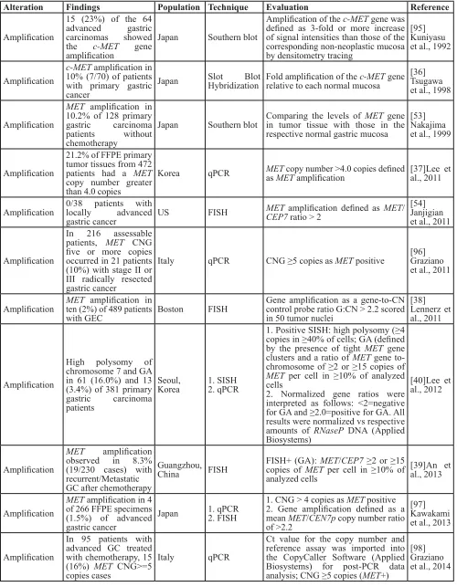
MET gene amplifications that result in protein overexpression and constitutive activation of the MET receptor kinase have been reported in NSCLC, gastric cancers and HCC [24]. Variable MET gene amplification rates were detected depending on the detection method (e.g., fluorescence in situ hybridization [FISH], single-nucleotide polymorphism [SNP] genotyping and quantitative polymerase chain reaction [qPCR]) and the different scoring criteria that define high amplification. In gastric cancer, MET gene amplification prevalence varies from 2 to 23% among studies limited by small sample sizes. In one study, a Southern blot using a [α-32P] dCTP-labeled MET-H probe (Oncor, Inc., Gaithersburg, MD, USA) detected MET amplification in 10% of chemotherapy-naïve primary gastric carcinomas compared with the surrounding normal mucosa [36]. In another study, 21.2% of formalin-fixed, paraffin-embedded (FFPE) primary tumor tissues exhibited MET
amplification defined as copy number >4 by qPCR [37].
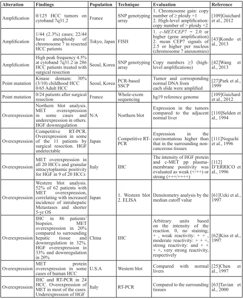
MET amplification was detected by FISH in 2% of patients with gastric cancer; in this case, MET amplification was defined as a MET/CEP7 ratio >2 [38]. Several studies with large cohorts correlated MET gene amplification with advanced tumor stage and poor prognosis in gastric cancer patients undergoing surgery with or without chemotherapy [39-41]. By contrast, MET amplification was barely detectable in patients with HCC who underwent resection: 4.5% by SNP genotyping array (Illumina), defined as a copy number ≥3, and 2.3% by FISH (Abbott Molecular), defined as MET/CEP7 ≥2.0 [42, 43].
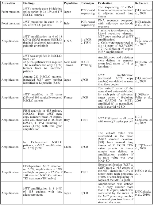
In NSCLC patients with wild-type EGFR, MET
Table 1: Molecular alterations of MET/HGF in human gastric cancer.
Alteration Findings Population Technique Evaluation reference
Amplification
15 (23%) of the 64 advanced gastric carcinomas showed the c-MET gene amplification
Japan Southern blot
Amplification of the c-MET gene was defined as 3-fold or more increase of signal intensities than those of the corresponding non-neoplastic mucosa by densitometry tracing
[95] Kuniyasu et al., 1992
Amplification
c-MET amplification in 10% (7/70) of patients with primary gastric cancer
Japan Slot Blot Hybridization Fold amplification of therelative to each normal mucosa c-MET gene [36]Tsugawa et al., 1998
Amplification
MET amplification in 10.2% of 128 primary gastric carcinoma patients without chemotherapy
Japan Southern blot Comparing the levels of in tumor tissue with those in the MET gene respective normal gastric mucosa
[53] Nakajima et al., 1999
Amplification
21.2% of FFPE primary tumor tissues from 472 patients had a MET
copy number greater than 4.0 copies
Korea qPCR METas MET copy number >4.0 copies defined amplification [37]Lee et al., 2011
Amplification 0/38 patients with locally advanced
gastric cancer US FISH
MET amplification defined as MET/
CEP7 ratio > 2
[54] Janjigian et al., 2011
Amplification
In 216 assessable patients, MET CNG five or more copies occurred in 21 patients (10%) with stage II or III radically resected gastric cancer
Italy qPCR CNG ≥5 copies as MET positive [96]Graziano et al., 2011
Amplification MET ten (2%) of 489 patients amplification in
with GEC Boston FISH
Gene amplification as a gene-to-CN control probe ratio G:CN > 2.2 scored in 50 tumor nuclei
[38] Lennerz et al., 2011
Amplification
High polysomy of chromosome 7 and GA in 61 (16.0%) and 13 (3.4%) of 381 primary gastric carcinoma patients
Seoul,
Korea 1. SISH2. qPCR
1. Positive SISH: high polysomy (≥4 copies in ≥40% of cells; GA (defined by the presence of tight MET gene clusters and a ratio of MET gene to-chromosome of ≥2 or ≥15 copies of
MET per cell in ≥10% of analyzed cells
2. Normalized gene ratios were interpreted as follows: <2=negative for GA and ≥2.0=positive for GA. All results were normalized vs respective amounts of RNaseP DNA (Applied Biosystems)
[40]Lee et al., 2012
Amplification
MET amplification observed in 8.3% (19/230 cases) with recurrent/Metastatic GC after chemotherapy
Guangzhou,
China FISH
FISH+ (GA): MET/CEP7 ≥2 or ≥15 copies of MET per cell in ≥10% of analyzed cells
[39]An et al., 2013
Amplification
MET amplification in 4 of 266 FFPE specimens (1.5%) of advanced gastric cancer
Japan 1. qPCR2. FISH
1. CNG > 4 copies as MET positive 2. Gene amplification defined as a mean MET/CEN7p copy number ratio of >2.2
[97] Kawakami et al., 2013
Amplification
In 95 patients with advanced GC treated with chemotherapy, 15 (16%) MET CNG>=5 copies cases
Italy qPCR
Ct value for the copy number and reference assay was imported into the CopyCaller Software (Applied Biosystems) for post-PCR data analysis; CNG ≥5 copies (MET+)
Amplification MET12 (6.1%) of 196 GC amplifications in patients
Shanghai,
China FISH
For MET analysis, tumors with MET
to CEP7 ≥2 or presence of ≥10% gene cluster were defined as amplified
[41]Liu et al., 2014
Point mutation
Juxtamembrane
domain: 1% (1/85) patients with primary gastric cancer
Korea 1. DHPLC 2. cold SSCP gastric carcinoma DNA compared to normal gastric tissue DNA [49]Lee et al., 2000
Overexpression
MET overexpression: 46.1% (59/128 patients with primary gastric carcinoma and without chemotherapy
Japan IHC
Tumors that were stained positively for membrane and cytoplasm were considered to be positive for the expression of the c-MET. Only distinct staining in more than 5% of tumor cells was recorded as positive immunoreactivity
[53] Nakajima et al., 1999
Overexpression
In the IHC study, c-MET overexpression in (71.1%) 32 of 45 patients in gastric carcinoma compared with matched normal gastric tissues
Taiwan IHC
The tumors were considered as positive immunreactivities if ≥ 5% of neoplastic cells showed distinct plasma membrane staining
[99]Huang et al., 2001
Overexpression
MET overexpression in 63% of 38 patients with locally advanced gastric cancer
US IHC
The percentage of positive tumor cells (scale 0%–100%) with staining intensity from 0 to 3+. Positive IHC expression is defined as 25% or more staining with intensity 2 or 3+
[54] Janjigian et al., 2011
Overexpression
MET protein expression: 104 (23.7%) of 438 patients with primary gastric carcinoma,94 (21.5%) with IHC 2+ and 10 (2.3%) cases with IHC 3+
Seoul,
Korea IHC
No membrane staining or membrane staining in <10% of tumour cells (score 0), faint/barely perceptible partial membrane staining in > 10% of tumour cells (score 1+), weak-to-moderate staining of the entire membrane in > 10% of tumour cells (score 2+), and strong staining of the entire membrane in > 10% of tumour cells (score 3+). Scores of 0 and 1+ were considered as negative for MET overexpression, and scores of 2+ and 3+ were considered as positive
[40]Lee et al., 2012
Overexpression
MET overexpression in 108 (21.8%) of 495 patients in advanced gastric carcinoma
Korea IHC
Both membranous and cytoplasmic staining was scored as follows: 0, no reactivity or faint staining; 1+, faint or weak staining; 2+, moderate staining; 3+, strong staining in >10% of tumor cells. MET overexpression was defined as 2+ or 3+ by membranous and cytoplasmic interpretation
[55]Ha et al., 2013
Overexpression
MET overexpression (IHC3+) in 9.6% (22/229 cases) with recurrent/Metastatic GC after chemotherapy
Guangzhou,
China IHC
The staining intensity and percentage of positive cells were assessed: 3+, ≥ 50% tumor cells with strong membrane/cytoplasm staining; 2+, ≥50% of tumor cells with moderate membrane/cytoplasm staining; 1+, ≥50% of tumor cells with weak membrane/cytoplasm staining; 0, No staining or ≤50% of tumor cells with membrane/cytoplasm staining of any intensity;
[39]An et al., 2013
Overexpression
MET overexpression (IHC2+ or 3+) in 12.3% (26/212 cases) of GC patients
Shanghai,
China IHC
DAKO HercepTest guideline used to semi-quantitatively score MET expression. MET+ defined by IHC2+/ IHC3+
and aCGH profiling [46-48]. These findings highlight the need for identification of lung cancer patients with MET
amplification who will benefit from combination therapy due to primary or acquired resistance, regardless of their
EGFR status.
Prevalence of MET mutations in cancers
Although MET mutations happen rarely in cancers, they may correlate with tumor development. Protein structure alterations, either via promotion of receptor dimerization or alteration of catalytic activity, can be attributed to increased kinase activity in MET mutants [15]. Missense mutations were detected in the MET juxtamembrane domain in only 1% of patients with primary gastric cancer using methods such as denaturing HPLC (DHPLC) (Transgenomics) or cold single-strand conformation polymorphism (SSCP; Novex); these mutations may contribute to tumorigenesis [49]. In NSCLC, somatic mutations in the MET juxtamembrane domain result in the deletion of exon 14, which is responsible for recruitment of the E3-ubquitin ligase, Cbl. This leads to the accumulation of abnormally spliced, activated MET unregulated by Cbl-induced degradation. This mutation was associated with elevated MET expression in primary tumors, which was detected in approximately 3% of NSCLC patients [28, 50]. Recently, polymorphisms in the juxtamembrane (R988C and T1010I) and sema (N375S) domains were detected in 1.7 and 4% of NSCLC patients, respectively, by PCR-based sequencing; however, no associations between MET mutations and clinical and pathological NSCLC features were observed [51, 52]. MET mutation was detected only in the kinase domain in 30% of childhood HCC cases by the SSCP method [27].
Prevalence of MET overexpression in cancers
Immunohistochemistry (IHC), reverse transcriptase PCR (RT-PCR), Western blot and enzyme-linked immunosorbent assay (ELISA) analyses have indicated that MET and HGF levels vary in tumors compared with surrounding normal tissues. MET overexpression was detected in vivo in 9.6 to 71% of human gastric carcinomas based on methodology and tissue type (Table 1). Notably, different antibodies that recognize various MET epitopes and domains have shown different membrane and/or cytoplasmic staining intensities by IHC. For example, 46.1% of primary gastric carcinoma patients exhibited cell membrane and cytoplasmic staining in >5% of tumor cells using a C-28 antibody (Santa Cruz Biotechnology) and the Dako ENVISION system [53]. In another study with the same antibody, 63% of patients showed positive MET expression defined as ≥25% of tumor cells with staining intensities of 2+ or 3+ [54]. MET overexpression
in gastric cancer ranged from 9.6 to 23.8%, as defined by IHC staining intensities of 2+ or 3+, via an SP44 rabbit monoclonal antibody from Ventana Medical System. MET IHC 3+ expression was associated with a shorter OS and PFS, and MET gene copy number detected by FISH correlated with MET protein expression detected by IHC in gastric cancer patients [39-41, 55].
The prevalence of HGF or MET in tissues has also been described in NSCLC tumors with wild-type EGFR. Positive intratumoral HGF expression was identified in 57% of 104 specimens using an anti-HGF antibody (sc-7949, Santa Cruz) according to the IHC Allred scoring system, and expression was associated with poor OS [56]. Using the same antibody in a separate study, HGF overexpression, in which ≥50% of tumor cells exhibited positive staining, was identified in 25% of 88 patients [57]. Western blot analysis also showed high HGF expression in NSCLC compared with normal lung tissue [58, 59]. HGF overexpression has been implicated in acquired resistance to EGFR inhibitors in patients with EGFR-mutant NSCLC [47]. Notably, the overall rate of MET overexpression measured via IHC varied widely from 13.7 to 61% in published studies based on cohorts of various sizes with different types of diseases and scoring systems (Table 2). Still, MET FISH-positive and IHC-positive patients reportedly have significantly shorter survival than MET-negative patients [57, 60].
Similarly, MET overexpression ranged from 20 to 100% in HCCs compared with surrounding normal hepatic tissue (Table 3). Western blot analysis showed MET overexpression in 52% of 62 HCC patients, which correlated with increased intrahepatic metastases and shorter (5-yr) OS [61]. Biopsy IHC analysis of 86 HCC patients revealed 20% with MET overexpression [62], whereas in a separate study employing RT-PCR, most (20/24) patients overexpressed MET [63]. Multiple studies reported that serum HGF concentrations detected
via ELISA after hepatectomy were higher as compared to normal tissues. Furthermore, HGF levels were correlated with tumor size, node cirrhosis, tumor recurrence or metastases in HCC patients [64].
Multiple studies have demonstrated that MET/HGF is associated with a poor prognosis. A study of 194 HCC patients showed that those with high MET expression with strong staining patterns (++) had significantly shorter 5-year survival than those with low expression with negative staining patterns (-) [65]. Thus, MET may represent a promising target for cancer therapies. MET and HGF alterations are indeed associated with clinical outcome, metastasis, invasion and disease severity in human cancers, and identification of patients with specific alterations may be critical to predict clinical response to targeted therapies. However, based on reported MET
Table 2: Molecular alterations of MET/HGF in human NSCLC.
Alteration Findings Population Technique Evaluation reference
Point mutation METsplice variant in 3 (1.7%) of 178 a somatic exon 14 deleting
NSCLC samples Japan
PCR-based sequencing
The sequencing of cDNA from tumor tissues compared to adjacent normal lung tissues
[51]Okuda et al., 2008
Point mutation MET4% of NSCLC patients mutations in exon 14 in Italy PCR-based sequencing DNA sequence compared with wild-type nucleotide sequence
[52]Ludovini et al., 2012
Amplification
MET amplification in 4 of 18 (22%) EGFR mutant NSCLCs that had developed resistance to gefitinib or erlotinib
U.S.A 1. qPCR2. FISH
1. relative to a reference, the Line-1 repetitive element:
MET copy number ≥4 (MET
amplification)
2. Cells were categorized as (1) ≤1 copy of MET/CEP7
; (2) ≥2 copies or ≥3 copies (MET amplification)
[48]
Engelman et al., 2007
Amplification
MET was amplified in NSCLCs from 9 of
43 (21%) patients with acquired TKI resistance but only 2 (3%) tumors from 62 untreated patients
New York
Taiwan aCGH Profiling
Amplification and deletions were defined as segment mean log2 ratios of >1 or less than -1
[46]Bean et al., 2007
Amplification Among 213 NSCLC patients, increased MET copy number
identified in 12 patients (5.6%) Japan qPCR
MET amplification
(increased MET copy number) was defined as more than three copies
[51]Okuda et al., 2008
Amplification MET(21%) of 106 surgically resected amplified in 22 cases
NSCLC patients France qPCR
The cut-off value of the normalized ratio established for each pair of reference/ target genes ( ß-globine and GAPDH for MET); amplified if its normalized ratio is over M +2 SD
[100]Beau-Faller et al., 2008
Amplification
FISH analysis in 435 primary NSCLCs. High MET gene copy number (mean ≥5 copies/ cell) was observed in 48 cases (MET+, 11.1%) including 18 cases (4.1%) with true gene amplification
Italy FISH METwith mean ≥5 copies per cell FISH-positive all cases [101]Cappuzzo et al., 2009
Amplification
In TKI-resistant NSCLC patients, c-MET amplification
in 17.2% (5/29) China qPCR
The cut-off value was established as the mean (M)+2 standard deviation (SD) from normal lung tissues of 53 EGFR TKI-naïve patients. A tumor sample was defined as amplification positive if its ratio value was over M+2×SD.
[102]Chen et al., 2009
Amplification
FISH-positive MET observed in 16.7%, amplification in 3.9% and high polysomy in 12.8% of 180 resected NSCLCs without TKI treatment (TKI naÏve)
Korea FISH
Gene amplification (MET to
CEP7 ratio 2; >15 copies of the MET signals in >10% of tumor cells; high polysomy (≥40% of cells displaying ≥4 copies of the MET signal)
[45]Go et al., 2010
Amplification
MET amplification in 8 (4%) of 183 patients with lung
adenocarcinoma Japan qPCR
Amplification was defined as a copy number more than 1.31 copies, which was calculated by the mean of the MET gene copy number measured plus two times of standard deviation
Amplification MET(15%) of TKIs resistant tumor amplification in 4/27 specimens
Boston, MA Hong Kong, China Guangzhou, China
1. qPCR 2. FISH
1. relative to a reference, the Line-1 repetitive element: MET copy number ≥4 (MET
amplification)
2. Cells were categorized as (1) ≤1 copy of MET/CEP7
; (2) ≥2 copies or ≥3 copies (MET amplification)
[47]Turke et al., 2010
Amplification
MET FISH-positive in 11.1% of 380 patients with surgically resected NSCLC (high polysomy, 8.7%; gene amplification, 2.4%)
Korea FISH
High polysomy (≥4 copies in ≥40% of cells); and gene amplification (presence of tight gene clusters and a ratio of MET to chromosome 7 of ≥2 or 15≥ copies of MET per cell in ≥10% of cells)
[60]Park et al., 2012
Amplification Increased number occurred in 18.0% of MET gene copy
61 NSCLC tissues China qPCR
the cut-off was set to 3 (MET
gene copy number ≥ 3) [44]Sun et al., 2013
Overexpression
In primary NSCLC carcinomas from 42 patients: Western blot analysis. MET increased 2 to 10 fold in 25%, HGF overexpressed 10-100 fold compared with adjacent normal tissue. In IHC, MET/HGF homogeneous expression in carcinomas
Italy 1. Western blot 2. IHC
1. The score relative to normal lung tissue of the same patient was as follows: (-), negative samples; (+), detectable expression as in the normal counterpart; ( + + ), 2 - 5-fold and (+ + +), more than 10-fold increase
[58]Olivero et al., 1996
Overexpression
Western blot analysis. MET overexpression in 104 patients: 34/47 (72%) in adenocarcinomas, 20/52 (38%) in squamous cell carcinomas. IHC in 104 patients: 56/104 (54%) of NSCLCs consisted of 36/47 (77%) of adenocarcinomas, 19/52 (37%) of squamous cell carcinomas
Japan 1. Western blot 2. IHC
1. Band intensity: (-), undetectable; (+) slight and (++) moderate was 50% level of the positive control; (+++), stronger than positive control
[103]
Ichimura et al., 1996
Overexpression High HGF expression in NSCLC Pittsburgh, USA quantitative Western blot
Bio-Rad assay was used to measure protein content of tumor extracts, and results from Western blots were expressed as nanograms of HGF per 40 ug of tumor protein
[59] Siegfried, 1997
Overexpression
In patients with small-sized lung adenocarcinomas, c-MET-positive in 69 of 131 cases (53%)
Japan IHC
The results were evaluated as positive when bundles of myofibroblasts were stained for c-MET in more than one microscopic area: occasional scattered c-MET-positive cells were considered negative
[104]
Tokunou et al., 2001
Overexpression 70% of the 166 primary NSCLC tissues showed strong HGF
expression Canada IHC
Tumors that showed similar or stronger staining levels as the normal epithelium were scored as high HGF expressors
Overexpression
In 88 patients with NSCLCs, 22 carcinomas (25.0%) were intratumoral HGF-positive, and 36 carcinomas (40.9%) were intratumoral c-MET positive
Japan IHC
1. intratumoral HGF-positive when ≥50% of the tumour cells positively stained for HGF
2. Staining intensity was classified as grade 0 (no staining), grade 1 (weak staining), grade 2 (moderately strong staining), grade 3 (very strong staining), or grade 4 (extremely strong staining). The intratumoral c-MET-positive when the intensity of c-MET-stained tumour cells was greater than grade 1
[57]Masuya et al., 2004
Overexpression
MET strongly expressed in 61% (14/23 cases) of NSCLCs and p-MET in invasive front of NSCLC
Chicago IHC
Immunohistochemical
staining intensity and extent of c-MET using the three-scale scoring system: negative (0), weak (1+), and strong (2+)
[106]Ma et al., 2005
Overexpression
In 130 primary NSCLCs, phospho-c-MET positive in 21.5% (28/130) of cases. MET positive in 74.6% of cases (97/130) and expressed at high levels in 36.1% of cases (47/130). HGF at high levels in 31.5% of cases (41/130)
Japan IHC
–, complete absence of staining or only focal weak staining; 1+, weak to moderate staining in less than 40% of cancer cells; 2+, weak to moderate staining in at least 40% of cancer cells; 3+, strong staining in at least 10% of cancer cells, among the specimens with weak to moderate staining in at least 40% of cancer cells. c-MET low (– or 1+) or c-MET high (2+ or 3+)
[107]
Nakamura et al., 2007
Overexpression HGF expression was higher in the TKIs resistant specimens
Boston, MA Hong Kong, China Guangzhou, China
IHC
The percentage of cancer cells with positive cytoplasmic and/or membranous staining (0–100%), and the modal intensity of the positively staining cells on a scale from 0 to 4+. The percentage and the intensity were multiplied to give a scoring index ranging from 0 - 400
[47]Turke et al., 2010
Overexpression
Positive expression of HGF in 104 specimens (57%). p-MET 1234/1235 in 12 (7%) specimens of 183 patients with lung adenocarcinoma
Japan IHC
The score for the positive staining cells were assigned according to the frequency of positive tumor cells (0, none; 1, <1/100; 2, 1/100 to 1/10; 3, 1/10 to 1/3; 4, 1/3 to 2/3; and 5, >2/3). Thereafter, 4 degrees for the intensity score were assigned according to the intensity of the staining (0, none; 1, weak; 2, intermediate; and 3, strong). The intensity score were then added to each other to obtain a total score, which ranged from 0 to 8. A positive expression when the score was 3 to 8.
given study, the use of different scoring systems and differences in cancer types. Standardized and optimized methods are needed to identify robust biomarkers that may assist in the selection of MET-positive patients for future clinical trials of MET-targeted therapies.
BIOMARKER VALIDATION IN MET
INHIBITOR CLINICAL TRIALS
Currently, there is no consensus on the optimal platforms to explore the relationship between MET status and drug efficacies in clinical trials. FISH and IHC assays, both traditional and commercialized approaches, require advanced technical skills and experienced experts or pathologists to analyze results. A validated biomarker detection strategy should be developed to identify a predictive or prognostic factor for these therapies. Here, we summarize the correlation between biomarker validation or clinical inclusion criteria and the therapeutic efficacy of selective MET/HGF kinase inhibitors or antibodies in clinical trials of multiple solid tumors, including GC, NSCLC and HCC.
MET as a therapeutic anti-cancer target
MET signaling dysregulation in cancer is associated with poor patient outcome. In gastric cancer, MET overexpression or amplification is correlated with tumor stage, metastasis, and shorter overall survival (OS) and progression-free survival (PFS). NSCLC patients may develop resistance to EGFR tyrosine kinase inhibitors (EGFR TKIs) via EGFR T790M mutation, HGF overexpression or MET amplification/overexpression [47, 48, 66]. Of note, MET copy number was increased in patients with EGFR TKI-resistant NSCLC compared with TKI-treatment-naïve patients [44-48]. MET activation may compensate for EGFR pathway inhibition via activation of the downstream PI3K pathway, and may correlate with acquired resistance to EGFR TKIs in patients with EGFR -mutant NSCLC [47].
MET/HGF has been regarded as a promising therapeutic target in cancer treatment, whose gene or protein status may be indicative of patient response to MET-targeted drugs. Numerous preclinical and clinical studies have demonstrated that antibody or small-molecule inhibitors targeting MET or HGF are effective anti-cancer therapies [5, 22, 67].
Overexpression MET IHC-positive in 13.7% of 380 patients with surgically
resected NSCLC Korea IHC
The intensity score: 0=no appreciable staining in the tumour cells, 1=faint/barely perceptible partial membrane staining, 2=weak to moderate staining of the entire membrane, and 3=strong staining of the entire membrane. The fraction score: 0=less than 5%, 1=from 5% to 25%, 2=from 26% to 50%, 3=from 51% to 75%, and 4=more than 75%. The total score was calculated by multiplying the intensity score and the fraction score, producing a total range of 0-12. For statistical analyses, scores of 0-3 were considered negative, and scores of 4-12 were considered positive
[60]Park et al., 2012
Overexpression 48% of samples (83 of 174) were MET positive. Poland IHC
The IHC scoring was done by one pathologist (B.R.A.) using the H-score assessment combining staining intensity (0–4) and the percentage of positive cells (0–100%). Each individual intensity level was multiplied by the percentage of cells, and all values were added to obtain the final IHC score, ranging from 0 to 400. median IHC score for the population as the cutoff point
[108]
Table 3: Molecular alterations of MET/HGF in human HCC.
Alteration Findings Population Technique Evaluation reference
Amplification 0/125 HCC tumors on cytoband 7q31.2 France SNP genotyping array
1. Chromosome gain: copy number of ≥ ploidy +1 2. High-level amplification: copy number of > ploidy +2
[109]Guichard et al., 2012
Amplification
1/44 (2.3%) cases; 22/44 have aneuploidy of chromosome 7 in resected HCC patients
Tokyo, Japan FISH
1. c-MET/CEP7 = 2.0 or higher (gene amplification) 2. mean CEP7 signals of 2.5 or higher per nucleus (chromosome 7 aneusomies)
[43]Kondo et al., 2013
Amplification
High peak frequency 4.5% at cytoband 7q31.2 in 286 HCC patients treated with surgical resection
Seoul, Korea SNP genotyping array Copy numbers ≥3 (high-level amplifications) [42]Wang et al., 2013
Point mutation Kinase domain: 30% (3/10) childhood HCC
0/65 Adult HCC Seoul, Korea PCR-based SSCP
Tumor and corresponding normal DNA from
each slide were amplified
[27]Park et al., 1999
Point mutation 0/24 patients after surgical resection France Whole-exomsequencing hg19 reference genome [109]Guichard et al., 2012
Overexpression
Northern blot analysis. MET overexpression in some cases and underexpression in others. HGF downregulation
N/A Northern blot
Expression in the tumors compared to the adjacent
normal liver [110]Selden et al., 1994
Overexpression
Competitive RT-PCR. Overexpression in some of the 11 patients by surgical resection. HGF undetectable
Japan Competitive RT-PCR
Expression in the carcinomatous higher than that in the surrounding non-cancerous tissues
[111]Noguchi et al., 1996
Overexpression
MET overexpression in all 20 HCCs and granular intracytoplasmic positivity for HGF in 9 of 20 HCCs
Italy IHC
The intensity of HGF protein and c-MET pp plasma-membrane positivity was evaluated as weak (+/++) or strong (+++/++++)
[112]
D’ERRICO et al., 1996
Overexpression
Western blot analysis. 52% of 62 patients with MET overexpression, correlating with increased incidence of intrahepatic Metastases and shorter 5-yr OS
Japan 1. Western blot 2. ELISA Densitometry analysis by the median cutoff value [61]Ueki et al., 1997
Overexpression
IHC in 86 patients’
biopsies. MET overexpression in 20%
compared to surrounding hepatic tissue and downregulation in 32%. HGF overexpression in 33% and downregulation in 20%
China IHC
Arbitrary units based on the intensity of the reaction. 0, no staining; + , weak reactivity: + + , moderate reactivity: + + +, strong reactivity: and + + + +, very strong reactivity, respectively
[62]Kiss et al., 1997
Overexpression MET protein overexpression in some
cases of human HCC U.S.A Western blot
Compared with normal
livers [25]Chen et al., 1997
Overexpression
IHC and RT-PCR in 24 HCC. Overexpression of MET in most of the cases. Underexpression of HGF
Overexpression
In 30 patients with HCC, MET over-expression in 19 cases and under-expression in 11 cases. HGF overexpression in 10 cases, and underexpression in 20 cases
Japan Western blot
Each value was obtained from the comparison with the level of ß-actin, and the mean values were calculated from three repeated measures. Expression level compared to non-tumor tissues
[113]Osada et al., 2005
Overexpression
Positive expression of c-MET protein in 27 cancerous regions (27/31) undergoing surgical resection. Higher preoperative concentration of serum HGF in the liver cancer patients
China 1. IHC2. ELISA
1. intensive positive (+++) when positive cells
comprised more than 50% of the total cells; moderately positive (++) when positive cells comprised 16–50%; weakly positive (+) when positive cells comprised 10– 15%; and negative (−) when positive cells comprised less than 10%
2. Compared with normal controls
[64]Wu et al., 2006
Overexpression
66.6% of 194 HCC patients with c-MET positive, 5-yr DFS: 61.6% vs 22.75% (c-MET- vs c-MET+)
China IHC
The extent of positive staining was scored as follows: 0, ≤10%; 1, >10–25%; 2, >25–50%; 3, >50–75%; and 4, >75%. The intensity was scored as follows: 0, negative; 1+, weak; 2+, moderate; and 3+, strong. The final score was obtained by multiplying the extent scores and intensity scores, producing a range from 0 to 12. Scores 9–12 were defined as strong staining pattern (++), scores 0–4 were defined as negative expression (−), and scores 6–8 were defined as intermediate staining pattern (+)
[65]Wang, et al., 2008
Overexpression
C-MET expression in 87.5% HCC patients undergoing hepatic resection. 29 of 40 patients (73%) had increased concentrations of portal HGF
Taiwan 1. IHC2. EISA
1. Immunoreactivity of c-MET was classified as negative when <10% of the cells stained positively, and positive when ≥10% of the cells stained positively 2. Posthepatectomy portal HGF levels compared to prehepatectomy portal HGF levels
[114]Chau et al., 2008
Overexpression
In 520 total patients, 282 c-MET+ patients (54.2%), correlating with shorter 7-yr OS
China IHC
Mean area of positive staining as cutoff value, c-MET high, >20% of tumor section
Volitinib
Volitinib is a highly selective small molecule, ATP-competitive MET kinase inhibitor being investigated as a monotherapy for MET-amplified cancers, such as gastric and lung cancer. Currently, this drug is in clinical development stage, including phase I studies in Australia and China, to test its efficacy against advanced cancers. Other phase I trials seek to test combinations of volitinib and docetaxel in gastric cancer (NCT02252913) and volitinib and gefitinib for EGFR TKI-resistant NSCLC in patients with mutant EGFR (NCT02374645).
In preclinical studies, biomarker analysis has shown that MET-amplified and MET-overexpressing tumor xenograft models are highly responsive to volitinib as a single agent or in combination with other therapies [68-71]. An in vivo study indicated that volitinib selectively inhibited the growth of MET-driven gastric and lung cancer cell line and primary tumor xenografts [68-71]. Anti-tumor efficacy in gastric cancer correlated with
MET gene amplification/overexpression and high levels of phosphorylated-MET (p-MET). Additionally, the combination of volitinib and docetaxel demonstrated efficacy in a MET-amplified gastric cancer cell line and in primary xenograft models [69]. However, correlations
between tumor growth inhibition and MET status were less clear in lung cancer compared with gastric cancer. This may be the result of heterogeneity or variation in p-MET or the activation of compensatory pathways (e.g., EGFR, KRAS) in lung cancer [71]. In particular, the combination of volitinib and taxotere had satisfactory efficacy in tumors less responsive to volitinib monotherapy [71]. The
in vivo efficacy of volitinib was tested in a model of EGFR TKI-resistant NSCLC (HCC872C4R) with acquired MET
gene amplification. Volitinib with gefitinib produced a synergistic effect compared with volitinib monotherapy, which produced a poor dose response [70]. Therefore, volitinib selectively inhibited tumor growth in a series of human tumor xenograft models with aberrant MET signaling.
MET-FISH was applied to an assessment of MET
amplification that involved labeling bacterial artificial chromosome (BAC) DNA with Spectrum Red (ENZO) and CEP7-Spectrum Green probe (Vysis Abbott). MET
amplification was defined as a MET/CEP7 ratio ≥2 or cluster signals in >10% of tumor cells. MET-IHC (SP44 Ventana Medical System, Roche) was used to detect MET overexpression, which was defined as a staining intensity of 2+ or 3+ (≥10% of tumor cells with membrane or cytoplasmic staining of moderate or strong intensity). MET-FISH or MET-IHC positivity was considered a Overexpression 15/59 (25.4%) cases high level of c-MET expression Tokyo, Japan IHC
4-point scoring system: 0 = no staining observed in invasive tumor cells; 1+ = weak, incomplete membrane staining in any proportion of the invasive tumor cells, or weak, complete circumferential membrane staining in fewer than 10% of cells; 2+ = weak but complete membrane staining in at least 10% of cells, or intense complete circumferential membrane staining in 30% or fewer of tumor cells; 3+ = intense complete ircumferential membrane staining in more than 30% of tumor cells. scores 0 and 1+ as c-MET low, and scores 2+ and 3+ as c-MET high
[43]Kondo et al., 2013
Overexpression
High expression of c-MET in 80.6% of 93 HCC patients. No correlation with clinicopathological factors, but correlated with PFS
Italy IHC
Combination of positive cell count and staining intensity used for scoring. Positive cell count, 0-10%, score 0; 11-25% score 1; 26-50%, score 2; 51-75% score 3; >75%, score 4. Staining intensity: negative score 0; faint yellow, score 1; yellow or deep yellow, score 2; brown or dark brown, score 3. High expression, Score ≥5
predictive marker for response to therapy and determined the patient selection criteria. Volitinib demonstrated robust
in vivo anti-tumor effects on predominantly MET-driven gastric and lung cancers in which the MET gene was amplified, while volitinib combined with chemotherapy could produce additional benefits in the treatment of tumors in which MET was a partial driver and where patients exhibited poor responses to volitinib monotherapy [68-71]. In an early clinical evaluation, a volitinib dose-escalation study was performed to evaluate the drug’s efficacy against advanced solid tumors, including gastric and lung cancers [72]. In 29 of 35 evaluable patients treated with 100-1000 mg volitinib daily or 300-400 mg twice daily for 21 days, 10% exhibited partial responses (PR), and 59% exhibited stable disease (SD) in at least one post-treatment. Anti-tumor volitinib activities were observed primarily in patients with papillary renal cell carcinoma (PRCC). Furthermore, data suggested that
MET gene copy number gain could be a biomarker for therapeutic response. The correlation between MET status and clinical efficacy will be further investigated to improve the patient selection criteria for later-stage clinical trials.
INc280
INC280 (Novartis) is a potent and highly selective MET kinase inhibitor currently being evaluated in early-stage clinical studies. INC280 inhibited cell proliferation, migration and apoptosis in MET-driven tumor cell lines. INC280 suppressed tumor growth in vivo in a dose-dependent manner, and was extremely well tolerated, especially in mouse xenograft models of MET-driven glioblastoma and gastric cancer [73, 74].
A phase I clinical study reported stable disease in 24% (8/33) of patients with MET-driven advanced solid tumors, including PRCC, NSCLC, HCC, gastric cancer and others (NCT01324479). INC280 showed preliminary anti-tumor efficacy as a single agent in 50% of patients with EGFR wild-type NSCLC with MET dysregulation, as confirmed by FISH (MET/centromere ratio ≥2.0 or MET
gene copy number ≥5) or IHC (MET H-score ≥150 or 50% of tumor cells with a staining intensity of 2+ or 3+) [75].
In MET-positive NSCLC tumors with mutant EGFR
that were found resistant to EGFR TKIs, a phase Ib/II study of INC280 in combination with gefitinib reported partial responses in 8/46 (17%) of evaluable patients with high MET status (IHC 3+ and/or MET copy number via
FISH ≥5) (NCT01610336). Overall response rates were 40% in patients with MET CN ≥5 and 38% in patients with MET IHC staining intensity of 3+ [76]. Unexpectedly, some NSCLCs with high MET expression (IHC 3+) and amplification exhibited tumor progression following treatment. Whether the platform, reagents and scoring system were appropriate for MET status assessment should be further evaluated in a phase II expansion study.
A phase I study of INC280 plus erlotinib in patients with MET-expressing NSCLC (NCT01911507) is ongoing. In HCC, a phase II clinical trial of INC280 as a first-line treatment is currently recruiting patients with tumors that harbor with activated MET pathways (NCT01737827).
rilotumumab
Rilotumumab (previously known as AMG 102; Amgen) is a fully human monoclonal HGF antibody that preferentially binds to the mature, active form of the protein [77]. Interaction with rilotumumab prevents HGF from binding to MET, which prevents subsequent signaling [78]. The antibody was tested in preclinical models and in clinical trials in multiple solid tumors, either as a monotherapy or in combination with other chemotherapeutics [79-83]. Based on preclinical data, rilotumumab has shown the anti-tumor activity in vitro
and in vivo studies, [79, 84]. In a phase I clinical trial, rilotumumab monotherapy was deemed safe and well tolerated in 40 patients with refractory advanced solid tumors. A total of 16/23 (70%) evaluable patients achieved stable disease as a best response with PFS from 7.9 to 40 weeks [81].
In a phase II trial (NTC00719550), rilotumumab in combination with the cytotoxic agents epirubicin, cisplatin or capecitabine (ECX) was evaluated in 121 patients with advanced or metastatic gastric or esophageal junction (G/ EGJ) cancer. Rilotumumab plus ECX had an anti-tumor efficacy compared to ECX alone, with modestly improved OS (median months, 10.6 vs. 8.9; hazard ratio [HR] = 0.70) and PFS (median months, 5.7 vs. 4.2; HR = 0.60) [85]. The MET-positive tumors were defined as such when at least 25% of tumor cells demonstrated membrane staining at any intensity using the MET4 monoclonal antibody and the verified MET immunohistochemistry pharmDx kit (Dako North America, Carpinteria, CA, USA). However, rilotumumab plus ECX significantly improved OS in patients with high MET expression compared with ECX alone (10.6 months [95% CI 8.0-13.4] vs. 5.7 months [4.2-10.4]). Conversely, MET-negative patients had slightly reduced OS when treated with rilotumumab plus ECX compared with ECX alone (11.1 months [6.9-13.2] vs. 11.5 months [5.5-20.5]). A similar trend was observed for PFS [85].
A phase III study to assess the efficacy and safety of rilotumumab in advanced gastric cancer did not confirm the phase II findings and was terminated due to futility results and casualties with an increased number of deaths as compared to chemotherapy [86]. Rilotumumab arm was not superior to placebo arm for OS (median months, 9.6
vs. 11.5; HR = 1.37) and PFS (median months, 5.7 vs. 5.7; HR = 1.30). OS, PFS and ORR were statistically worse in the rilotumumab arm. Significantly more patients in the placebo arm achieved 12-month OS (49.7% vs. 38.4%; P
to the rilotumumab arm. No benefit from treatment with rilotumumab was observed, including patients with higher MET expression, and a higher incidence of fetal adverse events occurred in the rilotumumab arm [86]. We suggest that defining MET positivity as IHC staining in 25% of tumor cells is not stringent enough to establish true MET overexpression, and could lead to negative results in clinical trials. Additionally, rilotumumab was recently reported to be only a partial, not full, HGF antagonist, resulting in HGF-induced MET phosphorylation. This may at least partially explain the poor responses and deaths in rilotumumab clinical trials, and has considerable implications for the use of this therapeutic antibody [87].
Onartuzumab
Onartuzumab (MetMAb; Roche/Genentech) is a single-armed humanized modified 5D5 anti-MET antibody that binds to the MET sema domain and effectively prevents HGF from binding to MET [88]. Preclinical studies have demonstrated inhibitory effects on glioblastoma U87 and pancreatic BxPC3 and KP4 tumor xenografts, including reduced cell proliferation and motility [88, 89]. In a phase I study, a complete response was observed in a patient with chemotherapy-refractory metastatic gastric cancer with high MET gene polysomy, high MET expression and evidence of autocrine HGF production [90]. In a randomized, double-blind phase II trial in EGFR-unselected NSCLC patients, no increased benefit in PFS (HR = 1.09, p = 0.69) or OS (HR = 0.80, p
= 0.34) was noted in patients receiving onartuzumab plus erlotinib vs. placebo plus erlotinib. However, in MET-positive (≥50% of tumor cells with a staining intensity of 2+ or 3+) NSCLC patients (52% of 137 individuals), onartuzumab plus erlotinib was associated with improved PFS (1.5 vs. 2.9 months, HR = 0.53, p = 0.04) and OS (3.8 vs. 12.6 months, HR = 0.37, p = 0.002). In contrast, MET-negative patients demonstrated worse OS after treatment with onartuzumab compared with patients treated with erlotinib alone. Despite improved PFS and OS in the MET-positive population, the ORR in this subset was not different from that of the placebo plus erlotinib arm [91], possibly because the inclusion criteria for MET-positive patients may not have been properly defined. A randomized phase III trial in patients with MET-positive advanced NSCLC who were to receive the standard chemotherapy has been terminated because treatment with onartuzumab in combination with erlotinib did not demonstrate a significant difference over erlotinib alone [92].
CONCLUSIONS
Because MET is involved in the regulation of tumor cell survival and metastasis, a better understanding of
individual patient sensitivities to MET inhibitors can help guide clinical trial design. Therefore, the key to success for MET-targeted therapies in human cancers may lie in MET-driven patient selection, which is determined by many factors.
First, an integrated platform that incorporates accurate, validated methods and reagent kits, such as antibodies and substrates for the characterization of MET alterations, will help to improve MET-driven population selection. For example, a phase III trial of onartuzumab showed a lack of efficacy in patients with advanced NSCLC, who were selected for the trial based on MET overexpression detected via IHC [91]. However, the IHC detection antibody, SP44, binds the cytoplasmic MET domain, whereas onartuzumab binds to the extracellular semaphorin domain. Reported IHC results vary widely because different antibodies that target distinct MET epitopes are used. It is critical to utilize specific antibodies for the detection of MET expression as a predictive biomarker to refine patient selection in clinical trials.
Next, MET-positive patient selection must also take into consideration of tumor stage and type, along with sample storage conditions, and must incorporate stringent clinical inclusion criteria. A single biomarker or a combination of biomarkers may serve as prognostic factors to fully inform patient selection. For example, in the phase III onartuzumab clinical trial, defining IHC MET positivity as 50% of tumor cells with moderate staining intensity might have resulted in the inclusion of patients with false-positive MET-overexpressing tumors.
In a phase II trial, however, IHC analysis correlated better with significant improvements in OS and PFS compared with FISH analysis. In this study, a MET/CEP7
ratio of two or more or a mean of ≥5 MET copies per cell were defined as MET FISH positivity, which may result in the inclusion of patients with no MET-amplified tumors [93]. These criteria could explain why NSCLC patients with MET FISH positivity exhibited negative responses to onartuzumab in a phase III trial [92]. Additionally, in rilotumumab studies, defining MET-positive tumors as those having at least 25% of tumor cells with membrane staining at any intensity may also have resulted in a high rate of false positives. These results together strongly suggest that testing for MET levels should include both MET overexpression assessment by IHC and MET gene amplification analysis by FISH for improved accuracy and reduced false positive detection rates.
treatment approach that targets both MET and other functionally redundant RTKs should also be considered.
Thus far, no MET inhibitors or antibodies have been approved for clinical use. Evidence is mounting that fighting MET-driven cancers may depend on many elements, such as the specific drug target, disease stage or type, optimal methods for tumor MET status assessment, and, perhaps most importantly, the identification of robust biomarkers as predictors of patient benefit from MET-targeting therapeutics.
CONFLICTS OF INTEREST
The authors declare that they have no conflicts of interest.
REFERENCES
1. Trusolino L, Bertotti A, Comoglio PM. MET signalling:
principles and functions in development, organ regeneration and cancer. Nat Rev Mol Cell Biol. 2010; 11: 834-848.
2. Gonzatti-Haces M, Seth A, Park M, Copeland T, Oroszlan S, Vande Woude GF. Characterization of the TPR-MET
oncogene p65 and the MET protooncogene p140
protein-tyrosine kinases. Proc Natl Acad Sci U S A. 1988; 85:
21-25.
3. Uehara Y, Minowa O, Mori C, Shiota K, Kuno J, Noda T, Kitamura N. Placental defect and embryonic lethality in mice lacking hepatocyte growth factor/scatter factor. Nature. 1995; 373: 702-705.
4. Sonnenberg E, Meyer D, Weidner KM, Birchmeier C. Scatter factor/hepatocyte growth factor and its receptor, the c-met tyrosine kinase, can mediate a signal exchange between mesenchyme and epithelia during mouse development. J Cell Biol. 1993; 123: 223-235.
5. Comoglio PM, Giordano S, Trusolino L. Drug development of MET inhibitors: targeting oncogene addiction and expedience. Nat Rev Drug Discov. 2008; 7: 504-516.
6. Rodrigues GA, Park M. Autophosphorylation modulates the
kinase activity and oncogenic potential of the Met receptor tyrosine kinase. Oncogene. 1994; 9: 2019-2027.
7. Ponzetto C, Bardelli A, Zhen Z, Maina F, dalla Zonca P, Giordano S, Graziani A, Panayotou G, Comoglio PM. A multifunctional docking site mediates signaling and
transformation by the hepatocyte growth factor/scatter factor receptor family. Cell. 1994; 77: 261-271.
8. Furge KA, Zhang YW, Vande Woude GF. Met receptor
tyrosine kinase: enhanced signaling through adapter proteins. Oncogene. 2000; 19: 5582-5589.
9. Sierra JR, Corso S, Caione L, Cepero V, Conrotto P, Cignetti A, Piacibello W, Kumanogoh A, Kikutani
H, Comoglio PM, Tamagnone L, Giordano S. Tumor angiogenesis and progression are enhanced by Sema4D produced by tumor-associated macrophages. J Exp Med.
2008; 205: 1673-1685.
10. Conrotto P, Valdembri D, Corso S, Serini G, Tamagnone L,
Comoglio PM, Bussolino F, Giordano S. Sema4D induces angiogenesis through Met recruitment by Plexin B1. Blood. 2005; 105: 4321-4329.
11. Yi S, Tsao MS. Activation of hepatocyte growth factor-met
autocrine loop enhances tumorigenicity in a human lung adenocarcinoma cell line. Neoplasia. 2000; 2: 226-234.
12. Graziani A, Gramaglia D, dalla Zonca P, Comoglio PM.
Hepatocyte growth factor/scatter factor stimulates the Ras-guanine nucleotide exchanger. J Biol Chem. 1993; 268: 9165-9168.
13. Maroun CR, Naujokas MA, Park M. Membrane targeting
of Grb2-associated binder-1 (Gab1) scaffolding protein through Src myristoylation sequence substitutes for Gab1 pleckstrin homology domain and switches an epidermal growth factor response to an invasive morphogenic program. Mol Biol Cell. 2003; 14: 1691-1708.
14. Xiao GH, Jeffers M, Bellacosa A, Mitsuuchi Y, Vande Woude GF, Testa JR. Anti-apoptotic signaling by
hepatocyte growth factor/Met via the phosphatidylinositol
3-kinase/Akt and mitogen-activated protein kinase pathways. Proc Natl Acad Sci U S A. 2001; 98: 247-252. 15. Organ SL, Tsao MS. An overview of the c-MET signaling
pathway. Ther Adv Med Oncol. 2011; 3: S7-S19.
16. Syed ZA, Yin W, Hughes K, Gill JN, Shi R, Clifford JL.
HGF/c-met/Stat3 signaling during skin tumor cell invasion: indications for a positive feedback loop. BMC Cancer. 2011; 11: 180.
17. Peschard P, Park M. Escape from Cbl-mediated downregulation: a recurrent theme for oncogenic deregulation of receptor tyrosine kinases. Cancer Cell. 2003; 3: 519-523.
18. Orian-Rousseau V, Morrison H, Matzke A, Kastilan T,
Pace G, Herrlich P, Ponta H. Hepatocyte growth factor-induced Ras activation requires ERM proteins linked to both CD44v6 and F-actin. Mol Biol Cell. 2007; 18: 76-83. 19. Puri N, Salgia R. Synergism of EGFR and c-Met pathways,
cross-talk and inhibition, in non-small cell lung cancer. J Carcinog. 2008; 7: 9.
20. Follenzi A, Bakovic S, Gual P, Stella MC, Longati P,
Comoglio PM. Cross-talk between the proto-oncogenes Met and Ron. Oncogene. 2000; 19: 3041-3049.
21. Yeh CY, Shin SM, Yeh HH, Wu TJ, Shin JW, Chang TY,
Raghavaraju G, Lee CT, Chiang JH, Tseng VS, Lee YC,
Shen CH, Chow NH et al. Transcriptional activation of the
Axl and PDGFR-alpha by c-Met through a ras- and
Src-independent mechanism in human bladder cancer. BMC Cancer. 2011; 11: 139.
22. Birchmeier C, Birchmeier W, Gherardi E, Vande Woude
GF. Met, metastasis, motility and more. Nat Rev Mol Cell Biol. 2003; 4: 915-925.
23. Maulik G, Shrikhande A, Kijima T, Ma PC, Morrison PT,
c-Met, in oncogenesis and potential for therapeutic inhibition. Cytokine Growth Factor Rev. 2002; 13: 41-59.
24. Smolen GA, Sordella R, Muir B, Mohapatra G, Barmettler A, Archibald H, Kim WJ, Okimoto RA, Bell DW, Sgroi DC, Christensen JG, Settleman J, Haber DA. Amplification
of MET may identify a subset of cancers with extreme
sensitivity to the selective tyrosine kinase inhibitor PHA-665752. Proc Natl Acad Sci U S A. 2006; 103: 2316-2321. 25. Chen Q, Seol DW, Carr B, Zarnegar R. Co-expression
and regulation of Met and Ron proto-oncogenes in human hepatocellular carcinoma tissues and cell lines. Hepatology. 1997; 26: 59-66.
26. Lubensky IA, Schmidt L, Zhuang Z, Weirich G, Pack S, Zambrano N, Walther MM, Choyke P, Linehan WM, Zbar
B. Hereditary and sporadic papillary renal carcinomas with c-met mutations share a distinct morphological phenotype.
Am J Pathol. 1999; 155: 517-526.
27. Park WS, Dong SM, Kim SY, Na EY, Shin MS, Pi JH, Kim BJ, Bae JH, Hong YK, Lee KS, Lee SH, Yoo NJ, Jang JJ et al. Somatic mutations in the kinase domain of the Met/hepatocyte growth factor receptor gene in childhood hepatocellular carcinomas. Cancer Res. 1999; 59: 307-310.
28. Kong-Beltran M, Seshagiri S, Zha J, Zhu W, Bhawe K,
Mendoza N, Holcomb T, Pujara K, Stinson J, Fu L, Severin C, Rangell L, Schwall R et al. Somatic mutations lead to an oncogenic deletion of met in lung cancer. Cancer Res. 2006; 66: 283-289.
29. Kentsis A, Reed C, Rice KL, Sanda T, Rodig SJ, Tholouli E, Christie A, Valk PJ, Delwel R, Ngo V, Kutok JL, Dahlberg SE, Moreau LA et al. Autocrine activation of the
MET receptor tyrosine kinase in acute myeloid leukemia. Nat Med. 2012; 18: 1118-1122.
30. Pennacchietti S, Michieli P, Galluzzo M, Mazzone M, Giordano S, Comoglio PM. Hypoxia promotes invasive growth by transcriptional activation of the met protooncogene. Cancer Cell. 2003; 3: 347-361.
31. Gambarotta G, Boccaccio C, Giordano S, Ando M, Stella
MC, Comoglio PM. Ets up-regulates MET transcription. Oncogene. 1996; 13: 1911-1917.
32. Boon EM, van der Neut R, van de Wetering M, Clevers H, Pals ST. Wnt signaling regulates expression of the receptor tyrosine kinase met in colorectal cancer. Cancer Res. 2002; 62: 5126-5128.
33. Migliore C, Martin V, Leoni VP, Restivo A, Atzori L, Petrelli A, Isella C, Zorcolo L, Sarotto I, Casula G, Comoglio PM, Columbano A, Giordano S. MiR-1 downregulation cooperates with MACC1 in promoting
MET overexpression in human colon cancer. Clin Cancer Res. 2012; 18: 737-747.
34. Jung KH, Park BH, Hong SS. Progress in cancer therapy
targeting c-Met signaling pathway. Arch Pharm Res. 2012;
35: 595-604.
35. Gherardi E, Birchmeier W, Birchmeier C, Vande Woude G.
Targeting MET in cancer: rationale and progress. Nat Rev
Cancer. 2012; 12: 89-103.
36. Tsugawa K, Yonemura Y, Hirono Y, Fushida S, Kaji M,
Miwa K, Miyazaki I, Yamamoto H. Amplification of the
c-met, c-erbB-2 and epidermal growth factor receptor gene in human gastric cancers: correlation to clinical features. Oncology. 1998; 55: 475-481.
37. Lee J, Seo JW, Jun HJ, Ki CS, Park SH, Park YS, Lim HY, Choi MG, Bae JM, Sohn TS, Noh JH, Kim S, Jang
HL et al. Impact of MET amplification on gastric cancer:
possible roles as a novel prognostic marker and a potential therapeutic target. Oncol Rep. 2011; 25: 1517-1524.
38. Lennerz JK, Kwak EL, Ackerman A, Michael M, Fox
SB, Bergethon K, Lauwers GY, Christensen JG, Wilner
KD, Haber DA, Salgia R, Bang YJ, Clark JW et al. MET amplification identifies a small and aggressive subgroup
of esophagogastric adenocarcinoma with evidence of responsiveness to crizotinib. J Clin Oncol. 2011; 29: 4803-4810.
39. An X, Wang F, Shao Q, Wang FH, Wang ZQ, Wang ZQ, Chen C, Li C, Luo HY, Zhang DS, Xu RH, Li YH. MET amplification is not rare and predicts unfavorable clinical
outcomes in patients with recurrent/metastatic gastric cancer after chemotherapy. Cancer. 2014; 120: 675-682.
40. Lee HE, Kim MA, Lee HS, Jung EJ, Yang HK, Lee BL,
Bang YJ, Kim WH. MET in gastric carcinomas: comparison between protein expression and gene copy number and impact on clinical outcome. Br J Cancer. 2012; 107: 325-333.
41. Liu YJ, Shen D, Yin X, Gavine P, Zhang T, Su X, Zhan P, Xu Y, Lv J, Qian J, Liu C, Sun Y, Qian Z et al. HER2, MET and FGFR2 oncogenic driver alterations define
distinct molecular segments for targeted therapies in gastric carcinoma. Br J Cancer. 2014; 110: 1169-1178.
42. Wang K, Lim HY, Shi S, Lee J, Deng S, Xie T, Zhu Z, Wang Y, Pocalyko D, Yang WJ, Rejto PA, Mao M, Park
CK et al. Genomic landscape of copy number aberrations
enables the identification of oncogenic drivers in
hepatocellular carcinoma. Hepatology. 2013; 58: 706-717. 43. Kondo S, Ojima H, Tsuda H, Hashimoto J, Morizane
C, Ikeda M, Ueno H, Tamura K, Shimada K, Kanai Y, Okusaka T. Clinical impact of c-Met expression and its
gene amplification in hepatocellular carcinoma. Int J Clin
Oncol. 2013; 18: 207-213.
44. Sun W, Song L, Ai T, Zhang Y, Gao Y, Cui J. Prognostic
value of MET, cyclin D1 and MET gene copy number in non-small cell lung cancer. J Biomed Res. 2013; 27: 220-230.
45. Go H, Jeon YK, Park HJ, Sung SW, Seo JW, Chung DH. High MET gene copy number leads to shorter survival in patients with non-small cell lung cancer. J Thorac Oncol. 2010; 5: 305-313.
46. Bean J, Brennan C, Shih JY, Riely G, Viale A, Wang
L, Chitale D, Motoi N, Szoke J, Broderick S, Balak M,
or without T790M mutations in EGFR mutant lung tumors
with acquired resistance to gefitinib or erlotinib. Proc Natl Acad Sci U S A. 2007; 104: 20932-20937.
47. Turke AB, Zejnullahu K, Wu YL, Song Y, Dias-Santagata D, Lifshits E, Toschi L, Rogers A, Mok T, Sequist L, Lindeman NI, Murphy C, Akhavanfard S et al. Preexistence and clonal selection of MET amplification in EGFR mutant
NSCLC. Cancer Cell. 2010; 17: 77-88.
48. Engelman JA, Zejnullahu K, Mitsudomi T, Song Y, Hyland C, Park JO, Lindeman N, Gale CM, Zhao X, Christensen J, Kosaka T, Holmes AJ, Rogers AM et al. MET amplification leads to gefitinib resistance in lung cancer by activating
ERBB3 signaling. Science. 2007; 316: 1039-1043. 49. Lee JH, Han SU, Cho H, Jennings B, Gerrard B, Dean M,
Schmidt L, Zbar B, Vande Woude GF. A novel germ line
juxtamembrane Met mutation in human gastric cancer. Oncogene. 2000; 19: 4947-4953.
50. Seo JS, Ju YS, Lee WC, Shin JY, Lee JK, Bleazard T, Lee J, Jung YJ, Kim JO, Shin JY, Yu SB, Kim J, Lee ER et al.
The transcriptional landscape and mutational profile of lung
adenocarcinoma. Genome Res. 2012; 22: 2109-2119. 51. Okuda K, Sasaki H, Yukiue H, Yano M, Fujii Y. Met gene
copy number predicts the prognosis for completely resected non-small cell lung cancer. Cancer Sci. 2008; 99: 2280-2285.
52. Ludovini V, Bianconi F, Pistola L, Pistola V, Chiari R, Colella R, Bellezza G, Tofanetti FR, Siggillino A, Baldelli E, Flacco A, Giuffrida D, Sidoni A et al. Optimization of
patient selection for EGFR-TKIs in advanced non-small
cell lung cancer by combined analysis of KRAS, PIK3CA,
MET, and non-sensitizing EGFR mutations. Cancer Chemother Pharmacol. 2012; 69: 1289-1299.
53. Nakajima M, Sawada H, Yamada Y, Watanabe A, Tatsumi
M, Yamashita J, Matsuda M, Sakaguchi T, Hirao T,
Nakano H. The prognostic significance of amplification
and overexpression of c-met and c-erb B-2 in human gastric carcinomas. Cancer. 1999; 85: 1894-1902.
54. Janjigian YY, Tang LH, Coit DG, Kelsen DP, Francone
TD, Weiser MR, Jhanwar SC, Shah MA. MET expression and amplification in patients with localized gastric cancer.
Cancer Epidemiol Biomarkers Prev. 2011; 20: 1021-1027.
55. Ha SY, Lee J, Kang SY, Do IG, Ahn S, Park JO, Kang WK,
Choi MG, Sohn TS, Bae JM, Kim S, Kim M, Kim S et al. MET overexpression assessed by new interpretation method
predicts gene amplification and poor survival in advanced
gastric carcinomas. Mod Pathol. 2013; 26: 1632-1641. 56. Onitsuka T, Uramoto H, Ono K, Takenoyama M,
Hanagiri T, Oyama T, Izumi H, Kohno K, Yasumoto K. Comprehensive molecular analyses of lung adenocarcinoma with regard to the epidermal growth factor receptor, K-ras, MET, and hepatocyte growth factor status. J Thorac Oncol. 2010; 5: 591-596.
57. Masuya D, Huang C, Liu D, Nakashima T, Kameyama K, Haba R, Ueno M, Yokomise H. The tumour-stromal
interaction between intratumoral c-Met and stromal hepatocyte growth factor associated with tumour growth and prognosis in non-small-cell lung cancer patients. Br J Cancer. 2004; 90: 1555-1562.
58. Olivero M, Rizzo M, Madeddu R, Casadio C, Pennacchietti
S, Nicotra MR, Prat M, Maggi G, Arena N, Natali PG,
Comoglio PM, Di Renzo MF. Overexpression and activation of hepatocyte growth factor/scatter factor in human non-small-cell lung carcinomas. Br J Cancer. 1996; 74: 1862-1868.
59. Siegfried JM, Weissfeld LA, Singh-Kaw P, Weyant RJ, Testa JR, Landreneau RJ. Association of immunoreactive
hepatocyte growth factor with poor survival in resectable non-small cell lung cancer. Cancer Res. 1997; 57: 433-439.
60. Park S, Choi YL, Sung CO, An J, Seo J, Ahn MJ, Ahn JS,
Park K, Shin YK, Erkin OC, Song K, Kim J, Shim YM et al. High MET copy number and MET overexpression: poor outcome in non-small cell lung cancer patients. Histol Histopathol. 2012; 27: 197-207.
61. Ueki T, Fujimoto J, Suzuki T, Yamamoto H, Okamoto E. Expression of hepatocyte growth factor and its receptor c-met proto-oncogene in hepatocellular carcinoma. Hepatology. 1997; 25: 862-866.
62. Kiss A, Wang NJ, Xie JP, Thorgeirsson SS. Analysis of
transforming growth factor (TGF)-alpha/epidermal growth factor receptor, hepatocyte growth Factor/c-met,TGF-beta receptor type II, and p53 expression in human hepatocellular carcinomas. Clin Cancer Res. 1997; 3: 1059-1066.
63. Tavian D, De Petro G, Benetti A, Portolani N, Giulini SM, Barlati S. u-PA and c-MET mRNA expression is co-ordinately enhanced while hepatocyte growth factor mRNA
is down-regulated in human hepatocellular carcinoma. Int J Cancer. 2000; 87: 644-649.
64. Wu F, Wu L, Zheng S, Ding W, Teng L, Wang Z, Ma Z, Zhao W. The clinical value of hepatocyte growth factor
and its receptor—c-met for liver cancer patients with hepatectomy. Dig Liver Dis. 2006; 38: 490-497.
65. Wang ZL, Liang P, Dong BW, Yu XL, Yu de J.
Prognostic factors and recurrence of small hepatocellular carcinoma after hepatic resection or microwave ablation: a retrospective study. J Gastrointest Surg. 2008; 12: 327-337.
66. Lutterbach B, Zeng Q, Davis LJ, Hatch H, Hang G, Kohl
NE, Gibbs JB, Pan BS. Lung cancer cell lines harboring
MET gene amplification are dependent on Met for growth
and survival. Cancer Res. 2007; 67: 2081-2088.
67. Ma PC, Maulik G, Christensen J, Salgia R. c-Met: structure, functions and potential for therapeutic inhibition. Cancer Metastasis Rev. 2003; 22: 309-325.
68. Cui Y, Dai G, Re Y, Zhou F, Fan S, Sai Y, Gu Y, Yan J, Li J, Qing W, Su WG. A novel and selective c-Met inhibitor
against subcutaneous xenograft and othotopic brain tumor
models Cancer Res. 2011; 71(8 Suppl):Abstract nr 3612. 69. Gavine PR, Fan S, Fu H, Han L. Volitinib (HMPL504), a