Evolution
Thesis by Benjamin R. Uy
In Partial Fulfillment of the Requirements for the degree of
Doctor in Philosophy
CALIFORNIA INSTITUTE OF TECHNOLOGY Pasadena, California
2016
ACKNOWLEDGEMENTS
ABSTRACT
Neural crest cells are unique to vertebrates and essential to the development and evolution of the craniofacial skeleton. Using a combination of DiI cell lineage tracing, transcriptomics, and analysis of key transcription factors of the Sox Family, I examined neural crest development in the sea lamprey, Petromyzon marinus, as the most basal extant vertebrate from which it is possible to get embryos. The results have uncovered distinct cranial and trunk neural crest subpopulations along the anterior-posterior axis of the lamprey embryo, with a clear separation between the two. However, no evidence of the presence of an intermediate vagal neural crest population was uncovered. Comparing cranial neural crest genes between lamprey and chick, either by examining individual candidate genes or whole genome transcriptome analysis, reveals significant changes in the cranial neural crest gene regulatory network of lamprey compared with chick. In
PUBLISHED CONTENT
1) *Haming, D., *Simoes-Costa, M., Uy, B., Valencia, J., Sauka-Spengler, T, Bronner, M.E., (2011) Expression of Sympathetic Nervous System Genes in Lamprey Suggests Their Recruitment for Specification of a New Vertebrate Feature. PLoS ONE 6(10): e26543. doi:10.1371/journal.pone.0026543 2) Uy, B., Simoes-Costa, M., Sauka-Spengler, T., and Bronner-ME. (2012)
Expression of Sox genes in lamprey development. Int. J. of Dev. Biol. 56: 377 – 383. doi: 10.1387/ijdb.113416bu
3) Modrell, M., Hockman, D. Uy, B., Sauka-Spengler, T., Bronner, M.E & Baker, C. (2014) A fate-map for cranial sensory ganglia in the sea lamprey. Dev. Biol. vol. 385, pp. 405-416. doi: 10.1016/j.ydbio.2013.10.021
4) B.R. Uy, M. Simoes-Costa, D.E. Koo, T. Sauka-Spengler, M.E. Bronner, (2015) Evolutionarily conserved role for SoxC genes in neural crest specification and neuronal differentiation. Dev. Biol., 397 (2015), pp. 282–292 doi:
TABLE OF CONTENTS
ACKNOWLEDGEMENTS ... iii
ABSTRACT... iv
PUBLISHED CONTENT ... v
TABLE OF CONTENTS ... vi
LIST OF FIGURES ... vii
CHAPTER I: INTRODUCTION ... 1
CHAPTER II: FATE MAPPING AND TRANSCRIPTOMICS OF NEURAL CREST SUBPOPULATIONS ... 13
I. EXPRESSION OF SYMPATHETIC NERVOUS SYSTEM GENES IN LAMPREY SUGGESTS THEIR RECRUITMENT FOR SPECIFICATION OF A NEW VERTEBRATE FEATURE ... 14
II. PROGRESSIVE REGIONALIZATION OF THE NEURAL CREST DURING VERTEBRATE EVOLUTION VIA ELABORATION OF GENE REGULATORY CIRCUITRY ... 38
CHAPTER III: ANCIENT EVOLUTIONARY ORIGIN OF THE VERTEBRATE ENTERIC NERVOUS SYSTEM FROM NEURAL CREST-DERIVED SCHWANN CELL PRECURSORS ... 63
CHAPTER IV: NOVEL ROLE OF SOX FACTORS IN NEURAL CREST FORMATION ... 81
I. EXPRESSION OF SOX GENES IN LAMPREY DEVLOPMENT ... 82
II. EVOLUTIONARILY CONSERVED ROLE FOR SOXC GENES IN NEURAL CREST SPECIFICATION AND NEURONAL DIFFERENTIATION ... 105
CHAPTER V: CONCLUSION ... 141
APPENDIX A: A FATE-MAP FOR CRANIAL SENSORY GANGLIA IN THE SEA LAMPREY ... 155
LIST OF FIGURES
Number Page
1. Lamprey as a model organism ... 12
2. DiI labeling of lamprey neural crest cells reveals absence of sympathetic ganglia during embryonic development ... 30
3. Expression of the transcription factor Phox2 in embryos of P. marinus ... 31
4. Expression of the helix-loop-helix transcription factor Hand in lamprey embryos ... 32
5. Expression of Ascl1 during embryonic development of the lamprey ... 33
6. DiI-labeling along the anterior-posterior body axis reveals distinct populations of cranial and trunk neural crest cells ... 56
7. Comparison of chick cranial-specific gene regulatory circuit with that of lamprey ... 57
8. Transcriptome analysis comparing lamprey cranial and trunk neural crest ... 58
9. Proposed model for evolution of neural crest subpopulations ... 59
10. Cranial Neural Crest contribute to the trigeminal ganglia ... 60
11. Trunk neural crest give rise to dorsal root ganglia and fin mesenchyme ... 60
12. Early formation of enteric neurons in the lamprey P. marinus ... 71
13. DiI Labeled cells in the neural tube contribute to enteric ganglia ... 72
14. Schwann cell precursors contribute to the ENS ... 73
15. Number of DiI-labeled neurons (Actub+) in 25 representative E30-35 embryos in which DiI had been injected into the neural tube at T22.5 ... 74
17. DiI labeling of the caudal hindbrain population shows contributions
to the branchial arches but not outflow tract or gut ... 76
18. Examining schwann cell markers in lamprey... 77
19. Phylogenetic Analysis of lamprey Sox genes ... 96
20. SoxB Expression ... 97
21. SoxD Expression ... 98
22. SoxE Expression ... 99
23. SoxF Expression ... 100
24. Maximum Likelihood phylogenetic tree of lamprey Sox genes ... 101
25. SoxC1–C4 are expressed in the neural plate border, neural tube, and neural crest derivatives in lamprey ... 127
26. Xenopus laevis SoxCs exhibit overlapping expression in the neural plate, neural crest, and facial region ... 128
27. Morpholino antisense oligonucleotides efficiently disrupt target protein translation ... 129
28. Loss of SoxC1, C3, and C4 in lamprey results in loss of neural crest specifier genes Sox10 and FoxD3... 130
29. Xenopus laevis Sox4 and Sox11 regulate neural crest specifier genes FoxD3, Sox10, and Twist. ... 131
30. Loss of SoxC genes in lamprey causes defects in the cranial ganglia and branchial arches ... 132
31. Xenopus laevis Sox4 and Sox11 knock-downs affect Neurogenin expression later in development ... 134
32. Phylogenetic analysis of the SoxC Family ... 135
33. SoxC1, C3, and C4 are expressed in premigratory neural crest and crest derivatives ... 136
34. Full Length SoxC proteins ... 137
35. Spatiotemporal development of lamprey cranial sensory ganglia ... 178
37. Pax3/7 is a specific marker for opV placode-derived neurons ... 181 38. Fate-mapping of opV and mmV placode-derived neurons reveals
two separate domains of mmV neuron precursors at late neurula stages ... 182 39. Fate-maps at E6-7 for lamprey epibranchial and lateral line
placode-derived ganglia ... 184 40. Neural crest-derived cells are found in cranial sensory ganglia and
C h a p t e r 1
Neural crest cells are responsible for essential derivatives in vertebrate development
Neural crest cells are a multipotent stem cell population that is unique to vertebrates and appears to have emerged at the base of the lineage. These cells give rise to a wide variety of derivatives, such as melanocytes, sensory ganglia, Schwann cells, craniofacial skeleton, connective tissue, chromaffin cells of the adrenal medulla, parafollicular cells of the thyroid, cells of the ventricular septum, and the aorticpulmonary septum. Because of their essential role in embryogenesis, defects in neural crest development are responsible for a variety of birth defects, diseases, and syndromes. Previous studies have employed fate mapping experiments and quail-chick heterospecific grafts to demonstrate neural crest cell fate and developmental potential along the anterior to posterior body axis (Le Douarin and Kalcheim 1999). The most anterior population is the cranial neural crest, emerging from dorsal neural tube of the posterior portion of the forebrain to the hindbrain. These cells give rise to craniofacial cartilage, bone, connective tissue, smooth muscle, neurons of the trigeminal ganglion, and glia. The next population is the vagal neural crest, which emerges adjacent to the 1st to 7th somite region. Vagal neural crest cells are essential for the formation of the
Studying Neural Crest in Lamprey
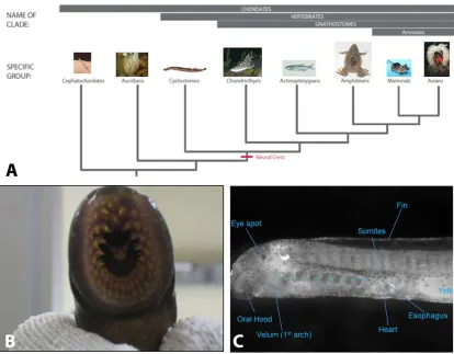
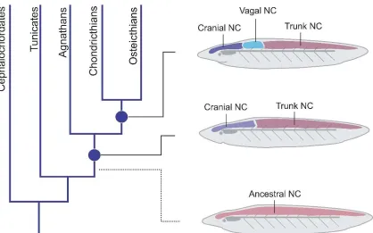
The chordate phylum can be separated into 3 subphyla: Urochordates (which includes tunicates, like Ciona intestinalis), Cephalochordates (e.g. amphioxus), and the vertebrates (Figure 1A). These subphylla all share common traits such as a notochord, hollow dorsal nerve cord, pharyngeal openings, endostyle, and a post-anal tail. Vertebrates can be further separated into jawed (agnathostomes) and jawless vertebrates (gnathostomes). The neural crest emerged from a vertebrate ancestor prior to this key separation. The sea lamprey (Petromyzon marinus), an extant jawless vertebrate, provides a unique opportunity for studying the transition from jawless to jawed vertebrates during early vertebrate evolution. Though lamprey possess many neural crest derivatives, they lack some key characteristics that are seen in jawed vertebrates such as an opposable jaw, a sympathetic nervous system, and a ventricular septum (Figure 1B). It has been postulated by “the new head hypothesis” that the invention of neural crest and placodes allowed early vertebrates to transition from being filter feeders to a larger predatory role by providing a complex craniofacial skeleton, muscularization of the gill apparatus for larger oxygen consumption, and a more complex peripheral sensory system (Glenn Northcutt 2005) (Figure1C).
development, the two-cell period lasts for approximately an hour and a half, providing sufficient time to quickly introduce the desired construct or treatment. Despite these advantages, some tradeoffs are made in lamprey. They only breed during the summer, have few identified tissue specific promoters, high levels of auto-florescence of the embryo due to high yolk content, and have few functioning antibodies due to lack cross reactivity. Nevertheless, the benefits of the knowledge that this ancient organism can shed on neural crest evolution are priceless and essential to understanding the role of neural crest.
Conservation of the Neural Crest Gene Regulatory Network in Vertebrates
al. 2010). Non-vertebrate chordates do not have bona fide neural crest but do have many of the transcription factors within their genomes, although these often differ in their spatial and temporal expression. In the Amphioxus, only the neural crest specifier gene Snail, but not other neural crest specifiers, is expressed in the dorsal neural tube in contrast to the other neural crest specifier genes (Meulemans and Bronner-Fraser 2004). Ascidians have some population of cells that express some neural plate and neural crest specifiers genes (Jeffery, Strickler et al. 2004, Abitua, Wagner et al. 2012) and migrate from the central nervous system (Stockli et al., 2015), perhaps representing a cell population that is transitional. In lamprey, many of these genes of the NC GRN have conserved function with only small differences in temporal expression. The exception is the late neural crest specifier genes Twist and Ets1, which are not expressed in lamprey premigratory neural crest, but only at later stages. Thus, it appears as though the proximal GRN elements were in place in the vertebrate common ancestor, and have been highly conserved across all vertebrates (Sauka-Spengler, Meulemans et al. 2007), with some small regulatory changes that may account for some differences in select derivatives. However, no invertebrate chordate has multipotent neural crest cells and they lack a regulatory cascade that includes Sox family members.
Essential role of Sox genes in Growth and Development
attention has been paid to their role in early embryonic development in any species, likely due to the fatal nature of early knockout studies. If examined earlier in both jawless and jawed vertebrates, these genes may play a role in regulation of neural crest specification.
By comparing gene function between jawed and jawless vertebrates, we can gain insight into their evolutionary history and origin via duplication events. Through Ohno’s 2R hypothesis, gnathostomes are thought to have undergone two rounds of genome wide duplication, at least one and possibly both of which are thought to have occurred prior to the divergence of jawed and jawless vertebrates (Dehal and Boore 2005). With gene duplications, a redundant copy of the gene is generated, allowing for modification and a potential for a greater genetic diversity. From these duplicated genes, selective pressure results in possible emergence of new functions (neofunctionalization), redistribution of functionality (subfunctionalization), or loss of the gene from the genome. It is thought that many paralog genes that were duplicated either through genome wide duplication or gene specific duplications have been lost, with only a subset retained. Therefore, not all genes in mammals will contain four paralogs. Given their importance, the Sox Family represents a prime candidate family to study duplication events prior to the separation of jawed and jawless vertebrates.
Neural Crest Regionalization in Vertebrates
arches 1 through 6 are derived from neural crest and mesoderm. Neural crest cells from rhombomeres 1 and 2 contribute to the cartilage, bone, and sensory elements of cranial nerves V, VII, IX, and X. The neural crest of the first branchial arch contributes to Meckel’s cartilage, the mandible, malleus, incus, and the sphenomandibular ligament, and cranial nerves V2/V3. In Treacher Collins Syndrome, the neural crest of the first branchial arch fails
to migrate, leading to mandibular hypoplasia and facial abnormalities, such as cleft palate and cleft lip. Trunk neural crest, which migrate along a dorsolateral route to form melancoytes and ventral pathways to contribute to neuronal and glial lineages, share some common neural crest derivatives with other axial levels. However, in vivo, grafts from the trunk level to cranial regions have demonstrated that trunk neural crest lack the ability to produce skeletal elements except under extremely long culture conditions (Le Douarin and Teillet 1973, McGonnell and Graham 2002). Thus, understanding the differences in regulatory elements of the cranial regions against the trunk may be essential in understanding skeletal and craniofacial development.
Neural crest contributions to the Enteric Nervous System in Vertebrates
Recently, in addition to the vagal neural crest, a population of neural crest cells from the trunk referred to as “Schwann cell precursors” (SCP) has been shown to contribute to a subpopulation of neurons in the gut (Uesaka et al., 2015). SCPs were first identified associated with peripheral nerves and contributing post-embryonically to melanocytes and parasympathetic ganglia (Espinosa-Medina, Outin et al. 2014). During ENS development, trunk neural crest-derived SCP migrate along spinal nerves and contribute to a minor portion (~20%) of enteric neurons (Uesaka, Nagashimada et al. 2015). These cells are Ret negative and contribute to submucosal plexuses in both the small intestine and large intestine while only in the submucosal plexuses of the small intestine. All chordates have an enteric nervous system but lamprey are the first chordate to have neural crest cells.
enteric nervous system. We speculate that these SCPs may represent an intermediate cell type in enteric nervous system formation. Finally, I explored the lamprey genome and examined the conservation of an important gene family involved in neural crest specification and migration, the Sox gene family. I found a conservation of many of these genes between lamprey and other vertebrates. In addition, the SoxC family serves a key role in early neural crest specification. Using these data, I have made inroads in understanding the evolution of neural crest as it emerges in vertebrates and transitions to jawed vertebrates.
Figure 1: Lamprey as a model organism:
C h a p t e r I I
C h a p t e r 2 - 1
EXPRESSION OF SYMPATHETIC NERVOUS SYSTEM GENES IN LAMPREY SUGGESTS THEIR RECRUITMENT FOR SPECIFICATION OF A NEW
VERTEBRATE FEATURE
Daniela Häming, Marcos Simoes-Costa, Benjamin Uy, Jonathan Valencia, Tatjana Sauka-Spengler, and Marianne Bronner-Fraser
Abstract
The sea lamprey is a basal, jawless vertebrate that possesses many neural crest derivatives, but lacks jaws and sympathetic ganglia. This raises the possibility that the factors involved in sympathetic neuron differentiation were either a gnathostome innovation or already present in lamprey, but serving different purposes. To distinguish between these possibilities, we isolated lamprey homologues of transcription factors associated with peripheral ganglion formation and examined their deployment in lamprey embryos. We further performed DiI labeling of the neural tube combined with neuronal markers to test if neural crest-derived cells migrate to and differentiate in sites colonized by sympathetic ganglia in jawed vertebrates. Consistent with previous anatomical data in adults, our results in lamprey embryos reveal that neural crest cells fail to migrate
Introduction
Lampreys are agnathans (jawless vertebrates) that have many essential vertebrate characteristics but lack the sympathetic nervous system and jaws. Morphologically, they resemble Cambrian era fossils [1], [2], suggesting a resemblance to the common ancestor of jawless (Agnatha) and jawed (Gnathostomata) vertebrates. Lamprey and hagfish, the only modern agnathans, are likely to be monophyletic, though there remains controversy on this point [3], [4]. As basal vertebrates, both occupy a critical phylogenetic position for understanding emergence of vertebrate traits. However, lamprey offers a significant advantage for developmental studies due of the accessibility and ease of obtaining embryos for experimental manipulation.
Neural crest cells are one of the defining features of vertebrates. This population of multipotent cells gives rise to a variety of different tissues and cell types including cartilage and bone of the facial skeleton, pigment cells, and sensory and peripheral ganglia, among other derivatives [5], [4]. The peripheral nervous system of jawed vertebrates is composed of sensory, parasympathetic, sympathetic, and enteric ganglia that form clusters of neurons that innervate peripheral structures and relay information back to the central nervous system. All of these sensory and autonomic ganglia are derived from the neural crest, together with a contribution of cranial placodes to the sensory ganglia of the head [6].
gene regulatory network underlying the formation and differentiation of neural crest cells is remarkably similar to higher vertebrates [10]–[12]. Interestingly, however, lampreys lack some key neural crest structures including dentine, bone and sympathetic neurons.
The sympathetic nervous system is a branch of the autonomic nervous system, responsible for the physiological modulation of inner organs in the absence of conscious control by the central nervous system (CNS). Lampreys and hagfishes lack the chain of sympathetic ganglia chain observed in gnathostomes [13]. Instead, their sympathetic innervation comes from preganglionic fibers that extend directly to the terminal plexus, similar to what is observed in amphioxus [14]. Nevertheless, lamprey and hagfish have been reported to have scattered chromaffin-like cells along blood vessels, the heart and cloaca [15]. Although these cells have been described as analogous to postganglionic neurons [14], it is not yet clear if they connect with the central nervous system and/or represent an evolutionary precursor to the gnathostome sympathetic nervous system [15].
There are a handful of characteristic markers for sympathetic neurons, including Phox2b, Ascl1 (Ash1), and dHand (hand2). Phox2b is a homeodomain transcription factor expressed in several types of neurons in the developing nervous system [16]. The bHLH transcription factor achaete-scute homolog 1 (Ascl1 formerly ash1) is a proneural gene that influences neuronal fate. Ascl1 is expressed in some domains of the
neuroepithelium of the forebrain and in precursors of sympathetic and enteric
development [18]. Here, we asked whether this suite of genes exists in lamprey and if so, where they were expressed.
To address this question, we isolated lamprey homologues of these genes and examined their expression patterns in embryos by in situ hybridization at various stages of development. The results show that all three genes are individually found in different areas of the head with only Phox2 expressed in cells at the trunk level. DiI labeling of presumptive neural crest cells failed to show a neural crest contribution to sites where sympathetic ganglia would be expected to coalesce. In contrast, DiI labeled neural crest cells contributed to both dorsal root ganglia and enteric ganglion cells of the gut. Taken together, the results raise the intriguing possibility that the transcriptional program responsible for migration to and/or differentiation within the site of sympathetic ganglion formation in gnathostomes was assembled through the recruitment of Phox2b, dHand and Ascl1 by precursors derived from the neural crest.
Results
While it has been established that lamprey lack an organized sympathetic nervous system [14], [19], some studies suggest that scattered sympathetic neurons and
chromaffin cells might be associated with blood vessels, hindgut, cloaca, and kidneys [15]. Furthermore, it has been suggested that lamprey have sympathetic
mapped the expression pattern of markers characteristic of differentiating sympathetic neurons.
DiI and neurofilament labeling of trunk neural crest in lamprey
In the trunk region of gnathostomes, neural crest precursors to sensory and sympathetic ganglia migrate from the dorsal neural tube along a ventral pathway to coalesce either next to the neural tube, to form dorsal root ganglia, or further ventrally adjacent to the dorsal aorta, to form sympathetic ganglia.
To test whether lamprey neural crest cells migrate ventrally to contribute to sensory and/or sympathetic ganglia as in gnathostomes, we first performed focal injections of the lipophilic dye, DiI, into the dorsal neural tube at trunk levels. In
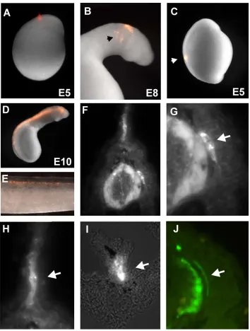
lamprey, the neural tube forms by secondary neurulation, where a solid rod-like structure transforms into a tube whose lumen forms by cavitation. At embryonic day 5, the neural rod elevates, gradually detaching from the dorsal epithelium. The first indication of neural crest precursors occurs at this time. The head morphologically extends and becomes visible at day 6, concomitant with cavitation. Neural crest primordia at this stage appear as bulges on the dorsal aspect of the newly formed neural tube [21]. Focal injections of DiI performed at any level of the trunk dorsal neural rod prior to cavitation (day 5.5 to day 6) (Figure 1A) failed to give rise to any migrating neural crest. In
Accordingly, we performed DiI-labeling at later times, after cavitation at day 6.5 to day 7, by injecting dye into the lumen of the neural tube (Figure 1D). This approach resulted in labeling of migrating neural crest cells. In embryos receiving neural tube injections at day 6.5 to 7 and examined through day 34, the labeled cells contributed to several neural crest derivatives at trunk and vagal levels (Figure 1E). These include dorsal root ganglia (DRGs) (Figure 1F and 1G) and the mesenchymal cells of the fin (Figure 1H), and enteric ganglia (Figure 1I). However, no structures resembling sympathetic ganglia were observed at any stage. These results suggest that lamprey neural crest cells contribute to dorsal root ganglia but fail to condense into sympathetic-like structures during embryonic development.
To examine neuronal differentiation, we performed immunostaining using
Expression pattern of transcription factors associated with sympathetic neuron differentiation in the lamprey
In gnathostomes, several transcription factors have been implicated in
sympathetic nervous system formation. These include the basic-helix-loop helix factors, dHand and Ascl1, as well as the homeodomain transcription factor Phox2b. To examine the presence and deployment of these lamprey genes during neural crest development, we cloned fragments of lamprey Hand, Phox2 and Ascl1 orthologues using 5′RACE and determined their expression patterns by in situ hybridization.
Phox2 transcripts also were detected in cells surrounding the yolk (E14, arrow onFigure 2G) and in the notochord.
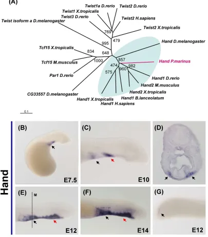
The Hand ortholog isolated from lamprey clusters to the base of the branch that contains both the D-Hand and E-hand gnathostome genes in our phylogenetic analysis (Figure 3A). Lamprey Hand is first observed at day 5 in the bilateral precursors that form the cardiac field (data not shown). From day 7 to day 10, there is additional staining visible in the anterior portion of the ventral mesenchyme (Figure 3D and 3F). At day 12, in
addition to the heart, the entire ventral mesenchyme that surrounds the endostyle and the notochord expresses Hand (Figure 3D and 3F). Hand transcripts were also detected at day 14 in the cardiac ganglia (Figure 3D and 3E) and in the posterior mesoderm (Figure 3G).
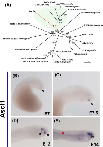
The lamprey Ascl1 fragment was within the branch that contains the Ascl1 gnathostome orthologues (Figure 4A). Expression of Ascl1 was first noted at day 6, with very faint expression observed in the pituitary gland (Figure 1B). This pattern was
maintained at day 7, accompanied by additional expression in the lens (Figure 4C). By day 10, Ascl1 was upregulated in the anterior lip mesoderm (day 10) and by day 12, transcripts also were detected in the VI cranial ganglion (Figure 4D). From day 11 onward, Ascl1 also was expressed faintly in the notochord (Figure 4G).
Hand, Ascl1 and Phox2 were co-expressed. Our results, in concert with the neurofilament and DiI data, demonstrate that the lamprey does not possess a sympathetic nervous system analogous to that in gnathostomes. We were also unable to identify any cells that might represent precursors to sympathetic postganglionic neurons of cyclostomes.
Discussion
We analyzed the expression patterns of three known marker genes of the
gnathostome sympathetic nervous system for their deployment in lamprey embryos. At 9 and 11 days after fertilization, we found expression of Hand, Ascl1 and Phox2 in the head region, with only Phox2 showing additional expression in a stream of cells that migrate toward the trunk. These cells may correspond to lateral line cells derived from ectodermal placodes. In ganglia, we see only Hand expressed in the cardiac ganglia and Phox2 in the epibranchial ganglia.
Interestingly, there was no overlapping expression of these “sympathetic” genes in any domain in the embryo. Consistent with this marker analysis, we found no
In gnathostomes, Ascl1 is generally required for development of autonomic neurons, with expression initiating earlier than Phox2b. Phox2b is also required for autonomic neurogenesis and, in a feedback loop, is required for maintenance of Ascl1 expression [22]. Ascl1 induces expression of pan-neuronal genes in neural crest precursor cells of the peripheral nervous system, but does not specify subtype specific expression of tyrosine hydroxylase (TH) or dopamine-β-hydroxylase (DBH), the enzymes responsible for the catalyzing synthesis of the neurotransmitter, nor-epinephrine [23]. Loss-of-function of dHand, another determinant of the sympathetic lineage, blocks neural crest cell differentiation into noradrenergic neurons, whereas its over-expression upregulates Phox2b, TH and DBH. Expression of dHand depends on Phox2b, but not Ascl1 [22].
Classical literature regarding the lamprey sympathetic nervous system is scarce and contradictory. Lampreys possess several neurotransmitters, including acetylcholine and noradrenaline, in the central nervous system. In addition, sequenced fragments from lamprey DNA reveal the presence of two adrenergic receptors [24] that, in gnathostomes, are the most abundant in the sympathetic nervous system. While this suggests the
SIF cells and chromaffin cells are closely related, and SIF cells are considered to be the intermediate in morphology between chromaffin cells and sympathetic neurons [28]. One intriguing possibility is that there may have been a shift from chromaffin and SIF cells to sympathetic neurons during gnathostome evolution, such that chromaffin and SIF cells represent the evolutionary precursor to sympathetic neurons. These three cell types are closely related lineage-wise, sharing a sympathoadrenal progenitor which co-expresses markers characteristic of both chromaffin cells and sympathetic neurons [28], [29].
Our DiI labeling experiments show a discrete neural crest stream that migrates towards the heart (the cardiac neural crest; data not shown) which could be the source of the progenitors that give rise to the cardiac SIF cell aggregation [26]. Indeed, there is expression of Hand in ganglia adjacent to the heart (figure 3F), although Phox2 is not present in the same structures. However, at the stages examined, we failed to observe migratory precursors that might give rise to the scattered chromaffin cells that are said to occur throughout the lamprey body.
Phox2, Hand and Ascl1 into a new gene battery allowed for the emergence of this new neural crest derivative.
Materials and Methods
DiI labeling in lamprey embryos
5 –7 day old embryos were dechorionated in 0.1xMMR and placed into agarose-coated petridishes. DiI solution (0.5 µg/µl, prepared in 0.3M sucrose) was filled into glass needles and injected into distinct neural crest population or into the entire neural tube. Embryos were analyzed for the injection into a discrete location or into the neural tube by fluorescence microscopy. Embryos were raised in petri dishes containing 0.1×MMR at 18°C and the migration of DiI stained cells was analyzed every day. Once the embryos had reached the desired stage they were fixed using 4%Paraformaldehyde in PBS at room temperature for 1hr.
Obtention of lamprey orthologs through 5′ Rapid Amplification of cDNA ends (RLM-5′ RACE)
Orthologs of Phox2b, dHand and Asc1 were identified by bioinformatic survey of the lamprey genomic sequences and cloned using RACE. Total RNA was extracted from embryos using the Ambion: RNAquous kit. RLM-5′ RACE was conducted on the total mRNA in accordance with Invitrogen: GeneRacer Kit. Total RNA was dephosphorylated through Calf Intestinal Phosphatase (CIP) treatment, decapped via Tobacco Acid
transcribed using random hexamer priming to form the cDNA template. The genes of interest were amplified using touch down PCR and cloned with TOPO TA Cloning Kit (Invitrogen).
Immunostaining of lamprey embryos
Immunostaining of lamprey embryos was performed as previously described [30]. Neurofilament (NF-M) antibody was used 1∶200 in blocking solution. As a secondary
antibody Alexa 488 anti mouse IgG2a was used 1∶1000 in blocking solution. Sections were degelatinized in 42°C PBS for 10 minutes and washed with PBSTr 2 times for 5 minutes. Afterwards the sections were blocked in 10% goat serum in PBSTr at 4°C for 5 hrs. The blocked sections were incubated overnight at 4°C with the neurofilament (NF-M) antibody 1∶200 in blocking solution. To remove unbound antibody the sections were washed 5 times for 10 minutes with PBSTr. As a secondary antibody Alexa 488 anti mouse IgG2a 1∶1000 in blocking solution was used for 2 hrs at room temperature. Unbound secondary antibody was washed of 3 times for 10 minutes in PBSTr followed by 2 washes for 10 minutes in PBS. For mounting, sections were dipped into distilled water a few times and mounted with Permaflour.
Whole-mount in situ hybridization on lamprey embryos
Whole-mount lamprey in-situ was performed as previously described [21].
Embedding and sectioning of lamprey embryos
Lamprey embryos were washed with PBSTr 3 times 15 min. Subsequently they were incubated in 15% sucrose in PBS for 3 hrs at room temperature, 7.5% gelatin and 15% sucrose for 12 at 37°C, and 20% gelatin for 4 hrs at 37°C. Subsequently embryos were positioned in 20% gelatin and frozen with liquid nitrogen. Embryos were sectioned at 8 – 10 µm with a Microm HM550 cryostat.
Phylogenetic Analysis
Alignments were built with the coding sequences retrieved from GenBank. Neighbor Joining (NJ) tree were constructed using ClustalX protocol from the DNA STAR package. The trees were visualized using Tree View v. 0.5.0.
Author Contributions
Figure 1. DiI labeling of lamprey neural crest cells reveals absence of sympathetic ganglia during embryonic development.
(A) Focal injections of DiI in the lamprey neural tube at day 5 result in labeling of migrating cephalic neural crest (arrow in B). However, focal injections into the posterior neural tube (C) fail to label trunk neural crest cells. D) Filling the lumen of the neural tube with DiI after cavitation produces labeled trunk neural crest cells in several neural crest derivatives (E). A section through an injected embryo (F) shows labeling of the dorsal root ganglia (DRG, arrow in G), the
Figure 2. Expression of the transcription factor Phox2 in embryos of P. marinus.
Figure 3. Expression of the helix-loop-helix transcription factor Hand in lamprey embryos. (A) Phylogenetic analysis suggests lamprey have one ortholog of both the dHand and eHand gnathostome genes. (B) Lamprey Hand expression is first observed in the cardiac field, and is conspicuous after 7 days of development. (B) At day 10, two domains of expression are clearly present: the heart (black arrow) and a part of the anterior mesenchyme (red arrow). (C)
Figure 4. Expression of Ascl1 during embryonic development of the lamprey.
References
1. Gess RW, Coates MI, Rubidge BS (2006) A lamprey from the Devonian period of South Africa. Nature 443: 981–984.
2. Janvier P (2006) Palaeontology: modern look for ancient lamprey. Nature 443: 921– 924.
3. Kuraku S, Hoshiyama D, Katoh K, Suga H, Miyata T (1999) Monophyly of lampreys and hagfishes supported by nuclear DNA-coded genes. Journal of molecular evolution 49: 729–735.
4. Osorio J, Retaux S (2008) The lamprey in evolutionary studies. Development Genes and Evolution 218: 221–235.
5. Le Douarin N (1982) The neural crest. Cambridge University Press.
6. D'Amico-Martel A, Noden DM (1983) Contributions of placodal and neural crest cells to avian cranial peripheral ganglia. Am J Anat 166: 445–468.
7. McCauley DW, Bronner-Fraser M (2006) Importance of SoxE in neural crest development and the evolution of the pharynx. Nature 441: 750–752.
8. Northcutt RG (1996) The agnathan ark: The origin of craniate brains. Brain Behavior and Evolution 48: 237–247.
9. Braun CB (1996) The sensory biology of the living jawless fishes: a phylogenetic assessment. Brain Behav Evol 48: 262–276.
11.Sauka-Spengler T, Bronner-Fraser M (2008) Evolution of the neural crest viewed from a gene regulatory perspective. Genesis 46: 673–682.
12.Nikitina N, Sauka-Spengler T, Bronner-Fraser M (2008) Dissecting early regulatory relationships in the lamprey neural crest gene network. Proceedings of the National Academy of Sciences of the United States of America 105: 20083–20088.
13.Nicol JAC (1952) Autonomic Nervous Systems in Lower Chordates. Biological Reviews of the Cambridge Philosophical Society 27: 1–50.
14.Johnels AG (1956) Lampetra planeri. On the peripheral autonomic nervous system of the trunk region of. Acta Zool. 37. : 251–286.
15.Gibbins I (1994) Comparative anatomy and evolution of the autonomic nervous system. In: Nilsson S, Holmgren S, editors. Comparative Physiology and Evolution of the Autonomic Nervous system: Academic Publishers. pp. 1–67.
16.Pattyn A, Morin X, Cremer H, Goridis C, Brunet JF (1997) Expression and interactions of the two closely related homeobox genes Phox2a and Phox2b during neurogenesis. Development 124: 4065–4075.
17.Lo LC, Johnson JE, Wuenschell CW, Saito T, Anderson DJ (1991) Mammalian Achaete-Scute Homolog-1 Is Transiently Expressed by Spatially Restricted Subsets of Early Neuroepithelial and Neural Crest Cells. Genes & Development 5: 1524–1537. 18.Schmidt M, Lin SY, Pape M, Ernsberger U, Stanke M, et al. (2009) The bHLH
transcription factor Hand2 is essential for the maintenance of noradrenergic properties in differentiated sympathetic neurons. Developmental Biology 329: 191–200.
20.Gobyrin VA, Leonteva GR (1965) Chromataffin Tissue and Sources of Catecholamines in Vertebrate Heart. Bulletin of Experimental Biology and Medicine. Ussr 59. : 92–&. 21.Sauka-Spengler T, Meulemans D, Jones M, Bronner-Fraser M (2007) Ancient
evolutionary origin of the neural crest gene regulatory network. Developmental cell 13: 405–420.
22.Rychlik JL, Hsieh M, Eiden LE, Lewis EJ (2005) Phox2 and dHAND transcription factors select shared and unique target genes in the noradrenergic cell type. Journal of Molecular Neuroscience 27: 281–292.
23.Guillemot F, Lo LC, Johnson JE, Auerbach A, Anderson DJ, et al. (1993) Mammalian Achaete-Scute Homolog-1 Is Required for the Early Development of Olfactory and Autonomic Neurons. Cell 75: 463–476.
24.Scofield MA, Deupree JD, Bylund DB (2002) Adrenergic receptor genes - cDNA and genomic library construction. Molecular Biotechnology 21: 171–197.
25.Owsiannikof T (1883) Über das sympatische Nervensystem der Flussneunaugen, nebst einigen histologischen Notizen über andere Gewebe desselben Thieres. Bull Acad Sci St-Petersb 28: 440–448.
26.Lignon JM (1979) Responses to Sympathetic Drugs in the Ammocoete Heart -
Probable Influence of the Small Intensely Fluorescent (Sif) Cells. Journal of Molecular and Cellular Cardiology 11: 447–465.
27.Burnstock G (1969) Evolution of the autonomic innervation of visceral and cardiovascular systems in vertebrates. Pharmacological reviews 21: 247–324.
29.Anderson DJ, Axel R (1986) A bipotential neuroendocrine precursor whose choice of cell fate is determined by NGF and glucocorticoids. Cell 47: 1079–1090.
C h a p t e r 2 - 2
PROGRESSIVE REGIONALIZATION OF THE NEURAL CREST DURING VERTEBRATE EVOLUTION VIA ELABORATION OF GENE REGULATORY
CIRCUITRY
Abstract
The neural crest of jawed vertebrates is divided into discrete subpopulations— cranial, vagal, trunk, and lumbosacral—distributed along the embryonic body axis with each following migratory pathways and forming a different set of derivatives. To gain insight into how these different neural crest territories may have arisen during vertebrate evolution, we analyzed the fate, behavior, and molecular identity of neural crest cells along the body axis of the sea lamprey (Petromyzon marinus), a basal jawless vertebrate. DiI fate-mapping revealed the presence of distinct cranial and trunk neural crest subpopulations, with a sharp transition occurring between them in the neural tube caudal to approximately somite 7. In contrast to jawed vertebrates, no intermediate population was apparent. We next performed comparative transcriptome analysis of lamprey cranial and trunk neural crest cells. The results revealed fewer cranial/trunk differences than observed in amniotes and several missing cranial crest-specific transcription factors. These results suggest that, in addition to the deeply conserved pan-vertebrate core neural crest gene regulatory network, there has been extensive elaboration of axial level specific regulatory circuits, likely contributing to the expansion of neural crest derived cell types during evolution of jawed vertebrates.
Introduction
Evolution of vertebrates is intimately linked to the advent of the neural crest, a multipotent embryonic stem cell population. Neural crest cells give rise to many defining vertebrate characters, including the craniofacial skeleton, skull, and peripheral ganglia (Green et al., 2015). In fact, according to the “New Head” hypothesis, acquisition of the neural crest in vertebrates facilitated active predation, thus enabling elaboration of the brain (Gans and Northcutt, 1983). Neural crest cells arise from the dorsal midline of the central nervous system, but subsequently leave the neural tube and migrate extensively to various locations. Upon arrival at their destinations, they differentiate into a wide range of derivatives that range from neurons and glia of the peripheral nervous system to pigment cells as well as bone and cartilage of the face (Simoes-Costa and Bronner, 2015).
cells. Finally, lumbosacral neural crest cells contribute to enteric glia of the rectum and portions of the posterior colon. Neural crest cells at all axial levels form melanocytes (Le Douarin 1982).
A pan-vertebrate neural crest (NC) gene regulatory network (GRN), invoking sequential deployment of signaling and transcriptional events, has been proposed to be responsible for formation of this unique cell type (Meulemans et al., 2004; Simoes-Costa and Bronner, 2015). Primarily studied at cranial levels, the NC GRN appears to be largely conserved across vertebrates (Sauka-Spengler et al., 2007), including the basal lamprey, a jawless vertebrate (agnathan). However, differences were noted in the position of key specifier genes, like cranial neural crest markers Ets1 and Twist, which seem to be deployed later in the lamprey GRN than in other vertebrates (Sauka-Spengler et al., 2007). This suggests that there may be significant regulatory differences underlying the molecular machinery controlling neural crest identity in agnathans and gnathostomes.
Results
Axial differences in the lamprey neural crest revealed by DiI fate-mapping
In lamprey, the neural tube forms by cavitation, similar to teleosts, transforming from a neural rod into a tube with a lumen. At embryonic day (E)5 (Tahara 20), the neural rod elevates, neural crest precursors arise and the head begins to extend. Cavitation initiates around E6 (T21), at which time the neural crest primordia appear as bulges on the dorsal neural tube.
To characterize the neural crest subpopulations in the lamprey, we fate mapped the premigratory neural crest along the anteroposterior axis of the embryo. A single focal spot of DiI was introduced into the dorsal midline at different positions along the body axis at E5 to E5.5 (T19-T20) (Figure 1A-C). Labelled embryos were then observed at different stages of development to follow the patterns of neural crest migration and differentiation into various derivatives. Consistent with previous reports (McCauley and Bronner-Fraser 2003, Haming, Simoes-Costa et al. 2011, Modrell, Hockman et al. 2014), we found that the cranial crest contributed to the head mesenchyme and cranial ganglia (Figure 1D), while the trunk neural crest differentiated into neurons of the dorsal root ganglia and mesenchymal cells of the dorsal fin (Figure 1E).
(Bronner-Fraser, 1993, Serbedzija 1992 et al., 1992, Selleck et al.,1993) (n = 32). DiI labeled cells possessed neurofilament-M labeled neuronal projections that were observed in the trigeminal ganglion, again similar to results in gnathostomes (Figure S1: E-H’).
We next analyzed whether at later stages, DiI labeled cells contributed to heart or neurons of the gut, as they do in gnathostomes. Despite hundreds of focal injections into the neural tube adjacent to approximately somites 1-3, no labeled cells were observed in the vicinity of the heart or aortic vessels. For focal injections into the dorsal neural tube adjacent to the region of approximately somites 4-7, we observed DiI-labeled cells within the branchial arches, but in no cases were these observed to enter the heart or gut.
In order to label trunk neural crest populations, we performed focal DiI injections in the dorsal neural tube adjacent to somites 8-30 at E5-E6.5. This resulted in labeling a small number of migrating trunk neural crest cells. Embryos were sectioned at E14 through E21 to look at the distribution of DiI-labeled cells. At all stages examined, the results were similar. We noted that a small number of cells had migrated and/or delaminated from the neural tube to form the dorsal root ganglia (Figure 1B & D). In addition, mesenchymal cells were observed in the dorsal fin, similar to their distribution pattern in frog and fish embryos (Collazo et al., 1993; Smith et al., 1994).
Partial conservation of cranial neural crest gene regulatory network in agnathans Our results suggest a clear distinction between the cranial and trunk subpopulations, with no obvious intervening population. Similarly, we failed to detect a “lumbosacral” neural crest population in lamprey. Thus, the ancestral state in stem vertebrates may been only to have distinct cranial and trunk neural crest populations. While our fate map analysis highlights important differences between the cranial and trunk neural crest cells, previous studies have shown that the lamprey lacks expression of cranial neural crest genes, such as Ets1 and Twist (Sauka-Spengler et al., 2008), in the migratory population. This raises the intriguing possibility that important changes in the molecular mechanisms controlling cranial neural crest development may have accompanied early evolution of jawed vertebrates. To explore this hypothesis, we surveyed the conservation of the cranial neural crest gene regulatory network in the lamprey.
migratory cranial neural crest (Figure 2B), a large number of cranial regulators appear to be absent from this cell population. In contrast to what has been described in amniotes, we were unable to detect genes like Brn3c, Lhx5, Dmbx1 in the cranial neural crest or anterior neural folds (Figure 2B). Although present in the genome, lamprey cranial neural crest cells appear to lack a large part of the cranial-specific regulatory circuit (Figure 2C), pointing to a high degree of regulatory divergence between the regulatory states of jawed and jawless neural crest.
The analysis of expression patterns of cranial genes in agnathans can provide important clues on how this regulatory sub-circuit evolved. Intriguingly, we found that genes from the cranial circuit that are absent from the early neural crest are co-expressed at later stages in the branchial arches. As previously described by Sauka-Spengler and colleagues (2008), Ets1 is transcribed by the differentiating post migratory branchial arches neural crest, cranial placodes, and dorsal root ganglia. Our analysis shows that transcripts of both Lhx5 and Dmbx1 are also present in the post migratory branchial arches neural crest (Figure 2D), raising the possibility that terminal regulatory circuits might have been re-deployed to play an earlier role in neural crest specification in gnathostomes. According to this scenario, genes involved in the differentiation of the neural crest in agnathans may have been co-opted by the specification program in gnathostomes (Green et al., 2015), possibly endowing the cranial neural crest with novel morphogenetic features.
Transcriptome analysis of the lamprey cranial neural crest
vertebrate evolution. To further investigate this hypothesis, we conducted an unbiased analysis of the lamprey cranial neural crest through comparative transcriptome analysis of cranial and trunk neural crest subpopulations.
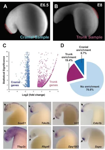
To obtain premigratory neural crest populations, we microdissected segments of cranial and trunk dorsal neural tubes at stages E6.5 and E8, respectively (Figure 3A-B), and extracted total RNA from these samples for transcriptome analysis. In this manner, we identified 811 genes significantly enriched in lamprey cranial neural crest (Figure 3C) when compared to the trunk. Consistent with our previous in situ hybridization analysis (Figure 2B), cranial genes in the cranial dataset included transcription factors Id2 (Id), Sox8 (SoxE1), and Tfap2b (Tfap2c). This indicated that our dataset was suitable for identification of cranial specific genes in the lamprey, and accordingly we employed it to conduct two types of analysis. First, we took a broader look at the conservation of vertebrate cranial neural crest genes in the lamprey by comparing our dataset with an amniote transcriptome. Second, we investigated the possibility that the lamprey has a cranial neural crest regulatory program that is distinct from those characterized in jawed vertebrates.
previous results showing low conservation of the cranial neural crest regulatory program in agnathans compared with gnathostomes.
Finally, we investigated the hypothesis that the lamprey might have a different set of cranial regulators involved in neural crest formation. To accomplish this, we examined the expression patterns of the 20 genes that were most enriched in our cranial neural crest dataset. Included in this group are Sox8 (SoxE1), Tfap2b (Tfap2c) (Figure 3E-F), and Id2 (Id), which are strongly expressed in the lamprey neural crest. However, we found that the other genes in that category, such as Tdo2b, Rbp4l, Vil1, Dpy19l3, Cdo1b, and Sspo, were indeed strongly restricted to the cranial regions, but absent from the neural crest (Figure 3G-L), being expressed in the central nervous system. Thus, we were unable to identify cranial neural crest genes that were lamprey specific. Taken together, these results support the hypothesis that agnathans possess a simpler cranial neural crest gene regulatory network, which might have important implications for the evolution of the neural crest subpopulations.
Discussion
the rostrocaudal body axis. To better understand how these subpopulations may have evolved, we also scrutinized the regulatory network of the lamprey cranial neural crest.
Our results confirm that cranial neural crest migratory pathways are similar to those observed in jawed vertebrates despite the lack of jaws. In contrast, posterior cranial neural crest cells arising from post-otic levels migrate ventrally and, upon reaching the branchial arches, move rostrally and caudally to occupy the mesenchyme of all arches. However, in contrast to jawed vertebrates, they fail to contribute to enter the heart/outflow tract or gut.
Focal injections of DiI into the neural tube caudal to the somite 7 level are only able to minimally label the trunk neural crest. In embryos assayed between E16 and E30, DiI labeled cells were observed in the mesenchyme of the fin, dorsal root ganglia, and along spinal nerves, and occasionally in the typhosole, but not in ventral locations corresponding to sympathetic ganglion formation. In general, the emigration of trunk neural crest cells from the neural tube occurs much later, after cavitation of the neural tube. These results suggest that, in contrast to the pre-otic neural crest in which migration pathways are highly conserved and perhaps subject to strong evolutionary constraint, there are marked differences at post-otic levels. This correlates with a lack of several critical post-post-otic neural crest derivatives like the cardiac septum, adrenal medulla, and sympathetic ganglia.
As a vertebrate innovation, the neural crest is present in all vertebrates examined to date including agnathans. In contrast, basal chordates like amphioxus completely lack neural crest cells. It has been proposed that urochordates may have some neural crest-like cells that have migratory properties and can form melanocytes (Jeffery et al., 2004). For example, it was shown that ectopic expression of Twist in a cephalic melanocyte lineage could cause this cell to become migratory, suggesting that is may represent a proto-neural crest cells (Abitua et al., 2012). Recently, a population of neurons, derived from precursors at the neural plate border that migrate along the paraxial mesoderm, appear to contribute to a type of sensory neuron in Ciona (Stofli et al., 2015), and thus possess both the ability to form peripheral neurons and to migrate, two important characteristics of the neural crest. However, only vertebrates have bona fide neural crest cells that form within the neural tube, migrate extensively and give rise to a multitude of derivatives.
our molecular analysis of the cranial neural crest reveals surprising differences in lamprey compared with gnathostome counterparts. These differences may help explain some of the evolutionary novelty that arose in the transition from jawless to jawed vertebrates. Our results show deep conservation of a few transcription factors in the lamprey cranial neural crest. SoxEs, Tfap2, and Id may be the rudiment of a larger, more complex gene regulatory network that was expanded during early vertebrate evolution with the incorporation of novel players (LMO4a, CSRNP, Brn3, Lhx5, Dmbx1, Ets1, Twist, and LZTS1) (Simoes-Costa and Bronner, under review). Our observation also shows that some of these “novel” genes are co-expressed at later stages of neural crest formation, consistent with the possibility that the elaboration of the cranial GRN might have taken place through cooption of parts of differentiation programs to earlier portions of the network.
The differences we observe in axial-specific genes contrasts with the deep conservation of the pan-neural neural crest program (Sauka-spengler et al., 2007). Thus, the pan-neural crest program was likely the ancestral molecular recipe to make neural crest, with the subsequent elaboration of the axial level specific regulatory programs conferring important differences in developmental potential to neural crest cells along the body axis. Given that lampreys lack several key neural crest derivatives, we postulate that the absence of these derivatives may be due gene regulatory differences associated with axial-level specific regulatory programs.
this ancestral state (although we cannot rule out that agnathans lost neural crest subpopulations during the course of evolution). The relative scarcity of cranial factors in the lamprey cranial neural crest might suggest that the GRN underlying this cell population has undergone extensive elaboration from a regulatory standpoint. Thus, we propose that regionalization of the neural crest, with both the emergence of the new subpopulations and the expansion of the cranial GRN, played a crucial part in vertebrate evolution, culminating in the rise of the gnathostomes.
Experimental Procedures:
DiI labeling in lamprey embryos
Embryonic day(E) 5 -7 day old embryos were manually dechorionated in 0.1xMMR and placed into agarose-coated Petri dishes. DiI solution [(Cell Tracker-CM-DiI Invitrogen) 0.5μg/μl, prepared in 0.3M sucrose] was injected into the respective regions along the rostrocaudal axis. Embryos were raised in 18mm agarose coated petri dishes containing 0.1xMMR at 18°C and the migration of DiI labeled cells were analyzed every day by fluorescence microscopy. Once the embryos had reached the desired stage, they were fixed using 4% paraformaldehyde in PBS at room temperature for 1hr or 16 hours at 4ºC.
Embedding, sectioning, and immunostaining of lamprey embryos
in 7.5% gelatin and 15% sucrose for 12 hours at 37°C. Embryos were placed into 7.5%
gelatin and 15% sucrose, positioned, and sectioned at 12-20 m with a Microm HM550 cryostat.
Immunostaining of lamprey embryos was performed as previously described (Nikitina et al, 2009). Anti-Neurofilament (NF-M, Mouse IgG2a, Invitrogen) antibody was used 1:200 and anti-type II collagen antibody supernatant (Developmental Studies Hybridoma Bank, II-II6B3, Mouse IgG1) was used at 1:10. Secondary antibodies were used at 1:500.Slides were mounted with Fluoromount-G (Southernbiotech).
In situ hybridization and histology.
Whole-mount in situ hybridization of lamprey embryos was performed using digoxigenin RNA probes according to Wilkinson (Xu et al., 1990), with following modifications: Prior to Proteinase K step, embryos equilibrated in the bleaching solution (0.5X SSC, 5% formamide, 10%H2O2), were exposed to direct light using light box for 10-15 minutes. The
concentration and the length of Proteinase K treatment (~20g/ml, 10 minutes) was the same for embryos of all stages. Hybridization and subsequent washes were carried out at 70°C in hybridization solution containing 50% formamide; 1.3X SSC; 5mM EDTA pH8.0; 200 g/ml yeast tRNA; 100g/ml heparin; 0.2% Tween-20 and 0.5% Chaps. The hybridization
Isolation and Cloning of the PCR product
Orthologs were identified by bioinformatic survey of the lamprey genomic sequences from the ensembl database. Total RNA was extracted from embryos using the Ambion: RNAquous kit. PCR was performed with Roche:GC-Rich PCR system according to protocol with a 50 C annealing temperature. LHX5 and DMBX1b were synthesized from GenScripts based on their ensembl sequences.
Extraction of the PCR product was conducted corresponding to Qiagen:QIAquick Gel Extraction Kit and cloned with Invitrogen: TOPO TA Cloning. The clones were selected against the metabolism of X-gal and the production of ß–galactosidase purified following the QIAprep spin miniprep kit and sent for sequencing. (Laragen Inc., Culver City, CA)
RNA-Seq and Transcriptome analysis
Biological samples were dissected from the cranial dorsal neural tubes of E6.5 and trunk neural tube of E8.5 lamprey embryos. Total RNA was extracted from embryos using the Ambion: RNAquous kit. RNA-Seq was performed at the Millard and Muriel Jacobs Genetics and Genomics Laboratory (California Institute of Technology, Pasadena, CA) at 35 million reads on 2 biological replicates and 1 whole embryo control for both the E6.5 cranial and E8.5 trunk neural tube samples.
1.3.1.OSX_ x86_64). DE-seq was used to calculate gene expression levels and identify differentially expressed transcripts.
Acknowledgments:
Figure 3: Transcriptome analysis comparing lamprey cranial and trunk neural crest.
Supplemental 1: Cranial Neural Crest contribute to the trigeminal ganglia. (A) Initial injection point at E5 and (B) at final stage of E16. (C) Initial injection point of an E5.5 embryo and D) final stage E16. (E-H’) At E16, DiI (red) labeling is observed in the trigeminal ganglia and transverse sections show that some cells express Neurofilament-M (Green).
References
1. Abitua, P.B., et al., Identification of a rudimentary neural crest in a non-vertebrate chordate. Nature, 2012. 492(7427): p. 104-7.
2. Bronner-Fraser, M., Mechanisms of neural crest cell migration. Bioessays, 1993. 15(4): p. 221-30.
3. Collazo, A., M. Bronner-Fraser, and S.E. Fraser, Vital dye labelling of Xenopus laevis trunk neural crest reveals multipotency and novel pathways of migration. Development, 1993. 118(2): p. 363-76.
4. Gans, C. and R.G. Northcutt, Neural crest and the origin of vertebrates: a new head. Science, 1983. 220(4594): p. 268-73.
5. Gitelman, I., Twist protein in mouse embryogenesis. Dev Biol, 1997. 189(2): p. 205-14.
6. Green, S.A., M. Simoes-Costa, and M.E. Bronner, Evolution of vertebrates as viewed from the crest. Nature, 2015. 520(7548): p. 474-82.
7. Haming, D., et al., Expression of sympathetic nervous system genes in Lamprey suggests their recruitment for specification of a new vertebrate feature. PLoS One, 2011. 6(10): p. e26543.
8. Harris, N.W., DG., In Situ Hybridisation. 1990, Cambridge University Press (CUP). 9. Horigome, N., et al., Development of Cephalic Neural Crest Cells in Embryos of Lampetra japonica, with Special Reference to the Evolution of the Jaw. Developmental Biology, 1999. 207(2): p. 287-308.
10. Jandzik, D., et al., Evolution of the new vertebrate head by co-option of an ancient chordate skeletal tissue. Nature, 2015. 518(7540): p. 534-7.
11. Jeffery, W.R., A.G. Strickler, and Y. Yamamoto, Migratory neural crest-like cells form body pigmentation in a urochordate embryo. Nature, 2004. 431(7009): p. 696-9.
12. Le Douarin, N.M., The Neural Crest. 1982.
13. Martinsen, B.J. and M. Bronner-Fraser, Neural crest specification regulated by the helix-loop-helix repressor Id2. Science, 1998. 281(5379): p. 988-91.
14. McCauley, D.W. and M. Bronner-Fraser, Neural crest contributions to the lamprey head. Development, 2003. 130(11): p. 2317-27.
15. Meulemans, D. and M. Bronner-Fraser, Gene-regulatory interactions in neural crest evolution and development. Dev Cell, 2004. 7(3): p. 291-9.
16. Modrell, M.S., D. Buckley, and C.V.H. Baker, Molecular analysis of neurogenic placode development in a basal ray-finned fish. genesis, 2011. 49(4): p. 278-294. 17. Nikitina, N., M. Bronner-Fraser, and T. Sauka-Spengler, The sea lamprey
Petromyzon marinus: a model for evolutionary and developmental biology. Cold Spring Harb Protoc, 2009. 2009(1): p. pdb emo113.
18. Sauka-Spengler, T. and M. Bronner-Fraser, Insights from a sea lamprey into the evolution of neural crest gene regulatory network. Biol Bull, 2008. 214(3): p. 303-14.
20. Sauka-Spengler, T. and M. Bronner-Fraser, A gene regulatory network orchestrates neural crest formation. Nature Reviews Molecular Cell Biology, 2008. 9(7): p. 557-568.
21. Sauka-Spengler, T., et al., Ancient Evolutionary Origin of the Neural Crest Gene Regulatory Network. Developmental Cell, 2007. 13(3): p. 405-420.
22. Selleck, M.A., T.Y. Scherson, and M. Bronner-Fraser, Origins of neural crest cell diversity. Dev Biol, 1993. 159(1): p. 1-11.
23. Serbedzija, G.N., M. Bronner-Fraser, and S.E. Fraser, Vital dye analysis of cranial neural crest cell migration in the mouse embryo. Development, 1992. 116(2): p. 297-307.
24. Simoes-Costa, M. and M.E. Bronner, Establishing neural crest identity: a gene regulatory recipe. Development, 2015. 142(2): p. 242-57.
25. Simoes-Costa, M. and M.E. Bronner, Reprogramming neural crest axial identity and cell fate. 2016.
26. Simoes-Costa, M.S., et al., Dynamic and differential regulation of stem cell factor FoxD3 in the neural crest is Encrypted in the genome. PLoS Genet, 2012. 8(12): p. e1003142.
27. Smith, M., et al., Trunk Neural Crest Origin of Caudal Fin Mesenchyme in the Zebrafish Brachydanio rerio. Proceedings of the Royal Society of London B: Biological Sciences, 1994. 256(1346): p. 137-145.
28. Stolfi, A., et al., Migratory neuronal progenitors arise from the neural plate borders in tunicates. Nature, 2015. 527(7578): p. 371-4.
C h a p t e r 3
ANCIENT EVOLUTIONARY ORIGIN OF THE VERTEBRATE
ENTERIC NERVOUS SYSTEM FROM SCHWANN CELL
PRECURSORS
Abstract
The enteric nervous system (ENS) of jawed vertebrates arises from vagal neural crest cells that migrate to the foregut and subsequently colonize and innervate the entire gastrointestinal tract. To gain insight into its evolutionary origin, we examined ENS development in the basal jawless vertebrate, the sea lamprey. Surprisingly, we found no evidence for the existence of a vagal neural crest population in the lamprey. Rather, DiI labeling showed that late-migrating cells, including those originating from the trunk neural tube and associated with nerve fibers, differentiated into neurons within the gut wall and typhlosole. We hypothesize that these trunk derived neural crest cells are homologous to Schwann cell precursors (SCPs) that have recently been shown to populate post-embryonic parasympathetic ganglia1,2, including enteric ganglia3 in mammalian embryos. Our results
The enteric nervous system is compsed of thousands of interconnected ganglia embedded within the walls of the gut4,5, making it the most complex portion of the peripheral
nervous system in amniotes. The ENS innervates the entire gastrointestinal tract to regulate muscle contraction, water balance, and gut secretions of jawed vertebrates. Classical transplantation experiments have demonstrated that the neurons and glia of the gut are largely derived from the “vagal” population of neural crest cells that arise within the post-otic portion of the hindbrain6,7. These cells emigrate from the hindbrain, enter the foregut, and undergo
the longest migration of any embryonic cell type to populate the entire length of the gut from foregut to hindgut.
The sea lamprey, Petromyzon marinus, is a jawless (agnathan) vertebrate, and a experimentally tractable representative of the cyclostomes, the sister group to all other (jawed) vertebrates, making lamprey an important model for identifying traits common throughout vertebrates. Lampreys possess migrating neural crest cells that give rise to many neural crest derivatives found in gnathostomes, including pigment cells, cartilage, sensory neurons, and glia, but they lack other neural crest-derived cell types that are present in gnathostomes like jaws and sympathetic chain ganglia8,9. Given that lamprey embryos lack
sympathetic ganglia, we sought to examine other components of the autonomic nervous system, with focus on the enteric nervous system. Adult lamprey have a simple ENS that includes ganglionated plexuses of serotonin (5-HT) producing cells10, as well as a smaller
number of catecholamine-containing neurons. However, the developmental origin of these enteric neurons is unknown.
Phox2b is expressed in enteric neurons in many jawed vertebrates8. Expression of lamprey
Phox2b is detectable along the gut from early stages [Tahara 258(T25)] but it is unclear
whether these cells will become enteric neurons. By stage T28 (E20), Phox2b expressing cells are associated with a depression in the gut which will become the typhlosole, a hematopoietic tissue that is associated with elements of the enteric nervous system (Fig. 1A-B). Differentiated neurons were first observed within the gut wall at approximately T28 or embryonic (E) day 20 (Fig. 1C-D). At this time point, we noted serotonergic neurons in more rostral portions of the gut. With time, the numbers increased, and 5-HT+ neurons were
noted progressively posteriorly, with particularly high cell numbers in the cloacal region, as reported previously11. By E30, there are approximately 80-100 neurons along the gut, in
association with the typhlosole and vagus nerve(Fig. 1E). Interestingly, differentiating neurons were often associated with nerve processes emanating ventrally from the spinal cord and along the vagus nerve that projects posteriorly along the gut (Fig. 1D). In addition to 5-HT+ neurons, we also noted the presence of 5-HT+ non-neuronal columnar cells that may
represent enterochromaffin cells (SupFig. 1A-B).
We next sought to determine the embryological origin of the neurons in the gut by performing lineage labeling with the lipophilic dye, DiI1,2. In chicken, the vagal neural crest
cells that contribute to the ENS arise from the hindbrain neural tube adjacent to somites 1-76. After exiting the neural tube, they migrate ventrally, invade the foregut, and then undergo
gnathostomes. Regardless of the exact injection site, dye labeled cells spread within the hindbrain and appeared to leave the neural tube at a single stream directly above the forming branchial arches. From this site, they progressed ventrally and then turned caudally to populate all of the branchial arches (SupFig. 2A-C), similar to previously reported migration patterns of cranial neural crest cells13,14. However, despite many focal injections throughout
the caudal hindbrain, we failed to find evidence of DiI-labeled cells entering the gut suggesting a lack of vagal crest in the lamprey. We next looked for expression of the vagal neural crest marker, ret, in lamprey, since it is required for vagal neural crest development in gnathostomes8. To this end, we cloned a lamprey ret homolog and examined its expression
pattern by in situ hybridization. Results show that lamprey ret is expressed in many parts of the embryo, including the typhlosole, but we did not observe expression in migrating neural crest cells (SupFig. 2 G-J), suggesting that enteric neurons might arise from a different cellular source.
In contrast to the vagal neural crest cells in gnathostome, migrating trunk neural crest cells normally fail to invade the immediately underlying gut due to the presence of repulsive signals like Slit that block their entry15. Recently, however, a secondary contribution to the
mammalian ENS has been uncovered that comes not from the vagal neural crest but rather from trunk neural crest-derived Schwann cell precursors (SCPs) associated with extrinsic nerves that contribute postnatally to calretinin-containing neurons of the mammalian gut3.
Intriguingly, this population persists in ret null mice3. To examine the possibility that a