Open Access
Methodology
TMA for all: a new method for the construction of tissue
microarrays without recipient paraffin block using custom-built
needles
Andréa Rodrigues Cordovil Pires*
†1, Felipe da Matta Andreiuolo
†2and
Simone Rabello de Souza
†2Address: 1Fonte Medicina Diagnóstica Ltda. and Fluminense Federal University, Rio de Janeiro, Niterói, Brazil and 2Fonte Medicina Diagnóstica
Ltda., Rio de Janeiro, Niterói, Brazil
Email: Andréa Rodrigues Cordovil Pires* - [email protected]; Felipe da Matta Andreiuolo - [email protected]; Simone Rabello de Souza - [email protected]
* Corresponding author †Equal contributors
Abstract
Background: TMAs are becoming a useful tool for research and quality control methods, mostly for immunohistochemistry and in situ hybridization.
Methods: A new technique that allows building TMA blocks with more than 300 tissue cores without using a recipient paraffin block for the tissue cores and without using a commercial TMA builder instrument is described. This technique is based on the construction of TMA needles modifying conventional hypodermic needles to punch tissue cores from donor blocks, which are attached by double-side adhesive tape on a computer-generated paper grid used to align the cores on the block mould, which is filled with liquid paraffin.
Results: More than two hundred TMA blocks were constructed using this method, utilized in immunohistochemistry and histochemistry as positive and negative controls and also in research.
Conclusion: This technique has the following advantages: it is easy to reproduce, affordable, quick and creates uniform blocks with more than 300 cores aligned, adherent and easy to cut, with negligible losses during cutting and immunohistochemistry and in situ hybridization procedures.
Background
Tissue microarrays (TMA) are becoming a useful tool for research and quality control on immunohistochemistry (IHC) and in situ hybridization methods [1-3]. The con-ventional construction of a TMA block involves the use of a commercial TMA builder instrument to punch the cores from donor blocks, and the transference of these tissue cores to a recipient block, producing blocks with even 1000 tissue cores. This technique has some disadvantages:
1. as tissue cores from different donor blocks are placed inside holes in the recipient block, to create and improve the adherence between tissue cores and the recipient block, the TMA block has to be lightly heated to melt both. During this phase cores may misalign or the fusion may not be complete, resulting in core losses;
2. the need for adhesive tape during microtome cutting by some commercial available TMA devices;
Published: 25 July 2006
Diagnostic Pathology 2006, 1:14 doi:10.1186/1746-1596-1-14
Received: 16 July 2006 Accepted: 25 July 2006
This article is available from: http://www.diagnosticpathology.org/content/1/1/14
© 2006 Pires et al; licensee BioMed Central Ltd.
3. the cores may be inserted too down in the recipient block, resulting in absence of the most profound cores in the first cuts and,
4. the commercial TMA builder instrument and its dispos-able needles are not afforddispos-able to many Pathology labora-tories worldwide.
This study was carried out in order to produce TMA blocks without the disadvantages listed above.
Methods
Needles (figure 1)
Conventional hypodermic Becton-Dickinson Preci-sionGlide® needles, 16 and 18 gauge (table 1), were
pre-pared for punching the donor blocks, as follows: we used a Dremel Multi-Pro® Rotary Tool Model 395, at low speed
(< 10,000 rpm), with a cutting wheel attached, to cut off the bevel and straighten the tip. Using the same tool, lat-eral openings (windows) were made, 1 mm away from the new tip, with 8–10 mm length and slightly wider than half the needles' width, so that the punched core could be safely removed through it. Then the external surface of the needle was sharpened with the sanding surface of the cut-ting wheel. The internal surface was also sharpened using either a shaft cone stone or a metal sharpener. Cores were manually punched out from the donor blocks, from regions selected on correspondent Hematoxylin-Eosin (H&E) slides overlaid. At this step it is important that the needle enters the block in a perpendicular way, so that the cores extracted will have a perfectly cylindrical shape. We alternatively adapted a hand-press grommet inserting machine, by means of removing the dies, and attaching one of the customized needles in their place, which ensures an even easier semi-automated movement, as well as a perpendicular angle between the needle and the donor block. The total initial cost was near USD$100.00 (table 2).
Paper grid (figure 1)
We designed a grid using the drawing software Corel Draw® the following way: 1 mm white circles were drawn
and aligned leaving 0,5 to 1 mm space between them, on a colorful background; the grid was printed in plain paper.
Mould (figure 2)
Paper grids were attached to stainless steel moulds by means of a double-face adhesive tape, Scotch 3 M®, 12
mm wide. The tape should be wider than the grid, so that the free border of the adhesive tape bottom surface could attach the paper grid into the mould and the whole upper surface should be available for attaching the tissue cores. Following, the extracted cores were attached to the tape, overlying the selected white circles on the paper grid, in an orderly fashion. We used 7,0 × 7,0 mm, 15,0 × 15,0 mm
and 25,0 × 15,0 mm moulds, according to the number of cylinders desired.
Blocks (figure 2)
Once all cores were attached to the mould, melted paraf-fin was gently poured into the mould. It is important to make sure that all cores are perfectly vertical at this step. From this point, blocks were handled according to routine histopathological procedures. Sections were cut on an American Optical standard rotatory microtome, 4 µm thick; each block provided 90–100 slides, depending on the tissue cores' length. There was no need to use adhe-sive-coated tape-sectioning. The design of each block was detailed in a TMA map, indicating the position and iden-tification of each core. We used normal tissues, different sized cores or position-specific blank cores for orientation during microscopy.
Slides (figure 2)
Hematoxylin-eosin (H&E) stained slides were obtained and processed in the conventional way. IHC was per-formed using standard avidin-biotin-peroxidase complex manual procedures using slides pre-treated with silane (3 amino-propyl-trietoxysilane) 3% in acetone, heat-medi-ated antigen retrieval by incubating slides in hot water bath at 96°C/204.8°F for 40 minutes and DAB as chro-mogen.
Results
Needles
Crafting the needles was easy and quick, requiring little skill to produce the bevel and lateral window – after a short period of learning and training (a few hours), every-one was able to produce good quality needles in less than 15 minutes. It is advisable to use protection eyeglasses and gloves in order to avoid harm from metal dust.
TMA blocks
The construction of TMA blocks using this technique was also quick (30 minutes to make one 25-cores block) and easy (once the exact areas are marked on H&E slides, the technician can punch the donor blocks and construct the TMA block following the designed map). This approach is interesting because with only a few conventional histopa-thology paraffin donor blocks, several almost identical TMA blocks can be done, and hundreds or thousands of slides obtained.
Immunohistochemistry
Initially, 79 different primary antibodies, monoclonal and polyclonal, were tested successfully for tittering/pro-tocol establishment (75 from Biogenex® and 4 from
Novo-castra®) using 320 slides from TMA blocks containing 25
sev-eral smaller TMA blocks (9 tissue cores, 3 × 3 grid) were designed to be used as routine IHC positive and negative controls, each one for a group of primary antibodies. One
TMA block with 9 tissue cores (3 × 3 grid) was designed to be used as histochemistry/special stains positive and neg-ative controls. Each block was cut until one tissue core
fin-Table 1: Needles measures and characteristics
External diameter Hub color Internal diameter Core area (mm2)
Gauge Millimeter Inches Millimeter
16 G1 1/2 1,60 × 40 white 0.047 1,19 1,1
18 G1 1/2 1,20 × 40 pink 0.033 0,83 0,53
21 G1 1/4 0,80 × 30 green 0.020 0,5 0,2
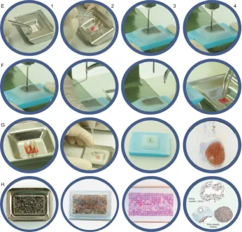
Needle customization and paraffin block punching Figure 1
ished, resulting in 90–100 control slides. These control slides were stored in plastic slide boxes at 4°C/39.2°F for no longer than 4 weeks. Routinely, each IHC case slide had two areas: the control TMA just below slide identifica-tion and the case secidentifica-tion just below the control TMA
(fig-ure 2). More than 200 IHC control TMA blocks with 9 cores (3 × 3 grid) have been confectioned up to the present. Larger TMA blocks have been produced using this technique for research or demonstration purposes, rang-ing from 81 (9 × 9 grid) to 325 cores (25 × 13 grid, figure
TMA block production and examples Figure 2
TMA block production and examples. TMA block production, step by step – grid positioning with double-side adhesive tape (E1 and E2); punching tissue core with hand-press grommet inserting machine (E3 to F3); positioning tissue core on the grid, over the double-side adhesive tape (F4); 3 × 3 grid complete with 9 tissue cores in the mould (G1); pouring liquid paraffin into the mould (G2); TMA block with 9 tissue cores (G3); slide with 9 tissue cores TMA for IHC control + case section [IHC with chromogranin A primary antibody, carcinoid tumor] (G4); 25 × 13 grid complete with 325 tissue cores in the mould (H1); TMA block with 325 tissue cores, 25 × 13 grid (H2); H&E slide with 325 tissue cores, 25 × 13 grid (H3); immunohistochemistry stains in cores done with 16G needle [PLAP, placenta, 4×] and 18G needle [cytokeratin AE1/AE3, teratoma, 4× and CD3, ton-sil, 4×] (H4).
Table 2: Costs
Item Unitary cost US$
DREMEL Multi-Pro® Rotary Tool Model 395 80.00
Hypodermic BD PrecisionGlide® needles 1.00
Double-face adhesive tape, Scotch 3 M® 2.00
Hand-press grommet inserting machine 17.00
Publish with BioMed Central and every scientist can read your work free of charge "BioMed Central will be the most significant development for disseminating the results of biomedical researc h in our lifetime."
Sir Paul Nurse, Cancer Research UK
Your research papers will be:
available free of charge to the entire biomedical community
peer reviewed and published immediately upon acceptance
cited in PubMed and archived on PubMed Central
yours — you keep the copyright
Submit your manuscript here:
http://www.biomedcentral.com/info/publishing_adv.asp
BioMedcentral 2). There were little core losses (<1%), most of them due
to consumption of one or more tissue cores during micro-tomy, rarely to section falling off from slides during tech-nical procedures. To avoid this irregular consumption, it is important to punch cores with near the same tissue length, so one should avoid donor blocks from which many slides have been cut or that bear too thin tissue specimens (at least 2 mm thick). Both 16 and 18 gauge needles yielded the same satisfactory results.
Discussion
Recently there have been reports of alternative methods for the manual construction of tissue microarrays [4-7]. This article describes another alternative that have been tested for over one year, which is inexpensive, easy to reproduce and quick to perform, and produces high-den-sity aligned TMA blocks that can be manipulated the same way any other paraffin tissue block. Following the present method, blocks do not break apart and there is a mini-mum core loss, precluding the use of an adhesive tape sec-tioning device. The initial cost is near USD$100.00; additional costs are negligible and include solely the even-tual purchase of extra hypodermic needles for customiza-tion and double-face adhesive tape. It is also possible to use bone marrow trephine biopsy needles or other types of needles, however hypodermic customized needles have proven to be much more cost-effective, and the lateral opening is useful. The use of the double-face adhesive tape to attach cores was recently described [8], and we have developed a grid that allows for a satisfactory align-ment of tissue fragalign-ments, and blocks thus obtained neces-sarily display tissue cores aligned and at the same cutting plane. Although the needles can be used manually, using the hand-press grommet inserting machine makes core punching easier, once it is absolutely perpendicular, pre-vents damage to the donor blocks and is faster.
Conclusion
We present an alternative method for the construction of high-density tissue microarray blocks that can be per-formed by any anatomic pathology laboratory, at low cost and requiring minimum skill and time.
Competing interests
The author(s) declare that they have no competing inter-ests.
Authors' contributions
All authors contributed equally to this work, in all its steps. All authors read and approved the final manuscript.
Acknowledgements
The authors thank Maurício Vincenzi for the photographs and Max Thiago Ferreira Xavier, Antonio Carlos Rodrigues do Nascimento, Ana Maria Rod-rigues, Celeste de Carvalho RodRod-rigues, Kátia Valéria Ferreira da Costa, Gisele Rabello de Souza and Alexander da Conceição Galvão for their
tech-nical support and dedication. Financial support provided by Fonte Medicina Diagnóstica Ltda.
References
1. van de Rijn M, Gilks CB: Applications of microarrays to histopa-thology. Histopathol 2004, 44:97-108.
2. Packeisen J, Korsching E, Herbst H, Boecker W, Buerger H: Demys-tified... Tissue microarray technology. J Clin Pathol: Mol Pathol
2003, 56:198-204.
3. Packeisen J, Buerger H, Krech R, Boecker W: Tissue microarrays: a new approach for quality control in immunohistochemis-try. J Clin Pathol: Mol Pathol 2002, 55:613-615.
4. Pan CC, Chen PCH, Chiang H: An easy method for manual con-struction of high density tissue arrays. Appl Immunohistochem Mol Morphol 2004, 12:370-372.
5. Hidalgo A, Pina P, Guerrero G, Lazos M, Salcedo M: A simple method for the construction of small format tissue arrays. J Clin Pathol 2003, 56:144-146.
6. Wang L, Deavers MT, Malpica A, Silva EG, Liu J: Tissue microarray: a simple and cost-effective method for high-throughput studies. Appl Immunohistochem Mol Morphol 2003, 11:174-176. 7. Datta MW, Kahler A, Macias V, Brodzeller T, Kajdacsy-Balla A: A
simple inexpensive method for the production of tissue microarrays from needle biopsy specimens – examples with prostate cancer. Appl Immunohistochem Mol Morphol 2005,
13:96-103.