An unusual case of high hyperdiploid childhood
ALL with cryptic
BCR
/
ABL1
rearrangement
Lizcova
et al.
C A S E R E P O R T
Open Access
An unusual case of high hyperdiploid childhood
ALL with cryptic
BCR
/
ABL1
rearrangement
Libuse Lizcova
1*, Zuzana Zemanova
1, Halka Lhotska
1, Jan Zuna
2, Lenka Hovorkova
2, Ester Mejstrikova
2,
Eva Malinova
1, Jana Rabasova
3, Ivan Raska
4, Lucie Sramkova
5, Jan Stary
5and Kyra Michalova
1Abstract
Background:Both high hyperdiploidy (HeH) and the translocation t(9;22)(q34;q11) are recurrent abnormalities in childhood B-cell acute lymphoblastic leukemia (ALL) and both are used in current classification to define different genetic and prognostic subtypes of the disease. The coexistence of these two primary genetic aberrations within the same clone is very rare in children with ALL. Here we report a new case of a 17-year-old girl with newly diagnosed ALL and uncommon cytogenetic and clinical finding combining high hyperdiploidy and a cryptic
BCR/ABL1fusion and an inherited Charcot-Marie-Tooth neuropathy detected during the induction treatment.
Results:High hyperdiploid karyotype 51,XX,+X,+4,+14,+17,+21 without apparent structural aberrations was
detected by conventional cytogenetic analysis and multicolor FISH. A crypticBCR/ABL1fusion, which was caused by the insertion of part of theABL1gene into the 22q11 region, was proved in HeH clone by FISH, RT-PCR and CGH-SNP array. In addition, an abnormal FISH pattern previously described as the deletion of the 3′BCRregion in someBCR/ABL1
positive cases was not proved in our patient.
Conclusion:A novel case of extremely rare childhood ALL, characterized by HeH and a crypticBCR/ABL1fusion, is presented and to the best of our knowledge described for the first time. The insertion ofABL1into theBCRregion in malignant cells is supposed. Clearly, further studies are needed to determine the genetic consequences and prognostic implications of these unusual cases.
Keywords:High-hyperdiploid childhood ALL, Ph-negative childhood ALL,BCR/ABL1fusion, FISH, CGH–SNP array
Background
Pediatric B-cell precursor acute lymphoblastic leukemia (BCP-ALL) is a heterogeneous disease on both the cyto-genetic and cyto-genetic levels [1]. A number of acquired chromosomal aberrations have been identified in the bone marrow cells of children with ALL, which provide diag-nostic and progdiag-nostic information that directly affects patient management [2].
The Philadelphia (Ph) chromosome, i.e., translocation t(9;22)(q34;q11), giving rise to the BCR/ABL1 fusion gene, is a rare finding in children with ALL, accounting for ~3% of cases, and is traditionally associated with a poor outcome [3]. However, implementation of tyrosine kinase inhibitors to standard therapy greatly improved
the survival of children with Ph + ALL [4]. In a small number of ALL patients, the Ph chromosome is not de-tected by conventional cytogenetics, but the BCR/ABL1 fusion is present [5]. In these cases, the fusion either arises as a cryptic rearrangement or is masked within a complex karyotype and can only be detected by molecular cytogenetic and/or molecular genetic methods. On the contrary, high hyperdiploidy (HeH), defined as the pres-ence of 51–67 chromosomes in the karyotype, is the most frequent cytogenetic finding in childhood ALL, occurring in 25%–30% of cases. It is characterized by a nonrandom gain of specific chromosomes and a clinically favorable prognosis [6,7].
Although HeH is a common finding in childhood ALL, there are rare instances of high-hyperdiploid patients car-rying other ALL-specific translocations, i.e., t(9;22)(q34; q11), t(12;21)(p13;q22), t(1;19)(q23;p13), and MLL rear-rangements. These cases comprise 1%–4% of the patients with high-hyperdiploid ALL [8]. We present a patient with
* Correspondence:[email protected]
1Center of Oncocytogenetics, Institute of Medical Biochemistry and
Laboratory Diagnostics, General University Hospital in Prague and First Faculty of Medicine, Charles University in Prague, Prague, Czech Republic Full list of author information is available at the end of the article
newly diagnosed ALL and a rare cytogenetic finding, com-bining HeH and an unusual BCR/ABL1fusion caused by the insertion of part of theABL1gene into the 22q11 re-gion. Moreover, the patient was diagnosed with an inher-ited neuropathy Charcot-Marie-Tooth syndrome during the antileukemic treatment.
Case presentation
A 17-year-old girl was diagnosed with common ALL (cALL) in January 2013 after experiencing one month of fatigue, bone pain at rest, and lymphocytosis in the periph-eral blood (lymphocytes 86%). A complete blood count showed 11.3 × 10⁹/L WBC, 86 g/L hemoglobin, and a platelet count of 42 × 109/L. Her bone marrow was infil-trated by lymphoblasts (96.8%) with an L1 morphology. Blasts had hyperdiploid DNA content (DNA index 1.089). Immunophenotypically blasts corresponded to cALL with aberrant expression of CD66c, high CD34 positivity and low expression of CD38 antigen. Immunophenotype was typical for BCR/ABL1 positivity [9]. BCR/ABL fusion gene was confirmed with multiplex RT-PCR.
The patient has been treated according to the EsPhALL 2010 protocol for BCR/ABL-positive ALL with a combination of chemotherapy and imatinib (Glivec). During induction, the patient developed very severe per-ipheral neurotoxicity with quadriparesis and paralytic ileus, which required major surgery with stoma. This critical clinical condition resulted in interruption of the chemotherapy treatment for two weeks. Surprisingly, the cause of this unexpected complication was inherited neuropathy Charcot-Marie-Tooth syndrome (CMT1A subtype), with a proven PMP22 mutation. Subsequently it was clear that the neurotoxicity was an abnormal reac-tion to Vincristine therapy and therefore further vinca alkaloid medication was contraindicated. At present, the patient is in continuous complete remission (18 months from the diagnosis) on maintenance chemotherapy with imatinib, she is after submerging of her stoma and her neurological status is improving.
For the cytogenetic analyses, bone marrow cells were cultured for 24 hours without stimulation, and chromo-somal preparations were made using standard techniques. In total, 25 metaphases were analyzed and the karyotypes were described according to An International System for Human Cytogenetic Nomenclature (ISCN 2013) [10].
To detect the BCR/ABL1 fusion gene, fluorescence in situ hybridization (FISH) was performed with commer-cially available locus-specific probes (Vysis LSI BCR/ABL Dual Color Dual Fusion Translocation Probe and Vysis LSI BCR/ABL ES Dual Color Translocation Probe; Abbott Molecular, Des Plaines, IL, USA). All available metaphases and 200 interphase nuclei were analyzed.
Other chromosomal aberrations were analyzed with multicolor FISH (mFISH) and comparative genomic
hybridization based array (aCGH) using the 24XCyte color kit (MetaSystems, Altlussheim, Germany) and an oligonucleotide CGH–single-nucleotide polymorphism (SNP) array (BlueGnome, Cambridge, UK) respectively. Array results were confirmed by FISH with the appro-priate bacterial artificial chromosome (BAC) probes (BlueGnome). All techniques were done according to manufacturer’s recommendations.
The fusion transcript BCR/ABL1was detected with re-verse transcription–polymerase chain reaction (RT–PCR), which was performed according to Europe Against Cancer program protocol [11]. The amount of cDNA was normal-ized to the expression of housekeeping gene GUS as de-scribed previously [12].
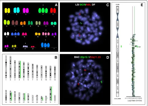
An m-BCR/ABL1fusion gene was detected with multi-plex RT–PCR. Moreover, we found also the genomic breakpoint between the BCR and ABL1 genes (BCR in-tron 1/ABL1 intron 1). The karyotype was described as follows: 51,XX,+X,+4,+14,+17,+21[22]/46,XX[3]. How-ever, FISH with the BCR/ABL Extra Signal and BCR/ ABL Dual Fusion Probes detected theBCR/ABL1fusion with an abnormal FISH signal pattern (1F2O1G), con-sistent with a breakpoint within m-BCR and the loss of the 3′BCRsignal from der(9) in ten out of eleven meta-phases and ~70% of the interphase nuclei (Figure 1). A detailed FISH signal analysis of the abnormal meta-phases revealed the fusion signal to be located on appar-ently normal chromosome 22 and theABL1signal in the 9q34 region of two copies of chromosome 9. The ex-pected deletion of the 3′BCR region (from the FISH re-sults) was not detected by array CGH and BAC probes and only other chromosomal abnormalities were found, including trisomies of chromosomes X, 4, 14, 17, and 21, cryptic deletion of the short arm of chromosome 20 [del (20) (p12.1p12.1)] and submicroscopic amplification of the 22q11.22 region (Figure 1).
Based on the cytogenetic and genetic results, it is clear that the BCR/ABL1fusion in our patient arose from the submicroscopic insertion of part of the ABL1 gene into chromosome 22. It is based on the facts that: (1) no clas-sical or variant Ph translocation was detected by conven-tional cytogenetic/mFISH; (2) loss of the 3′BCR region, although expected from interphase FISH signal pattern, was not confirmed by array CGH or by FISH with BAC probes for this region, i.e. entire BCR signal remained on chromosome 22 and was not translocated on chromosome 9; and (3) only the BCR/ABL1 fusion was proved at the RNA as well as DNA level, while the re-ciprocal ABL1/BCR fusion was not found by any of these approaches.
Our results show that only parallel cytogenetic, molecu-lar cytogenetic, and molecumolecu-lar genetic analyses can pro-vide detailed information about cryptic and prognostically significant aberrations that could not have been achieved
Lizcovaet al. Molecular Cytogenetics2014,7:72 Page 2 of 5
with any of these techniques used independently. The metaphase FISH results were important for the correct in-terpretation of the interphase FISH findings, because only this analysis could identify the location of the BCR/ABL1 fusion on der(22). The current inclusion of array techniques into routine practice could lead us to reevaluate the real oc-currence of deletions of either the 3′BCRor 5′ABL1region inBCR/ABL1-positive cases (especially with cryptic inser-tions) previously described by FISH analyses.
Several patients with CML, or less frequently with ALL, have been reported with cryptic insertions of part of the
BCRregion intoABL1at 9q34 or rarely ofABL1intoBCR at 22q11 [13,14]. However, to our knowledge, this is the first published case of such an aberration in childhood high-hyperdiploid ALL with inherited Charcot-Marie-Tooth neuropathy. In general, ALL-specific rearrange-ments are extremely rare in patients with HeH and it is difficult to identify the primary abnormality. In most cases
(as in our patient), both aberrations were presented simul-taneously in abnormal cells. However, few cases of HeH Ph-positive ALL have been published in the literature [15,16], and these patients displayed not only a Ph-positive HeH clone, but also cells with 46 chromosomes and t(9;22) (q34;q11) as the sole abnormality, suggesting that the Ph chromosome was the primary aberration.
There is no difference in the gain of specific chromo-somes (namely X, 4, 6, 8, 10, 14, 17, 18 a 21) between patients with high-hyperdiploid ALL and commonly en-countered ALL aberrations, and those with high-hyperdiploid ALL. The only exception is the trisomy of chromosome 2, which was previously described as a quite frequent chromosome gain in patients with trans-location t(9;22)(q34;q11) [6,16]. However, trisomy of chromosome 2 was not found in our patient, nor was revealed as a frequent trisomy in six other currently de-scribed cases with Ph positive HeH ALL [8].
E
A
B
BAC 20p12.1/22q11.23D
LSI BCR/ABL DF
C
Figure 1Results of molecular cytogenetic analysis.Multicolor FISH(A)and array CGH(B)showing trisomies of chromosomes X, 4, 14, 17 and 21(A, B)and submicroscopic aberrations: deletion of chromosome 20 and amplification of chromosome 22(B). BCR/ABL1 positive metaphase hybridized with BCR/ABL dual fusion (DF) probe demonstrating apparent loss of green 3′BCR signal, i.e. 1F2O1G FISH pattern (caused by the insertion of theABL1gene into theBCRregion, thus entire BCR signal remained on chromosome 22)(C). FISH with BAC probes RP11-80O7 (orange) and RP11-400P21 (green) with evidence of normal finding of 22q11.23 region matching to 3′BCR locus (two orange signals) and deletion of 20p12.1 (one green signal)
From a clinical perspective, patients with HeH and well-known translocations are considered to represent a bio-logically distinct subgroup, which may have independent prognostic implications [17]. From previously reported cases, it seems that the prognostic impact of the transloca-tion could override the beneficial effect of HeH [17,18]. However, brief follow-up period does not allow to assess any conclusion about the impact of this finding on prog-nosis in our patient nor to predict the effect of the inher-ited neuropathy.
Conclusion
In conclusion, a novel case of extremely rare childhood ALL, characterized by HeH and a cryptic BCR/ABL1 fu-sion, is presented and described for the first time and the insertion ofABL1into theBCR region in malignant cells is supposed. Although several cases of childhood ALL with HeH and general ALL-specific aberrations have been pub-lished, the exact pathogenetic mechanisms of the disease in these patients have not been clarified. Clearly, further studies of these uncommon cases are necessary to deter-mine the genetic consequences and real prognostic impli-cations of these phenomena.
Consent
Written informed consent was obtained from the patient’s parents for publication of this Case report and any accom-panying images. A copy of the written consent is available for review by the Editor-in-Chief of this journal.
Abbreviations
BCP-ALL:B-cell precursor acute lymphoblastic leukemia; CML: Chronic myeloid leukemia; HeH: High hyperdiploidy; FISH: Fluorescence in situ hybridization; mFISH: Multicolor FISH; RT-PCR: Reverse transcription-polymerase chain reaction; CGH-SNP array: Comparative genomic hybridization-single nucleotide polymorphism array; BAC probes: Bacterial artificial chromosome probes.
Competing interests
The authors declare that they have no competing interests.
Authors’contributions
LL performed FISH experiments, interpreted the data and wrote the manuscript. ZZ carried out mFISH/mBAND analysis and participated in interpretation of the data. HL performed and analyzed array CGH experiments. JZ and LH were responsible for molecular analysis. EM done the immunophenotypic analyses. EM carried out FISH analyses. JR performed conventional cytogenetic analysis. IR participated in interpretation of the data and supervision of the manuscript. LS and JS treated the patient, collected samples and provided patient’s data. KM supervised the manuscript. All authors read and approved the final manuscript.
Acknowledgements
This work was supported by grants from the Czech Ministry of Health RVO-VFN64165, and IGA NT14350-3 and the Czech Science Foundation GACR-P302/12/G157.
Author details
1Center of Oncocytogenetics, Institute of Medical Biochemistry and
Laboratory Diagnostics, General University Hospital in Prague and First Faculty of Medicine, Charles University in Prague, Prague, Czech Republic.
2
CLIP-Childhood Leukaemia Investigation Prague, Department of Paediatric Haematology and Oncology, Second Faculty of Medicine, Charles University,
Prague and University Hospital Motol, Prague, Czech Republic.3Department
of Medical Genetics, Faculty Hospital Hradec Kralove, Hradec Kralove, Czech Republic.4Institute of Cellular Biology and Pathology, First Faculty of
Medicine, Charles University in Prague, Prague, Czech Republic.5Department of Paediatric Haematology and Oncology, Second Faculty of Medicine, Charles University, Prague and University Hospital Motol, Prague, Czech Republic.
Received: 25 July 2014 Accepted: 12 October 2014
References
1. Moorman AV, Ensor HM, Richards SM, Chilton L, Schwab C, Kinsey SE, Vora A, Mitchell CD, Harrison CJ:Prognostic effect of chromosomal abnormalities in childhood B-cell precursor acute lymphoblastic leukaemia: results from the UK Medical Research Council ALL97/99 randomised trial.Lancet Oncol2010,
11:429–438.
2. Harrison CJ:The genetics of childhood acute lymphoblastic leukemia. Baillieres Best Pract Res Clin Haematol2000,13:427–439.
3. Carroll WL, Raetz EA:Clinical and laboratory biology of childhood acute lymphoblastic leukemia.J Pediatr2012,160:10–18.
4. Schultz KR, Carroll A, Heerema NA, Bowman WP, Aledo A, Slayton WB, Sather H, Devidas M, Zheng HW, Davies SM, Gaynon PS, Trigg M, Rutledge R, Jorstad D, Winick N, Borowitz MJ, Hunger SP, Carroll WL, Camitta B, and from the Children’s Oncology Group:Long-term follow-up of imatinib in pediatric Philadelphia chromosome-positive acute lymphoblastic leukemia: Children’s Oncology Group Study AALL0031.Leukemia2014,
28:1467–1471.
5. van Rhee F, Kasprzyk A, Jamil A, Dickinson H, Lin F, Cross NC, Galvin MC, Goldman JM, Secker-Walker LM:Detection of the BCR-ABL gene by reverse transcription/polymerase chain reaction and fluorescence in situ hybridization in a patient with Philadelphia chromosome negative acute lymphoblastic leukaemia.Br J Haematol1995,90:225–228.
6. Paulsson K, Johansson B:High hyperdiploid childhood acute lymphoblastic leukemia.Genes Chromosomes Cancer2009,48:637–660. 7. Zemanova Z, Michalova K, Sindelarova L, Smosek P, Brezinova J,
Ransdorfova S, Vavra V, Dohnalova A, Stary J:Prognostic value of structural chromosomal rearrangements and small cell clones with high hyperdiploidy in children with acute lymphoblastic leukemia.Leuk Res
2005,29:273–281.
8. Paulsson K, Forestier E, Andersen MK, Autio K, Barbany G, Borgström G, Cavelier L, Golovleva I, Heim S, Heinonen K, Hovland R, Johannsson JH, Kjeldsen E, Nordgren A, Palmqvist L, Johansson B, Nordic Society of Pediatric Hematology and Oncology (NOPHO), Swedish Cytogenetic Leukemia Study Group (SCLSG), NOPHO Leukemia Cytogenetic Study Group (NLCSG):High modal number and triple trisomies are highly correlated favorable factors in childhood B-cell precursor high hyperdiploid acute lymphoblastic leukemia treated according to the NOPHO ALL 1992/2000 protocols.Haematologica2013,98:1424–1432.
9. Hrusak O, Porwit-MacDonald A:Antigen expression patterns reflecting genotype of acute leukemias.Leukemia2002,16:1233–1258. 10. Shaffer LG, McGowan-Jordan J, Schmidt M:ISCN 2013: An International
System for Human Cytogenetic Nomenclature.Basel: S. Karger; 2013. 11. Gabert J, Beillard E, van der Velden VH, Bi W, Grimwade D, Pallisgaard N,
Barbany G, Cazzaniga G, Cayuela JM, Cavé H, Pane F, Aerts JL, De Micheli D, Thirion X, Pradel V, González M, Viehmann S, Malec M, Saglio G, van Dongen JJ:Standardization and quality control studies of‘real time’ quantitative reverse transcriptase polymerase chain reaction of fusion gene transcripts for residual disease detection in leukemia a Europe Against Cancer program.Leukemia2003,17:2318–2357.
12. Beillard E, Pallisgaard N, van der Velden VH, Bi W, Dee R, van der Schoot E, Delabesse E, Macintyre E, Gottardi E, Saglio G, Watzinger F, Lion T, van Dongen JJ, Hokland P, Gabert J:Evaluation of candidate control genes for diagnosis and residual disease detection in leukemic patients using‘real time’quantitative reverse transcriptase polymerase chain reaction (RQ PCR) a Europe against cancer program.Leukemia2003,17:2474–2486. 13. Primo D, Tabernero MD, Rasillo A, Sayagués JM, Espinosa AB, Chillón MC,
Garcia-Sanz R, Gutierrez N, Giralt M, Hagemeijer A, San Miguel JF, Orfao A:
Patterns of BCR/ABL gene rearrangements by interphase fluorescence in situ hybridization (FISH) in BCR/ABL + leukemias: incidence and underlying genetic abnormalities.Leukemia2003,17:1124–1129.
Lizcovaet al. Molecular Cytogenetics2014,7:72 Page 4 of 5
14. Robinson HM, Martineau M, Harris RL, Barber KE, Jalali GR, Moorman AV, Strefford JC, Broadfield ZJ, Cheung KL, Harrison CJ:Derivative chromosome 9 deletions are a significant feature of childhood Philadelphia chromosome positive acute lymphoblastic leukaemia.Leukemia2005,
19:564–571.
15. Heerema NA, Harbott J, Galimberti S, Camitta BM, Gaynon PS, Janka-Schaub G, Kamps W, Basso G, Pui CH, Schrappe M, Auclerc MF, Carroll AJ, Conter V, Harrison CJ, Pullen J, Raimondi SC, Richards S, Riehm H, Sather HN, Shuster JJ, Silverman LB, Valsecchi MG, Aricò M, Acute Lymphoblastic Leukemia Study Groups, ALL-BFM; CoALL; AIEOP; DCLSG; FRALLE; CCG; DFCI; POG; St Jude; UKALL:Secondary cytogenetic aberrations in childhood Philadelphia chromosome positive acute lymphoblastic leukemia are nonrandom and may be associated with outcome.Leukemia2004,18:693–702.
16. Chilton L, Buck G, Harrison CJ, Ketterling RP, Rowe JM, Tallman MS, Goldstone AH, Fielding AK, Moorman AV:High hyperdiploidy among adolescents and adults with acute lymphoblastic leukaemia (ALL): cytogenetic features, clinical characteristics and outcome.Leukemia2014,
28:1511–1518.
17. Moorman AV, Richards SM, Martineau M, Cheung KL, Robinson HM, Jalali GR, Broadfield ZJ, Harris RL, Taylor KE, Gibson BE, Hann IM, Hill FG, Kinsey SE, Eden TO, Mitchell CD, Harrison CJ, United Kingdom Medical Research Council’s Childhood Leukemia Working Party:Outcome heterogeneity in childhood high-hyperdiploid acute lymphoblastic leukemia.Blood2003,
102:2756–2762.
18. Forestier E, Johansson B, Gustafsson G, Borgström G, Kerndrup G, Johannsson J, Heim S:Prognostic impact of karyotypic findings in childhood acute lymphoblastic leukaemia: a Nordic series comparing two treatment periods. For the Nordic Society of Paediatric Haematology and Oncology (NOPHO) Leukaemia Cytogenetic Study Group.Br J Haematol2000,110:147–153.
doi:10.1186/s13039-014-0072-9
Cite this article as:Lizcovaet al.:An unusual case of high hyperdiploid
childhood ALL with crypticBCR/ABL1rearrangement.Molecular Cytogenetics20147:72.
Submit your next manuscript to BioMed Central and take full advantage of:
• Convenient online submission
• Thorough peer review
• No space constraints or color figure charges
• Immediate publication on acceptance
• Inclusion in PubMed, CAS, Scopus and Google Scholar
• Research which is freely available for redistribution