BOSNIAN JOURNAL OF BASIC MEDICAL SCIENCES 2005; 5 (2): 20-26&
Abstract
A decision for operative versus nonoperative management of thoracolumbar fractures should NEV-ER be based solely on one factor. Only after a thorough physical, neurological, and spinal examina-tion, and an assessment of a patient’s prior activity, social and educational background and patient’s expectations, one should review the patient’s radiographs and CT scans to determine risks and benefi ts of operative versus nonoperative care. Both treatment options are discussed in this paper. As a surgical option our preference is short-segment instrumentation and fusion. Careful and ap-propriate patient selection and an excellent operative technique insure the minimum complications.
KEY WORDS: thoracolumbar spine fractures, load-sharing classifi cation, short segment fusion
Treatment Options
for Thoracolumbar
Spine Fractures
Eldin E. Karaiković¹*, Hector O. Pacheco²
. Assistant Professor, Northwestern University, and Director of the Spine Center and Lead Physician, Orthopaedic Surgery, Evanston Northwestern Healthcare, Chicago, Illinois, U.S.A.
. Assistant Professor, Chief of Spine Surgery, Department of Orthopaedics and Rehabilitation, Texas Tech University, El Paso, Texas.
BOSNIAN JOURNAL OF BASIC MEDICAL SCIENCES 2005; 5 (2): 20-26
Th ere are three fundamental questions that every spine surgeon must answer when treating a patient with a spine fracture. First, should the fracture be treated operatively or nonoperatively. Second, if an opera-tive treatment is recommended, how many spinal seg-ments should be instrumented and/or fused (short vs. long segment fusion). Lastly, which surgical approach should be undertaken: anterior, posterior or combined.
Fracture anatomy should never be used as a single criterion to determine fracture treatment because if there is no neurological deficit, any fracture can be treated either operatively or nonoperatively. The ma-jority of the spine fractures, especially in the thoracic spine can be successfully treated nonsurgically with bracing or adequate length of the bed rest. Patient selection is fundamental for nonoperative manage-ment. Besides anatomical characteristics of a fracture other factors critical for fracture management in-clude patient’s age, general health, expectations, and compliance. An overactive, debilitated, demented or noncompliant patient who is unwilling or unable to follow treatment instructions is particularly at risk.
NON-OPERATIVE TREATMENT
The treatment for the majority of the thoracolumbar fractures is non-operative. Non-operative treatment options are no bracing, bracing with early ambulation, bracing with delayed ambulation or defi ned period of bed rest with bracing.
BED REST
Treatment with bed rest is not a preferable choice any more. Prolonged bed rest even for a few days increases a risk of deep venous thrombosis (DVT), pulmonary complications and pressure decubiti formation. If for any reason patients with spine fractures should be on a prolonged bed rest a chemical and/or mechani-cal DVT prophylaxis with aggressive pulmonary toilet should be initiated. Patients with spinal cord injuries treated with or without surgical stabilization are espe-cially prone to these complications. Careful daily skin inspection especially around bony prominences and skin care with washing, massage, powdering and ap-plication of lotions are essential. Because of the above mentioned complications related to prolonged re-cumbency, bed rest is no longer considered as an ac-cepted initial treatment method. If a fracture can not be maintained stable enough in a brace and patient
mobilized as soon as a brace is applied, an indication for surgical stabilization should be strongly considered.
BRACING
It is accepted that compression fractures with up to degree of kyphosis can be successfully treated with bracing. Some fracture types as a fl exion-distrac-tion fractures with bony involvement (“bony seat belt fractures, bony Chance fracture”) can be successfully treated with bracing. Flexion-distraction injuries of the thoracolumbar spine with complete disruption of the ligamentous structures and, no or minimal bony in-volvement (“soft seat belt injuries, soft tissue Chance fractures”) are not suitable for bracing and should be stabilized surgically. A minority of authors has made the attempts to popularize bracing for certain types of burst fractures but that has not been widely accepted. For the majority of the thoracolumbar fractures use of a high profi le thoracolumbar orthosis (TLSO) is recom-mended. High profi le TLSO’s extend from the sternum to the pubis. Th ese orthoses are made of fi berglass of dif-ferent thicknesses that successfully restrict most of the motion in the thoracolumbar spine. TLSO’s are made as a “clam shell” with anterior and posterior parts inter-connecting together and secured with Velcro fastening straps. Th is orthosis design feature allows easy removal for hygiene and skin care. Low profi le TLSO’s extend from the xyphoid to pubis and are mostly used for post-operative bracing of the lower lumbar spine, and very rarely for non-operative treatment of lower lumbar frac-tures. If a fracture is localized in the upper thoracic spine or cervicothoracic junction a TLSO brace with cervi-cal extension (CTLSO) is recommended. Th e CTLSO brace prevents most of the fl exion, extension and lateral bending. Jewett braces can be used in the treatment of the thoracolumbar fractures but their weight due to met-al bars, often exceeds the weight of TLSO and for this reason have fallen out of favor for fracture management.
CASTING
BOSNIAN JOURNAL OF BASIC MEDICAL SCIENCES 2005; 5 (2): 20-26Surgical Treatment of
Thoracolumbar Fractures
PATIENT SELECTION
Once the decision for surgical treatment is made a surgeon has to answer two questions: ) how many spinal segments should one instrument and perform either short or long segment fusion, and ) which sur-gical approach should one use either anterior, poste-rior or combined. Details regarding the patient’s age, general health, compliance, future endeavors, and the surgeon’s expertise in a particular system are essential component to treatment decision-making. For exam-ple, a young active patient with no medical problems whose life expectancy is long should be considered dif-ferently that a patient in elderly with multiple medical problems and sedentary lifestyle. Anterior approach through thoracotomy or laparotomy might not be suitable if not being dangerous for a patient with pul-monary or other medical complications, and a surgical treatment should be tailored to the patient condition. If a surgical treatment is chosen, in order to optimize a satisfactory surgical outcome a surgeon should un-derstand the mechanical properties of any given im-plant and determine a patient’s willingness and abil-ity to comply with treatment recommendations. At the same time, the patient should understand basic principles of the spinal fi xation system used and con-comitantly take the responsibility for his/her recov-ery. Th e patient should be aware that noncompliance with postoperative instruction could lead to failure of the device and possible need for surgical revision. Fracture location influences a choice of the length of fusion and instrumentation. Because the thoracic spine is a less mobile area, posterior long segment in-strumentation is generally the treatment of choice for surgical instrumentation, as the addition of a few segments generally does not limit functional motion. Posterior short segment or anterior instrumentation, or both, is therefore rarely done in the thoracic spine.
RADIOGRAPHIC FRACTURE ASSESSMENT
Load Sharing Classifi cation - Analysis of load-sharing through the fracture site
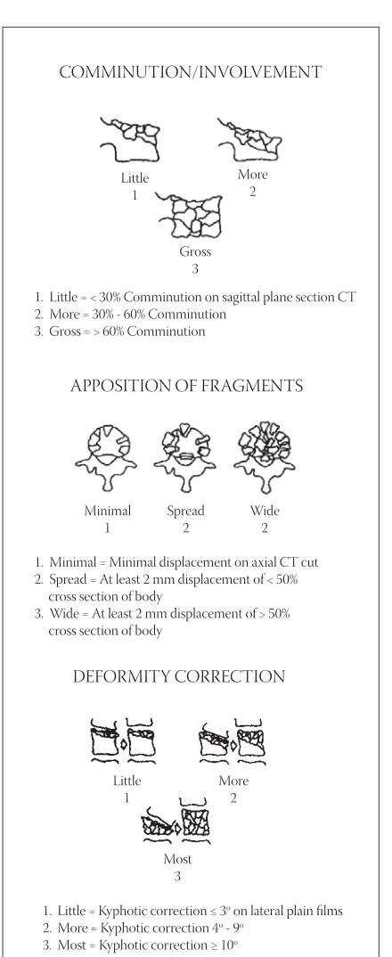
All currently used classifications accept the fact that imaging techniques provide only a static view of spinal displacement. Based on our data analysis, we recog-nized that by preoperatively quantifying the commi-nution of the most injured vertebral body (regardless of the mechanism of injury, and without being col-umn-specific regarding the comminution), one can
predict with great accuracy, the occurrence of a post-operative pedicle screw fracture for spine fractures treated with short segment instrumentation and fu-sion. The Load-Sharing Classification of spinal frac-tures is generated by the review of preoperative plain radiographs, and sagittal and axial CT scans, which provide data regarding three separate characteristics of the fracture site. These are: comminution, apposi-tion of fragments and deformity correcapposi-tion (Figure ). All three factors used in our system quantify the comminu-tion of the vertebral body that occurred during the injury. Each of these factors is subdivided into three de-grees of severity and graded by awarding point for mild, points for moderate, and points for most severe. Every fracture regardless of the mech-anism can be graded from a minimum total of three points to a maximum total of nine points. The Load-Sharing Classification by itself does not recommend a decision for operative or nonopera-tive treatment of a specific spine fracture. However, it does help the surgeon to understand the qual-ity of load-sharing transferred across the fracture site and the spinal implant after surgical fixation.
Translational displacement
Translational displacement represents lateral or ante-rior-posterior disruption of the sagittal, coronal or axial spinal column alignment. It indicates serious multiple spinal ligament disruption, which we use to define a fracture-dislocation. It ranges from subtle to severe. Careful clinical evaluation of local swelling, identifying a palpable defect in supraspinal and interspinal ligaments, and evaluation of radiographs is necessary (Figure ).
Surgical Treatment Options
SURGICAL GOALS
Main goals of surgical treatment are stabilization of un-stable fractures and early patient mobilization in order to prevent complications associated with prolonged bed rest and posttraumatic deformities. Proper opera-tive fracture reconstruction must restore spinal balance in all three planes over as few segments as possible.
SURGICAL DECOMPRESSION
BOSNIAN JOURNAL OF BASIC MEDICAL SCIENCES 2005; 5 (2): 20-26
had occurred and posttraumatic deformity did not exist. On the other hand, neurological deterioration com-monly develops in neurologically intact patients with progressive deformity after an unstable spinal fracture that was not stabilized surgically early after the injury. Neural recovery has been observed in many of these pa-tients after direct decompression even after many years. Patients with neurological defi cit. In patients with cord injury, decompression is usually performed concomi-tantly with spinal instrumentation, generally via the same approach used for fracture repair. However, anterior
fragments compressing the spinal cord can be also suc-cessfully removed from a posterior approach, through a posterolateral or posterior transpedicular approach, if posterior instrumentation is selected for fracture re-pair., Isolated decompressive laminectomy without fracture stabilization has no benefi cial eff ect with up to occlusion of the spinal canal. Laminectomy without stabilization further destabilizes the spine fracture and is contraindicated in most cases of thoracolumbar fractures secondary to high development of iatrogenic kyphosis and need for instrumentation. Inadequate
decompres-FIGURE . Th e Load-Sharing Classifi cation of Th oracolumbar Fractures (. Th e amount of involve-ment/comminution – as best seen on sagittal CT reconstructions of the fracture site. Th is factor describes the amount and extent of comminution of the most injured vertebra. Comminution controls collapse if nonoperative treatment is selected, and infl uences load-sharing if operative treatment is selected. . Th e apposition/displacement of fragments
BOSNIAN JOURNAL OF BASIC MEDICAL SCIENCES 2005; 5 (2): 20-26sion of the spinal canal can cause long-standing radicu-lar symptoms and/or spinal cord myelopathy much later from the time of injury. Possible consequences of chronic neurological compromise include arachnoidi-tis, syringomyelia, and late-onset vascular compromise.
LENGTH OF FUSION: SHORT VERSUS LONG SEGMENT FUSION
In preoperative planning of a surgical treatment of a spine fracture, the surgeon must determine the length of fusion and the surgical approach that will achieve stable fixation but preserve maximal spinal motion postoperatively. The fracture location (thoracic, tho-racolumbar, or lumbar spine) infl uences the choice of length of fi xation. Longer fi xation in the upper or mid-dle thoracic spine does not reduce the patient’s mobil-ity much, because this part of the spine is already natu-rally limited in motion from its connection with the ribs.
SHORT SEGMENT FUSION
Short-segment fixation (instrumentation one level above and one level below the damaged vertebra) and fusion limits the levels of fusion. The introduc-tion of the pedicle-screw-based implants, anterior spinal implants, and better understanding of fracture biomechanics have led to the ability to perform short-segment instrumentation and fusion from either ante-rior or posteante-rior approach to preserve motion segments.
LONG SEGMENT FUSION
Long-segment fi xation and fusion involves instrumen-tation two or more levels above and below the fracture. In our practice, only patients with grotesque, severely displaced, and comminuted fracture dislocations are treated with long-segment instrumentation and/or combined anterior and posterior procedures, and for pa-tients who apt to be noncompliant in brace-wear, based on their premorbid personality, their injuries, or both.
PEDICLE SCREW FIXATION
Pedicle screw fixation has improved the stability of the spinal fusion constructs, produced better correc-tion of the fracture, allowed an applicacorrec-tion of short segment instrumentation and fusion, produced better retention of intraoperative correction of spinal defor-mity, and has nearly eliminated nonunion as a com-plication.,, Use of pedicle screws in fi xation of the thoracic and lumbar spine is widespread and there use is popularized even in the cervical spine. Although complications associated with placement of pedicle screws are multiple (nerve root, spinal cord, and vessel
injuries, etc.), their incidence is very rare with the use of a proper technique., We recommend using a technique of direct visualization of the pedicle during placement, the so called “funnel technique”. Th e accu-rate placement of the pedicle screws is confi rmed using intraoperative fl uoroscopy. Th e “funnel technique” was proved to be very accurate based on anatomical and clinical data. Placement of pedicle screw based solely on a tactile feedback, landmarks, or fl uoroscopy is danger-ous and can produce grave consequences to a patient.
SURGICAL APPROACH: POSTERIOR VS. ANTERIOR
Th e fundamental principle of load-sharing between the implant system and injured spinal column provides good bony apposition at any internally fi xed fracture site to permit healing and prevent implant failure., A preoperative analysis of bone fracture anatomy, as well as individual characteristics of the patient, guide the selection of successful candidates for either an-terior and/or posan-terior instrumentation and fusion, and for long- or short-segment instrumentation.
BOSNIAN JOURNAL OF BASIC MEDICAL SCIENCES 2005; 5 (2): 20-26
POSTERIOR APPROACH
Posterior approach, especially short-segment fusion can be successfully used only if the support through the anterior and middle columns is adequate to transfer residual load that extends the capacity of the posterior instrumentation. Only when pedicle screws are ap-plied posterior can short segment instrumentation be utilized. Strict analysis of the fracture character us-ing the principles of Load-Sharus-ing Classification will allow one to apply posterior short-segment instrumen-tation and fusion. Utilization of hook and wire con-structs require long- or longer-segment fusion due to their inherent instability in short-segment constructs.
ANTERIOR APPROACH
In highly comminuted vertebral body injury at the tho-racolumbar junction (a total point of or higher on the Load-Sharing Classifi cation scheme), anterior strut
grafting and instrumentation is superior to fi xation with a posterior instrumentation with pedicle screws. An-terior spinal fusion surgery is a safe procedure which can be used with confi dence when the patient’s disor-der dictates its use. Th e total complication rate for all complications that were directly attributed to the ante-rior approach is .. Serious complications, such as death (.), paraplegia (.), and deep wound infec-tion (.) are rare. Any anterior device (screw-rods and a plate) must be applied laterally on the vertebral bodies, with bicortical purchase assuring no instru-mentation penetrating into the spinal canal or contra-lateral foramen, and performed under intraoperative radiographic control. The success of any anterior in-strumentation depends on adequate vertebral body reconstruction. Autologous bone struts are preferable (tricortical iliac crest or fi bula), although allografts, and ti-tanium or carbon fi ber cages, can be used successfully.,,
Conclusions
BOSNIAN JOURNAL OF BASIC MEDICAL SCIENCES 2005; 5 (2): 20-26() An H.S., Cotler J.M., Balderston R.A. Complications of treatment of fractures and dislocations of the thoracolumbar spine. In Balderston RA, An HS: Complications in spinal surgery. W.B. Saunders, . () Boeger T.O., Limb D., Dickson R.A. Does ‘canal clearance’ aff ect
neurological outcome after thoracolumbar burst fractures? J. Bone Joint Surg. ; B:-.
() Gaines R.W. Th e use of pedicle-screw based internal fi xation for the operative treatment of spinal disorders. J Bone Joint Surg. ; A:-.
() Gertzbein S.D. Neurologic deterioration in patients with tho-racic and lumbar fractures after admission to the hospital. Spine ;:-.
() Kim N.H., Lee H.M., Chun I.M. Neurologic injury and recovery in patients with burst fracture of the thoracolumbar spine. Spine ; : -.
() Kaneda K., Abumi K., Fujiya M. Burst fractures with neurological defi cits of the thoracolumbar spine: Results of anterior decompres-sion and stabilization with anterior instrumentation. Spine ; :-.
() Karaikovic E.E., Daubs M.D., Madsen R., Gaines R.W. Jr. Mor-phologic characteristics of human cervical pedicles. Spine ; ():-.
() Karaikovic E.E., Gaines R.W. Trauma: anterior versus posterior re-construction in thoracic and lumbar fractures. Curr. Opin. Orthop. ;:-.
() Karaikovic E.E., Gaines R.W.Jr. Short segment fi xation using VSP plates and pedicle screws for trauma. Spinal Instrumentation Tech-niques (Editor: Courtney W. Brown), Scoliosis Research Society, March .
() Karaikovic E.E., Gaines R.W.Jr. Load-Sharing Classifi cation: Prevent-ing implant failure followPrevent-ing surgical treatment. In: Revision Spine Surgery (Editors:J.Y. Margulies, M. Aebi, J-PC Farcy), Mosby, , -.
() Karaikovic E.E., Gaines R.W.Jr. Load-Sharing Classifi cation of spine fractures. In Controversies in Spine Surgery (Eds.: T. Zdeblick, E. Benzel, P. Anderson, C. Stillerman), Quality Medical Publishing, , -.
() Karaikovic E.E., Gaines R.W.Jr. Decision making in revisions of tho-racolumbar spinal fractures. In Th e Failed Spine (Editor: S. Boden), National Spine Network, Lippincott-Raven, ; -. (). Karaikovic E.E., Kaneda K., Akbarnia B.A., Gaines R.W.Jr. Kaneda
instrumentation for spinal fractures. In: The Textbook of Spinal Surgery (Editors: K.H. Bridwell and R.L. DeWald), Second Edition, Lippincott-Raven, ; -.
(). McCormack T., Karaikovic E.E., Gaines R.W.Jr. Th e Load-Sharing Classifi cation of spine fractures. Spine ; ():-. () McLain Fr., Sparling E., Benson D.R. Early failure of short-segment
pedicle instrumentation for thoracolumbar fractures. A preliminary report. J. Bone Joint Surg. ;()A:-.
() Mosekilde L. Age-related changes in bone mass, structure, and strength – eff ects on loading. Zeitschrift fur Rheumatologie , S:-.
() Parker J.W., Lane J.R., Karaikovic E.E., Gaines R.W. Successful short-segment instrumentation and fusion for thoracolumbar spine frac-tures, A consecutive ⁄-year series. Spine ;():-. () Rechtine G.R. Nonsurgical treatment of thoracic and lumbar
frac-tures. Instructional course Lectures ; :-.
() Riska E.B., Myllynen P., Bostman O. Anterolateral decompression for neural involvement in thoracolumbar fractures. J. Bone Joint Surg. ; B:-.
() Vieu M., Tarbox B., Wonglertsiri S., Karaikovic E.E, Yingsakmong-kol W., Gaines R.W. Th oracic pedicle screw instrumentation using the “Funnel technique”. Part II: Clinical experience, J. Spinal Disor-ders and Tech. ; ():-.
() Yingsakmongkol W., Karaikovic E.E, Gaines R.W. Th e accuracy of pedicle screw placement in the thoracic spine using the “Funnel technique”. Part I: A cadaver study, J. Spinal Disorders and Tech ; ():-.