An Observational Cohort Study of
Clostridium difficile
Ribotype
027 and Recurrent Infection
Krishna Rao,a,bPeter D. R. Higgins,a,cVincent B. Younga,b,d
aDepartment of Internal Medicine, University of Michigan, Ann Arbor, Michigan, USA
bDivision of Infectious Diseases, University of Michigan, Ann Arbor, Michigan, USA
cDivision of Gastroenterology, University of Michigan, Ann Arbor, Michigan, USA
dDepartment of Microbiology and Immunology, University of Michigan, Ann Arbor, Michigan, USA
ABSTRACT RecurrentClostridium difficileinfection (rCDI) frequently complicates recov-ery from CDI. Accurately predicting rCDI would allow judicious allocation of limited resources, but published models have met with limited success. Thus, biomarkers predic-tive of recurrence have been sought. This study tested whether PCR ribotype
indepen-dently predicted rCDI. Stool samples from nonpregnant inpatients ⱖ18 years of age
with diarrhea were included from October 2010 to January 2013 after the patients
tested positive forC. difficile in the clinical microbiology laboratory. Per guidelines, the
rCDI was defined as a positive test for C. difficileat⬎2 weeks butⱕ8 weeks from the
index episode. For each sample, a single colony ofC. difficilewas isolated by anaerobic
culture, confirmed to be toxigenic by PCR, and ribotyped. Simple logistic regression and multiple logistic regression were used to model the primary outcome of rCDI, incorpo-rating a wide range of clinical parameters. In total, 927 patients with 968 index episodes of CDI were included, with 110 (11.4%) developing rCDI. Age and use of proton pump inhibitors or concurrent antibiotics did not increase the risk of rCDI. Low serum bilirubin levels and ribotype 027 were associated with increased risk of rCDI on unadjusted analy-sis, with health care-associated CDI being inversely associated. In the final multivariable model, ribotype 027 was the strongest independent predictor of rCDI (odds ratio, 2.17;
95% confidence interval, 1.33 to 3.56;P⫽0.002). Ribotype 027 is an independent
pre-dictor of rCDI.
IMPORTANCE CDI is a major public health issue, with over 400,000 cases per year in the United States alone. Recurrent CDI is common, occurring in approximately one in five individuals after a primary episode. Although interventions exist that could reduce the risk of recurrence, deployment in all patients is limited by cost, invasive-ness, and/or an undetermined long-term safety profile. Thus, clinicians need risk stratification tools to properly allocate treatments. Because prior research on clinical predictors has failed to yield a reliable, reproducible, and effective predictive model to assist treatment decisions, accurate biomarkers of recurrence would be of great value. This study tested whether PCR ribotype independently predicted rCDI, and the data build upon prior research in showing that ribotype 027 is associated with rCDI.
KEYWORDS Clostridium difficile, biomarkers, clinical decision making, molecular epidemiology, ribotyping
C
lostridium difficileinfection (CDI) is responsible for over 400,000 cases of infectious colitis and over 30,000 deaths per year in the United States alone (1). Even among those who recover, recurrent CDI is common and affects approximately 20% of patients, many of whom are readmitted or have further recurrences (1). The estimated cost of recurrent CDI alone in the United States is up to $2.8 billion annually (2). AlthoughReceived17 January 2018Accepted2 May 2018 Published23 May 2018
CitationRao K, Higgins PDR, Young VB. 2018. An observational cohort study ofClostridium difficileribotype 027 and recurrent infection. mSphere 3:e00033-18.https://doi.org/10.1128/ mSphere.00033-18.
EditorBrandi M. Limbago, Centers for Disease Control and Prevention
Copyright© 2018 Rao et al. This is an open-access article distributed under the terms of theCreative Commons Attribution 4.0 International license.
Address correspondence to Krishna Rao, [email protected].
Infection with Clostridium difficile ribotype 027 increases the risk of subsequent recurrence. @PulseTsar
Clinical Science and Epidemiology
crossm
on September 8, 2020 by guest
http://msphere.asm.org/
newer therapies that reduce the risk of recurrent CDI, such as the use of fidaxomicin (3), monoclonal antibodies (4), and fecal microbiota transplantation (FMT) (5, 6), are avail-able, their widespread deployment in all patients is limited by cost (7) and/or unde-termined safety profiles (8). Thus, clinicians are in need of tools to achieve stratification of patients for risk of recurrence and consequently to better allocate limited resources. Models utilizing clinical variables alone to predict recurrent CDI in patients present-ing with an index episode have been developed (9–11). However, when validation of these models in external cohorts was attempted, they failed to make accurate predic-tions (12). There is evidence that biomarkers based on the immune response (13–15), the microbiota (16, 17), or the infecting strain (18–22) are associated with recurrence. The hope is that the use of such biomarkers will improve the predictive performance of clinical models. Here, in an observational cohort study, we tested the hypothesis that
infection with specificC. difficilestrains, as determined by the PCR ribotype, is
associ-ated with a greater risk of recurrence. We specifically focus on the ribotype 027 strain, given its importance in the hospital setting (23, 24), where our study took place, compared to the outpatient, community setting, where different strains may predom-inate (24).
(Parts of this work were previously presented at the Anaerobe 2016 conference in Nashville, TN, on 14 July 2016.)
RESULTS
Descriptive and unadjusted statistics.Selected results from the baseline patient
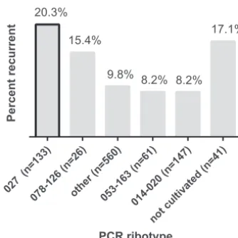
characteristics and outcomes are summarized in Table 1. In total, 899 patients with 968 index episodes of CDI were included, with 110 (11.4%) developing recurrent CDI. Notably, our cohort had slightly more women (54.3%) and was predominantly white (85.2%). The majority of patients were on proton pump inhibitors (PPIs) and were receiving concurrent antibiotics for an infection other than CDI and/or had hospital-associated CDI (HA-CDI). The breakdown of recurrent CDI by ribotype is shown in Fig. 1.
We were able to culture and ribotypeC. difficilefrom 927 (95.7%) stool samples. Among
those, infection with ribotype 027 had the largest risk of recurrence (20.3%), followed by infection with ribotype 078-126 (15.4%). There were 79 (8.2%) deaths within 30 days of diagnosis.
In the initial bivariable analysis, only HA-CDI, serum bilirubin, diagnosis of CDI by presence of toxin(s) A/B by enzyme immunoassay (EIA), and infection with ribotype 027 were significantly associated with recurrent CDI (Table 1). Notably, prior CDI, index CDI episode severity, age, PPIs, and concurrent antibiotic use were not associated with recurrent CDI. No other ribotype was associated with recurrence. Multiple variables were significantly associated with ribotype 027 (Table 2), and these were considered for adjustment in the multivariable model.
Multivariable modeling.The final multivariable model is shown in Table 3, which
was arrived at by both the forward and backward selection procedures described in Materials and Methods. Interactions between variables in the final model were tested, and none of the interactions were significant. Thus, after adjustment for HA-CDI and serum bilirubin, ribotype 027 remained a significant independent predictor of recurrent
CDI (odds ratio [OR], 2.17; 95% confidence interval [CI], 1.33 to 3.56;P⫽0.002). Adding
back into the model several variables demonstrated to associate with recurrence in other studies, specifically, age, PPI use, and concurrent antibiotics, did not affect this relationship between ribotype 027 and recurrence (data not shown). Additionally, adding back in other potential confounders associated with ribotype 027 on bivari-able analysis (Tbivari-able 2) did not change the point estimates or the significance of the association between ribotype 027 and recurrence (data not shown). We further ex-plored HA-CDI, since the inverse association with recurrence was unexpected. Variables common among hospitalized, sick patients were associated with HA-CDI (obesity, congestive heart failure, and renal disease), but none of these affected the inverse association between HA-CDI and rCDI and were not included for adjustment in the models (data not shown). When patients who had died within 30 days were excluded
on September 8, 2020 by guest
http://msphere.asm.org/
from the model, the association between ribotype 027 and recurrent CDI remained
significant (OR, 2.45; 95% CI, 1.47 to 4.09;P⫽0.001). The Hosmer-Lemeshow test did
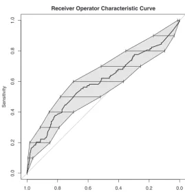
not suggest poor model fit (P⫽0.172). The receiver operator characteristic (ROC) curve
for the model is shown in Fig. 2. As suggested by the curve’s bootstrapped confidence intervals crossing the 50% line and the AUC value of 0.59, the model’s predictive ability was poor overall.
DISCUSSION
The results from this study support the hypothesis that the infecting strain of the index CDI episode contributes to the risk of subsequent recurrent CDI. Specifically, the data identify infection with ribotype 027 as an independent predictor of recurrence even when adjusted for potential confounders. This result is in line with prior studies conducted from 2001 to 2017 that have also shown such an association. Three of these were much smaller studies (20–22), and two larger ones of comparable size to the current study were conducted outside the United States (18, 19). Thus, our results bolster the claim that ribotype 027 infection is associated with increased risk of rCDI. Notable strengths of our study included its large size, use of a validated ribotyping protocol, and careful attention to variable construction and analysis.
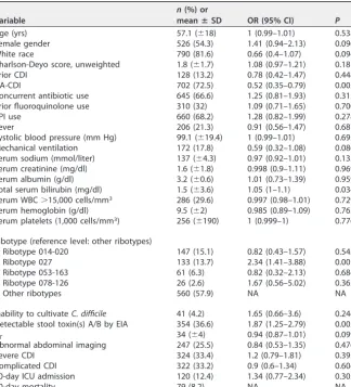
TABLE 1 Selected baseline characteristics, outcomes, and unadjusted analysis versus recurrent CDI (968 index episodes; 110 recurrences)a
Variable
n(%) or
meanⴞSD OR (95% CI) P Age (yrs) 57.1 (⫾18) 1 (0.99–1.01) 0.538 Female gender 526 (54.3) 1.41 (0.94–2.13) 0.096 White race 790 (81.6) 0.66 (0.4–1.07) 0.094 Charlson-Deyo score, unweighted 1.8 (⫾1.7) 1.08 (0.97–1.21) 0.181 Prior CDI 128 (13.2) 0.78 (0.42–1.47) 0.448 HA-CDI 702 (72.5) 0.52 (0.35–0.79) 0.002 Concurrent antibiotic use 645 (66.6) 1.25 (0.81–1.93) 0.313 Prior fluoroquinolone use 310 (32) 1.09 (0.71–1.65) 0.700 PPI use 660 (68.2) 1.28 (0.82–1.99) 0.278
Fever 206 (21.3) 0.91 (0.56–1.47) 0.687
Systolic blood pressure (mm Hg) 99.1 (⫾19.4) 1 (0.99–1.01) 0.692 Mechanical ventilation 172 (17.8) 0.59 (0.32–1.08) 0.086 Serum sodium (mmol/liter) 137 (⫾4.3) 0.97 (0.92–1.01) 0.133 Serum creatinine (mg/dl) 1.6 (⫾1.8) 0.998 (0.9–1.11) 0.969 Serum albumin (g/dl) 3.2 (⫾0.6) 1.01 (0.73–1.39) 0.959 Total serum bilirubin (mg/dl) 1.5 (⫾3.6) 1.05 (1–1.1) 0.034 Serum WBC⬎15,000 cells/mm3 286 (29.6) 0.997 (0.98–1.01) 0.729 Serum hemoglobin (g/dl) 9.5 (⫾2) 0.985 (0.89–1.09) 0.763 Serum platelets (1,000 cells/mm3) 256 (⫾190) 1 (0.999–1) 0.776
Ribotype (reference level: other ribotypes)
Ribotype 014-020 147 (15.1) 0.82 (0.43–1.57) 0.542 Ribotype 027 133 (13.7) 2.34 (1.41–3.88) 0.001 Ribotype 053-163 61 (6.3) 0.82 (0.32–2.13) 0.684 Ribotype 078-126 26 (2.6) 1.67 (0.56–5.02) 0.362
Other ribotypes 560 (57.9) NA NA
Inability to cultivateC. difficile 41 (4.2) 1.65 (0.66–3.6) 0.244 Detectable stool toxin(s) A/B by EIA 354 (36.6) 1.87 (1.25–2.79) 0.002
CT 34 (⫾4) 0.94 (0.87–1.01) 0.099
Abnormal abdominal imaging 247 (25.5) 0.84 (0.53–1.35) 0.476 Severe CDI 324 (33.4) 1.2 (0.79–1.81) 0.393 Complicated CDI 322 (33.2) 0.9 (0.6–1.34) 0.604 30-day ICU admission 120 (12.4) 1.34 (0.77–2.34) 0.303
30-day mortality 79 (8.2) NA NA
aComorbidities with nonsignificantPvalues not shown in this table: immunosuppression, AIDS, lymphoma, solid-organ tumor, metastatic cancer, obesity, liver disease, peptic ulcer disease, hypertension, prior myocardial infarction, congestive heart failure, peripheral vascular disease, prior stroke, dementia, chronic pulmonary disease, rheumatologic disorder, diabetes, chronic kidney disease, and depression. CDI, Clostridium difficileinfection; CI, confidence interval;CT, PCR cycle threshold; EIA, enzyme immunoassay; HA, hospital associated; ICU, intensive care unit; NA, not applicable; OR, odds ratio; PPI, proton pump inhibitor; WBC, white blood cell count.
on September 8, 2020 by guest
http://msphere.asm.org/
Knowledge of the infecting strain could be an important tool for clinicians who aim to stratify patients with respect to risk for recurrent CDI. However, our model with an AUC value of only 0.59 is comparable in performance to models reported from prior studies (11). This suggests that much work needs to be done to improve model performance, possibly through incorporation of additional, novel biomarkers. It is unlikely that ribotype information alone would have much clinical utility, but it might still be useful in an integrative model alongside clinical variables, microbiological factors, and host-level biomarkers. We utilized PCR ribotyping to distinguish strains, but approaches such as whole-genome sequencing may reveal other genome-derived biomarkers of adverse outcomes such as severity and recurrence. Evaluations of the performance of published models have not yet shown an AUC value above 90%, the level where reliable allocations of expensive and/or invasive treatments such as those employing fidaxomicin, monoclonal antibodies, or FMT can be made (25, 26). Further-more, ribotyping may be too coarse a typing method to lead one to general
conclu-sions, as strains that are categorized as ribotype 027 strains can have variablein vitro
characteristics, such as sporulation (27). Finally, host factors also influence the risk of recurrent CDI and the relative importance of these versus strain needs to be deter-mined.
There are notable results from our study that differ from results from prior studies.
027 (n=133
)
078 -126
(n=26)
othe r (n=
560 )
053 -163
(n=61)
014 -020
(n=147 )
not c ultiv
ated (n=41)
PCR ribotype
Percen
t
re
c
u
rr
e
n
t 20.3%
15.4%
9.8% 8.2% 8.2% 17.1%
FIG 1 PCR ribotype of index CDI episode and subsequent recurrent CDI risk. Infection with ribotype 027 carries the highest risk of recurrent CDI.
TABLE 2 Selected results from simple logistic regression of predictors versus infection with ribotype 027a
Variable OR (95% CI) P
Age (yrs) 1.03 (1.02–1.05) ⬍0.001
Charlson-Deyo score, unweighted 1.26 (1.14–1.39) ⬍0.001
HA-CDI 0.52 (0.35–0.76) 0.001
Solid-organ tumor 2.03 (1.31–3.14) 0.002 Prior myocardial infarction 2.08 (1.28–3.38) 0.003 Congestive heart failure 2.01 (1.26–3.21) 0.004 Diabetes mellitus 1.63 (1.1–2.41) 0.016 Chronic kidney disease 1.7 (1.15–2.52) 0.008 Concurrent antibiotic use 1.75 (1.14–2.67) 0.010
PPI use 1.52 (0.997–2.31) 0.052
Mechanical ventilation 0.54 (0.28–1.01) 0.055 Serum albumin (g/dl) 0.52 (0.38–0.71) ⬍0.001 Serum WBC⬎15,000 cells/mm3 2.05 (1.4–2.98) ⬍0.001 Detectable stool toxin(s) A/B by EIA 3.29 (2.25–4.81) ⬍0.001 30-day ICU admission 2.07 (1.28–3.34) 0.003
aCI, confidence interval; EIA, enzyme immunoassay; HA, hospital associated; ICU, intensive care unit; OR, odds
ratio; PPI, proton pump inhibitor; WBC, white blood cell count.
on September 8, 2020 by guest
http://msphere.asm.org/
We identified associations with recurrence that not previously been described, includ-ing serum bilirubin and HA-CDI, and these need to be independently validated. HA-CDI in particular was inversely associated with recurrence, and we could not explain this within the scope of the current study. One possibility is that those with HA-CDI were less prone to CDI and recurrence for other reasons and developed CDI only with the increased colonization pressure of the hospital setting. This would imply they were less likely to have a history of prior CDI outside the current hospital admission. This was true in our study, where only 10.7% of patients with a current index HA-CDI episode had had a prior CDI episode(s), compared to 19.9% of those without a current HA-CDI episode
having had one or more prior CDI episodes (P⬍0.001). However, adjustment for prior
CDI did not affect the inverse relationship between HA-CDI and recurrence (OR for
recurrence with HA-CDI, 0.51; 95% CI, 0.34 to 0.77;P⫽0.001). As before, excluding the
patients who died, a possible source of bias against increased recurrence, did not
significantly alter these results (OR, 0.59; 95% CI, 0.29 to 1.1;P⫽0.002).
It is also notable that our study failed to validate the contention that age, PPI use, or concurrent antibiotic use is associated with recurrence. Prior studies have consis-tently demonstrated such associations (28). Age is also associated with ribotype 027 infection, although this does not explain the lack of association seen in our study (29). Although it is not clear why age did not associate with recurrence in our study, PPI and
TABLE 3 Final multivariable model of recurrent CDI
Variable OR (95% CI) P
HA-CDI 0.53 (0.35–0.82) 0.004
Serum bilirubin (mg/dl) 1.05 (1.01–1.1) 0.024
Ribotype 027 2.17 (1.33–3.56) 0.002
Receiver Operator Characteristic Curve
Specificity
Sensitivity
0.0
0.2
0.4
0.6
0.8
1.0
1.0 0.8 0.6 0.4 0.2 0.0
FIG 2 Receiver operator characteristic curve for the final multivariable model of recurrent CDI. The shaded area and bars represent bootstrapped confidence intervals (CIs) for specificity at each of the respective levels of sensitivity. The area under the curve (AUC) was 0.59 (95% CI, 0.53 to 0.66). The portion of highest specificity (left side of the curve) is primarily responsible for raising the AUC above 0.5.
on September 8, 2020 by guest
http://msphere.asm.org/
concurrent antibiotic use were highly prevalent in our cohort and, thus, the discrimi-natory ability of statistical tests may have been compromised.
Our study was also limited by its having been conducted at a single center, by the use of retrospective data extraction, and by the high likelihood of misclassification bias. The high risk of misclassification bias occurred because our hospital is a tertiary care referral center. There is a probable failure to detect recurrence occurring in referral center areas that are not served by our hospital’s microbiology laboratory. That is, inpatients living further away who had developed HA-CDI and had then experienced disease recurrence were less likely to have had the recurrence diagnosed at our center, thus resulting in an erroneous protective association with rCDI. This is a threat to the internal validity of our study’s results regarding HA-CDI and rCDI, and the results thus require external validation. Our center uses multistep testing in the clinical microbiol-ogy laboratory; thus, many subjects were diagnosed by PCR and not toxin detection. The optimal testing methodology for CDI and the possible association of toxin detec-tion with increased mortality are hotly debated without a clear consensus among experts or guidelines (30), but this could have influenced our results. Additionally, only one isolate from each sample was recovered, and prior studies have shown that mixed
infections with different C. difficile strains can occur (31). Finally, we did not have
isolates from the recurrent episodes and thus could not determine if the recurrence involved the same strain, though that determination is beyond the scope of this study, which was focused on the risk of recurrence based on the infecting strain.
Conclusions.Overall, our study data suggest that infection with PCR ribotype 027
during a nonrecurrent index episode of CDI is independently associated with subse-quent recurrent CDI. The overall predictive ability of this finding is poor; thus, novel biomarkers for recurrent CDI should be sought to aid clinicians. Since the infecting
strain has prognostic implications but ⬙ribotype⬙ is a rather coarse classification, the
increased resolution of whole-genome sequencing means that it is a potential tool for future investigations that may uncover other useful, genome-derived biomarkers that improve the performance of predictive models for recurrent CDI.
MATERIALS AND METHODS
Patients and clinical data.The University of Michigan Institutional Review Board approved this study. We selected subjects for inclusion from a previously described cohort of 981 patients (32). Briefly, in that cohort, stool samples from nonpregnant patients that were submitted to the clinical microbiology laboratory and that tested positive for presence of toxigenicC. difficile(testing details below) were prospectively and consecutively included between October 2010 and January 2013. Initial laboratory testing was performed at the discretion of the inpatient team. From that cohort, we selected only patients with a primary, nonrecurrent (i.e., index) episode of CDI, i.e., patients with whom the episode had not occurred within 8 weeks of a prior episode (33). However, we included those with a history of CDI at⬎8 weeks prior to the current episode. We defined recurrent CDI as positive stool testing for toxigenicC. difficile⬎2 weeks butⱕ8 weeks from the index episode as suggested by the Centers for Disease Control and Prevention (33), again with testing driven by the clinical team. All testing was done by the University of Michigan Clinical Microbiology Laboratory.
Data were extracted from the chart as previously described (32). Briefly, we classified CDI cases as hospital-associated (HA-CDI) cases if symptoms developed⬎72 h after admission (33). We also collected demographics, medical history, unweighted Charlson-Deyo comorbidity scores (34), vital signs, and laboratory test results. Since our goal was to identify factors available to a clinician at the time of diagnosis of the index episode, we limited our search for the above variables to⫾48 h from diagnosis. Severe CDI was defined as a white blood cell count (WBC) ofⱖ15,000 cells/mm3and/or an elevation of serum creatinineⱖ1.5 times the premorbid value (35). Complicated CDI was defined as the presence of hypotension, shock, ileus, or megacolon (35). We separately assessed whether abdominal radiographic imaging was abnormal (evidence of colitis, colonic thickening, ileus, distension, perforation, or perito-nitis).
Microbiology.The clinical microbiology laboratory tested stool samples for toxigenicC. difficilewith a two-step algorithm. The initial step used the C. Diff Quik Check Complete test forC. difficileglutamate dehydrogenase (GDH) and toxin A or B by the use of an enzyme immunoassay (Techlab, Inc., Blacksburg, VA). All initial step results that were discordant (GDH positive and toxin negative [GDH⫹/toxin⫺] or
GDH⫺/toxin⫹) were reanalyzed (reflexed) using a real-time PCR for thetcdBgene and the GeneOhm Cdiff
assay (BD, Franklin Lakes, NJ) run on a Cepheid SmartCycler system (Cepheid, Sunnyvale, CA). Where available, PCR cycle threshold (CT) values were obtained through query of the SmartCycler database.
Confirmation of positive tests was attempted by anaerobic culture on
on September 8, 2020 by guest
http://msphere.asm.org/
fructose agar at 37°C, and isolates were ribotyped using a high-throughput, fluorescent PCR ribotyping protocol previously validated at multiple sites and described elsewhere (36).
Statistical analysis.All analyses were conducted in R version 3.3.1 (R Foundation for Statistical Computing, Vienna, Austria), and a two-tailedPvalue of⬍0.05 was considered significant for all analyses. The strategy for handling variable constructions and missing values, including use of imputation with the R package missForest version 1.4 (37), was previously described (32). Bivariable relationships were assessed using simple logistic regression for the outcome of recurrent CDI but also for comparisons between ribotype 027 and other variables, since only ribotype 027 was significantly associated with recurrence (see the Results section). To build multivariable models testing our hypothesis that ribotype was associated with recurrence, we started with a base model that included⬙ribotype⬙as the sole predictor. We subsequently built adjusted models via stepwise addition and included variables that had a likelihood ratio test withPvalues of⬍0.05. Special attention was paid to any variables that were significant with respect to the initial bivariable, unadjusted analysis performed either with recurrence or with ribotype 027. We also performed backwards elimination starting with a full model and compared the final models arrived at by both strategies. Finally, the analysis was repeated by excluding those patients who had died within 30 days of diagnosis to assess if inclusion of these patients introduced bias. The final model’s fit was further assessed by the Hosmer-Lemeshow test (S. R. Lele, J. L. Keim, and P. Solymos, R package ResourceSelection, https://cran.r-project.org/web/packages/ResourceSelection/ ResourceSelection.pdf) and by calculating the area under the receiver operator characteristic (AUROC) curve. To obtain 95% confidence intervals for the ROC curve, bootstrapping using 10,000 replicates was employed with the R packagepROC(38). Interactions between variables in the final model were assessed and included if significant (P⬍0.05).
ACKNOWLEDGMENTS
This work was supported by grants from the Claude D. Pepper Older Americans Independence Center (grant number AG-024824), the Michigan Institute for Clinical and Health Research (grant number 2UL1TR000433), and the National Institute of Allergy and Infectious Diseases at the National Institutes of Health (grant numbers U19-AI090871, R21-AI120599, and U01-AI124255). The funders had no role in study design, data collection and analysis, decision to publish, or preparation of the manuscript.
All authors report no conflicts of interest.
REFERENCES
1. Lessa FC, Mu Y, Bamberg WM, Beldavs ZG, Dumyati GK, Dunn JR, Farley MM, Holzbauer SM, Meek JI, Phipps EC, Wilson LE, Winston LG, Cohen JA, Limbago BM, Fridkin SK, Gerding DN, McDonald LC. 2015. Burden of Clostridium difficileinfection in the United States. N Engl J Med 372: 825– 834.https://doi.org/10.1056/NEJMoa1408913.
2. Rodrigues R, Barber GE, Ananthakrishnan AN. 2017. A comprehensive study of costs associated with recurrent Clostridium difficile infection. Infect Control Hosp Epidemiol 38:196 –202.https://doi.org/10.1017/ice .2016.246.
3. Crook DW, Walker AS, Kean Y, Weiss K, Cornely OA, Miller MA, Esposito R, Louie TJ, Stoesser NE, Young BC, Angus BJ, Gorbach SL, Peto TEA, Study 003/004 Teams. 2012. Fidaxomicin versus vancomycin for Clos-tridium difficileinfection: meta-analysis of pivotal randomized controlled trials. Clin Infect Dis 55:S93–S103.https://doi.org/10.1093/cid/cis499. 4. Wilcox MH, Gerding DN, Poxton IR, Kelly C, Nathan R, Birch T, Cornely
OA, Rahav G, Bouza E, Lee C, Jenkin G, Jensen W, Kim YS, Yoshida J, Gabryelski L, Pedley A, Eves K, Tipping R, Guris D, Kartsonis N, Dorr MB; MODIFY I and MODIFY II Investigators. 2017. Bezlotoxumab for preven-tion of recurrent Clostridium difficile infecpreven-tion. N Engl J Med 376: 305–317.https://doi.org/10.1056/NEJMoa1602615.
5. Chapman BC, Moore HB, Overbey DM, Morton AP, Harnke B, Gerich ME, Vogel JD. 2016. Fecal microbiota transplant in patients with Clostridium difficile infection: a systematic review. J Trauma Acute Care Surg 81: 756 –764.https://doi.org/10.1097/TA.0000000000001195.
6. Lofgren ET, Moehring RW, Anderson DJ, Weber DJ, Fefferman NH. 2014. A mathematical model to evaluate the routine use of fecal microbiota transplantation to prevent incident and recurrentClostridium difficile infection. Infect Control Hosp Epidemiol 35:18 –27.https://doi.org/10 .1086/674394.
7. Bartsch SM, Umscheid CA, Fishman N, Lee BY. 2013. Is fidaxomicin worth the cost? An economic analysis. Clin Infect Dis 57:555–561.https://doi .org/10.1093/cid/cit346.
8. Kelly CR, Ihunnah C, Fischer M, Khoruts A, Surawicz C, Afzali A, Aroniadis O, Barto A, Borody T, Giovanelli A, Gordon S, Gluck M, Hohmann EL, Kao D, Kao JY, McQuillen DP, Mellow M, Rank KM, Rao K, Ray A, Schwartz MA,
Singh N, Stollman N, Suskind DL, Vindigni SM, Youngster I, Brandt L. 2014. Fecal microbiota transplant for treatment ofClostridium difficile infection in immunocompromised patients. Am J Gastroenterol 109: 1065–1071.https://doi.org/10.1038/ajg.2014.133.
9. Zilberberg MD, Reske K, Olsen M, Yan Y, Dubberke ER. 2014. Develop-ment and validation of a recurrentClostridium difficilerisk-prediction model. J Hosp Med 9:418 – 423.https://doi.org/10.1002/jhm.2189. 10. Hu MY, Katchar K, Kyne L, Maroo S, Tummala S, Dreisbach V, Xu H, Leffler
DA, Kelly CP. 2009. Prospective derivation and validation of a clinical prediction rule for recurrentClostridium difficileinfection. Gastroenter-ology 136:1206 –1214.https://doi.org/10.1053/j.gastro.2008.12.038. 11. D’Agostino RB, Collins SH, Pencina KM, Kean Y, Gorbach S. 2014. Risk
estimation for recurrentClostridium difficileinfection based on clinical factors. Clin Infect Dis 58:1386 –1393.https://doi.org/10.1093/cid/ciu107. 12. Stevens V, Khader K, Nelson RE, Jones M, Brown K, Rubin M, Samore M. 2015. Evaluation of existing clinical prediction rules to identify patients at risk of recurrent Clostridium difficile infection using electronic health record data from the Veterans Affairs Health System, p S4 –SS4. Oxford University Press, New York, NY.
13. Gupta SB, Mehta V, Dubberke ER, Zhao X, Dorr MB, Guris D, Molrine D, Leney M, Miller M, Dupin M, Mast TC. 2016. Antibodies to toxin B are protective againstClostridium difficileinfection recurrence. Clin Infect Dis 63:730 –734.https://doi.org/10.1093/cid/ciw364.
14. Kyne L, Warny M, Qamar A, Kelly CP. 2001. Association between anti-body response to toxin A and protection against recurrentClostridium difficile diarrhoea. Lancet 357:189 –193. https://doi.org/10.1016/S0140 -6736(00)03592-3.
15. Leav BA, Blair B, Leney M, Knauber M, Reilly C, Lowy I, Gerding DN, Kelly CP, Katchar K, Baxter R, Ambrosino D, Molrine D. 2010. Serum anti-toxin B antibody correlates with protection from recurrentClostridium difficile infection (CDI). Vaccine 28:965–969. https://doi.org/10.1016/j.vaccine .2009.10.144.
16. Chang JY, Antonopoulos DA, Kalra A, Tonelli A, Khalife WT, Schmidt TM, Young VB. 2008. Decreased diversity of the fecal microbiome in
on September 8, 2020 by guest
http://msphere.asm.org/
rentClostridium difficile-associated diarrhea. J Infect Dis 197:435– 438.
https://doi.org/10.1086/525047.
17. Seekatz AM, Rao K, Santhosh K, Young VB. 2016. Dynamics of the fecal microbiome in patients with recurrent and nonrecurrentClostridium difficileinfection. Genome Med 8:47.https://doi.org/10.1186/s13073-016 -0298-8.
18. Eyre DW, Walker AS, Wyllie D, Dingle KE, Griffiths D, Finney J, O’Connor L, Vaughan A, Crook DW, Wilcox MH, Peto TEA; Infections in Oxfordshire Research Database. 2012. Predictors of first recurrence ofClostridium difficileinfection: implications for initial management. Clin Infect Dis 55:S77–S87.https://doi.org/10.1093/cid/cis356.
19. Petrella LA, Sambol SP, Cheknis A, Nagaro K, Kean Y, Sears PS, Babakhani F, Johnson S, Gerding DN. 2012. Decreased cure and increased recur-rence rates forClostridium difficileinfection caused by the epidemic C. difficile BI strain. Clin Infect Dis 55:351–357.https://doi.org/10.1093/ cid/cis430.
20. Stewart DB, Berg A, Hegarty J. 2013. Predicting recurrence ofC. difficile colitis using bacterial virulence factors: binary toxin is the key. J Gastro-intest Surg 17:118 –124.https://doi.org/10.1007/s11605-012-2056-6. 21. Marsh JW, Arora R, Schlackman JL, Shutt KA, Curry SR, Harrison LH. 2012.
Association of relapse ofClostridium difficiledisease with BI/NAP1/027. J Clin Microbiol 50:4078 – 4082.https://doi.org/10.1128/JCM.02291-12. 22. Pichenot M, Héquette-Ruz R, Le Guern R, Grandbastien B, Charlet C,
Wallet F, Schiettecatte S, Loeuillet F, Guery B, Galperine T. 2017. Fidax-omicin for treatment ofClostridium difficileinfection in clinical practice: a prospective cohort study in a French university hospital. Infection 45:425– 431.https://doi.org/10.1007/s15010-017-0981-8.
23. McDonald LC, Killgore GE, Thompson A, Owens RC, Jr, Kazakova SV, Sambol SP, Johnson S, Gerding DN. 2005. An epidemic, toxin gene-variant strain of Clostridium difficile. N Engl J Med 353:2433–2441.https://doi.org/10.1056/ NEJMoa051590.
24. Davies KA, Ashwin H, Longshaw CM, Burns DA, Davis GL, Wilcox MH; EUCLID tudy roup. 2016. Diversity of Clostridium difficile PCR ribotypes in Europe: results from the European, multicentre, prospective, biannual, point-prevalence study of Clostridium difficile infection in hospitalised patients with diarrhoea (EUCLID), 2012 and 2013. Euro Surveill 21.
https://doi.org/10.2807/1560-7917.ES.2016.21.29.30294.
25. Cobo J, Merino E, Martínez C, Cózar-Llistó A, Shaw E, Marrodán T, Calbo E, Bereciartúa E, Sánchez-Muñoz LA, Salavert M, Pérez-Rodríguez MT, García-Rosado D, Bravo-Ferrer JM, Gálvez-Acebal J, Henríquez-Camacho C, Cuquet J, Pino-Calm B, Torres L, Sánchez-Porto A, Fernández-Félix BM; Nosocomial Infection Study Group. 2018. Prediction of recurrent clostridium difficile infection at the bedside: the GEIH-CDI score. Int J Antimicrob Agents 51:393–398.https://doi.org/10.1016/j.ijantimicag.2017.09.010.
26. Escobar GJ, Baker JM, Kipnis P, Greene JD, Mast TC, Gupta SB, Cossrow N, Mehta V, Liu V, Dubberke ER. 2017. Prediction of recurrent Clostridium difficile infection using comprehensive electronic medical records in an integrated healthcare delivery system. Infect Control Hosp Epidemiol 38:1196 –1203.https://doi.org/10.1017/ice.2017.176.
27. Carlson PE, Jr, Walk ST, Bourgis AET, Liu MW, Kopliku F, Lo E, Young VB,
Aronoff DM, Hanna PC. 2013. The relationship between phenotype, ribotype, and clinical disease in human Clostridium difficile isolates. Anaerobe 24:109 –116.https://doi.org/10.1016/j.anaerobe.2013.04.003. 28. Abou Chakra CN, Pepin J, Sirard S, Valiquette L. 2014. Risk factors for
recurrence, complications and mortality inClostridium difficileinfection: a systematic review. PLoS One 9:e98400.https://doi.org/10.1371/journal .pone.0098400.
29. Aronoff DM. 2014. Editorial commentary: Host-pathogen interactions in Clostridium difficileinfection: it takes two to tango. Clin Infect Dis 58: 1401–1403.https://doi.org/10.1093/cid/ciu141.
30. Fang FC, Polage CR, Wilcox MH. 2017. Point-Counterpoint: What is the optimal approach for detection of Clostridium difficile infection? J Clin Microbiol 55:670 – 680.https://doi.org/10.1128/JCM.02463-16. 31. Behroozian AA, Chludzinski JPBS, Lo ES, Ewing SA, Waslawski S, Newton
DWP, Young VBPMD, Aronoff DMMD, Walk STP. 2013. Detection of mixed populations of Clostridium difficilefrom symptomatic patients using capillary-based polymerase chain reaction ribotyping. Infect Con-trol Hosp Epidemiol 34:961–966.https://doi.org/10.1086/671728. 32. Rao K, Micic D, Natarajan M, Winters S, Kiel MJ, Walk ST, Santhosh K,
Mogle JA, Galecki AT, LeBar W, Higgins PD, Young VB, Aronoff DM. 2015. Clostridium difficileribotype 027: relationship to age, detectability of toxins A or B in stool with rapid testing, severe infection, and mortality. Clin Infect Dis 61:233–241.https://doi.org/10.1093/cid/civ254. 33. McDonald LC, Coignard B, Dubberke E, Song X, Horan T, Kutty PK; Ad
Hoc Clostridium difficile Surveillance Working Group. 2007. Recommen-dations for surveillance ofClostridium difficile-associated disease. Infect Control Hosp Epidemiol 28:140 –145.https://doi.org/10.1086/511798. 34. Deyo RA, Cherkin DC, Ciol MA. 1992. Adapting a clinical comorbidity
index for use with ICD-9-CM administrative databases. J Clin Epidemiol 45:613– 619.https://doi.org/10.1016/0895-4356(92)90133-8.
35. Cohen SH, Gerding DN, Johnson S, Kelly CP, Loo VG, McDonald LC, Pepin J, Wilcox MH; Society for Healthcare Epidemiology of America, Infectious Diseases Society of America. 2010. Clinical practice guidelines for Clos-tridium difficile infection in adults: 2010 update by the Society for Healthcare Epidemiology of America (SHEA) and the Infectious Diseases Society of America (IDSA). Infect Control Hosp Epidemiol 31:431– 455.
https://doi.org/10.1086/651706.
36. Martinson JNV, Broadaway S, Lohman E, Johnson C, Alam MJ, Khaleduz-zaman M, Garey KW, Schlackman J, Young VB, Santhosh K, Rao K, Lyons RH, Walk ST. 2015. Evaluation of portability and cost of a fluorescent PCR ribotyping protocol forClostridium difficileepidemiology. J Clin Micro-biol 53:1192–1197.https://doi.org/10.1128/JCM.03591-14.
37. Stekhoven DJ, Bühlmann P. 2012. MissForest—non-parametric missing value imputation for mixed-type data. Bioinformatics 28:112–118.
https://doi.org/10.1093/bioinformatics/btr597.
38. Robin X, Turck N, Hainard A, Tiberti N, Lisacek F, Sanchez JC, Müller M. 2011. pROC: an open-source package for R and S⫹ to analyze and compare ROC curves. BMC Bioinformatics 12:77.https://doi.org/10.1186/ 1471-2105-12-77.