Bosn J Basic Med Sci 2012; 12 (2): 64-68Abstract
Th e objective of our study was to investigate changes in cell morphology and viability after sonoporation. Sonoportion was achieved by ultra-sound ( kHz) exposure on adherent human prostate cancer DU cells in the cell culture dishes with the presence of microbubble contrast agents and calcein (a cell impermeant dye). We investigated changes in cell morphology immediately after sonoporation under scanning elec-tron microscope (SEM) and changes in cell viability immediately and h after sonoporation under fl uorescence microscope. It was shown that various levels of intracellular calcein uptake and changes in cell morphology can be caused immediately after sonoporation: smooth cell sur-face, pores in the membrane and irregular cell surface. Immediately after sonoporation, both groups of cells with high levels of calcein uptake and low levels of calcein uptake were viable; h after sonoporation, group of cells with low levels of calcein uptake still remained viable, while group of cells with high levels of calcein uptake died. Sonoporation induces diff erent eff ects on cell morphology, intracellular calcein uptake and cell viability. © Association of Basic Medical Sciences of FBIH. All rights reserved
KEY WORDS: sonoporation, molecular delivery, drug delivery, ultrasound, low frequency ultrasound, microbubble contrast agents, cell morphology
Diff erent eff ects of sonoporation on
cell morphology and viability
Ji-Zhen Zhang1, Jasdeep K. Saggar2, Zhao-Li Zhou3, Bing-Hu1*
1 Department of Ultrasound In Medicine, Shanghai Jiao Tong University Affi liated 6th People’s Hospital, Shanghai Institute of Ultrasound
in Medicine, 600 Yi Shan Road, Shanghai 200233, China. 2 Department of Medical Biophysics, University of Toronto, 610 University
Avenue (Room 10-610), Toronto, Ontario, Canada M5S 3E2. 3 Central Research Institute, Shanghai, Pharmaceuticals Holding Co., Ltd.
Division of Antitumor Pharmacology, State Key Laboratory of Drug Research, Shanghai Institute of Materia Medica, Chinese Academy of Sciences, Shanghai 201203, China
INTRODUCTION
Conventional drug delivery systems, such as systemic ad-ministration via intravenous injection or oral administra-tion, are often not sufficient for delivery of therapeutic compounds such as proteins and genes [, ]. A recent de-velopment in delivery systems for therapeutic compounds is ultrasound (US)-aided intracellular delivery [-]. It has been demonstrated that US can achieve efficient intracel-lular delivery of a variety of drugs and/or genes [-]. So-noporation is defined as the formation of transient, non-specific pores or openings in the cellular membranes upon US exposure was commonly considered as the main mechanism of action for efficient drug delivery [-]. However, several studies have recently reported hetero-geneity in the levels of both small- and macro- molecular uptake by sonoporation [-]. Cells with various lev-els of molecular uptake can be generally divided into two
groups: cells with high levels of molecular uptake and those with low levels of molecular uptake. The exact mecha-nism is still not fully understood. Zarnitsyn et al. [] pre-sented a theoretical model that determined membrane pore size as a function of calcein (a cell impermeant dye) uptake where calcein uptake is directly related to pore size (i.e. greatest calcein uptake in cells with the largest pores). In the current study, US was applied to adherent cells in the cell culture dishes in order to establish a model of het-erogeneity in sonoporation. The possible mechanism of action was studied by observing changes in cell morphol-ogy immediately after sonoporation using scanning elec-tron microscope (SEM) and cell viability immediately and h after sonoporation using fluorescence microscope.
MATERIALS AND METHODS
Cell lines
Human prostate cancer DU cell lines were purchased from the American Type Culture Collection (ATCC, Manassas, VA, USA). Cells were cultured as monolay-ers and grown to confl uence on cell culture dishes ( mm in diameter) in RPMI- media (GIBCO, USA) supplemented with (v/v) heat-inactivated fetal bovine serum (FBS; GIBCO, USA), mmol/L glutamine, IU/
* Corresponding author: Bing-Hu,
Department of Ultrasound In Medicine, Shanghai Jiao Tong University Affi liated 6th People’s Hospital, Shanghai Institute of Ultrasound in Medicine, 600 Yi Shan Road, Shanghai 200233, China. Tel: 086-21-64369181; Fax: 086-21-54488254
e-mail: [email protected]
Bosn J Basic Med Sci 2012; 12 (2): 65-68
JIZHEN ZHANG ET AL.: DIFFERENT EFFECTS OF SONOPORATION ON CELL MORPHOLOGY AND VIABILITY
mL penicillin, μg/mL streptomycin, and mmol/L HEPS (pH .) at oC, CO, and relative humidity.
Cell pre-treatment
Three ml cell culture media (fresh RPMI- with FBS) containing (v/v) of the microbubble contrast agent-Sonovue (Bracco International B.V., Italy) and μM calcein ( Da, radius=. nm; A green fluorescent and cell membrane impermeant stain, Sigma, USA) was added into the cell culture dishes containing adherent human prostate cancer DU cells before sonication.
Ultrasound apparatus and exposure
Ultrasound was generated at kHz by a function gen-erator and amplifier (Shanghai Institute of Ultrasound in Medicine, Shanghai, China) that controlled the trans-ducer via matching transformer (Shanghai Institute of Ul-trasound in Medicine, Shanghai, China). The transducer was calibrated using laser interferometry as described by Wu et al. []. Acoustic power of mW, duty cycle and s exposure time were chosen for sonication treat-ment. Transducer tip (fl at and round with a diameter of mm) was fixed by a holder and faced vertically upwards. A cell culture dish was placed just above the transducer surface with a thin layer of gel between them (Figure).
Cell morphology observation
To view cell morphology, we imaged adherent cells using
scanning electron microscope (SEM) (Quanta , Philips, Netherlands). Briefly, before sonication ml of fresh cell media (RPMI- with FBS) containing (v/v) of the microbubble contrast agent-Sonovue and μM cal-cein, was added into the cell culture dish containing adherent human pros-tate cancer DU cells. Immediately ( sec after sonication) cell culture me-dia was discarded and ml of EM-grade glutaraldehyde (Sigma, USA) was added. Preparations for SEM were per-formed using established techniques.
Cellular viability assessment
To identify cellular viability, propidium iodide (PI) (Sigma, USA), which is able to stain the nuclei of nonviable, mem-brane-compromised cells with red fluo-rescence, was added to the cell culture dishes containing adherent human pros-tate cancer DU cells min after soni-cation producing a fi nal concentration of μM. Propidium iodide (PI) was left on cells for a total of min at room temperature, thereafter the cell culture media containing PI and calcein was removed and cells were washed twice with phosphate buffer solution (PBS; GIBCO, USA) and ml of fresh RPMI- cell culture media (with FBS) was added prior to being assayed by fluorescence micro-scope (ZX, OLYMPUS, Japan). Merged image was used to show PI staining (red) of cell with calcein uptake (green).
RESULTS
Levels of intracellular calcein uptake immediately after sono-poration
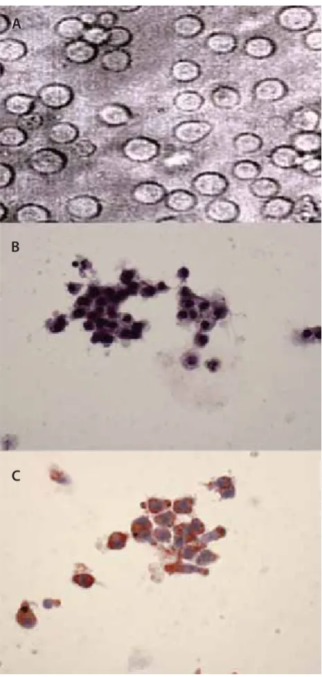
Several studies have reported that US exposure on adherent cells in the presence of microbubble contrast agents can in-duce cell detachment and sonoporation [, ]. Consistent with these studies, the substrate was partially cleared of cells following US exposure in the presence of the microbubble contrast agent-Sonovue (Figure A). Adherent cells, which have not been washed away but line the border between oc-cupied and empty region, emitted green fl uorescence under fl uorescence microscope duo to the uptake of calcein (Fig-ure B). No green fl uorescence was detected for cells far way from the sonciation-induced detachment (Figure B). It was also shown that cells with calcein uptake can be roughly di-vided into two subgroups: cells with high levels of calcein up-take (strong green fl uorescence, Figure B) and cells with low
Bosn J Basic Med Sci 2012; 12 (2): 66-68 JIZHEN ZHANG ET AL.: DIFFERENT EFFECTS OF SONOPORATION ON CELL MORPHOLOGY AND VIABILITYlevels of calcein uptake (weak green fl uorescence, Figure B). Changes in cell morphology immediately after sonoporation By using scanning electron microscope (SEM), it was shown that cells far away from the vacanted regions displayed rich and homogeneous distribution of microvilli on the cellular surfaces (Figure A). While, various changes in cell surface morphology for those cells surrounding the vacanted re-gions could be detected: smooth surface (Figure B), pores
in membrane (Figure C) and ir-regular cell surface (Figure D).
Changes in cell viability after sono-poration
Under fluorescence microscope, it was shown that immediately after sonoporation, both groups of cells with high levels of calcein uptake and low levels of calcein uptake were viable as evidenced by no staining with PI (Figure A); h after sonoporation, group of cells with low levels of cal-cein uptake still remained viable (Figure A-B, negative PI stain-ing as indicated by the long ar-row), while group of cells with high levels of calcein uptake died (Figure A-B, PI staining changed from negative into positive as indicated by the short arrow).
DISCUSSION
This study showed different in-tracellular calcein uptake and changes in cell morphology and viability after sonoporation. Fur-thermore, group of cells with low levels of calcein uptake re-mained viable h after sonopora-tion, while group of cells with high levels of calcein uptake died. Sonoporation is defi ned as forma-tion of transient, nonspecifi c pores or openings in the cellular mem-branes upon US exposure []. Acoustic cavitation produced by
US exposure is believed to be the main physical mechanism caused by US exposure [, ]. Acoustic cavitation is the process entail-ing bubbles formation, growth, vibration or even collapse in the medium under US activation []. Microbubble con-trast agents could act as the acoustic cavitation nuclei and produce acoustic cavitation under ultrasound exposure []. It is currently believed that mechanical wounding on cells due to acoustic cavitation is the predominant mechanism of action of sonoporation []. Several studies have investigated the changes in cell morphology immediately after sonication
FIGURE 2. Diff erent levels of intracellular calcein uptake immediately after sonoporation im-aged by fl uorescence microscope. (A, bright fi eld) The substrate is partially cleared of cells by sonication(as indicated by the circle). (B) Cells lining at the border between occupied and empty region showed roughly two diff erent levels of calcein uptake: low levels of calcein uptake (weak green fl uorescence as indicated by short arrow) or high levels of uptake (strong green fl uorescence as indicated by long arrow). Cells far away from the vacant region showed no calcein uptake.
Bosn J Basic Med Sci 2012; 12 (2): 67-68
JIZHEN ZHANG ET AL.: DIFFERENT EFFECTS OF SONOPORATION ON CELL MORPHOLOGY AND VIABILITY
[, , ]. However, results vary and most are focused on observing the pores in the membrane. In our study, we ob-served diff erent changes in cell morphology immediately af-ter sonoporation; these included: smooth surface, pores in the membrane and irregular cell surface . It is noteworthy that in our study smooth surface was the most common change while pores were rarely seen. One possible explanation is that the pores may have been resealed before cell fi xation []. Our study further showed that some cells containing high levels of calcein uptake died h after sonopora-tion; while those with low levels of calcein uptake sur-vived. It has been reported that fractions of cells with various levels of molecular uptake of calcein can be af-fected by changing the US exposure intensity []. There-fore, combined with the study by Zarnitsyn et al. [], it is suggested that levels of molecular uptake of calcein may be consistent with the levels of cell membrane wounds . The exact mechanism regarding how cellular membrane wounding results in delayed cellular death is still un-known. Several studies have reported that there is an im-mediate calcium ion influx following mechanical wound-ing (includwound-ing sonication) [, ]. Hutcheson et al. [] successfully rescued up to of cells with high levels of calcein uptake from apoptosis by the addition of a calcium ionic chelator. However, several studies also show that im-mediate calcium ion influx after sonication is vital to the wound repairing [-]. Th erefore, calcium ion infl ux after sonication may play a complex role in sonoporation []. However, there is still one main limitation in our study since we did not measure the accurate distribution of the acous-tic fi eld in our experimental setup. Nonetheless, the aim of our study is not to optimize the exposure parameters, but
to observe the changes in cell morphology of cells with mo-lecular uptake under this simple experimental model [, ].
CONCLUSION
Our study showed different ef-fects of sonoporation on in-tracellular calcein uptake, cell morphology and viability. It was suggested that various changes in cell morphology may be respon-sible for diff erent levels of intracel-lular calcein uptake and changes in cell viability after sonoporation.
ACKNOWLEDGEMENTS
The authors would like to thank the following Profes-sors: Qian Cheng (Institute of Acoustics, Tongji Univer-sity, Shanghai, China) for ultrasound parameters calibra-tion and Wen-de Shou (Shanghai Institute of Ultrasound in Medicine, Shanghai, China) for acoustic theory consultation.
DECLARATION OF INTEREST
This work was supported in part by the National Natu-ral Science Foundation of China (grant ) and Shanghai Science and the Technology Commit-tee Basic Research Program (grant JC). All authors have read and approved this manu-script. Neither the submitted paper nor any similar pa-per, in whole or in part has been or will be published in any other primary scientific journal. No conflict of interest exists in the submission of this manuscript.
REFERENCES
[] Langer R. Drug delivery and targeting. Nature ; ( Sup-pl):-.
[] Feril LB, Jr. Ultrasound-mediated gene transfection. Methods Mol Biol ;:-.
[] Mitragotri S. Healing sound: the use of ultrasound in drug de-livery and other therapeutic applications. Nat Rev Drug Discov ;():-.
[] Pua EC, Zhong P. Ultrasound-mediated drug delivery. IEEE Eng Med Biol Mag ; ():-.
[] Zderic V. Ultrasound-enhanced drug and gene delivery: a review. Conf Proc IEEE Eng Med Biol Soc ; :.
[] Li W, Liu S, Ren J, Xiong H, Yan X, Wang Z. Gene transfection to retinal ganglion cells mediated by ultrasound microbubbles in vitro.
Bosn J Basic Med Sci 2012; 12 (2): 68-68 JIZHEN ZHANG ET AL.: DIFFERENT EFFECTS OF SONOPORATION ON CELL MORPHOLOGY AND VIABILITYAcad Radiol ;():-.
[] Negishi Y, Matsuo K, Endo-Takahashi Y, Suzuki K, Matsuki Y, Tak-agi N, et al. Delivery of an angiogenic gene into ischemic muscle by novel bubble liposomes followed by ultrasound exposure. Pharm Res ; ():-.
[] Mohan P, Rapoport N. Doxorubicin as a molecular nanotheranos-tic agent: eff ect of doxorubicin encapsulation in micelles or nano-emulsions on the ultrasound-mediated intracellular delivery and nuclear traffi cking. Mol Pharm ;():-.
[] Kudo N, Okada K, Yamamoto K. Sonoporation by single-shot pulsed ultrasound with microbubbles adjacent to cells. Biophys J ; ():-.
[] Schlicher RK, Radhakrishna H, Tolentino TP, Apkarian RP, Zarnit-syn V, Prausnitz MR. Mechanism of intracellular delivery by acous-tic cavitation. Ultrasound Med Biol ; (): -.
[] Fan Z, Kumon RE, Park J, Deng CX. Intracellular delivery and cal-cium transients generated in sonoporation facilitated by microbub-bles. J Control Release ;():-.
[] Guzman HR, Nguyen DX, McNamara AJ, Prausnitz MR. Equilib-rium loading of cells with macromolecules by ultrasound: eff ects of molecular size and acoustic energy. J Pharm Sci ; ():-.
[] Hutcheson JD, Schlicher RK, Hicks HK, Prausnitz MR. Saving cells from ultrasound-induced apoptosis: quantifi cation of cell death and uptake following sonication and eff ects of targeted calcium chelation. Ultrasound Med Biol ; ():-.
[] Mehier-Humbert S, Bettinger T, Yan F, Guy RH. Plasma mem-brane poration induced by ultrasound exposure: implication for drug delivery. J Control Release ;():-.
[] Zarnitsyn V, Rostad CA, Prausnitz MR. Modeling transmembrane transport through cell membrane wounds created by acoustic cavi-tation. Biophys J ;():-.
[] Wu Xian-mei Qian Meng-lu. Vibration measuring technique using laser interferometer for calibration of transducers. TECHNICAL ACOUSTICS ;():-.
[] Ohl CD, Wolfrum B. Detachment and sonoporation of adherent HeLa-cells by shock wave-induced cavitation. Biochim Biophys Acta ;(-):-.
[] Zarnitsyn VG, Prausnitz MR. Physical parameters infl uencing op-timization of ultrasound-mediated DNA transfection. Ultrasound Med Biol ;():-.
[] Tachibana K, Uchida T, Ogawa K, Yamashita N, Tamura K.
Induction of cell-membrane porosity by ultrasound. Lancet ;():.
[] Forbes MM, Steinberg RL, O'Brien WD, Jr. Examination of inertial cavitation of Optison in producing sonoporation of chinese ham-ster ovary cells. Ultrasound Med Biol ; (): - [] Lai CY, Wu CH, Chen CC, Li PC. Quantitative relations of acoustic
inertial cavitation with sonoporation and cell viability. Ultrasound Med Biol ; ():-.
[] Apfel RE. Acoustic cavitation: a possible consequence of biomedi-cal uses of ultrasound. Br J Cancer Suppl ; :-. [] Karshafi an R, Bevan PD, Williams R, Samac S, Burns PN.
Sonopor-ation by ultrasound-activated microbubble contrast agents: eff ect of acoustic exposure parameters on cell membrane permeability and cell viability. Ultrasound Med Biol ;():-. [] Schlicher RK, Hutcheson JD, Radhakrishna H, Apkarian RP,
Praus-nitz MR. Changes in cell morphology due to plasma membrane wounding by acoustic cavitation. Ultrasound Med Biol ; ():-.
[] Zhou Y, Kumon RE, Cui J, Deng CX. The size of sonopora-tion pores on the cell membrane. Ultrasound Med Biol ; ():-.
[] Deng CX, Sieling F, Pan H, Cui J. Ultrasound-induced cell mem-brane porosity. Ultrasound Med Biol ;():-. [] Kumon RE, Aehle M, Sabens D, Parikh P, Han YW, Kourennyi
D, et al. Spatiotemporal eff ects of sonoporation measured by real-time calcium imaging. Ultrasound Med Biol ; ():-. [] Kumon RE, Aehle M, Sabens D, Parikh P, Kourennyi D, Deng CX.
Ultrasound-induced calcium oscillations and waves in Chinese hamster ovary cells in the presence of microbubbles. Biophys J ; (): L-.
[] Hassan MA, Campbell P, Kondo T. The role of Ca(+) in ultra-sound-elicited bioeffects: progress, perspectives and prospects. Drug Discov Today ;(-):-.
[] Kodama T, Tomita Y, Koshiyama K, Blomley MJ. Transfection eff ect of microbubbles on cells in superposed ultrasound waves and behavior of cavitation bubble. Ultrasound Med Biol ; ():-.
Bosn J Basic Med Sci 2012; 12 (2): 69-73
Abstract
Th e objective of our study was to investigate the eff ect of Aliskiren, a renin inhibitor, on the deoxycorticosterone (DOCA) induced myocardial fi brosis in a rat model and its underlying mechanism. A total of Sprague-Dawley (SD) rats underwent right nephrectomy and were ran-domly assigned into groups: control group (CON group: silicone tube was embedded subcutaneously); DOCA treated group (DOC group: mg of DOCA was subcutaneously administered); DOCA and Aliskiren (ALI) treated group (ALI group: mg of DOCA and mg/ kg/d ALI were subcutaneously and intragastrically given, respectively). Treatment was done for weeks. Sirius red staining was employed to detect the expression of myocardial collagen, and the myocardial collagen volume fraction (CVF) and perivascular collagen volume area (PVCA) were calculated. Radioimmunoassay was carried out to measure the renin activity (RA) and content of angiotensin II (Ang II) in the plasma and ventricle. Western blot assay was done to detect the expressions of extracellular signal-regulated kinase / (ERK/), phosphory-lated ERK/ (PERK/) and matrix metalloproteinase (MMP-). In the DOC group and ALI group, the CVF and PVCA were signifi cantly increased; the RA and Ang II levels in the plasma and ventricle were remarkably lowered when compared with the CON group. Th e RA and Ang II levels in the ventricle of the ALI group were signifi cantly lower than those in the DOC group. Moreover, the expressions of ERK/, PERK/ and MMP were the lowest in the CON group, but those in the ALI group were signifi cantly reduced as compared to the DOC group. ALI can inhibit the DOCA induced myocardial fi brosis independent of its pressure-lowing eff ect, which may be related to the suppres-sion of RA and Ang II production, inhibition of ERK/ phosphorylation and MMP expressuppres-sion in the heart.
© Association of Basic Medical Sciences of FBIH. All rights reserved
KEY WORDS: Aliskiren, deoxycorticosterone, myocardial fi brosis, renin- angiotensin - aldosterone system
Anti-fi brotic eff ect of Aliskiren in rats with
deoxycorticosterone induced myocardial
fi brosis and its potential mechanism
Likun Ma1*, Jinsheng Hua1, Lifeng He1, Qian Li1, Junling Zhou1, Jiangtao Yu2
1 Department of Cardiology, Anhui Provincial Hospital, No 17 Lujiang Road, Hefei 230001, China. 2 Department of Cardiology, Zentralklinik
Bad Berka GmbH, Robert-Koch-Allee 9, Bad Berka, DE- 99437, Germany.
INTRODUCTION
Myocardial fibrosis is a common pathological feature shared by several heart diseases at the end stage. To pre-vent or even reverse myocardial fi brosis has been a key goal in the prevention and treatment of severe cardiovascular events including heart failure, arrhythmia and sudden car-diac death. Studies have demonstrated that renin - angio-tensin - aldosterone system (RAAS) plays important roles in the regulation of myocardial collagen metabolism and the occurrence of myocardial fi brosis [, ]. Evidence shows Aliskiren (ALI), a new rennin inhibitor, can not only lower the blood pressure, but improve the myocardial fi brosis and subsequent remodeling via its infl ammatory and anti-oxidative eff ects [, ]. Currently, the anti-fi brotic eff ect of ALI and its potential mechanism are less studied. Th e
pres-ent study aimed to investigate the cardioprotective effect of ALI on the deoxycorticosterone (DOCA) induced myo-cardial fi brosis and its potential mechanism in a rat model.
MATERIALS AND METHODS
Reagents
ALI (Novartis, Switzerland), DOCA (Sigma, USA), pri-mary antibodies against β-actin, extracellular signal kinase / (ERK/), phosphorylated ERK/ (PERK/) and metalloproteinase- (MMP-) (Santa Cruz, USA), second-ary antibodies, two - quinolinecarboxylic acid (BCA) (Bei-jing Zhongshan Golden-Bridge Biotech, China), electro-chemiluminescence (ECL) kit (Pierce), Sirius red (Beijing Haide Biotech, China) and radioimmunoassay kit (Beijing Yuanzi Biotech, China) were used in the present study.
Grouping and modelling
A total of Sprague-Dawley (SD) male rats weighing ~ g were purchased from the Animal Center of An-hui Medical University. Th e investigation conforms with the Guide for Care and Use of Laboratory Animals published by
* Corresponding author: Likun Ma,
Department of Cardiology of Anhui Provincial Hospital, No 17 Lujiang Road, Hefei 230001, China
Tel:+86551-2283339; Fax : +86551-2282121 e-mail: [email protected]
Bosn J Basic Med Sci 2012; 12 (2): 70-73 LIKUN MA ET AL.: ANTIFIBROTIC EFFECT OF ALISKIREN IN RATS WITH DEOXYCORTICOSTERONEINDUCED MYOCARDIAL FIBROSIS AND ITS POTENTIAL MECHANISM
the US National Institutes of Health (NIH Publication, th edition). Animals were randomly assigned into groups (n= per group): control (CON) group, DOCA (DOC) group and ALI (ALI) group. Following anesthesia with intra-peritoneal chloral hydrate ( mg/kg), right nephrec-tomy was done in these animals. One week after surgery, animals were given access to NaCl and received following treatments. In the CON group, silicone tube was embedded subcutaneously in the left lower abdomen. In the DOC group, mg of DOCA were embedded in the left lower
abdo-men. In the ALI group, mg of DOCA were embedded in the left lower abdomen and animals were intragastrically treated with ALI at mg/kg/d for weeks. In addition, ani-mals in the CON group and DOC group were also intragas-trically treated with normal saline for consecutive weeks.
Measurement of blood pressure and sample collection
At the end of treatment, rats were intraperitoneally anes-thetized with chloral hydrate followed by cannulation of right carotid artery for the measurement of mean arte-rial blood pressure (MABP) using a multi-channel physi-ological recorder. Th en, ml of blood were collected into the anti-coagulated tube and rats were sacrifi ced by exsan-guinations. Th oracotomy was performed and the heart col-lected. The atrium, major vessels and connective tissues were removed and the left ventricle was divided into two: one was fi xed in paraformaldehyde followed by process-ing for histological stainprocess-ing for collagen; the other was stored at -oC for the detection of protein expression by western
blot assay. Blood was centrifuged at rpm/min for min and the plasma was collected and stored at -oC for use.
Detection of myocardial fi brosis
Th e heart tissues were embedded in the paraffi n and cut into sections followed by Sirius red staining for collagen. A to-tal of fi elds without blood vessels were randomly selected from each section and representative photographs were captured with Nikon camera followed by analysis using Image-Pro plus . image analysis system. Th e collagen area and total area of each fi eld were measured, and myocardial collagen volume fraction (CVF) was calculated as collagen area / total area. Th e CVFs from fi elds were averaged and used as the final CVF of this sample. In addition, fields with small blood vessels at cross section were randomly col-lected, and the perivascular collagen volume area (PVCA) and lumen area (LA) determined. The PVCA was nor-malized by the LA (PVCA/LA). Th e PVCAs from fi elds were averaged and used as the fi nal PVCA of this sample.
Detection of renin and angiotensin II by radioimmunoassay
Briefly, in each group, mg of ventricle were
homoge-nized, and then the homogenate and plasma were indepen-dently divided into two: one was kept at oC for h which
may facilitate the binding of renin (RA) to angiotensinogen producing angiotensin I (Ang I) (experiment group); the other was stored at oC serving as controls. Th e Ang I level
in the experiment group and control group was measured by radioimmunoassay according to the manufacturer’s in-structions. The difference in Ang I between experiment group and control group was normalized by the time of in-cubation as the production rate of Ang I. The production rate of Ang I in unit time was defi ned as the renin activity of this sample and the unit was ng /ml/h. According to the manufacturer’s instructions, the Ang II level in the plasma and ventricle was also measured and its unit was pg/ml.
Detection of ERK/, PERK/ and MMP expressions in the ventricle by Western blot assay
Th e ventricle was homogenized and total protein extracted followed by detection of protein concentration. Th en, the protein concentration of different sample was adjusted to the same level and μg of proteins were subjected to polyacrylamide gel electrophoresis. Th e protein was trans-ferred onto nitrocellulose membrane. Following washing and blocking overnight, the membrane was treated with primary antibody (:) at room temperature for h un-der continuous shaking. After washing, the membrane was incubated with secondary antibody (:) at room tem-perature for h under continuous shaking. Visualization was done using ECL kit and X-ray fi lm was obtained. Th e bands were scanned using Bio-Rad image system and the optical density (OD) was determined followed by analysis with Quantity-one. Th e OD of target genes was normalized by that of β-actin as the relative expression of target genes.
Statistical analysis
Statistical analysis was performed using SPSS ver-sion .. Qualitative data were expressed as mean ± standard deviation (X±s). Means among groups were compared with one way analysis of variance, and com-parisons of rate were done with chi square test. A val-ue of p<. was considered statistically significant.
RESULTS
MABP in diff erent groups
Bosn J Basic Med Sci 2012; 12 (2): 71-73
LIKUN MA ET AL.: ANTIFIBROTIC EFFECT OF ALISKIREN IN RATS WITH DEOXYCORTICOSTERONE INDUCED MYOCARDIAL FIBROSIS AND ITS POTENTIAL MECHANISM
Collagen content in the ventricle of diff erent groups
Following Sirius red staining, the collagens were scarlet and non-collagen tissues yellow (Figure . and .). Th e CVF and PVCA in diff erent groups are shown in Table . When com-pared with the control group, the CVF and PVCA in the DOC group and ALI group were markedly increased (p<. and p<., respectively). However, the CVF and PVCA in the ALI group was dramatically lower than those in the DOC group (p<. and p<., respectively). Th is fi nding implies ALI improves the DOC induced myocardial fi brosis.
RA and Ang II levels in the plasma and ventricle
The RA level of the plasma and ventricle of DOC group and ALI group was significantly lower than those in the CON group (p<. or p<.). Signifi cant diff erence in the RA level was found in the ventricle between DOC group and ALI group (p<.), but marked diff erence was absent in the RA level of the plasma (p>.). Th e Ang II level of the plasma and ventricle of the DOC group and ALI group was also significantly decreased when compared with CON group (p<.). Significant difference in the Ang II
level was observed between ALI group and DOC in the ventricle (p<.) but not in the plasma (p>.)(Table ).
Protein expressions of ERK/, PERK/ and MMP- in the ventricle
The protein expressions of ERK/, PERK/ and MMP- are shown in Figure . Analysis showed the expres-sions of ERK/, PERK/ and MMP- in the CON group were the lowest, followed by ALI group, and those in the DOC group the highest. Significant difference in the ex-pressions of ERK/, PERK/ and MMP- was found between any two groups (p<.). Moreover, the expres-sions of ERK/, PERK/ and MMP- in the ALI group were significantly higher than those in the CON (p<.) but lower than those in the DOC group (p<. or p<.).
DISCUSSION
In the present study, the DOCA induced myocardial fi-brosis rat model was employed and the anti-fibrotic
ef-FIGURE 1. Staining of collagen in the ventricle (×400; A: control; B: DOCA; C: ALI)
FIGURE 2. Staining of blood vessels in the ventricle (×400; A: control; B: DOCA; C: ALI)
CON group DOC group ALI group (n=13) (n=11) (n=12) CVF 4.71±0.60 14.35±1.41* 8.11±1.13**#
PVCA 16.94±1.02 26.17±1.95** 21.22±1.31**##
TABLE 1. CVF and PVCA in diff erent groups (X±s)
Note: * p <0.05 and ** p<0.01 vs CON group; #p <0.05 and ##p<0.01 vs DOC group.
CON group DOC group ALI group (n=13) (n=11) (n=12) Plasma RA 47.10±4.00 2.40±0.46** 2.22±0.21** Ventricle RA 6.13±0.20 3.17±0.30* 2.17±0.20*#
Plasma Ang II 17.98±4.70 10.13±3.10* 8.05±2.60* Ventricle Ang II 4.11±0.87 1.80±0.34* 1.12±0.22*#
TABLE 2. RA and Ang II levels in the plasma and ventricle of dif-ferent groups (X±s)
Bosn J Basic Med Sci 2012; 12 (2): 72-73 LIKUN MA ET AL.: ANTIFIBROTIC EFFECT OF ALISKIREN IN RATS WITH DEOXYCORTICOSTERONEINDUCED MYOCARDIAL FIBROSIS AND ITS POTENTIAL MECHANISM
fect of ALI was investigated. Our findings revealed ALI could improve the myocardial fibrosis demonstrated by Sirius red staining, reduce the RA and Ang II levels in the ventricle and the expressions of ERK/, PERK/ and MMP as compared to the DOCA treated animals. Myocardial fi brosis refers to the aberrant deposition of ex-tracellular matrix (ECM) in the heart, and characterized by increase of collagen in the interstitium, imbalance and irregu-lar arrangement of diff erent types of collagen. Th e imbalance between the synthesis and degradation of collagen is a major cause of myocardial fi brosis. Under the pathological condi-tions, the proliferation and phenotype of cardiac fi broblasts change and these cells produce a large amount of collagens re-sulting in imbalance between type I and type II collagens and subsequent deposition of collagens between myocardial cells. MMPs play critical role in the myocardial fi brosis through af-fecting the degradation of ECM. MMP can degrade normal collagens such as gelatin, elastin, collagen type IV collagen, type V collagen and mucin but has no infl uence on abnor-mal collagens []. In the myocardial fi brosis, the myocardial interstitium is occupied by abnormal collagens (mainly type I and III collagens). Under pathological conditions, the MMP- expression is increased and then degrades the normal colla-gens. Th us, the myocardial interstitium between myocardial cells loses and a large amount of abnormal collagens gener-ated. Westermann et al. [] investigated the effect of ALI on the rat myocardial infarction. Th eir results showed ALI could reduce the deposition of collagen in the infarction re-gion and subsequent myocardial fibrosis via reducing the MMP- activity. Our results also revealed the MMP- ex-pression was markedly elevated in rats with DOCA induced
myocardial fibrosis accompanied by increased extracellu-lar deposition of collagen. However, following ALI treat-ment, the MMP- expression and deposition of collagen were markedly decreased and myocardial fi brosis improved. In addition, studies have shown that RAAS plays an impor-tant role in the regulation of collagen metabolism in the heart and blood vessels, and Ang II and aldosterone are two major eff ector proteins [, ]. Th ere is evidence that angiotensin-converting enzyme inhibitors (ACEI), angiotensin receptor blockers (ARB) and aldosterone receptor antagonists can significantly improve the fibrosis []. However, long-term application of ACEI and ARB may lead to the Ang II escape []. It has been confirmed that Ang II can be synthesized via both ACE-dependent and ACE-independent pathways. Continuous use of ACEI and ARB may cause accumulation of Ang I and activate ACE-independent pathways resulting in increase in Ang II. Of note, - of Ang II in the tissues is synthesized via ACE-independent pathways []. ALI, a renin inhibitor, acts on the fi rst rate-limiting step of RAAS chain and blocks the activation of RAAS leading to the de-crease in Ang II and aldosterone production []. Th us, theo-retically, ALI has potent anti-fi broric eff ect on myocardial fi brosis. Singh et al. [] showed, in streptomycin induced hyperglycemia rats, the ALI was superior to benazepril and losartan in improving myocardial fi brosis and apoptosis of myocardial cells. In the present study, the blood pressure was not dramatically lowered following administration of ALI, but the DOCA induced myocardial fi brosis was obvi-ously improved. Studies showed the ventricular RAAS plays a more critical role in the myocardial fi brosis than circulation RAAS does [, ]. Our study further confi rmed the RA and
Bosn J Basic Med Sci 2012; 12 (2): 73-73
LIKUN MA ET AL.: ANTIFIBROTIC EFFECT OF ALISKIREN IN RATS WITH DEOXYCORTICOSTERONE INDUCED MYOCARDIAL FIBROSIS AND ITS POTENTIAL MECHANISM
Ang II in the circulation were largely unchanged followed ALI treatment, but those in the heart were dramatically de-creased. These findings suggest the anti-fibrotic effect of ALI is related to the suppression of local Ang II production. Currently, the mechanisms underlying the anti-fi brotic eff ect of ALI are not completely clear. Our results showed the anti-fi brotic eff ect of ALI may be attributed to the suppression of phosphorylation of ERK/ signaling pathway. In recent years, studies show the Ang II/angiotensin receptor (ATR) mediated myocardial fi brosis is related to the ERK/ signal-ing pathway []. Th e binding of Ang II to ATR can activate the phospholipase C on the cell membrane resulting in the hydrolysis of phosphatidylinositol phosphate and produc-tion of diacylglycerol. Th e later promotes the release of Ca+
in the sarcoplasmic reticulum and endoplasmic reticulum. Ca+ can act as a second messenger to activate ERK/
sig-naling pathway. Th e activation of ERK/ signaling pathway may facilitate the transcription of early response genes (such as c-fos) in the cardiac fibroblasts, skeletal muscle α-actin gene, β-myosin heavy chain gene and embryonic contrac-tile protein gene and promote the expression of growth fac-tors in fi broblasts. In addition, a large amount of ECM and collagen are produced, the proliferation of fi broblasts pro-moted and expressions of Iα and IIIα increased resulting in myocardial fi brosis [, ]. Our results showed the ex-pressions of ERK/ and PERK/ in the ALI group were markedly decreased when compared with the DOC group. We speculate that ALI can decrease the production of
An-gII and local RA to reduce the Ang II/ATR induced activa-tion of ERK/, which fi nally exerts the anti-fi brotic eff ects.
CONCLUSION
Evidence shows that RAAS plays important roles in the oc-currence of myocardial fibrosis and ALI has anti-fibrotic effect via its anti-inflammatory and anti-oxidative effects. Th e present study demonstrates that ALI as an inhibitor of
renin can improve the DOCA induced myocardial fi brosis, which may be attributed to the suppression of myocardial RAAS, decrease in Ang II level and inhibition of phosphory-lation of ERK/ signaling pathway and MMP- expression. However, more studies are required to confi rm our results.
ACKNOWLEDGEMENTS
Th is study was supported by the International Cooperation Projects of Department of Science & Technology of Anhui Province () (No: )
DECLARATION OF INTEREST
We declare no confl ict of interest
REFERENCES
[] Ma TK, Kam KK, Yan BP, Yam YY. Renin-angiotensin-aldosterone system blockade for cardiovascular diseases: current status. Br J Pharmacol ; ():-.
[] Susic D, Varagic J, Frohlich ED. Cardiovascular eff ects of inhibition of renin- angiotensin-aldosterone system components in hyperten-sive rats given salt excess. Am J Physiol Heart Circ Physiol ; (): H-.
[] Gradman AH, Schmieder RE, Robert L, Nussberger J, Chiang Y, Bedigian MP. Aliskiren, a novel orally eff ective renin Inhibitor, pro-vides dose-dependent antihypertensive effi cacy and placebo-like tolerability in hypertensive patients. Circulation ; (): -.
[] Westermann D, Riad A, Lettau O, Roks A, Savvatis K, Becher PM, et al. Renin inhibition improves cardiac function and remodeling after myocardial infarction independent of blood pressure. Hyper-tension ; ():-.
[] Li J, Schwimmbeck PL, Tschope C, Leschka S, Husmann L, Rutschow S, et al. Collagen degradation in a murine myocarditis model: relevance of matrix metalloproteinase in association with infl ammatory induction. Cardiovasc Res. ;():-. [] Johar S, Cave AC, Narayanapanicker A, Grieve DJ, Shah AM.
Aldo-sterone mediats angiotensin II-induced interstitial cardiac fi brosis via a Nox-containing NADPH oxidase. Th e FASEB Journal ; ():-.
[] Brilla CG. Aldosterone and myocardial fibrosis in heart failure. Herz ; (): - .
[] Athyros VG, Mikhailidis DP, Kakafi ka AI, Tziomalos K, Karagi-annis A. Angiotensin II reactivation and aldosterone escape phe-nomena in renin-angiotensin-aldosterone system blockade: is oral renin inhibition the solution? Expert Opin Pharmacother. ; (): -.
[] Xu J, Oscar A, Carretero B, Peng H, Shesely EG, Xu J, et al. Local angiotensin II aggravates cardiac remodeling in hypertension. Am J Physiol Heart Circ Physiol ; (): H-.
[] Nussberger J, Wuerzner G, Jensen C, Brunner HR. Angiotensin II suppression in humans by the orally active renin inhibitor aliskiren (SPP): comparison with enalapril. Hypertension ; (): E-.
[] Singh VP, Le B, Khode R, Baker KM, Kumar R. Intracellular angio-tensin II production in diabetic rats is correlated with cardiomyo-cyte apoptosis, oxidative stress, and cardiac fi brosis. Diabetes ; (): -.
[] Zhong JC, Basu R, Guo D, Chow FL, Byrns S, Schuster M, et al. An-giotensin-converting-enzyme suppresses pathological hypertro-phy, myocardial fi brosis, and cardiac dysfunction. Circulation ; (): -.
[] Olson ER, Shamhart PE, Naugle JE, Meszaros JG. Angiotensin II-induced extracellular signal-regulated kinase / activation is medi-ated by protein kinase Cdelta and intracellular calcium in adult rat cardiac fi broblasts. Hypertension ; ():-.
[] Tang W, Wei Y, Le K, Li Z, Bao Y, Gao J, et al. Mitogen-activated protein kinases ERK /- and p-GATA pathways mediate the Ang II-induced activation of FGF gene in neonatal rat cardiomyo-cytes. Biochem Pharmacol ; ():-.
Bosn J Basic Med Sci 2012; 12 (2): 74-81Abstract
Trefoil Factor Family (TFF) plays an essential role in the intestinal epithelial restitution, but the relationship between TFF and gastric cancer (GC) is still unclear. Th e present study aimed to determine the role of TFF in repairing gastric mucosa and in the pathogenesis of GC. Th e TFF expression in diff erent gastric mucosas was measured with immunohistochemistry. Th en, siRNA targeting TFF or plasmids
ex-pressing TFF gene were transfected into BGC cells, SGC cells and GES- cells. Th e cell proliferation was detected with MTT assay and apoptosis and cell cycle measured by fl ow cytometry.
From normal gastric mucosa to mucosa with dysplasia and to gastric cancer, the TFF expression had a decreasing trend. Down-regulation of TFF expression signifi cantly reduced the apoptosis of three cell lines and markedly facilitated their proliferation but had no signifi cant eff ect on cell cycle. Over-expression of TFF could promote apoptosis of three cell lines and inhibit proliferation but had no pronounced eff ect on cell cycle. TFF can inhibit proliferation and induce apoptosis of GC cells in vitro.
© Association of Basic Medical Sciences of FBIH. All rights reserved
KEY WORDS: Trefoil Factor Family , gastric cancer, apoptosis, in vitro
TFF1 inhibits proliferation and induces
apoptosis of gastric cancer cells in vitro
Yanli Ge#, Junjie Zhang#, Jianchun Cao#, Qiong Wu, Longe Sun, Likun Guo, Zhirong Wang*
Department of Gastroenterology, Tongji Hospital, Tongji University, Shanghai200065, China
INTRODUCTION
Although the incidence of gastric cancer (GC) has de-clined over the past years worldwide, especially in western countries, it remains the second leading cause of cancer-related death and accounts for . of cancer deaths globally []. About GC cases and cancer deaths are estimated to have occurred in worldwide []. More than one-half of GC patients have lymph node metastases when they are initially diagnosed or operated, which usually results in poor prognosis [-]. Therefore, it is important to investigate the pathogen-esis of GC, fi nd eff ective measures to prevent and treat GC. Th e trefoil factor family (TFF), which comprises gastric pep-tides pS/TFF and spasmolytic peptide (SP)/TFF and in-testinal trefoil factor (ITF)/TFF, plays an essential role in the intestinal epithelial restitution []. Th ey are small (~ kDa) protease-resistant proteins and are abundantly secreted onto the mucosal surface by mucus-secreting cells in the
gastroin-testinal tract. Th e TFFs share an absolutely conserved distinct motif of six cysteine residues that defi ne a so-called “trefoil” domain, which is also known as a “P” domain []. At certain physiological conditions, in the presence of a tissue-specifi c distribution, TFF plays an important role in mucosal protec-tion and wound healing. But in malignant tissues, TFF is high-ly expressed and correlated stronghigh-ly with the genesis, metas-tasis and invasion of tumor cells. Th ese indicate that TFF may be a common mediator of oncogenic responses to diff erent stimuli. Th e biological functions of TFF involve complex reg-ulatory processes. TFF was fi rst discovered in breast cancer cell line MCF- in [-]. In normal tissues, the main site of expression of TFF is the mucosal epithelial cells of gastric body and antrum in a site-specifi c fashion. However, under pathological conditions, such as ulceration, the TFF-expres-sion of site-specifi c fashion is absent, TFF can be identifi ed in any damaged mucosas, and its expression is up-regulated to participate in gastrointestinal epithelial reconstruction and repair process []. However, there is lack of TFF ex-pression in human GC. To determine the function of TFF, the mouse TFF gene was inactivated. Th e antral and pyloric gastric mucosa of mpS-null mice was dysfunctional and exhibited severe hyperplasia and dysplasia. All homozygous mutant mice developed antropyloric adenoma, and de-veloped multifocal intraepithelial or intramucosal carcino-mas. Th ese results indicate that TFF is essential for the
nor-# These authors contributed equally to this work. * Corresponding author: Zhirong Wang, Department of Gastroenterology, Tongji Hospital, Tongji University, Shanghai 200065, China Tel: +8621-66111329; Fax: +8621-56050502 E-mail: [email protected]
Bosn J Basic Med Sci 2012; 12 (2): 75-81
YANLI GE ET AL.: TFF1 INHIBITS PROLIFERATION AND INDUCES APOPTOSIS OF GASTRIC CANCER CELLS IN VITRO
mal diff erentiation of antral and pyloric gastric mucosa and may function as a gastric-specifi c tumor suppressor gene []. Th e aim of our study is to determine the role of TFF in re-pairing gastric mucosa and in the pathogenesis of GC. To this end, the TFF expression was detected in different gastric mucosas. Then, siRNA targeting TFF and plas-mids expressing TFF were transfected into normal gastric mucosal epithelial cells (GES- cells []), highly malig-nant GC cell line (BGC cells) and moderately maligmalig-nant GC cell line (SGC cells), respectively, to investigate the effect of TFF on the biological behaviors of gastric mucosal epithelial cells, which provides evidence for fur-ther investigation and application of TFF target fur-therapy.
MATERIALS AND METHODS
Sample collection and immunohistochemistry
GC tissues and adjacent normal and atypical hyperplasia gastric mucosas were obtained from patients undergo-ing gastroscopy at Tongji Hospital of Tongji University. Th e characteristics of these patients are shown in Table . All antibodies used for immunohistochemistry were pur-chased from Zhongshan Goldenbridge Biotechnology CO., LTD (Beijing, China). All other chemicals and reagents were commercially available and had the highest purity. Tis-sues were fi xed in formaldehyde in phosphate buff ered saline (PBS), embedded in paraffi n, and cut into -μm sec-tions. Sections were heated at oC overnight, deparaffi nzed
with xylene twice, and rinsed in a decreasing ethanol series (-) for min/solution. Samples were treated with HO for min to inactivate endogenous peroxidase. Antigen retrieval was done with . M Na-citrate buffer (pH .) in a microwave oven for min. Sections were in-cubated at oC overnight in moist chambers with primary
antibody (:) and then with biotinylated secondary anti-body (Zhongshan Goldenbridge Biotechnology CO., LTD., Beijing, China, SP-Kit) for min at room temperature, fol-lowed by incubation with streptavidin peroxidase. Visual-ization was done with diaminobenziding tetrachloride and counterstaining was performed with haematoxylin. In nega-tive controls, the primary antibody was replaced with PBS. Sections were observed under a light microscope and positive cells had brown granules in cytoplasm. Five fi elds were randomly selected from each section at high mag-nification, and cells were counted in each field, fol-lowed by calculation of percentage of positive cells. Sec-tions with positive cells of > was regarded as positive.
Cell culture
Two gastric adenocarcinoma cell lines (BGC cells and SGC cells) (Cell bank of Chinese Academic of
Scienc-es), as well as normal gastric epithelial cell line (GES- cells) (Tumor Institute of Beijing Medical University, China) were maintained in RPMI medium (Gibco BRL, USA) supple-mented with fetal bovine serum (FBS; Hangzhou Sijiqing, China) and μg/ml streptomycin and penicillin G (Am-resco, USA) at oC under humidifi ed CO. Passaging
was performed every days by trypsinization (Sigma, USA). Synthesis of TFF-siRNA and Cell Transfection mRNA sequence of TFF was obtained from GeneBank. With Invitrogen's online design software BLOCK-iTTM RNAi Designer, three sites (stealth , stealth , stealth ), and they were selected and designed to be three sets of targeting stealth siRNA sequence. Th e selection and design were based on three principles: avoiding the ' and ' end non-coding region, selecting the sequences with G/C ratio between and and using BLAST to exclude other cod-ing sequences. Stealth siRNA sequences are shown in Table . When the cell confl uence reached about ~, transfec-tion of Stealth siRNAs and stealth RNA negative control was performed with LipofectamineTM according to manu-facturer’s instructions (Invitrogen, USA). Cells transfected with Stealth siRNAs was defined as Stealthgroup, those transfect with Stealth negative control as negative control group, and those without transfection as blank control group. Detection of TFF mRNA expression by RT-PCR Total RNA was extracted with Trizol reagent (Invitrogen, USA). About μg of RNA were used for reverse transcrip-tion with random primers to synthesize fi rst strand cDNA, followed by conventional PCR amplification with μl of cDNA as template. Th e forward primers, reverse primers and anticipated size of products and GAPDH are shown in Table . Th e volume of reaction system was μl, and reaction con-ditions were as follows: denaturation at oC for min;
an-n Age (yr) (mean ± SD)
Sex Male Female Normal gastric mucosa 15 56±13 7 8 Atypical hyperplasia gastric mucosa 15 55±8 9 11 GC mucosa 15 56±11 12 8
TABLE 1. Characteristics of 45 patients
siRNA target point Sequences of stealth siRNAs stealth_115 5’-CACUGUACACGUCUCUGUCUGGGCC-3’ stealth_144 5’-AAACCACAAUUCUGUCUUUCACGGG-3’ stealth_268 5’-AAAUUCACACUCCUCUUCUGGAGGG-3’
TABLE 2. Sequences of stealth siRNAs
gene Primers (5’-3’) Size (bp)
TFF1 Forward: ATGGCCACCATGGAGAACAA 159
Reverse: ATTTGCACACTGGGAGGGCG
GAPDH Forward: ACCACAGTCCATGCCATCAC 452 Reverse: TCCACCACCCTGTTGCTGTA
Bosn J Basic Med Sci 2012; 12 (2): 76-81 YANLI GE ET AL.: TFF1 INHIBITS PROLIFERATION AND INDUCES APOPTOSIS OF GASTRIC CANCER CELLS IN VITROnealing at oC for min; extension at oC for min. Th e
products were then subjected to agarose gel electrophoresis, and images were captured to analyze the quality of RNA.
Determination of cell proliferation with MTT assay
Cell viability was determined by MTT assay. Different cell lines were seeded at /ml into -well plates, and
then divided into blank control group, blank transfec-tion group, stealth_ group, stealth_ group and stealth_ group. At h after transfection, the cell pro-liferation was determined by MTT assay every other h for days. In brief, at designed time points, MTT was added into each well at a final concentration of . mg/ ml followed by incubation for h at oC. Th e formazan
was dissolved by addition of dimethylsulfoxide (DMSO) and absorbance (A) was measured with microplate reader (Bio-Rad, USA) at nm. Th e inhibition rate (IR) was cal-culated formulas follow: IR = (-Aexperiment/Acontrol)×.
Detection of cell cycle by fl ow cytometry
At h after transfection, cells (× cells) were harvested
and washed twice in cold PBS. Cells were fi xed in etha-nol and washed in cold PBS. Th en, the cells were suspend-ed in ml of propidium iodide (PI, Sigma, USA) solution containing μg/ml PI, μg/ml RNAase (Sigma, USA), . (w/v) sodium citrate and . (v/v) Triton X. Cells were incubated at room temperature in dark for at least min, and analyzed by flow cytometry (Beckman, USA).
Detection of cell apoptosis by fl ow cytometry
At h after transfection, × cells were harvested.
Ac-cording to the instructions in Annexin V-FITC kit (Nanjing Keygen Biotech. Co. Ltd., China), cells were suspended in μl of binding buffer and μl of An-nexin V-FITC followed by addition of μl of PI and subsequent incubation for min at room tempera-ture in dark. Apoptosis was detected by flow cytometry.
Construction of plasmid TFF-pcDNA. and cell transfection
Human plasmid TFF-pcDNA. was constructed and identified by Shanghai Shuiyuan Biotechnology Com-pany. Cells were divided into TFF-pcDNA. trans-fection group, pcDNA. negative control group and blank control group. Cells were seeded into -well plates. When cell confluence reached about , trans-fection was performed with LipofectamineTM ac-cording to manufacturer’s instructions (Invitrogen, USA).
Detection of TFF protein expression by western blot
Total protein was extracted and μg of proteins were subjected to polyacrylamide gel electrophoresis. Th en, the
proteins were transferred onto PVDF membrane, which were blocked for h at oC in non-fat milk and then
incubated with mouse anti-human TFF or GAPDH monoclonal antibody (Santa Cruz, USA; :) for . h at room temperature. After washing in PBST thrice ( min for each), the membrane was incubated with HRP-conjugated goat anti-mouse secondary antibody (Santa Cruz, USA; :) at room temperature for . h. Follow-ing rinsFollow-ing in PBS, visualization was done and images were captured with a Touching gel imaging system. In negative control group, primary antibody was replaced with PBS.
Determination of cell proliferation with MTT assay
Different cell lines were inoculated at /ml into -well
plates, and then divided into blank control group, TFF-pcDNA.-transfection group and pcDNA. transfec-tion group. At h after transfectransfec-tion, cell proliferatransfec-tion was measured with MTT assay every other h for days. The procedures of MTT assay were abovementioned.
Detection of cell cycle by fl ow cytometry
At h after transfection, cells (× cells) were harvested
and processed with above procedures. Cell cycle was mea-sured by fl ow cytometry.
Detection of cell apoptosis by fl ow cytometry
At h after transfection, × cells were harvested and
pro-cessed with above procedures. Cell apoptosis was detected by fl ow cytometry.
Statistical analysis
SPSS version .. statistical software was employed for statistical analysis and data were expressed as means ± standard deviation (X±s). Independent sample t-test was used to compare data between two groups and one-way ANOVA to compare date among multiple groups. If there were significant differences, a further least significant dif-ference method would be used for pairwise comparison. A value of p <. was considered statistically significant.
RESULTS
TFF Expressionin diff erent mucosal tissues
Bosn J Basic Med Sci 2012; 12 (2): 77-81
YANLI GE ET AL.: TFF1 INHIBITS PROLIFERATION AND INDUCES APOPTOSIS OF GASTRIC CANCER CELLS IN VITRO
GC, the TFF expression had a gradually decreasing trend, and significant difference in TFF expression was noted among groups (Table , Figure ). Th e positive expression rate of TFF was . in males (/) and . in females (/) showing no significant difference. The positive ex-pression rate of TFF was . in patients aged ≥ years (/) and . in those aged < years (/) showing no marked difference. These results suggest that TFF ex-pression was independent of both age and gender (Table ).
Stealth siRNA inhibited mRNA expression of TFF
Results from RT-PCT showed stealth siRNA significantly inhibited TFF expression in a time dependent manner. At h after transfection, the inhibition of TFF
expres-sion was present, and then reached a maximal level at h after transfection but became to reduce at h after trans-fection. Different stealth siRNAs inhibited the TFF ex-pression when compared with control group and blank transfection group. Th e inhibitory eff ect of stealth_ was the most obvious and, at h after transfection, the inhibi-tion rate of TFF expression in GES- cells, BGC cells and SGC cells was ., . and ., respec-tively. The inhibitory effect was comparable among dif-ferent cell lines. Although TFF expression in negative control group was slightly lower than that in control group, there was no significant difference (p>.) (Figure ).
Down-regulation of TFF reduced apoptosis rate and promoted cell proliferation but had no eff ect on cell cycle
MTT assay showed that the proliferation of three cell lines undergoing transfection with stealth siRNAs increased significantly at h and h after transfection (p<.), and the increase in steath_ group was the most obvi-ous and reached a peak level at h after transfection. Th e proliferation remained comparable among cells at h, h and h after transfection (p>.) (Table , Figure ). Th e apoptosis rate of three cell lines was signifi cantly reduced at h after TFF stealth siRNA transfection when compared with control group (p<.). Th e reduction of apoptosis rate in stealth_ group was most obvious and the apoptosis rate in BGC cells, SGC cells and GES- cells was reduced by ., . and ., respectively. However, there was no
*p < 0.05 vs normal gastric mucosa
Group Total Positive
(N)
Negative (N)
Positive rate (%)
gender
male 28 23 5 82.1
female 27 21 6 77.8
age
≥60 yr 31 24 7 77.4
<60 yr 24 20 4 83.3
gastric mucosas
normal gastric mucosa 15 15 0 100
dysplasic gastric
mucosa 20 16 4 85.0*
GC 20 13 7 65.0*
TABLE 4. TFF1 protein expression in diff erent patients
FIGURE 1. Immunohistochemistry for TFF1 in diff erent gastric mucosas. A: normal gastric mucosa. B: dysplasic gastric mucosa. C: GC.
FIGURE 2. TFF1 mRNA expression in stealth_144 group at 48 h after transfection. M: DNA Marker; 1: GES-1 cells with transfection; 2: control GES-1 cells; 3: BGC-823 cells with transfection; 4: con-trol BGC-823 cells; 5: SGC7901 cells with transfection; 6: concon-trol SGC7901 cells
*p < 0.05 vs control group
group BGC823 SGC7901 GES-1
A value IR% A value IR% A value IR%
control 1.107±0.02 1.122±0.04 0.931±0.05
blank
trans-fection 1.113±0.10 -0.5 1.098±0.23 2.1 0.902±0.12 1.2
Bosn J Basic Med Sci 2012; 12 (2): 78-81 YANLI GE ET AL.: TFF1 INHIBITS PROLIFERATION AND INDUCES APOPTOSIS OF GASTRIC CANCER CELLS IN VITROmarked diff erence in apoptosis among cells transfected with diff erent stealth siRNAs .Th ere was no signifi cant alteration in cell cycle among stealth_ group, stealth_ group and stealth_ group at h after transfection (p>.) (Table ).
TFF protein expression after transfection with TFF-pcD-NA.
When compared with TFF expression before transfec-tion, the TFF protein expression in GES- cells, BGC cells and SGC cells was markedly increases (p<.) in a time dependent manner. Increase of TFF expression was noted as early as h after transfection, but the TFF expression was similar to that in control group (p>.). TFF expression reached a peak level at h after transfec-tion but began to reduce at h after transfectransfec-tion (Figure )
Over-expression of TFF promoted apoptosis, inhibited cell pro-liferation but had no eff ect on cell cycle
MTT assay showed that cell proliferation reduced after TFF-pcDNA. transfection,. Th e IR was the most obvious at h after transfection (p<.). Th e IRs of GES- cells, BGC
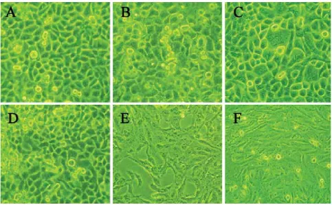
FIGURE 3. Stealth siRNA promotes cell proliferation at 72 h after transfection (×200) A: control SGC7901 cells. B: SGC7901 transfected with stealth_144. C: control BGC823 cells. D: BGC823 transfected with stealth_144. E: control GES-1 cells. F: GES-1 transfected with stealth_144.
FIGURE 4. TFF1 protein expression at 48 h after transfection with TFF1-pcDNA3.1. 1: GES-1 cells with transfection; 2: control GES-1 cells; 3: BGC-823 cells with transfection; 4: control BGC-823 cells; 5: SGC7901 cells with transfection; 6: control SGC7901 cells Group Cell cycle Apoptosis
rate
G0/G1 S G2/M
Black Control 56.72±2.11 36.78±1.10 6.5±0.89 4.83±0.56 Stealth
nega-tive control 57.07±1.68 35.43±1.21 7.2±0.26 5.03±0.38 Stealth_115 57.27±1.32 35.86±1.38 6.87±0.35 2.96±0.51* Stealth_144 55.98±1.43 36.42±1.31 7.6±0.28 2.83±0.25* Stealth_268 56.43±1.69 37.21±1.38 6.36±0.36 3.02±0.14* TABLE 6. Cell cycle and apoptosis rate of BGC823 cells at 72 h after stealth siRNA transfection
*p < 0.05 vs control group
Groups BGC823 SGC7901 GES-1 A IR% A IR% A IR%
Control 0.960±0.13 1.074±0.08 0.876±0.05
pcDNA3.1
blank 0.942±0.08 1.8 1.027±0.14 2.1 0.868±0.21 1.2
TFF1-pcD-NA3.1 0.648±0.07* 32.5 0.754±0.11* 29.8 0.667±0.08* 23.8 TABLE 7. Eff ect of pcDNA3.1 transfection on proliferation of BGC823 cells, SGC7901 cells and GES-1 cells
Bosn J Basic Med Sci 2012; 12 (2): 79-81
YANLI GE ET AL.: TFF1 INHIBITS PROLIFERATION AND INDUCES APOPTOSIS OF GASTRIC CANCER CELLS IN VITRO
cells and SGC cells were ., . and ., respec-tively. At h and h after transfection, the IR was also dramatically reduced when compared with control group (p<.). At h and h after transfection, there was no signifi cant alteration in the IR (p>.) (Table , Figure ). The apoptosis rate of TFF-pcDNA. transfected cells markedly increased at h after transfection when com-pared with control group (p<.). The apoptosis rate increased from . ± . to . ± . in GES- cells, from . ± . to . ± . in BGC cells, and from . ± . to . ± . in SGC cells (Table ). Af-ter TFF-pcDNA. transfection, the proportion of cells in G and G/M phase markedly decreased, while that in S-phase significantly increased, indicating that cells were arrested in S phase. The cell cycle distribution was mark-edly diff erent from that in control group (p<.) (Table ).
DISCUSSION
Under physioligical conditions, human TFF is mainly pressed in epithelial cells of gastrointestinal tract. TFF ex-pression has regional and cellular selectivity, and, in nor-mal tissues, TFF is expressed in the gastric antrum, the crypt of gastric body and mucosa. Low TFF expression is also noted in the small intestine, colon and breast epiderm. However, under pathological conditions, the specifi city in TFF expression is absent. When the gastrointestinal cosa is injured, TFF may be expressed in the injured mu-cosa of gastrointestinal tract, and its expression is rapidly up-regulated to participate in the reconstruction and repair process of epithelial cells in gastrointestinal tract. Our study found that, in normal gastric mucosa, the TFF expression was mainly found in gastric epithelial cells, gastric pits and glands, which was consistent with previous study. In addition, TFF is now considered as a tumor suppressor, and may be a stomach-specifi c suppressor factor. In humans, about of GC patients show loss of human pS expression. It is gen-erally believed that GC is preceded by a precancerous prog-ress with the following well-recognized steps: normal gastric mucosa, chronic active inflammation, multifocal atrophy (gland loss), intestinal metaplasia, complete type-intestinal metaplasia, incomplete type-dysplasia [, ]. The accu-mulation of many genetic and molecular changes is closely
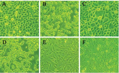
FIGURE 5. TFF1-pcDNA3.1 transfection inhibited cell proliferation (48 h; ×200) A:control SGC7901 cells; SGC7901 cells with TFF1-pcD-NA3.1 transfection; C: control BGC823 cells; D: BGC823 cells with TFF1-pcDTFF1-pcD-NA3.1 transfection; E:control GES-1 cells; F: GES-1 cells with TFF1-pcDNA3.1 transfection
Group Cell Cycle Apoptosis
Rate
G0/G1 S G2/M
Black Control 56.72±2.11 36.78±1.10 6.5±0.89 4.83±0.56 pcDNA3.1
control 55.23±1.72 37.29±3.11 7.48±0.45 4.57±0.23
TFF1-pcD-NA3.1 46.67±2.89* 47.23±2.65* 6.10±0.54 14.38±0.89* TABLE 8. Changes in cell cycle and apoptosis rate of BGC823 cells at 48 h after TFF1-pcDNA3.1 transfection (%)