INTRODUCTION
Coronary artery ectasia (CAE) is an abnormal dilatation of coronary arteries in which the ectatic segment exceeds the diameter of the normal adjacent segments or the diameter of the patient’s largest coronary vessel by 1.5 times [1]. It is a rare clinical entity. Th e incidence of CAE ranges from 0.3 to 5.3 in coronary angiographic series [1,2]. Severity of CAE is defi ned by the involvement of a single vessel or multiple vessels [3]. CAE is characterized by inappropriately localized or diff use dilatation of the coronary vasculature. Th e main coronary angiographic characteristics of CAE are impaired coronary blood fl ow, delayed antegrade coronary dye fi lling, segmen-tal back fl ow phenomenon and stasis with local deposition of dye in dilated coronary segments [4]. Patients with CAE may
be totally asymptomatic or present with atherosclerotic heart disease-like symptoms, about 50 of CAE patients present with atypical anginal pain, as the ectatic lesions can interfere with the coronary blood fl ow and CAE may cause myocardial ischemia or myocardial infarction without signifi cant coro-nary artery stenosis due to intracorocoro-nary thrombosis within the ectatic segment and distal embolization of this thrombotic material [5]. Th e etiology and pathophysiology of CAE remains unclear. It is believed that CAE is a specifi c form of athero-sclerotic coronary artery disease (CAD), and in approximately 20-30 of patients, it maybe congenital in origin. In most cases, CAE is found to coexist with CAD, whereas in 10-20 of cases, it is associated with cocaine abuse, toxins, infl amma-tion, infections, infl ammatory diseases, cardiac lymphoma and connective tissue disorders such as Kawasaki disease, systemic lupus erythematosus, Marfan syndrome, and Ehlers-Danlos syndrome [6-10]. In histopathological appearance of the disease, marked destruction and reduction of the medial elastic fi bers with disruption of the internal and external elastic lamina, smooth muscle hyalinization of the coronary *Corresponding author: Ismail Biyik,
Uşak Devlet Hastanesi, Kardiyoloji Kliniği, Posta Kodu: 64100 Uşak, Turkiye, Phone: +905424173209
E-mail: [email protected]
Submitted: 13 April 2014 / Accepted: 23 July 2014
coronary artery ectasia
Ibrahim Faruk Akturk1*, Ismail Biyik2, Ahmet Arif Yalcin1, Nilgun Isiksacan3, Omer Celik1, Derya Ozturk1, Mehmet Erturk1
1Department of Cardiology, Mehmet Akif Ersoy Thoracic and Cardiovascular Surgery Training and Research Hospital, Istanbul, Turkey. 2Department of Cardiology, Usak State Hospital, Usak, Turkey. 3Department of Biochemistry, Mehmet Akif Ersoy Thoracic and Cardiovascular
Surgery Training and Research Hospital, Istanbul, Turkey
Abstract
Coronary artery ectasia (CAE) is characterized by inappropriate dilatation of the coronary vasculature. Th e mechanisms of CAE are not well known. Insulin-like growth factor-1 (IGF-1) may make endothelial cells and smooth muscle cells more sensitive to the eff ects of growth hor-mone. In the present study, we hypothesized that IGF-1 may have an impact on the formation of ectasia and aneurysm in arterial system, and aimed to investigate the associations between the presence of CAE and serum IGF-1 levels in patients undergoing coronary angiography. Th e study included 2.980 subjects undergoing elective diagnostic coronary angiography. We selected 40 patients diagnosed with CAE as CAE group and 44 subjects with absolutely normal coronary arteries were assigned as normal control group. IGF-1 levels were measured in both groups of patients. Groups were similar in terms of age, sex and coronary artery disease risk factors. Th e serum IGF-1 levels were signifi cantly higher in CAE patients with 109.64±54.64 ng/mL than in controls with 84.76±34.01 ng/mL (p=0.016). HDL levels were lower in ectasia group with 41.5±10.7 mg/dL than controls with 47.7±10.4 mg/dL (p=0.018). By means of logistic regression analysis, high IGF-1 and low HDL levels were found to be independent risk factors for the presence of CAE (p<0.02, p<0.016, respectively). Th e study revealed that there was a positive correlation between serum IGF-1 levels and presence of CAE, and high IGF-1 levels and low HDL levels were independent risk factors for the presence of CAE. Future studies are needed to confi rm these results.
KEY WORDS: insulin like growth factor 1, IGF-1; coronary artery ectasia, CAE
fi bro-muscular media, excessive nitric oxide (NO) production which leads to hyalinization by indirect acetylcholine produc-tion have been found [6-10]. Severe coronary wall infl amma-tion may play a role in CAE pathogenesis [10,11]. Insulin-like growth factor-1 (IGF-1) has anti-infl ammatory and pro-repair-ing properties that make it antiatherogenic [12,13]. Circulatpro-repair-ing IGF-1 is mainly released by the liver under the regulation of growth hormone and executes all of its physiological eff ects via binding to its receptor [13]. Up to date, several studies have already described the importance of IGF-1 on atherosclerosis with its large biological eff ects. Although the results of these trials are inconclusive; in general, there is an inverse relation between IGF-1 levels and atherosclerosis, and IGF-1 reduces oxidative stress, infl ammation and atherogenesis in the vascu-lature and plays a major role in vasodilatory responses by reg-ulating (NO) production in the endothelium [13,14]. On the other hand, some cross-sectional and prospective studies sug-gest a positive association between IGF-1 and its binding pro-teins and atherosclerosis [15,16]. Accumulating evidence now indicates that IGFs and their regulatory proteins, secreted by cells of the cardiovascular system, are growth promoters for arterial cells and mediators of cardiovascular diseases and IGF-1 molecule may make endothelial cells (EC) and smooth muscle cells more sensitive to the eff ects of growth hormone (GH) [14-18]. Total circulating IGF-1 concentrations are not infl uenced by diurnal or circadian variation, which is an advantage for biomarker research [19]. Th e underlying mecha-nisms of CAE formation have not been entirely explained yet. Role of IGF-1 molecule on the development of CAE has not been studied up to date. In the present study, we hypothesized that IGF-1 may have an impact on the formation of ectasia and aneurysm in arterial system, and aimed to investigate the asso-ciations between the presence of CAE and serum IGF-1 levels in patients undergoing coronary angiography.MATERIALS AND METHODS
Subjects
Th e present observational, case-control comparative study was conducted in a high volume tertiary heart center. Approximately, 3000 subjects undergoing elective diagnos-tic coronary angiography in our institution were scanned to fi nd patients with apparent CAE. We selected 40 patients diagnosed with CAE, and they were labeled as CAE group, and we selected 44 patients with absolutely normal coronary arteries; those were assigned as normal control group. Patients with coronary aneurysms associated with balloon angioplasty, coronary stent placement, brachytherapy or atherectomy, Kawasaki disease, known collagen vascular diseases, patients with previous myocardial infarction, undergoing coronary
artery by-pass grafting surgery, patients with disorders aff ect-ing IGF-1 levels such as diabetes mellitus on insulin therapy, poorly controlled diabetes mellitus, acromegaly, growth hor-mone defi ciency, patients on steroid therapy and patients diag-nosed with malignant disease were excluded from the study. A detailed medical history was obtained from all patients and a complete physical examination was performed. A detailed transthoracic echocardiography was performed by two expe-rienced specialists. Th e diagnosis of hypertension was estab-lished if a systolic blood pressure was 140 mmHg or higher; or a diastolic blood pressure of 90 mmHg or higher, measured in at least three separate measurements; or the use of anti-hy-pertensive medication. Th e diagnosis of diabetes mellitus was established by a fasting blood glucose of 126 mg/dL or higher, or the use of anti-diabetic medication. Fasting blood glucose of 100 mg/dL to 126 mg/dL was defi ned as impaired fasting glucose. Hyperlipidemia was defi ned as total cho-lesterol levels of 200 mg/dL or higher, or a history of statin use except in the last 3 month. Patients who were smoking before hospitalization were accepted as smokers. Th e study protocol was approved by the local ethics committee and all patients signed a written informed consent. Th e study was conducted in accordance with the Declaration of Helsinki, good clinical practice (GCP) and International Conference on Harmonization (ICH) guidelines.
Blood sampling and biochemical measurements
Coronary angiography and transthoracic
echocardiography
Coronary angiography was performed by the Judkins technique through femoral artery access. Coronary angio-grams were analyzed by two experienced interventional cardiologists who were blinded for the laboratory mea-surements and clinical status of the participants. CAE was defi ned as the segmental or diff use dilatation of the coronary arteries more than 1.5 fold of the diameter of the adjacent segments of the same artery or of diff erent arteries. CAE was classifi ed according to a classifi cation system proposed by Markis et al. in 1976 [3]. According to this classifi cation, CAE was graded as follows type 1, diff use ectasia of ≥ 2 coronary arteries; type 2, diff use ectasia in one coronary artery and localized ectasia in another coronary artery; type 3, diff use ectasia of one coronary artery, and type 4, localized or seg-mental ectasia of only one coronary artery [3]. Transthoracic echocardiography was performed before discharge using a System V device (General Electric, Horten, Norway) with a 2.5 MHz phased-array transducer. Th e left ventricular ejection fraction (LVEF) was measured using the modifi ed Simpson’s rule [21].
Statistical analysis
Statistical calculations were performed with Number Cruncher Statistical System 2007 Statistical Software pro-gram for Windows (Utah, USA). Besides standard descriptive statistical calculations (mean and standard deviation, median, interquartile range), in the comparisons between groups, one way ANOVA test was used. Independent Samples T test was used in the comparison of two groups. Chi square test and Fisher’ exact test were used during the evaluation of quali-tative data. Logistic regression analysis was used to identify factors that may aff ect presence of CAE. A p value< 0.05 was accepted to be statistically signifi cant. Because no study on the associations between IGF-1 and CAE has been reported to date, partly referencing other previous IGF-1 and CAE studies, we arbitrarily calculated that at least 40 patients with CAE were required for 80 power and 5 signifi cance assum-ing that 20 to 41 diff erence between groups for IGF-1 is esti-mated to be.
RESULTS
Demographic and clinical characteristics of the patients and the results of laboratory investigations were presented in Table 1 and 2, respectively. Both groups were similar in terms of sex, age and cardiovascular risk factors such as smoking, family history of CAD, presence of diabetes, hypertension, alcohol abuse and metabolic syndrome (Table 1).
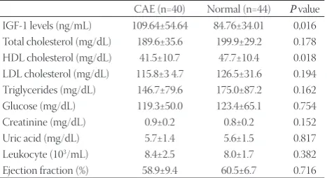
Serum IGF-1 levels were signifi cantly higher in patients with CAE than in patients with normal coronary arteries (p=0.016), and serum HDL level was statistically lower in CAE group (p=0.018) (Table 2).
Although, serum aspartate transaminase (AST) levels were in normal ranges in both groups, AST levels were signifi -cantly higher in patients with CAE compared to the patients with normal coronary arteries. In the present study, we found no correlation between the degree of CAE and IGF-1 levels (Table 3).
In logistic regression analysis, high IGF-1 levels and low HDL levels were found to be independent risk factors for the presence of CAE (p<0.02, p<0.016, respectively) (Table 4).
TABLE 1. Demographic and clinical characteristics of study groups.
CAE (n=40) Normal (n=44)
Age (years) 58.6±10.3 55.4±9.5
Gender (M) (n %) 28 (70.0) 25 (56.8)
BMI (kg/m2) 29.0±4.7 28.8±4.0
Diabetes mellitus (n %) 8 (20.0) 11 (25.0)
Hypertension (n %) 21 (52.5) 18 (40.9)
Alcohol (n %) 7 (17.5) 2 (4.5)
Smoking (n %) 12 (30.0) 13 (29.5)
Family history of CAD (n %) 21 (52.5) 18 (40.9)
Metabolic syndrome (n %) 13 (32.5) 13 (29.5)
TABLE 2. Laboratory fi ndings of study groups.
CAE (n=40) Normal (n=44) P value
IGF-1 levels (ng/mL) 109.64±54.64 84.76±34.01 0,016
Total cholesterol (mg/dL) 189.6±35.6 199.9±29.2 0.178
HDL cholesterol (mg/dL) 41.5±10.7 47.7±10.4 0.018
LDL cholesterol (mg/dL) 115.8±3 4.7 126.5±31.6 0.194
Triglycerides (mg/dL) 146.7±79.6 175.0±87.2 0.162
Glucose (mg/dL) 119.3±50.0 123.4±65.1 0.754
Creatinine (mg/dL) 0.9±0.2 0.8±0.2 0.152
Uric acid (mg/dL) 5.7±1.4 5.6±1.5 0.817
Leukocyte (103/mL) 8.4±2.5 8.0±1.7 0.382
Ejection fraction (%) 58.9±9.4 60.5±6.7 0.716
TABLE 3. Logistic regression analysis of variables.
B S.E. Wald df p Exp (B)
Constant 0.23 0.241 0.91 1 0.034 1.26
95.0% C.I. EXP (B)
B S.E. p Exp (B) Lower Upper
IGF-1 0.02 0.01 0.02 1.02 1.00 1.03
HDL −0.07 0.03 0.016 0.93 0.88 0.99
TABLE 4. Correlations between disease severity and IGF-1 levels.
Disease severity n (%) IGF-1 (ng/mL) p value
Class 1 8 (20) 100.19±38.87
Class 2 9 (22.5) 75.08±25.96 0.102
Class 3 6 (15) 120.25±37.7
Class 4 17 (42.5) 128.64±68.5
Diff use 23 (57.5) 95.6±37.34
DISCUSSION
In our study we found a positive correlation between serum IGF-1 levels and the presence of CAE, and high IGF-1 levels and low HDL levels were independent risk factors for the presence of CAE. Th e serum IGF1 levels were signifi -cantly higher in patients with CAE but we found no correla-tion between disease severity and IGF-1 levels. In addicorrela-tion, we found a negative correlation between HDL levels and CAE, and HDL levels were lower in patients with CAE.
Several studies have evaluated the traditional cardiovas-cular risk factors in patients with CAE, and male dominance, younger age, hypertension, dyslipidemia, smoking, cocaine use, low prevalence of diabetes and persisting congenital anomaly, which included bicuspid aortic valve, aortic root dil-atation, ventricular septal defect, pulmonary stenosis or cya-notic congenital heart disease have been implicated [6]. Th e frequent coexistence of CAE with CAD and histopathological fi ndings resembling those of atherosclerosis have led to the conclusion that the mechanism underlying the pathogene-sis of CAE is a variant of atheroscleropathogene-sis but there are some diff erences between CAE and CAD. CAE is associated with risk factors such as diabetes mellitus and age, elevated infl am-matory parameters, changes in extracellular matrix remodel-ing, matrix metalloproteinase (MMP)-3 5A polymorphism, increased plasma levels of MMP-3, more pronounced involve-ment of right coronary artery, lower incidence of increased carotid intima-media thickness, decreased endothelium inde-pendent dilatation, possible association with vein involve-ment and possible hazardous eff ects of nitrate treatinvolve-ment [6]. Th e pathogenesis of CAE has not yet been clear. Th ere are also obvious similarities between the pathogenesis of CAE and CAD. Regarding the high coexistence of CAE and CAD, pos-itive remodeling described as enlargement of the area within the external elastic membrane may play a role in the pathogen-esis of CAE [22]. On the other hand, histological examinations have revealed signifi cant destruction and reduction of the media elastic fi bers with disruption of the internal and exter-nal elastic membrane, usually not matching the degree of the involvement of vessel intima, the loss of musculoelastic arterial wall components in CAE, which was noticed to be unrelated to local atheromatous burden [1,3,6,9]. Non-atherosclerotic forms of CEA have been described with an intact vessel intima, but with extensive media degeneration and smooth muscle cell replacement by hyalinized collagen [1,3,6,9]. Th us, a functional loss of the musculoelastic components of the coronary artery media is considered to be the predominant aspect in the pathogenesis of CAE [1,3,6,9]. Chronic overstim-ulation of endothelium by NO or NO donors and enhanced NO production have also been documented and have been suspected to be an underlying pathophysiological mechanism
of CAE [23]. Lamblin et al. [24] have proposed other possible culprits in CAE pathogenesis: the system of metalloprotein-ases, which are actively involved in the proteolysis of the extra-cellular matrix proteins. Chronic vascular infl ammation has been stressed as the common denominator in all cases with CAE [1, 3, 6, 9]. Conventional infl ammatory markers like cyto-kines, tumor necrosis factor (TNF), interleukins and T helper (Th ) lymphocyte activation have been found elevated in CAE patients, and the abovementioned markers are considered as good markers of systemic infl ammation [25,26]. On the other hand, markers like infl ammatory cells e.g. leucocytes count, monocyte count and C-reactive protein are closely linked to the presence of CAE [27]. Furthermore, levels of soluble adhe-sion molecules (e.g. intracellular adheadhe-sion molecules - ICAM and vascular cell adhesion molecules - VCAM) were also found to be higher in isolated CAE as well as in CAE with occlusive CAD compared with occlusive coronaries without CAE; these fi ndings suggest that more severe coronary wall infl ammation may play a role in CAE pathogenesis [11]. On the other hand, Liu et al. recently reported that IGF-1 can res-cue endothelial nitric oxide synthase (e-NOS) activity and decrease ICAM-1 and VCAM-1 secretion infl uenced by CRP and can diminish the eff ect of CRP [28].
smooth muscle hyalinization of the coronary fi bro-muscular media may contribute to vascular positive remodeling and development of CAE [6-10]. Th is suggested mechanism for development of CAE may be supported by other clinical trials. Lindholt et al. showed that baseline serum IGF-1 correlated pos-itively with abdominal aortic aneurysm (AAA) size and growth rate [35]. Yeap et al. showed that higher IGF-1 and an increased ratio of IGF-1/IGF binding protein (IGFBP) 3 are associated with AAA, while IGFBP-1 is independently associated with increased aortic diameter, and components of the IGF-1 system may contribute to, or be a marker for, aortic dilation in elderly patients [36]. Casini et al. have reported that acromegaly wasassociated with aortic ectasia, suggesting the GH and IGF-1 excess might have eff ects on the cardiovascular system [37]. Oshino et al. have reported that a signifi cantly higher prevalence of cerebral aneurysm was detected in male patients with acro-megaly, and this fi nding indicates that excess growth hormone or IGF-1 aff ects the cerebral vascular wall, resulting in aneurysm formation [38]. Ramos-Mozo et al. reported that IGFBP-1 has been identifi ed by a protein array approach as a potential novel biomarker of AAA [39]. Th ere are numerous studies on the associations between CAE and hematologic parameters such as mean platelet volume, red cell distribution width, neutrophil/ lymphocyte ratio, platelet/lymphocyte ratio and infl ammatory markers such as YKL 40, hs-CRP, matrix metalloproteinases [6,9,10]. Most of these studies support the idea that infl amma-tion may play a role in CAE pathogenesis, which may be related to increasing eff ect of IGF-1 on infl ammation.
In the present study, we also found signifi cantly lower HDL levels in patients with CAE compatible with previous studies. In previous studies, Sudhir et al. and Altiparmak et al. pointed out lower HDL levels in patients with CAE [40,41]. Buchart et al. reported that there was no relationship between IGF-1 and HDL levels [42]. Friedrich et al. pointed that IGF-1 reduces HDL levels [43] but Elbornsson et al. showed that IGF-1 increases HDL-1 levels [44]. We found that there was no asso-ciation between IGF-1 and low HDL levels in this very study.
Th e present study has some limitations. It was a single cen-tered study and limited to native vessels. A major limitation was a small number of patients involved. We only measured free IGF-1 but were not able to measure IGFBPs, which also represents an important limitation of the study.
We cannot yet speculate whether IGF-1 itself would be the only pathogenic factor in the development of CAE but it may be an important factor in the pathogenesis of CAE.
In our study, we have found a positive correlation between IGF-1 levels and CAE. Our fi ndings suggest that elevated lev-els of IGF-1 may have a pivotal role in the development of CAE, and may indicate the direction for further investigation aiming on the development of novel diagnostic and therapeu-tic approaches for CAE.
CONCLUSIONS
Th e present study revealed that there was a positive cor-relation between serum IGF-1 levels and the presence of CAE, and high IGF-1 levels and low HDL levels were independent risk factors for the presence of CAE. Future studies are needed to confi rm these results.
DECLARATION OF INTEREST
Th e authors declare no confl icts of interest, and have received no fi nancial support for the research.
REFERENCES
[1] Manginas A, Cokkinos DV. Coronary artery ectasias: imaging, functionalassessment and clinical implications. Eur Heart J 2006; 27:1026-31. http://dx.doi.org/10.1093/eurheartj/ehi72.
[2] Swaye PS, Fisher LD, Litwin P, Vignola PA, Judkins MP, Kemp HG, et al. Aneurysmal coronary artery disease. Circulation 1983; 67:134-138. http://dx.doi.org/10.1161/01.CIR.67.1.134
[3] Markis JE, Joff e CD, Cohn PF, Feen DJ, Herman MV, Gorlin R. Clinical significance of coronary arterial ectasia. Am J Cardiol 1976;37:217-222. http://dx.doi.org/10.1016/0002-9149(76)90315-5. [4] Krüger D, Stierle U, Herrmann G, Simon R, Sheikhzadeh A.
Exercise-induced myocardial ischaemia in isolated coronary artery ectasias and aneurysms (“dilated coronaropathy”). J Am Coll Cardiol 1999;34:1461-1470. http://dx.doi.org/10.1016/ S0735-1097(99)00375-7.
[5] Hartnell GG, Parnell BM, Pridie RB. Coronary artery ectasia. Its prevalence and clinical signifi cance in 4993 patients. Br Heart J 1985; 54:392-395. http://dx.doi.org/10.1136/hrt.54.4.392.
[6] Yetkin E, Waltenberger J. Novel insights into an old controversy: is coronary artery ectasia a variant of coronary atherosclero-sis? Clin Res Cardiol 2007;96:331-9. http://dx.doi.org/10.1007/ s00392-007-0521-0.
[7] Satran A, Bart BA, Henry CR, Murad MB, Talukdar S, Satran D, et al. Increased prevalence of coronary artery aneurysms among cocaine users. Circulation 2005;111:2424-9. http://dx.doi.org/10.1161/01. CIR.0000165121.50527.DE.
[8] Antoniadis AP, Chatzizisis YS, Giannoglou GD. Pathogenetic mechanisms of coronary ectasia. Int J Cardiol 2008; 130:335-343. http://dx.doi.org/10.1016/j.ijcard.2008.05.071.
[9] Boles U, Rakhit R, Shiu MF, Patel K, Henein M. Coronary artery ectasia as a culprit for acute myocardial infarction: review of pathophysiology and management. Anadolu Kardiyol Derg. 2013;13:695-701.
[10] Brunetti ND, Salvemini G, Cuculo A, Ruggiero A, De Gennaro L, Gaglione A, et al. Coronary artery ectasia is related to coronary slow fl ow and infl ammatory activation. Atherosclerosis 2014; 233:636-640. http://dx.doi.org/10.1016/j.atherosclerosis.2014.01.018. [11] Turhan H, Erbay AR, Yasar AS,Aksoy Y, Bicer A, Yetkin G, et al. Plasma
soluble adhesion molecules; intercellular adhesion molecule-1, vas-cular cell adhesion molecule-1 and E-selectin levels in patients with isolated coronary artery ectasia. Coron Artery Dis 2005; 16:45-50. http://dx.doi.org/10.1097/00019501-200502000-00009.
[12] Sukhanov S, Higashi Y, Shai SY, Vaughn C, Mohler J, Li Y, et al. IGF-1reduces infl ammatory responses, suppresses oxidative stress, anddecreases atherosclerosis progression in ApoE-defi cient mice.
ArteriosclerTh romb Vasc Biol 2007; 27:2684-2690. http://dx.doi.
org/10.1161/ATVBAHA.107.156257.
[14] Burchardt P, Gozdzicka-Jozefi ak A, Zurawski J, Nowak W, Durzynska J, Link R, et al. Are Elevated Levels of IGF-1 Caused by Coronary Arteriesoclerosis?: Molecular and Clinical Analysis. Protein J 2010; 29:538-544. http://dx.doi.org/10.1007/ s10930-010-9288-7.
[15] Andreassen M, Raymond I, Kistorp C, Hildebrandt P, Faber J, Kristensen LO. IGF1 as predictor of all cause mortality and car-diovas¬cular disease in an elderly population. Eur J Endocrinol 2009;160:25-31. http://dx.doi.org/10.1530/EJE-08-0452.
[16] Schuler-Luttmann S, Monnig G, Enbergs A, Schulte H, Breithardt G, Assmann G, et al. Insulin-like growth factor-binding protein-3 is associated with the presence and extent of coronary
arteriosclero-sis. Arterioscler Th romb Vasc Biol 2000;20:E10–E15. http://dx.doi.
org/10.1161/01.ATV.20.4.e10.
[17] Vaessen N, Heutink P, Janssen JA,Witteman JC, Testers L, Hofman A, et al. A polymorphism in the gene for IGF-1; functional proper-ties and risk for type 2 diabetes and myocardial infarction. Diabetes 2001; 50:637-42. http://dx.doi.org/10.2337/diabetes.50.3.637.
[18] Moschos SJ, Mantzoros CS. Th e role of IGF system in cancer: from
basic to clinical studies and clinical applications. Oncology 2002; 63:317-32. http://dx.doi.org/10.1159/000066230.
[19] Rajpathak SN, Gunter MJ, Wylie-Rosett J, Ho GY, Kaplan RC,
Muzumdar R, et al. Th e role of insulin-like growth factor-I and
its binding proteins in glucose homeostasis and type 2 diabetes. Diabetes Metab Res Rev 2009; 25:3-12. http://dx.doi.org/10.1002/ dmrr.919.
[20] Krebs A, Wallaschofski H, Spilcke-Liss E, Kohlmann T, Brabant G, Völzke H, et al. Five commercially available insulin-like growth fac-tor I (IGF-I) assays in comparison to the former Nichols Advantage IGF-I in a growth hormone treated population. Clin Chem Lab Med 2008; 46:1776-83. http://dx.doi.org/10.1515/CCLM.2008.349. [21] American Society of Echocardiography Recommendations for
Use of Echocardiography in Clinical Trials. A Report from the American Society of Echocardiography’s Guidelines and Standards
Committee and Th e Task Force on Echocardiography in Clinical
Trials. J Am SocEchocardiogr 2004; 17:1086-1119.
[22] Glagov S, Weisenberg E, Zarins CK, Stankunavicius R, Kolettis GJ. Compensatory enlargement of human atherosclerotic coronary arteries. N Engl J Med 1987;316: 1371-1375 http://dx.doi.org/10.1056/ NEJM198705283162204.
[23] Sorrell VL, Davis MJ, Bovee AA. Origins of coronary artery ectasia. Lancet 1996;347:136-137. http://dx.doi.org/10.1016/ S0140-6736(96)90335-9.
[24] Lamblin N, Bauters C, Hermant X, Lablanche JM, Helbecque N, Amouyel P. Polymorphisms in the promoter regions of MMP-2, MMP-3, MMP-9 and MMP-12 genes as determinants of aneu-rysmal coronary artery disease. J Am Coll Cardiol 2002;40:43-48. http://dx.doi.org/10.1016/S0735-1097(02)01909-5.
[25] Adiloglu AK, Can R, Nazlı C, Ocal A, Ergene O, Tinaz G, et al. Ectasia and severe atherosclerosis: relationships with chlamydia pneumonia, helicobacter pylori, and infl ammatory markers. Tex Heart Inst J 2005; 32:21-7.
[26] Triantafyllis AS, Kalogeropoulos AS, Rigopoulos AG, Sakadakis EA, Toumpoulis IK, Tsikrikas S, et al. Coronary artery ectasia and
infl ammatory cytokines: link with a predominant Th -2 immune
response? Cytokine 2013; 64:427-32. http://dx.doi.org/10.1016/j. cyto.2013.05.003.
[27] Li JJ, Nie SP, Qian XW, Zeng HS, Zhang CY. Chronic infl amma-tory status in patients with coronary artery ectasia. Cytokine 2009; 46:61-4. http://dx.doi.org/10.1016/j.cyto.2008.12.012.
[28] Liu SJ, Zhong Y, You XY, Liu WH, Li AQ, Liu SM. Insulin-like growth factor 1 opposes the eff ects of C-reactive protein on endo-thelial cell activation. Mol Cell Biochem. 2014; 385:199-205. http:// dx.doi.org/10.1007/s11010-013-1828-y.
[29] Delafontaine P, Song YH, Li Y. Expression, regulation, and func-tion of IGF-1, IGF-1R, and IGF-1 binding proteins in blood
ves-sels. Arterioscler Th romb Vasc Biol 2004;24:435-44. http://dx.doi.
org/10.1161/01.ATV.0000105902.89459.09.
[30] Song H, Mowbray AL, Sykes MC, Jo H. Emerging role of IGF-1R in stretch-induced neointimal hyperplasia in venous grafts.
Arterioscler Th romb Vasc Biol 2007;27:1679-1681. http://dx.doi.
org/10.1161/ATVBAHA.107.148189.
[31] Cruzado MC, Risler NR, Miatello RM, Yao G, Schiff rin EL, Touyz RM. Vascular smooth muscle cell NAD(P)H oxidase activ-ity during the development of hypertension: Eff ect of angiotensin II and role of insulinlike growth factor-1 receptor transactiva-tion. Am J Hypertens2005; 18:81-87. http://dx.doi.org/10.1016/j. amjhyper.2004.09.001.
[32] Shigematsu S, Yamauchi K, Nakajima K, Iijima S, Aizawa T, Hashizume K. IGF-1 regulates migration and angiogenesis of human endothelial cells. Endocr J1999;46:S59-S62. http://dx.doi. org/10.1507/endocrj.46.Suppl_S59.
[33] Che W, Lerner-Marmarosh N, Huang Q, Osawa M, Ohta S, Yoshizumi M, et al. Insulin-like growth factor-1 enhances infl am-matory responses in endothelial cells: role of Gab1 and MEKK3 in TNF-a´-induced c-Jun and NF-B activation and adhesion molecule expression. Circ Res2002; 90:1222-1230. http://dx.doi. org/10.1161/01.RES.0000021127.83364.7D.
[34] Wickman A, Jonsdottir IH, Bergstrom G, Hedin L. GH and IGF-I regulate the expression of endothelial nitric oxide synthase (eNOS) in cardiovascular tissues of hypophysectomized female rats. Eur J Endocrinol. 2002;147:523-533. http://dx.doi.org/10.1530/ eje.0.1470523.
[35] Lindholt JS, Martin-Ventura JL, Urbonavicius S, Ramos-Mozo P, Flyvbjerg A, Egido J, et al. Insulin-like growth factor I - a novel bio-marker of abdominal aortic aneurysms. Eur J VascEndovascSurg 2011; 42:560-2. http://dx.doi.org/10.1016/j.ejvs.2011.07.013. [36] Yeap BB, Chubb SA, McCaul KA, Flicker L, Ho KK, Golledge J, et al.
Associations of IGF1 and its binding proteins with abdominal aortic aneurysm and aortic diameter in older men. Eur J Endocrinol 2012; 166:191-7. http://dx.doi.org/10.1530/EJE-11-072.
[37] Casini AF, Neto LV, Fontes R, França RF, Xavier SS, Gadelha MR. Aortic root ectasia in patients with acromegaly: experience at a sin-gle center. Clin Endocrinol (Oxf ) 2011; 75:495-500. http://dx.doi.org /10.1111/j.1365-2265.2011.04067.
[38] Oshino S, Nishino A, Suzuki T, Arita H, Tateishi A, Matsumoto K, et al. Prevalence of cerebral aneurysm in patients with acro-megaly. Pituitary 2013; 16:195-201. http://dx.doi.org/10.1007/ s11102-012-0404-x.
[39] Ramos-Mozo P, Rodriguez C, Pastor-Vargas C, Blanco-Colio LM, Martinez-Gonzalez J, Meilhac O, et al. Plasma profi ling by a protein array approach identifi es IGFBP-1 as a novel biomarker of abdom-inal aortic aneurysm. Atherosclerosis 2012; 221:544-50. http://dx. doi.org/10.1016/j.atherosclerosis.2012.01.009.
[40] Sudhir K, Ports TA, Amidon TM, Goldberger JJ, Bhushan V, Kane JP, et al. Increased prevalence of coronary ectasia in hetero-zygous familial hypercholesterolemia. Circulation. 1995; 91:1375-80. http://dx.doi.org/10.1161/01.CIR.91.5.1375.
[41] Altıparmak IH, Kaya Z, Sezen H, Aydın MS, Demirbağ R, Aksoy N. Th e relation of serum paraoxonase-1 activity with isolated coronary artery ectasia: an observational study. Anadolu Kardiyol Derg. 2012; 12:307-312.
[42] Burchardt P, Tabaczewski P, Goździcka-Józefi ak A, Siminiak T, Szczepaniak A, Banaszak A, et al. Association between insulin like growth factor-1 and lipoprotein metabolism in stable angina patients on statin therapy: a pilot study. Kardiol Pol. 2012; 70:1017-22. [43] Friedrich N, Nauck M, Schipf S, Völzke H, Brabant G,
Wallaschofski H. Cross-sectional and longitudinal associations between insulin-like growth factor I and metabolic syndrome: a general population study in German adults. Diabetes Metab Res Rev. 2013; 29:452-62. http://dx.doi.org/10.1002/dmrr.2412.