Article
RET Functions as a Dual-Specificity Kinase that
Requires Allosteric Inputs from Juxtamembrane
Elements
Graphical Abstract
Highlights
d
The JM segment enhances RET catalytic domain activity
d
Structural visualization of activation-loop phospho-S909
engaging the HRD motif
d
Phospho-S909 arises from an intrinsic RET dual-specificity
kinase activity
d
RET
a
C hydrophobic pocket is a potential drug-targetable
allosteric site
Authors
Iva´n Plaza-Menacho, Karin Barnouin,
Rachael Barry, ...,
Rube´n J. Martı´nez-Torres, Pascal Meier,
Neil Q. McDonald
Correspondence
[email protected] (I.P.-M.),
[email protected] (N.Q.M.)
In Brief
Receptor tyrosine kinases exhibit a
plethora of activation mechanisms
despite highly homologous catalytic
domains. Plaza-Menacho et al. find that
RET tyrosine kinase activation and
signaling require allosteric inputs from
juxtamembrane elements as well as
dual-specificity activity.
Accession Numbers
5FM3
5FM2
Plaza-Menacho et al., 2016, Cell Reports17, 3319–3332 December 20, 2016ª2016 The Author(s).
Cell Reports
Article
RET Functions as a Dual-Specificity
Kinase that Requires Allosteric Inputs
from Juxtamembrane Elements
Iva´n Plaza-Menacho,1,7,8,*Karin Barnouin,2Rachael Barry,5Annabel Borg,3Mariam Orme,5Rakhee Chauhan,1 Stephane Mouilleron,4Rube´n J. Martı´nez-Torres,1Pascal Meier,5and Neil Q. McDonald1,6,*
1Structural Biology Laboratory
2Protein Analysis and Proteomics
3Protein Production Facility
4Structural Biology Science Technology Platform
The Francis Crick Institute, 1 Midland Road, London NW1 1AT, UK
5The Breast Cancer Now Toby Robins Research Centre, Mary-Jean Mitchell Green Building, Institute of Cancer Research,
SW3 6JB London, UK
6Department of Biological Sciences, Institute of Structural and Molecular Biology, Birkbeck College, Malet Street, WC1E 7HX London, UK
7Present address: Structural Biology and Biophysics, Biozentrum, University of Basel, Klingelbergtrasse 50/70, 4056 Basel, Switzerland
8Lead Contact
*Correspondence:[email protected](I.P.-M.),[email protected](N.Q.M.)
http://dx.doi.org/10.1016/j.celrep.2016.11.061
SUMMARY
Receptor tyrosine kinases exhibit a variety of
activa-tion mechanisms despite highly homologous
cata-lytic domains. Such diversity arises through coupling
of extracellular ligand-binding portions with highly
variable intracellular sequences flanking the tyrosine
kinase domain and specific patterns of
autophos-phorylation sites. Here, we show that the
juxtamem-brane (JM) segment enhances RET catalytic domain
activity through Y687. This phospho-site is also
required by the JM region to rescue an otherwise
catalytically deficient RET activation-loop mutant
lacking tyrosines. Structure-function analyses
identi-fied interactions between the JM hinge,
a
C helix, and
an unconventional activation-loop serine
phosphory-lation site that engages the HRD motif and promotes
phospho-tyrosine conformational accessibility and
regulatory spine assembly. We demonstrate that
this phospho-S909 arises from an intrinsic RET
dual-specificity kinase activity and show that an
equivalent serine is required for RET signaling in
Drosophila
. Our findings reveal dual-specificity and
allosteric components for the mechanism of RET
activation and signaling with direct implications for
drug discovery.
INTRODUCTION
Vertebrates have close to 60 receptor tyrosine kinases (RTKs) that respond to a diverse set of extracellular polypeptide ligands by stimulating their intrinsic tyrosine kinase function. RTKs play key roles during embryogenesis and cellular homeostasis; they
are also crucial at the origin and progression of many types of cancer (Lemmon and Schlessinger, 2010). Recent progress on the structural basis for EGFR, IR, and FGFR activation has emphasized the importance of RTK-specific or ‘‘private’’ mech-anisms of activation for their catalytic domains involving flanking regions and asymmetrical and symmetrical arrangements of dimeric and higher-order oligomeric states (Bae and Schles-singer, 2010; Cabail et al., 2015; Jura et al., 2011; Lemmon et al., 2014). The activation mechanism operating in RET in these terms is currently unclear.
In the current RET paradigm for ligand-dependent RET activa-tion, autophosphorylation (autoP) of several tyrosine residues within the cytoplasmic domain is required for cell signaling (Air-aksinen et al., 1999; Plaza-Menacho et al., 2006). For other RTKs, such as the IR and FGFR2, ligand-dependent stimulation leads to kinase activation and phosphorylation of specific tyro-sine residues, which relieve repressivecis-inhibitory interactions to enhance catalytic activity and to promote binding of phos-photyrosine-binding domain (PTB)- and Src homology 2 (SH2)-domain-containing proteins to transmit downstream signals (Chen et al., 2007; Hubbard, 1997). While the latter role for phos-phorylation has been demonstrated for RET, its effect on cata-lytic activation has been only recently elucidated. In vitro, phos-phorylation of the canonical RET activation loop has little effect on catalytic activity (Knowles et al., 2006; Plaza-Menacho et al., 2011). Indeed, RET activation-loop tyrosines Y900 and Y905 should not be considered activating, because they un-dergo delayed autoP and are not catalytically required (Plaza-Menacho et al., 2011, 2014a). A similar situation is found for the EGFR and non-RTK ACK1 (Lougheed et al., 2004; Zhang et al., 2006). In these cases, allosteric mechanisms have been identified to stimulate receptor activity independent of activation segment phosphorylation.
(Perrinjaquet et al., 2010). In addition, phosphorylation at RET S696 by protein kinase A (PKA) has also been reported. Mutation of S696 affected the ability of RET to activate the small GTPase RAC1 and stimulate formation of cell lamellipodia (Fukuda et al., 2002). Homozygous knockin mice carrying this mutation lacked enteric neurons in the distal colon, resulting from a migration defect of enteric neural crest cells (Asai et al., 2006), indicating a physiological role for a PKA-RET functional crosstalk. How-ever, structural and molecular information about allosteric mech-anisms promoted by the JM region on RET kinase activity are lacking. Taking into account that the role of the JM segment of EGFR family members is distinct from that of typical RTKs because it enhances, rather than inhibits, the catalytic activity (Li et al., 2003; Thiel and Carpenter, 2007), the nature of this coupling between the JM segment and catalytic domain for RET has not been properly explored.
In this study, we define flanking elements and phospho-sites required for RET catalytic domain activation and signaling. We show that the JM segment functions to increase RET catalytic domain activity through Y687. Structufunction analyses re-vealed a crosstalk among the JM hinge,aC helix, and serine phosphorylated activation loop. We demonstrate that the previ-ously unreported S909 phospho-site arises from a dual-speci-ficity RET kinase activity, unique among RTKs. We show that an equivalent serine inDrosophilaRET is required for signaling in vivo. Further structural and biochemical examination revealed an RETaC hydrophobic pocket as a potential drug-targetable allosteric site.
RESULTS
The JM Segment Increases RET Tyrosine Kinase Activity
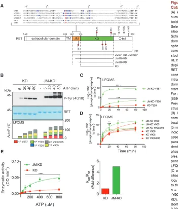
To define the functional impact of the JM segment on RET tyro-sine kinase activity, we used purified recombinant RET kinase domain (KD; residues 705–1013) and RET KD with the JM segment (JM-KD; residues 661–1012; seeFigure 1A) and per-formed a series of biochemical experiments. First, we measured the enzymatic parameters of RET JM-KD and RET KD against an exogenous peptide (Figures S1A and S1B). RET JM-KD showed a 5-fold increased catalytic efficiency (kcat/KMconstant) toward the substrate, indicating increased RET enzymatic activity promoted by the JM region. To support these results further, we performed in vitro time-course autoP assays using saturating concentrations of ATP (5 mM) and MgCl2(10 mM) for 0–80 min (Figures 1B, upper panel andS1D). Western blot (WB) analysis demonstrated increased kinetics and total phosphorylation by RET JM-KD, as indicated by levels of phospho-tyrosine 4G10 antibody. The temporal sequence of RET autoP was also evalu-ated by label-free quantitative mass spectrometry (LFQMS) following a previously described protocol (Plaza-Menacho et al., 2014a). LFQMS analysis identified tyrosine residues— Y687, Y826, Y900, and Y905—which upon RET catalytic activa-tion were efficiently phosphorylated in a time-dependent fashion (Figure 1B, lower panel). Signal log2ratios of phosphorylated peptides standardized to their non-phosphorylated counterparts were plotted relative to a zero time point (Figures 1C and 1D). As indicated by the kinetics of saturation, JM segment Y687 undergoes faster autoP than activation-loop Y900 and Y905.
Furthermore, enhanced phosphorylation kinetics for Y900 and Y905 by RET JM-KD were observed compared with RET KD. In particular, a significant difference was observed in the kinetics of the double-phosphorylated activation-loop peptide. Examination of the total cumulative phosphorylation for each site demonstrated that fully phosphorylated RET JM-KD was achieved between 20 and 40 min compared with the 80–90 min required for RET KD (Fig-ure 1B). Taken together, these data demonstrated that the JM segment increases RET catalytic activity presumably through an allosteric means. Contrary to the EGFR (Jura et al., 2009), the JM region did not promote the formation of RET dimers in solution at protein concentrations used in the biochemical assays as as-sessed by dynamic light scattering (DLS;Figure S1C). The JM segment had no appreciable impact on the stability of RET KD as reported by thermal shift experiments (Figure S1C). However, the apparent affinity for ATP measured by isothermal titration calo-rimetry (ITC) was affected by 2-fold (RET JM-KD Kd= 37.5± 3.1mM, RET KD Kd= 64.3±10mM;Figure S1C). In line with these results, RET JM-KD also displayed increased enzyme kinetic parameters for ATP (Figures 1E andS1C).
Mapping JM Elements Required for RET Catalytic Activation
To map key residues within the RET JM region, we generated a series of deletions and performed biochemical analyses. First, enzymatic assays were performed using an ABL-derived pep-tide, used previously as a good surrogate substrate for RET. Comparison of the catalytic efficiency (kcat/KM) among the different RET JM-KD deletions demonstrated that full-length JM segment starting at residue 661 (JM661) was required to achieve maximal catalytic activity (Figures 2A and 2B). Time-course autoP assays (0–80 min) using RET phospho-specific antibodies were performed to validate the enzymatic assays and LFQMS data. More rapid and elevated phosphorylation levels were observed by RET JM661 as indicated by total phos-pho-tyrosine and phospho-specific RET Y905 and Y981 anti-bodies, respectively (Figures 2C andS2C). In this context, the shorter RET JM698 behaved similarly to RET KD, showing slower kinetics. Faster Y905 and Y981 autoP was observed by RET JM661 compared with RET JM678, and even more signifi-cantly with RET JM698 or RET KD. These results confirmed an increased RET catalytic activity because of the JM segment and implicate the region between residues 661 and 697. Further truncations targeting the transition toward the RET catalytic core, especially residues 705–712, were evaluated in expression analyses and in autoP assays. While the recombinant RET cata-lytic domain starting at residue 709 was stable in solution, the construct starting from residue 713 gave rise to an unstable pro-tein (Figure S2A). The RET catalytic domain starting at residue 709 displayed slower kinetics of phosphorylation compared with that beginning at residue 705 (Figure S2B).
this artificial system could lead to forced dimerization by the GST modules to dominate and override the effect of JM segment on RET activity, these data indicate that dimerization is not driving the increased activity promoted by the JM region in solution.
The JM Segment Increases RET Catalytic Activity without Affecting Substrate Presentation
The JM segment could potentially increase RET autoP by pro-moting a better substrate. To assess whether the JM segment also influences the substrate presentation properties of RET, we performed phosphorylation rescue experiments intrans us-ing catalytically deficient RET K758M variants as substrates (Plaza-Menacho et al., 2014a). Consistent with earlier experi-ments, RET JM-KD-containing residues 661–677 were more active against catalytically deficient RET intracellular domain
(ICD) K758M (i.e., substrate) than RET KD (Figure S3A). A recip-rocal experiment was then performed using an active RET ICD against a catalytically deficient RET K758M in either JM-KD or KD context (Figure S3B). No significant differences were observed between the two substrate variants with or without the JM segment, indicating that the JM region makes RET kinase a better enzyme and not a better substrate for autoP.
JM Segment Y687 Promotes RET Catalytic Activity
The activating JM segment spanning residues 661–697 contains Y687, a known autoP site. To evaluate the functional role of this phospho-site in RET catalytic activity, we made Y687F mutant variants. AutoP assays of wild-type (WT) or Y687F mutants in a RET JM661 or JM678 context were compared with RET JM698 and showed a significant detrimental effect for Y687F A
B
E
C
D
Figure 1. The JM Segment Enhances RET Catalytic Domain Activity In Vitro
(A) Alignment of selected JM sequences from human RTKs highlighting conserved residues in bold, serine residues in blue, and tyrosine in red. Selected acidic side chains at an equivalent po-sition to Y687 of RET are also shown in red. Schematic diagram of discrete RET functional domains together with the phospho-sites (red spheres) analyzed in this study. White spheres correspond to sites outside the scope of this study. Residue numbering corresponds to human RET9 sequence (NP_065681.1). Dashed arrow depicts the transition from the JM segment to the RET catalytic core. Lower panel depicts RET constructs used in this study as indicated: RET intracellular domain (ICD; 661–1,072), RET kinase domain (KD; 705–1,013), and RET JM-KD variants starting at 661, 678, and 698, respectively. For crystallization purposes, an RET JM659-KD (659–1,013) construct was used in this study (*). Previously solved RET catalytic domain crystal structures used an RET KD (705–1,013) construct. (B) Western blot (WB) analyses of purified re-combinant RET JM-KD and RET KD (2.5 mM) treated with saturating concentrations of ATP (5 mM) and MgCl2(10 mM) for 0–80 min using the
indicated antibody. Total amount of protein was assessed by Coomassie blue staining (upper panel). Lower panel shows a global time-depen-dent analysis by LFQMS (showing accumulative phosphorylation for each site) of the same sam-ples. Data are representative of multiple inde-pendent experiments (n): n > 6 for WB and n = 3 for LFQMS.
(C and D) Phosphorylation kinetics of individual sites from (B) is shown. Data represent the mean log2ratios of phosphorylated peptides normalized
to their non-phosphorylated counterparts±SEM, n = 3. Statistics for RET phospho-Y687, -Y826, -Y900, -Y905, and -Y900/Y905 (JM-KD versus KD): ****p < 0.0001, ***p = 0.009, two-way ANOVA Bonferroni test (black asterisks); *p < 0.05, **p < 0.005, multiple t test Sidak Bonferroni method (gray asterisks).
mutants using both total phospho-tyrosine and phospho-specific RET Y905 and Y981 antibodies, respectively. In RET JM661, the effect of the Y687F mutant was reduced, suggesting 661–678 could partially compensate for the loss of Y687. Next, a phos-pho-specific polyclonal antibody was raised against a phospho-Y687 peptide. As expected, the phospho-specific phospho-Y687 antibody showed no signal for Y687F mutants nor RET JM698, but increased signal for WT RET JM661 compared with RET JM678 (Figures 3A andS3C). Previous data showed no impact of Y687 on RET ICD activity (Plaza-Menacho et al., 2014a). A dependency on Y687 is seen only in the absence of RET C-terminal (CT)
sequences. One explanation would be if the JM and CT segments were in a spatially close proximity and could exhibit a compen-satory effect masking a Y687 functional role (seeFigure 5and A
C
ATP (min)
RET P-Tyr (4G10) P-Tyr905 RET
P-Tyr981 RET
kDa - 5 20 40 80 120
JM661-KD
45 45
45
45
5 20 40 80 120
-JM678-KD
5 20 40 80 120
JM698-KD
5 20 40 80 120
KD
RET P-Tyr905 RET
P-Tyr (4G10) P-Tyr981 RET ATP (min)
kDa - 5 20 40 80
GST-RET
JM661-KD JM678-KD JM698-KD KD
1 - 1 5 20 40 80 - 1 5 20 40 80 - 1 5 20 40 80 D
70
70
70
70
Enzymatic activity (OD min )
-1
ABL peptide (mg ml )-1 B
Figure 2. Mapping Key JM-Segment Elements Required for RET Catalytic Activation
(A) Enzymatic assay performed with purified recombinant (1mM) RET JM-KD (with differing lengths) and RET KD varying the concentration of ABL peptide (sequence EAIYAAPFAKKK). Data are mean±SEM and represent n = 3. (B) Catalytic efficiency constants (kcat/KM, fold difference) from (A).
(C) WB analyses of purified recombinant RET JM-KD (2.5mM, JM residues 661–705, RET JM661-KD), RET JM678-KD, RET JM698-KD, and RET KD treated with ATP (5 mM) and MgCl2(10 mM) for 0–120 min using the indicated
antibodies.
(D) WB analyses of purified recombinant GST-RET fusions treated with ATP (5 mM) and MgCl2(10 mM) for 0–80 min using the indicated antibodies. Total
amount of protein was assessed by Coomassie blue staining.
A
B
ATP (min)
RET P-Tyr (4G10) P-Tyr905 RET P-Tyr687 RET kDa - 5 10 40 80 - 5 10 40 80 - 5 10 40 80 - 5 10 40 80 - 5 10 40 80
C
WT Y687F WT Y687F WT
RET P-Tyr981 RET
P-Tyr (4G10) P-Tyr687 RET
P-Tyr905 RET
5 10 40 80
-kDa
45 45
45
45
5 10 40 80
- - 5 10 40 80 - 5 10 40 80 - 5 10 40 80 JM661-KD ATP (min)
WT Y900F Y905F Y900/905F Y981F
5 15 30 90
- WT
RET P-Tyr905 RET P-Tyr687 RET JM661-KD
ATP (min)
P-Tyr (4G10) P-Tyr981 RET KD
5 15 30 90
- YY/FF -YYY/FFF5 15 30 90 - 5 WT15 30 90 -YY/FF5 15 30 90 kDa
45
45
45
45
45
P-Tyr981 RET
45 45 45
45 45
JM661-KD JM678-KD JM698-KD
45
Figure 3. JM-Y687 Promotes RET Catalytic Activity and Rescues a Catalytically Deficient RET Activation-Loop Mutant Lacking Tyrosine
(A) WB analysis of purified recombinant RET JM661-KD, JM678-KD, and JM-698-KD (2.5mM) WT and Y687F mutants stimulated with ATP (5 mM) and MgCl2(10 mM) for 0–80 min using the indicated antibodies.
(B) WB analysis of purified recombinant RET JM661-KD (2.5mM) WT and indicated Y/F mutants (Y900F, Y905F, Y900/905F, Y981F) with ATP (5 mM) and MgCl2(10 mM) for 0–80 min using the indicated antibodies.
(C) WB analysis of purified recombinant RET JM661-KD and RET KD (2.5mM) WT, Y900/905F (YY/FF), and Y687/900/905F (YYY/FFF) mutants as indicated stimulated with ATP (5 mM) and MgCl2(10 mM) for 0–90 min using the
Discussion). Next, we assessed the effect of single Y900F, Y905F, and Y981F and double Y900/905F mutants on RET JM-KD activ-ity. In contrast with the detrimental effect observed for Y687F mu-tants, replacement of the other phospho-sites (Y/F) did not disrupt RET autoP (Figures 3B andS3D). Of note, double activation-loop RET JM-KD Y900/905F mutant showed significant lower levels of Y981 phosphorylation despite no effect on total phosphorylation. More importantly, RET JM661-KD Y900/905F showed WT levels of phospho-Y687 and total phosphorylated RET kinase, indi-cating the JM segment is able to rescue the activity incisof the catalytically deficient RET KD Y900/905F mutant (Plaza-Menacho et al., 2011, 2014a). These data demonstrate that crosstalk (i.e., rescue) between the JM segment and the activation-loop is required for RET catalytic function. Further evidence of coupling between the JM and activating segments was obtained by testing a triple RET JM-KD Y687F/Y900/905F mutant for tyrosine kinase activity (Figures 3C andS3E). Crucially, RET JM-KD Y687F was not able to rescue the catalytically deficient Y900/905F mutation. Altogether, these data demonstrate that Y687 is required for a proper allosteric input by the JM segment on RET catalytic activity able to overcome and stabilize a Y900/905F-deficient activation-loop mutant.
Crystallographic Identification of an Unexpected Activation-Loop Phospho-S909
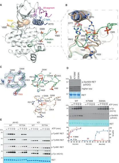
We have determined two similar crystal structures of a construct containing the RET JM region and KD (amino acids 659–1013) at 3.3 and 2.95 A˚, respectively (Table 1). Both crystal structures con-tained the PP1 tyrosine kinase inhibitor in the nucleotide-binding pocket, had an ordered proximal portion of the RET JM segment, and had a hyper-phosphorylated status with four sites phosphor-ylated (Y809, Y905, S909, and Y928) (Figures 4A and 4B). The structures differ slightly in resolution and in the occupancy of the phospho-S909 site. The enhanced multi-site phosphorylation status was surprising when compared with previously solved crystal structures of mono-phosphorylated RET catalytic domain (see PDB: 2IVT, 2IVU, 2IVV, and 4CKI), but consistent with biochemical data, indicating higher RET JM-KD levels of tyrosine kinase activity compared with RET KD (Figures 1and2). Residue Y809 is located within the RET hinge connecting the N-lobe and C-lobe of the RET KD, whereas Y905 and S909 are within the RET activation loop, and Y928 follows the WMAIE motif at the end of the activation segment between helixes a4 and a5 (Hanks et al., 1988). The presence of these phosphorylation sites impacts mainly on the activation-loop conformation detaching it from the body of the catalytic core without affecting the conformation of the hinge, as described later (Figures 4A, 4B, andS4A). Previ-ously solved phosphorylated RET KD crystal structures (PDB: 2IVT, 2IVV, and 2IVU) showed phospho-Y905 tethers several basic side chains including R770 from theaC helix and residues R897 and K907 from the activation loop. In the crystal structures presented in this study, phospho-S909 displaces phospho-Y905 and adopts an approximate equivalent position by engaging acti-vation segment residues R897 and R912, as well as R873 from the HRD motif instead (Figures 4B, 4C, andS4B). In this situation, Y905 does not engage the side chain of theaC helix R770; as a consequence, phospho-Y905 projects away from the body of the RET kinase to mimic a fully solvent-accessible conformer. The second unexpected phosphorylation site at Y928 is posi-tioned beneath the tethered phospho-S909 and is likely to further disrupt interactions of phospho-Y905 with the activation loop. Phospho-Y928 forms hydrogen bonds with side chains of R873 (HRD motif) and activation loop R897 at the top and with H926 from beneath. Its partially buried position indicates the acti-vation loop must have adopted an accessible conformation to fully expose Y928 to undergo phosphorylation. These data are consistent with a recent study where we showed enhanced sub-strate presentation intrans(i.e., activation-loop out conformer) in solution by an oncogenic RET M918T mutant targeting the P+1 substrate-binding pocket (Plaza-Menacho et al., 2014a).
Phospho-S909 Arises from an Intrinsic RET Dual-Specificity Kinase Activity
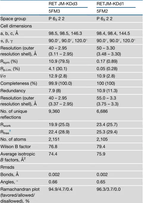
Full-length RET and RET ICD are known to be serine phosphor-ylated in cells and in vitro, respectively (Plaza-Menacho et al., 2014a; Takahashi et al., 1993). S909 is invariant in all RET se-quences and is found only within a minority of RTKs in the human kinome (e.g., FGFR4, ROR1, and HER3). We did not detect S909 phosphorylation by mass spectrometry; however, when we used a specific antibody against an RET phospho-S909 epitope (pSQG), weak basal phospho-serine activity was observed for Table 1. Data Collection and Refinement Statistics
RET JM-KDd3 RETJM-KDd1
5FM3 5FM2
Space group P 632 2 P 632 2
Cell dimensions
a, b, c, A˚ 98.5, 98.5, 146.3 98.4, 98.4, 144.5
a,b,g 90.0, 90.0, 120.0 90.0, 90.0, 120.0
Resolution (outer resolution shell), A˚
40 – 2.95 (3.11 – 2.95)
50 – 3.30 (3.48 – 3.30)
Rsym(%) 10.9 (79.5) 0.17 (0.89)
Rp.i.m.(%) 4.1 (30.1) 0.05 (0.28)
I/s 12.9 (2.8) 10.9 (2.8)
Completeness (%) 99.9 (100.0) 100 (100)
Redundancy 7.9 (8) 10.9 (11.3)
Resolution (outer resolution shell), A˚
40 – 2.95 (3.37 – 2.95)
55.0 – 3.3 (3.75 – 3.3)
No. of unique reflections
9,360 6,686
Rwork 19.9 (25.0) 23.4 (25.7)
Rfreea 22.4 (28.9) 25.3 (29.4)
No. of atoms 2,151 2,105
Wilson B factor 76.8 79.4
Average isotropic
Bfactors, A˚2
74.4 75.9
Rmsds
Bonds, A˚ 0.002 0.002
Angles, 0.66 0.65
Ramachandran plot (favored/allowed/ disallowed), %
94.9/4.7/0.4 96.3/3.7/0.0
aA total of 5% of the data were set aside to compute R
ATP (min)
p-Tyr (4G10) p-Tyr905 RET
RET
10
- 5 20 40 80 kDa
50 50
50
10
- 5 20 40 80- 5 10 20 40 80- 5 10 20 40 80 WT R770A WT R770A
JM-KD KD
E
50
p-Tyr981 RET p-Tyr687 RET 50
R770
Y905 S909
D771
R912
Y928
H926 K907
R873
R897 Y905
R897
H926 Y905
S909
R770
R897
R908 H926
Y905
Y900
K907 R912
S909
Y905
Y928 R912
S896
R897 R873
R912
S896
R897 E903
E903 D898
D898
S896 D898
S904
B
D A
JM-segment
αC helix
Hinge
Y928 S909
R770 Y809
Activation loop
Y905
C
PP1
p-Ser909 RET (pSQG)
RET
-50
WT K758M S909A kDa
-50
ATP (min)
15
- 5 30 60 90
WT
kDa
50
50
50 p-Tyr
(4G10)
RET
15
- 5 30 60 90 - 5 15 30 60 90
K758M S909A
p-Ser909 (pSQG)
-50
higher exp
**** ****
****
**** ********
Figure 4. Crystallographic Identification of an Unexpected Activation-Loop Phospho-S909 Reveals Intrinsic RET Dual-Specificity Activity
(A) Cartoon representation of phosphorylated RET JM-KD structure bound to PP1 inhibitor. Selected residues (including phosphorylated side chains) and secondary structure elements are depicted with discrete colors: JM-segment residues D707 to K716 (magenta),aC helix (purple), hinge residues (yellow), and activation segment residues (green) are shown.
(B) Close-up of the activation-loop conformation and side chains in (A) (green) superposed with RET KD (tint wheat, PDB: 2IVV).
RET WT (Figure 4D, upper panel). RET S909 phosphorylation was dependent on the catalytic status of the receptor as indi-cated by lack of signal of a kinase-dead K758M mutant and was highly specific for S909 (i.e., no signal by a S909A mutant). These data suggested that, contrary to a constitutive phosphor-ylation event intranson S909 by an unknown serine-threonine ki-nase as we initially hypothesized, RET could be a dual-specificity kinase that can autophosphorylate on S909. To test this hypoth-esis, we performed time-course autoP assays with RET ICD WT versus K758M and S909A mutants (Figure 4D, lower panel). As predicted from previous results, RET K758M showed no tyrosine kinase activity compared with RET WT and S909A mutant, which showed similar time-dependent tyrosine autoP (Figure S4C). Crucially, when a phospho-specific RET S909 epitope antibody was used, a time-dependent effect was seen only for RET WT, which showed phospho-serine levels saturating at 60–90 min af-ter stimulation. In contrast, no signal was seen in the case of RET K758M or S909A mutants. Taken together, these data demon-strated that phospho-S909 arises from an intrinsic RET dual-specificity kinase activity not previously reported for an RTK.
Structure-Function Validation of RET JM-KD Crystal Structure
To interpret the increased JM-KD kinase activity from the new structure, we considered whetheraC R770 side chain, which co-ordinates phospho-Y905 in the RET KD structure, could instead make contacts with the JM segment, thereby stabilizing a more active conformer independently of phospho-Y905. Assessing the functional impact of an R770A mutant in the context of both RET JM-KD and RET KD, we found the mutant was selectively im-pairing RET JM-KD activity but had not a measureable detrimental effect on RET KD (Figures 4E andS4F). These data implicate R770 in engaging the JM segment to increase RET catalytic domain activity. Second, we evaluated whether S909 was required for RET tyrosine kinase activity in vitro. Surprisingly, we did not observe any significant effect of the S909A mutant on RET activity in either enzyme kinetics using peptide substrates or in autoP assays (Figures S4C and S4D). Further enzymatic experiments using purified recombinant RET KD with increasing concentra-tions of activation-loop-derived S909 phospho- and non-phos-pho-peptides confirmed these results further (Figure S4E). These data indicate that analogously to RET KD tyrosine autoP sites, S909 is not intrinsically required for catalytic activity. We also considered that redundancy of S909 with phospho-Y905 could mask such a critical role. The latter possibility was further excluded by the lack of any functional effect observed by an RET JM-KD Y905F/S909A double mutant (Figure S5A). An equally plausible explanation is that multi-site phosphorylation of the RET activation segment could play a role in releasing
phos-pho-Y905 or even phospho-S909 acting as a docking or adaptor site required for downstream signaling (seeDiscussion).
Structural Identification and Functional Validation of RETaC Hydrophobic Patch
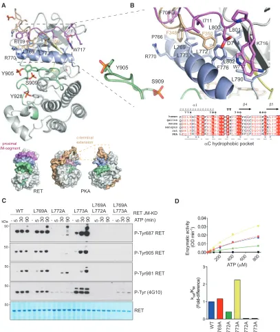
The JM-KD structure revealed contacts from a short segment of the proximal JM region (residues D707 to W717) with a hydropho-bic patch composed of residues from different structural elements includingb4 (L790),b5 (L800, L801, L802), andaC helix (L769, L772, L773, F776, L779) (Figures 5A and 5B). ThisaC hy-drophobic patch is present in many tyrosine and serine-threonine kinases and is frequently a site of regulation to assemble a func-tional regulatory (R) spine (Kannan et al., 2007; Kovacs et al., 2015; Thompson et al., 2009). Intramolecular contacts with this hydrophobic patch arise from interaction with either N- or C-ter-minal sequences flanking the KD (Jura et al., 2011). We noticed a passing similarity between RET aC hydrophobic patch-JM-segment interaction and the PIF pocket-hydrophobic motif inter-action found in AGC kinases first described for the PDK1 serine-threonine kinase (where PIF is defined as the PDK1-interacting fragment) and PKA (Kannan et al., 2007; Biondi et al., 2000). Superposition of the RET JM-KD crystal structure with the PKA catalytic domain (PDB: 1ATP) suggests an equivalence between residues of the PKA hydrophobic motif located at its C terminus to contact theaC helix with those observed in the RET JM segment that engage the hydrophobicaC patch (Figures 5A and 5B). Our interest in this similarity was stimulated by the development of selective drugs against the PDK1 PIF pocket, suggesting the potential for targeting the same region of RET by chemical inhib-itors as an alternative route to RET nucleotide pocket inhibition.
To biochemically probe the role of thisaC hydrophobic patch on RET tyrosine kinase activity in vitro, we engineered individual mutants L769A, L772A, and L773A and double mutants L769/ 772A and L769/773A, and assessed the effect on autoP and enzyme kinetic assays (Figures 5C and 5D). Out of the three sin-gle-point mutants, L772A had a profound detrimental effect compared with WT and L769A, whereas L773A had a marked gain-of-function effect on RET kinase activity. The proximity of L772 and L773 on theaC helix and their opposing effects suggests a subtle conformational alteration of aC would be important for R-spine assembly and hence RET activation. We note that the insulin receptor kinase L1045 (structural equivalent to L773 of RET) directly contacts JM segment Y984 side chain stabilizing an auto-inhibited form (Li et al., 2003). By analogy, L773 could also potentially engage Y687 bound in a similar manner; this would explain why a L773A mutant stimulates RET activity (seeDiscussion). In contrast, L772A gave rise to a loss-of-function effect. We hypothesize that an RET L772A mutant would not create a constitutively active RET (based on
(C) The 2Fo-Fc electron density map of phosphorylated activation-loop from PDB: 5FM2 is shown as blue mesh countered at 1s. Cartoon representation of basic residues engaged by either phospho-S909/Y928 from the JM-KD structure (upper panel, PDB: 5FM2) or phospho-Y905 (from PDB: 2IVV).
(D) WB analyses using a specific antibody against RET phospho-S909 epitope (pSQG) using recombinant RET ICD WT, K758M, and S909A (upper panel) and in vitro time-course autoP assay in the presence of ATP (5 mM) and MgCl2(10 mM) for 0–90 min (lower panel). Total RET protein was evaluated by Coomassie blue
staining. Quantitation of WB data ofFigure 4D is depicted. Data represent the mean of autoP (percentage) normalized to total protein±SEM of the indicated antibodies, n = 3. Statistics: ****p < 0.0001, two-way ANOVA Bonferroni test versus control (WT).
(E) WB analysis of in vitro time-course autoP assay using RET JM661-KD and RET KD core wild-type (WT) and R770A mutants after adding ATP (5 mM) and MgCl2
A B
C D
Figure 5. Structure-Function Analysis of RETaC Hydrophobic Patch
(A) Upper panel shows a cartoon representation of RET JM-KD structure, colored according toFigure 4A, with the superposition of PKA C-terminal residues (PDB: 1ATP). Lower panel shows two views of a surface representation of PKA catalytic subunit together with one of the RET JM-KD structures. The PKA C-terminal segment (pale brown),aC helix (purple), and activation loop (green) are depicted together with selected residues.
(B) Close-up of a superposition of RETaC hydrophobic patch contact residues from the proximal JM-residues (D707 to W717) together with the C-terminal hydrophobic motif (FTDF) from PKA. Selected residues and secondary structural elements are depicted as in (A); some residues have been omitted for clarity. Alignment of RET sequences from different species indicating secondary structural elements and key residues (*) implicated in theaC hydrophobic patch (lower panel).
(C) WB analysis of in vitro time-course phosphorylation assay using RET JM661-KD WT and indicated mutants after addition of ATP (5 mM) and MgCl2(10 mM) for
0–80 min using the indicated antibodies.
the BRAF paradigm, seeDiscussion), but would rather impact on the catalytically required K758-E775-D892 tether and would therefore have a similar impact to the loss-of-function K758M mutant. Double mutants L769/772A and L769/773A were both impaired in their catalytic activity. The contribution of residues from the proximal JM region (i.e., D707, F709, and I711) was also evaluated, revealing lack of functional effect (Figures S6A and S6B). These data suggest that other N-terminal residues from the JM segment not captured in the crystal structure may be relevant for this interaction (e.g., Y687). Alternatively, residues proximal to the transition between JM segment and catalytic domain boundary could also be implicated (seeRET W717 Con-tributes to the Assembly of the JM Hinge and R-Spine and Discussionsections). These data implicate residues L772 and L773 from theaC helix as key determinants in achieving an active conformer and may potentially, by analogy to PDK1, provide an alternative druggable pocket to target within RET.
RET W717 Contributes to the Assembly of the JM Hinge and R-Spine
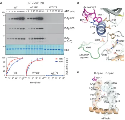
Further structural examination of the aC hydrophobic patch highlighted W717, a highly conserved residue preceding the b-1 strand in many protein kinases including SRC, BTK, EGFR, and BRAF that separates the JM segment from the core catalytic domain (Figure 1A). In the case of BRAF (W342), this residue is important for capping the R-spine in an active conformation and is preceded by a set of phosphorylated residues that are important for dimerization (Hu et al., 2013). In RET, W717 is pre-ceded by a short sequence that engages theaC hydrophobic pocket that contains also the translocation site found in onco-genic RET fusions. To test the function of W717, we generated W717A and W717F mutants in the context of RET JM-KD and found that, contrary to W717F, the W717A mutant had a pro-found detrimental effect on RET phospho-tyrosine activity compared with WT (Figure 6A). These data indicate that W717 is required for RET catalytic activity. One plausible scenario is that W717 would be required for the proper alignment of the JM-proximal hydrophobic motif (DALKIL) to theaC hydrophobic patch (i.e., PIF-like pocket). Although correct in principle, this is unlikely based on the lack of effect in RET activity seen by alanine mutants targeting the DxLxI motif sequence (Figure S6), which suggest that residues farther up in the JM segment (e.g., Y687) make important contacts with the catalytic core (Figure 3). Alter-natively, W717 would be required for docking onto and proper alignment of the R-spine in the active conformation, following the BRAF paradigm (Figure 6A). We hypothesize that mutating W717 by alanine and not by phenylalanine will perturb the R-spine side chain stacking and, as a consequence, impact on RET activ-ity. Examination of the crystal structure (Figure 6B) revealed that in RET the R-spine is composed of four hydrophobic residues orig-inating from theaF helix connecting N- and C-lobes; these resi-dues include H872 (from the catalytic HRD motif), F893 (from the DFG motif), L779 (aC helix), and L790 (b-4 strand). W717 caps from the top the R-spine in a linear tetrad compatible with an active DFG in conformation of the kinase (Taylor and Kornev, 2011). It is further preceded by D714 adjacent to the fusion site between L712 and E713, which forms an important salt bridge with aC K780, a specific feature lacking in previously solved
RET catalytic domain crystal structures. The combined effect of both W717 docking and the D714-K780 tether locks the hinge be-tween the proximal JM segment and N-terminal residues of the catalytic core. In this scenario it is plausible also to hypothesize that perturbation of the hinge by the W717A mutant, contrary to hydrophobic motif DxLxIx alanine mutants, results in a non-compatible JM-proximal segment alignment with N-terminal residues of the catalytic core and a consequent alteration of the R-spine. How perturbation of the R-spine linear architecture re-sults in catalytic inefficiency is likely an indirect effect on both the catalytic (C)-spine and the catalytically required K758 (b-3 strand)-E775 (aC helix)-D892 (DFG motif) tether, which links both spines and the nucleotide moiety (Figure 6C). From these data we conclude that W717 is an important residue for RET func-tion by playing a role in the assembly of the JM hinge and R-spine.
Activation-Loop Serine Phosphorylation Is Required for RET Signaling In Vivo
S909 is a novel autoP site that arises from intrinsic dual-specificity kinase activity exhibited by RET in vitro (Figure 4D). Functional assays of a S909A mutant excluded a direct role of S909 on RET catalytic activity (Figures S4C and S4D). These results are consis-tent with data for mutants targeting RET KD tyrosine autoP sites (Figure 3B) and suggest phospho-S909 could act as a docking or alternatively a substrate site for effector proteins important for RET signaling. Given the high conservation of S909 in all RET se-quences and its consistent occupancy, we assessed whether S909 had a role in RET downstream signaling. We therefore used aDrosophilaRet2B(dRet2B;dRet M955T) fly model to assess whether mutation at residue S946 (equivalent to human RET S909) could influence in vivo the aberrant phenotype promoted by onco-genicdRet2B. We employed a pUAST-attB fly vector system to allow specific site insertion of the transgene into the fly genome (Bischof et al., 2007). Overexpression ofdRet2Bin the developing eye using the glass multiple reporter (GMR) Gal4-815 promoter (GMR-Gal4-815 > dRet2B) led to extensive mispatterning and positioning of ommatidia resulting in a ‘‘rough eye’’ phenotype in the adult fly compared with the driver-line control. When we gener-ated a transgenic fly expressing a double mutant dRet2B/S946A
(dRet M955T/S946A), the aberrant rough eye phenotype was
toward further complexity through interplay with RET phospho-tyrosine sites, or alternatively as a docking and/or phospho-site for a yet unknown effector that impacts on RET signaling.
DISCUSSION
To identify unique features of RET tyrosine kinase activation, we have applied biochemical, structural, and biophysical analyses, together with an in vivo model for RET hyper-activation. We
show that the JM segment functions to increase RET tyrosine KD activity without affecting substrate presentation intrans. Fully phosphorylated RET JM-KD appears rapidly, between 20 and 40 min, compared with the 80–120 min required for core RET KD (Figure 1B). This increased activity promoted by the JM segment, contrary to the EGFR (Jura et al., 2009; Red Brewer et al., 2009), does not appear to result from stable dimer formation in solution (Figure S1C) and is independent of ‘‘forced’’ dimeriza-tion through the presence of a GST tag (Figure 2D). Our results are
B
C
E775
K758
D892
R-spine C-spine
W717
L790 L779
F893
D933 H872
A756
V738
V882 L881
L812 I880
I944 L940
αF helix ATP (min)
RET JM661-KD
- 1 5 15 30 60 90 - 1 5 15 30 60 90 - 1 5 15 30 60 90
WT W717F W717A
A
RET P-Tyr905
P-Tyr (4G10) P-Tyr687
-kDa 5050
50
50
Time (min)
WT W717F W717A
RET
autoP
(%)
****
**
****
W717
K780
L790
L779
F893
H872
D993 S909
Y905
Y928
αF
αC JM-segment
Activation segment
D714
Figure 6. RET W717 Contributes to the Assembly of the JM Hinge and R-Spine
(A) WB analysis of in vitro time-course autoP assay using RET JM661-KD WT and indicated mutants (1mM) after addition of ATP (5 mM) and MgCl2(10 mM) for 0–
90 min using the indicated antibodies. Quantitation of RET phospho-Y687 and -Y905 signal is shown; data represent mean±SEM, n = 3, ****p < 0.0001, **p = 0.0014, two-way ANOVA Bonferroni test.
(B) Cartoon representation of RET JM-KD crystal structure secondary structural elements, colored according toFigure 4A. Close-up of JM-hinge-composing residues (D714, K780, and W717) and R-spine-assembling residues (L779, L790, F893, H872, and D993) is shown; further selected residues and secondary structural elements are depicted as inFigure 4A. Some residues and structural elements have been omitted for clarity.
(C) View of the C- and R-spines of RET JM-KD crystal structure as per text (seeResultsandDiscussionfor R-spine); in addition, the catalytic triad K758 (b-3
consistent with VEGFR2, where in solution the kinetics of autoP is significantly enhanced by the JM region (Solowiej et al., 2009). Comparing these data with the slower overall phosphorylation ki-netics of RET ICD (Plaza-Menacho et al., 2014a) suggests the C-terminal (CT) segment could act as a negative regulator of RET catalytic domain activity and restrain by competition JM segment activating input. The RET JM region appears not to play acis-inhibitory role as observed for KIT and MET (Chan et al., 2003; Hubbard, 2004) but is likely to stabilize an active form of RET in a manner that is dependent on Y687. We argue that autoP is not prevented by a non-phosphorylated conformer of JM segment on Y687, but that timely phosphorylation of JM segment on Y687 leads to a conformation contributing to a more active RET kinase. This is supported by the observation that Y687 is required for the JM segment to rescue a catalytically deficient RET KD lacking both activation-loop tyrosines (Y900/ 905F). Furthermore, a RET JM-KD Y687E mutant (mimicking a constitutive phospho-Y687) showed a significant decrease on tyrosine kinase activity, which indicated that Y687 is a tightly regu-lated autoP site. We hypothesize that constitutive phosphoryla-tion on Y687 results in a detrimental effect on activity because of the lack of required contacts between JM segment and RET
GMR GMR >dRet 2B S946A
A
765 2B
765 >dRet S946A
B
- + + + + + + +
P-Tyr (total)
P-Tyr1015 RET
P-Tyr1062 RET
C D
dRet M955T M955T S946A
2B GMR>dRet
2B
765 >dRet
dRet pGal4
--
-+ WTM955TM955T/S946A WTM955TM955T/S946A
150 kDa
150
150 RET
150 kDa
Figure 7. An Activation-Loop Serine Phos-pho-Site Is Required for RET Signaling In Vivo
(A) Ectopic expression ofdRet2B
(dRet M955T) and dRet2B
S946Ain the retina from theGMR-Gal4 815 promoter.
(B) Expression ofdRet2B
from theGal4 765promoter led to ectopic vein formation. S946A mutation rescued the wing defects. Scale bar, 500mM. (C) Quantification of data shown inFigure 6B. The percentage of wings with ectopic veins was deter-mined from three distinct transformants for each genotype. The number of individual flies counted from each transformant was 19, 30, and 23 for dRet2Band 35, 17, and 23 fordRet2BS946A. **p < 0.01, one-way ANOVA Bonferroni test.
(D) WB analyses of S2 insect cells ectopically ex-pressing dRet WT, M955T, and M955T/S946A, together with an Actin-promoter-driven Gal4 construct using the indicated antibodies.
catalytic core prior and during kinase acti-vation (Figure S5B). Furthermore, the de-pendency seen by RET JM-KD, but not RET KD, onaC R770 implicates its side chain in engaging the JM segment to in-crease RET catalytic domain activity. Further evidence for theciseffect of the JM segment in RET activation includes: (1) phosphorylation rescue experiments
in trans using as substrate catalytically
deficient versions (i.e., K758M) of RET JM-KD and RET KD (Figure S3B) did not show significant differences because of the JM segment, and (2) the presence of the RET JM-segment effectively rescues an otherwise catalytically-deficient RET mutant bearing a dou-ble-tyrosine (Y900/905F) substitution in the activation-loop. Note this is contrary to the rescue experiment intransof catalyti-cally deficient K758M kinase versions, which cannot be rescued in
cisby either JM segment (Figure S3) or oncogenic mutations (Plaza-Menacho et al., 2014a).
The trajectory of the proximal part of the RET JM-segment resembles to some extent that seen in the IR JM-KD crystal struc-ture (PDB: 1P14) (Li et al., 2003). In the IR crystal strucstruc-ture, JM-Y984 docks into theaC hydrophobic patch incis(adopting an equivalent position to RET F776) and forms a network of hydrogen bonds between residues from theaC helix and prox-imal JM segment. These interactions provide steric restraints preventingaC from assuming a catalytically competent position. A recent study has shown, however, that the JM-IR can also adopt a JM-out conformer contacting theaC of a second recep-tor molecule and is able to stabilize an active catalytic dimer (Cabail et al., 2015). This role for the IR JM-segment intrans
(Hu et al., 2013) to make intermolecular contacts with theaC hy-drophobic patch from a second molecule. This resembles intrans
an extended stretch of the proximal JM region seen in our RET JM-KD crystal structure. Our interpretation is that in the absence of a stable RET KD dimer in solution or in the crystal, the JM-segment collapses onto theaC helix incisin the crystal lattice. Our data pointed also at the critical RETaC hydrophobic pocket as being sensitive to allosteric input from JM-segment elements, possibly including Y687. This hydrophobic pocket has the poten-tial to be targeted by small molecules, because there are prece-dents for PDK1 where allosteric inhibitors against an equivalent site are already available (Busschots et al., 2012).
Chromosomal translocations involving the RET exons 12–21 are found in human thyroid and lung cancers (Nikiforov and Niki-forova, 2011; Plaza-Menacho et al., 2014b). These gene rear-rangements fuse a variety of unrelated coiled-coil proteins within the same RET intron, thereby removing exons 1–11, including the JM segment. Our data are consistent with a scenario in which removing the JM segment, rather than eliminating an autoinhibi-tory element, replaces it with a more potent dimerizing motif that stabilizes a RET dimer independently of ligand and transmem-brane region. For the IR, its JM segment extends away from the kinase core pivoting about a conserved VPDEWE motif to engage a second kinase molecule via contacts to theaC helix. A network of salt-bridge interactions at the pivot point involves the VPDEWE motif to help stabilize a conformation associated with an activated IR tyrosine kinase. The equivalent sequence for RET, EDPKWE, contains the fusion site (between L712 and E713) of many RET translocations that eliminate RET exons 1–11 (Nikiforov and Nikiforova, 2011). Such fusions add a dimeric coiled-coil region just prior to D714 and W717 that would lock permanently the JM hinge and R-spine into a DFG-in conformer resulting in a hyperactive RET, no longer localized at the plasma membrane. These findings have important drug discovery and therapeutic implications as perturbation of the JM hinge and consequent effect on adequate R-spine assembly could be a new drug-targetable strategy against oncogenic RET.
The JM-KD crystal structure shows two unexpected phos-phorylation sites, Y928 and S909, both invariant RET residues. Both are in close proximity and engage basic residues otherwise found in the core RET KD structures engaged by phospho-Y905 (Figures 4A and 4B;Figure S4B). In particular, the unconven-tional activation-loop S909 phospho-site engages the HRD motif in what it seems to be a unique active conformation promoting both regulatory-spine assembly and accessibility to phospho-tyrosine binding modules. As a consequence, Y905 is displaced, adopting a solvent-accessible conformer competent for a signaling function rather than playing an activating role.
Analyses of RET sequences flanking the invariant protein ki-nase RD motif establish it as tyrosine kiki-nase (HRDLAARN or HRDLRAAN) rather than a serine-threonine kinase (H/YRDLXXN) (Hanks et al., 1988; Lindberg et al., 1992). There are precedents for dual-specificity kinase activity among the cyclin-dependent kinase (CDK), mitogen-activated protein kinase (MAPK), gly-cogen synthase kinase (GSK3), CDC-like kinase (CLK) group of protein kinases (CMGC) that include the mitogen-activated pro-tein kinase (MAPK) and DYRK kinase members. The latter exam-ples phosphorylate exclusively serine and threonine side chains
in their substrates but are able to autoP on tyrosine (Himpel et al., 2000). More recently, a non-receptor tyrosine kinase Syk has been shown to exhibit dual-specificity kinase activity (Heizmann et al., 2010). In our study, robust biochemical examination re-vealed that S909 is an autoP site that arises from an intrinsic RET dual-specificity kinase activity. This is consistent with a recent study reporting that RET can phosphorylate AP2 intrans
on threonine (Bagheri-Yarmand et al., 2015). This dual-specific activity can generate phospho-S909 in vitro. However, phos-phorylation on RET S909 was not catalytically required (Figures S4C–S4E), consistent with other serine-to-alanine mutants tar-geting the JM segment (Figure S5C). Further efforts are needed to establish whether phospho-S909 can act as a docking for a yet-unknown effector protein involved in RET signaling. Interest-ingly, when we evaluated an RET S909D mutant, a marked in-crease in RET JM-KD phospho-tyrosine activity was observed (Figure S5B). These data suggest that a phosphorylation event on S909 intranscan contribute to RET tyrosine kinase activity and signaling in vivo. To explore further this hypothesis, we em-ployed an in vivo model and found that mutating this residue in
Drosophila Ret (dRet) has a deleterious effect on the dRet2B
(dRet M955T) oncogenic phenotype, a well-established model
of transformation. In particular, dRet M955T/S946Aefficiently rescued the phenotype ofdRet2Busing alternative promoters.
Overall, our structure-function analyses and in vivo experi-ments have revealed complex eleexperi-ments in the mechanism of RET activation and signaling. Allosteric inputs from the JM-segment and activation-loop S909 contribute to kinase function. We show that phospho-S909 is an autoP site arising from an intrinsic dual-specificity RET kinase activity and appears to play key roles in oncogenic signaling. Our study also suggests that targeting the aC hydrophobic pocket together with the JM hinge using small molecules to manipulate RET kinase activ-ity may be a productive approach for either blocking oncogenic forms of RET or stimulating RET activity in Hirschsprung’s dis-ease (HSCR) and neurodegenerative Parkinson’s disdis-ease (PD).
EXPERIMENTAL PROCEDURES
Expression and Purification of Recombinant Protein
Protein expression was carried out using Sf9 insect cells following a previously described protocol (Knowles et al., 2006). Codon optimized human RET9 iso-form intracellular domain (ICD residues 661–1072), different lengths versions of the RET JM-KD (661 to 698–1012) and RET KD core (705–1013) WT, and the indicated mutants proteins were purified following a protocol previously described (Plaza-Menacho et al., 2014a).
Mass Spectrometric Label-free Quantitation
Mass spectrometry procedures were performed as previously described (Plaza-Menacho et al., 2014a).
Autophosphorylation Assays, SDS-PAGE, and Western Blotting
Enzymatic Kinase Assays
Enzyme kinetic experiments were performed as previously described ( Plaza-Menacho et al., 2014a).
ITC
ITC experiments were performed as previously described (Plaza-Menacho et al., 2014a).
Dynamic Light Scattering and ThermoFluor Assays
To determine protein stability, we performed thermal shifts assays as previ-ously described (Plaza-Menacho et al., 2010, 2014a). Molecular weight deter-mination in solution was performed by DLS using different RET protein concentrations.
Crystallization, Diffraction, Data Collection, and Processing
Crystals of the phosphorylated RET JM-catalytic domain (residues 659– 1013) were grown at 22C by vapor diffusion in sitting drops containing crystal 1 (5FM2), 1mL protein stock solution (6 mg/ml) mixed with 1mL reservoir solution (1.5 M ammonium sulfate, 0.1 M BIS-TRIS propane [pH 7.0]); the protein stock solution also contained 2.5 mM ATP and 5 mM MgCl2. Crystal 2 (5FM3) comprised 0.8mL protein stock solution
(5 mg/ml) mixed with 0.8mL reservoir solution (1.2 ammonium sulfate, 0.1 M tri-sodium citrate [pH 5.43]). The crystals were cryoprotected in 25% glycerol in reservoir solution for several minutes and flash frozen in liquid nitrogen, and X-ray datasets were collected at the I-24 beamline of the Diamond Light Source Synchrotron (Oxford, UK). Data collection and refinement statistics are summarized inTable 1. The dataset was indexed with MOSFLM and scaled with SCALA (Winn et al., 2011). Molecular replacement was carried out using the atomic coordinates of the phosphor-ylated RET KD (PDB: 2IVT) in PHASER (McCoy et al., 2007). Refinement was carried out by using Phenix (Adams et al., 2010). Model building was carried out in COOT (Emsley et al., 2010). Model validation used PROCHECK (Vaguine et al., 1999), and figures were prepared using the graphics program PYMOL (http://www.pymol.org).
DrosophilaExperiments
pUASTattB-dRetM955T (dRet2B
) and double mutant pUASTattB-dRetM955T/S946Aconstructs were generated by site-directed mutagenesis us-ing the followus-ing primers: M955T forward 50-GTGCCCGTCAAGTGGACG GCTCCGGA-30, M955T reverse 50- TCCGGAGCCGTCCACTTGACGGGCAC-30, S946A forward 50- GCCTATTTAAAGAGAGCCCGAGATCGTGTGCCC-30, and S946A reverse 50- GGGCACACGATCTCGGGCTCTCTTTAAATAGGC. Transgenic flies were generated using P-element-mediated (pUAST) transgene-sis by BestGene.Drosophilastocks and crosses were maintained at 25C, un-less stated otherwise. For ectopic expression of the various transgenes in the developingDrosophilawing,dRettransgenic flies were crossed with the Gal4 C-765 driver (36523, Bloomington). Adult wings were dissected, mounted, and imaged at 43magnification using the EVOS cell imaging system. For ectopic expression of thedRet2B
anddRet2B
S946Ain the developing eye, the transgenic flies were crossed with the GMR-Gal4 815 (weak) driver and maintained at 18C. Eye phenotypes were analyzed by light microscopy of whole mounts.
Ectopic Expression in S2 Cells
pUASTattB-dRetWT, M955T, and M955T/S947A constructs (400 ng) were co-transfected together with an Actin-promoter-driven Gal4 plasmid (400 ng) as indicated into S2 cells using Effectene and following manufac-turer’s instructions. Data represent three independent experiments.
Statistical Analyses
Graphs and statistical analyses were done using Prism GraphPad.
ACCESSION NUMBERS
The crystallographic coordinates and structure factors for the RET JM-KD crystal structures reported in this paper are PDB: 5FM3 and 5FM2, respec-tively (http://www.pdb.org).
SUPPLEMENTAL INFORMATION
Supplemental Information includes six figures and can be found with this article online athttp://dx.doi.org/10.1016/j.celrep.2016.11.061.
AUTHOR CONTRIBUTIONS
I.P.-M. and N.Q.M. planned the project, designed experiments, analyzed the data, and wrote the paper. I.P.-M. performed all biochemical and crystallo-graphic experiments, assembled the initial draft of the paper, and prepared all figures. K.B. performed the mass spectrometry analyses. A.B. and R.C. assisted directly with baculovirus production. R.B. and M.O. performedDrosophila exper-iments under supervision of I.P.-M. and P.M. S.M. assisted with data processing and structure determination. R.J.M.-T. performed the ITC experiments.
ACKNOWLEDGMENTS
We thank our Lincoln’s Inn Fields Laboratories (Francis Crick Institute) colleagues Sara Kisakye Nambozo, Roger George, and Svend Kjaer (Protein Production Facility) for their technical assistance in the production of baculoviruses; Nicola O’Reilly (Peptide Production Facility) for timely supply of peptides; and Andrew Purkiss-Trew (Structural Biology Laboratory) for helping with data collection and structure determination. We also thank Tilman Schirmer (Biozentrum, Uni-versity of Basel) for helpful comments on the manuscript and Ross Cagan (Icahn School of Medicine, Mount Sinai, NY) for providing initial pUAST-dRetplasmids. We thank Diamond Light Source Synchrotron (Oxford, UK) for allowing X-ray dataset collection at the I-24 beamline. N.Q.M. acknowledges that this work was supported by the Francis Crick Institute, which receives its core funding from Cancer Research UK (FC001115), the UK Medical Research Council (FC001115) and the Wellcome Trust (FC001115); by the NCI/NIH (grant reference 5R01CA197178); by the Association for Multiple Endocrine Neoplasia Disorders MTC Research Fund. P.M. acknowledges NHS funding to the NIHR Biomedical Research Centre.
Received: March 22, 2016 Revised: October 2, 2016 Accepted: November 20, 2016 Published: December 20, 2016
REFERENCES
Adams, P.D., Afonine, P.V., Bunko´czi, G., Chen, V.B., Davis, I.W., Echols, N., Headd, J.J., Hung, L.W., Kapral, G.J., Grosse-Kunstleve, R.W., et al. (2010). PHENIX: a comprehensive Python-based system for macromolecular struc-ture solution. Acta Crystallogr. D Biol. Crystallogr.66, 213–221.
Airaksinen, M.S., Titievsky, A., and Saarma, M. (1999). GDNF family neurotrophic factor signaling: four masters, one servant? Mol. Cell. Neurosci.13, 313–325.
Asai, N., Fukuda, T., Wu, Z., Enomoto, A., Pachnis, V., Takahashi, M., and Cos-tantini, F. (2006). Targeted mutation of serine 697 in the Ret tyrosine kinase causes migration defect of enteric neural crest cells. Development 133, 4507–4516.
Bae, J.H., and Schlessinger, J. (2010). Asymmetric tyrosine kinase arrange-ments in activation or autophosphorylation of receptor tyrosine kinases. Mol. Cells29, 443–448.
Bagheri-Yarmand, R., Sinha, K.M., Gururaj, A.E., Ahmed, Z., Rizvi, Y.Q., Huang, S.C., Ladbury, J.E., Bogler, O., Williams, M.D., Cote, G.J., and Gagel, R.F. (2015). A novel dual kinase function of the RET proto-oncogene negatively regulates activating transcription factor 4-mediated apoptosis. J. Biol. Chem. 290, 11749–11761.
Biondi, R.M., Cheung, P.C., Casamayor, A., Deak, M., Currie, R.A., and Alessi, D.R. (2000). Identification of a pocket in the PDK1 kinase domain that interacts with PIF and the C-terminal residues of PKA. EMBO J.19, 979–988.
Busschots, K., Lopez-Garcia, L.A., Lammi, C., Stroba, A., Zeuzem, S., Piiper, A., Alzari, P.M., Neimanis, S., Arencibia, J.M., Engel, M., et al. (2012). Sub-strate-selective inhibition of protein kinase PDK1 by small compounds that bind to the PIF-pocket allosteric docking site. Chem. Biol.19, 1152–1163.
Cabail, M.Z., Li, S., Lemmon, E., Bowen, M.E., Hubbard, S.R., and Miller, W.T. (2015). The insulin and IGF1 receptor kinase domains are functional dimers in the activated state. Nat. Commun.6, 6406.
Chan, P.M., Ilangumaran, S., La Rose, J., Chakrabartty, A., and Rottapel, R. (2003). Autoinhibition of the kit receptor tyrosine kinase by the cytosolic juxta-membrane region. Mol. Cell. Biol.23, 3067–3078.
Chen, H., Ma, J., Li, W., Eliseenkova, A.V., Xu, C., Neubert, T.A., Miller, W.T., and Mohammadi, M. (2007). A molecular brake in the kinase hinge region reg-ulates the activity of receptor tyrosine kinases. Mol. Cell27, 717–730.
Emsley, P., Lohkamp, B., Scott, W.G., and Cowtan, K. (2010). Features and development of Coot. Acta Crystallogr. D Biol. Crystallogr.66, 486–501.
Fukuda, T., Kiuchi, K., and Takahashi, M. (2002). Novel mechanism of regula-tion of Rac activity and lamellipodia formaregula-tion by RET tyrosine kinase. J. Biol. Chem.277, 19114–19121.
Hanks, S.K., Quinn, A.M., and Hunter, T. (1988). The protein kinase family: conserved features and deduced phylogeny of the catalytic domains. Science 241, 42–52.
Heizmann, B., Reth, M., and Infantino, S. (2010). Syk is a dual-specificity ki-nase that self-regulates the signal output from the B-cell antigen receptor. Proc. Natl. Acad. Sci. USA107, 18563–18568.
Himpel, S., Tegge, W., Frank, R., Leder, S., Joost, H.G., and Becker, W. (2000). Specificity determinants of substrate recognition by the protein kinase DYRK1A. J. Biol. Chem.275, 2431–2438.
Hu, J., Stites, E.C., Yu, H., Germino, E.A., Meharena, H.S., Stork, P.J., Kornev, A.P., Taylor, S.S., and Shaw, A.S. (2013). Allosteric activation of functionally asymmetric RAF kinase dimers. Cell154, 1036–1046.
Hubbard, S.R. (1997). Crystal structure of the activated insulin receptor tyro-sine kinase in complex with peptide substrate and ATP analog. EMBO J.16, 5572–5581.
Hubbard, S.R. (2004). Juxtamembrane autoinhibition in receptor tyrosine ki-nases. Nat. Rev. Mol. Cell Biol.5, 464–471.
Jura, N., Endres, N.F., Engel, K., Deindl, S., Das, R., Lamers, M.H., Wemmer, D.E., Zhang, X., and Kuriyan, J. (2009). Mechanism for activation of the EGF re-ceptor catalytic domain by the juxtamembrane segment. Cell137, 1293–1307.
Jura, N., Zhang, X., Endres, N.F., Seeliger, M.A., Schindler, T., and Kuriyan, J. (2011). Catalytic control in the EGF receptor and its connection to general ki-nase regulatory mechanisms. Mol. Cell42, 9–22.
Kannan, N., Haste, N., Taylor, S.S., and Neuwald, A.F. (2007). The hallmark of AGC kinase functional divergence is its C-terminal tail, a cis-acting regulatory module. Proc. Natl. Acad. Sci. USA104, 1272–1277.
Knowles, P.P., Murray-Rust, J., Kjaer, S., Scott, R.P., Hanrahan, S., Santoro, M., Iba´n˜ez, C.F., and McDonald, N.Q. (2006). Structure and chemical inhibition of the RET tyrosine kinase domain. J. Biol. Chem.281, 33577–33587.
Kovacs, E., Zorn, J.A., Huang, Y., Barros, T., and Kuriyan, J. (2015). A struc-tural perspective on the regulation of the epidermal growth factor receptor. Annu. Rev. Biochem.84, 739–764.
Lemmon, M.A., and Schlessinger, J. (2010). Cell signaling by receptor tyrosine kinases. Cell141, 1117–1134.
Lemmon, M.A., Schlessinger, J., and Ferguson, K.M. (2014). The EGFR family: not so prototypical receptor tyrosine kinases. Cold Spring Harb. Perspect. Biol.6, a020768.
Li, S., Covino, N.D., Stein, E.G., Till, J.H., and Hubbard, S.R. (2003). Struc-tural and biochemical evidence for an autoinhibitory role for tyrosine 984 in the juxtamembrane region of the insulin receptor. J. Biol. Chem. 278, 26007–26014.
Lindberg, R.A., Quinn, A.M., and Hunter, T. (1992). Dual-specificity protein kinases: will any hydroxyl do? Trends Biochem. Sci.17, 114–119.
Lougheed, J.C., Chen, R.H., Mak, P., and Stout, T.J. (2004). Crystal structures of the phosphorylated and unphosphorylated kinase domains of the Cdc42-associated tyrosine kinase ACK1. J. Biol. Chem.279, 44039–44045.
McCoy, A.J., Grosse-Kunstleve, R.W., Adams, P.D., Winn, M.D., Storoni, L.C., and Read, R.J. (2007). Phaser crystallographic software. J. Appl. Cryst.40, 658–674.
Nikiforov, Y.E., and Nikiforova, M.N. (2011). Molecular genetics and diagnosis of thyroid cancer. Nat. Rev. Endocrinol.7, 569–580.
Perrinjaquet, M., Vilar, M., and Iba´n˜ez, C.F. (2010). Protein-tyrosine phospha-tase SHP2 contributes to GDNF neurotrophic activity through direct binding to phospho-Tyr687 in the RET receptor tyrosine kinase. J. Biol. Chem. 285, 31867–31875.
Plaza-Menacho, I., Burzynski, G.M., de Groot, J.W., Eggen, B.J., and Hofstra, R.M. (2006). Current concepts in RET-related genetics, signaling and thera-peutics. Trends Genet.22, 627–636.
Plaza-Menacho, I., Morandi, A., Robertson, D., Pancholi, S., Drury, S., Dows-ett, M., Martin, L.A., and Isacke, C.M. (2010). Targeting the receptor tyrosine kinase RET sensitizes breast cancer cells to tamoxifen treatment and reveals a role for RET in endocrine resistance. Oncogene29, 4648–4657.
Plaza-Menacho, I., Morandi, A., Mologni, L., Boender, P., Gambacorti-Passer-ini, C., Magee, A.I., Hofstra, R.M., Knowles, P., McDonald, N.Q., and Isacke, C.M. (2011). Focal adhesion kinase (FAK) binds RET kinase via its FERM domain, priming a direct and reciprocal RET-FAK transactivation mechanism. J. Biol. Chem.286, 17292–17302.
Plaza-Menacho, I., Barnouin, K., Goodman, K., Martı´nez-Torres, R.J., Borg, A., Murray-Rust, J., Mouilleron, S., Knowles, P., and McDonald, N.Q. (2014a). Oncogenic RET kinase domain mutations perturb the autophosphorylation tra-jectory by enhancing substrate presentation in trans. Mol. Cell53, 738–751.
Plaza-Menacho, I., Mologni, L., and McDonald, N.Q. (2014b). Mechanisms of RET signaling in cancer: current and future implications for targeted therapy. Cell. Signal.26, 1743–1752.
Red Brewer, M., Choi, S.H., Alvarado, D., Moravcevic, K., Pozzi, A., Lemmon, M.A., and Carpenter, G. (2009). The juxtamembrane region of the EGF recep-tor functions as an activation domain. Mol. Cell34, 641–651.
Solowiej, J., Bergqvist, S., McTigue, M.A., Marrone, T., Quenzer, T., Cobbs, M., Ryan, K., Kania, R.S., Diehl, W., and Murray, B.W. (2009). Characterizing the effects of the juxtamembrane domain on vascular endothelial growth factor receptor-2 enzymatic activity, autophosphorylation, and inhibition by axitinib. Biochemistry48, 7019–7031.
Takahashi, M., Asai, N., Iwashita, T., Isomura, T., Miyazaki, K., and Mat-suyama, M. (1993). Characterization of the ret proto-oncogene products ex-pressed in mouse L cells. Oncogene8, 2925–2929.
Taylor, S.S., and Kornev, A.P. (2011). Protein kinases: evolution of dynamic regulatory proteins. Trends Biochem. Sci.36, 65–77.
Thiel, K.W., and Carpenter, G. (2007). Epidermal growth factor receptor juxta-membrane region regulates allosteric tyrosine kinase activation. Proc. Natl. Acad. Sci. USA104, 19238–19243.
Thompson, E.E., Kornev, A.P., Kannan, N., Kim, C., Ten Eyck, L.F., and Taylor, S.S. (2009). Comparative surface geometry of the protein kinase family. Pro-tein Sci.18, 2016–2026.
Vaguine, A.A., Richelle, J., and Wodak, S.J. (1999). SFCHECK: a unified set of procedures for evaluating the quality of macromolecular structure-factor data and their agreement with the atomic model. Acta Crystallogr. D Biol. Crystal-logr.55, 191–205.
Winn, M.D., Ballard, C.C., Cowtan, K.D., Dodson, E.J., Emsley, P., Evans, P.R., Keegan, R.M., Krissinel, E.B., Leslie, A.G., McCoy, A., et al. (2011). Over-view of the CCP4 suite and current developments. Acta Crystallogr. D Biol. Crystallogr.67, 235–242.