R E S E A R C H A R T I C L E
Open Access
Comparison of our self-designed rotary
self-locking intramedullary nail and interlocking
intramedullary nail in the treatment of long bone
fractures
Bailian Liu
1, Ying Xiong
1, Hong Deng
2, Shao Gu
1, Fu Jia
1, Qunhui Li
1, Daxing Wang
1*, Xuewen Gan
1and Wei Liu
1Abstract
Objective:The purpose of this study is to compare the clinical effects of our self-designed rotary self-locking intramedullary nail (RSIN) and interlocking intramedullary nail (IIN) for long bone fractures.
Methods:A retrospective study was performed in 1,704 patients who suffered bone fractures and underwent RSIN
or IIN operation in our hospital between March 1999 and March 2013, including 494 with femoral fractures, 572 with humeral fractures, and 638 with tibial fractures. Among them, 634 patients were followed up for more than 1 year. The operative time, intraoperative blood loss, postoperative complications, healing rate, and the excellent and good rate of functional recovery were compared between two groups.
Results:Compared with IIN group, RSIN group exhibited significantly shorter operative time and less intraoperative blood loss no matter for humeral, femoral, or tibial fractures (allp< 0.001). The healing rate in patients with more than 1 year follow-up was significantly higher in RSIN group for femoral and tibial fractures (bothp< 0.05). In RSIN group, no nail breakage or loosening occurred, but radial nerve injury and incision infection were respectively observed in one patient with humeral fracture. In IIN group, nail breakage or loosening occurred in 7 patients with femoral fractures and 16 patients with tibial fractures, radial nerve injury was observed in 8 patients with humeral fractures, and incision infection was present in 2 patients with humeral fractures and 1 patient with femoral fracture. The complication rate of IIN group was significantly higher than that of RSIN group (p< 0.05). However, there were no significant differences in the excellent and good rate of shoulder, elbow, knee, and ankle joint functional recovery between RSIN group and IIN group.
Conclusion:RSIN may be a reliable and practical alternative method for the treatment of long bone fractures.
Keywords:Rotary self-locking intramedullary nail, Interlocking intramedullary nail, Retrospective analysis, Long bone fractures
Background
Long bone fractures, including tibial, femoral, and hu-meral fractures, are common traumatic injuries, account-ing for approximately 4% of emergency department visits in the USA every year [1]. The management of long bone fractures remains challenging and often controversial. Re-cently, closed or open reduction and internal fixation with
interlocking intramedullary nailing (IIN) has been recom-mended as a standard approach for treatment of long bone fractures because it has the advantages of fixation stability, anti-rotation, and anti-contraction, contributing to a high healing rate and a low incidence of complica-tions [2-4]. However, the use of interlocked nail also has some disadvantages: it is a demanding step for accurate insertion of the distal locking screws, leading to increased radiation exposure and delayed operative time [5,6]; rota-tor cuff tear and shoulder impingement are easily caused for fixation of humeral fractures, resulting in shoulder * Correspondence:[email protected]
1
Department of Orthopaedics, Yan’an Hospital, No. 245 Renmin East Road, Kunming, Yunnan 650051, China
Full list of author information is available at the end of the article
pain and shoulder function dysfunction [7]. Because of ex-cessive bending stress in femur and tibia, the IIN nail has the potential risk of breakage for fixation of tibial and fem-oral fractures [8,9].
To overcome the drawbacks of IIN for clinical fixation of long bone fractures, from 1995 to 1998, our team de-veloped self-locking intramedullary nails (RSIN) for fix-ation of the humeral, femoral and tibial fractures. RSIN can be directly screwed into the medullary cavity with the side-locking tag to achieve anti-rotation. The pur-pose of present study was to retrospectively review the clinical and therapeutic outcomes of IIN and our RSIN for treatment of long bone fractures.
Methods
Patients
Totally, 1,704 patients (approved by ethics committee of Yan’an Hospital) suffering humeral (n= 494), femoral (n= 572), or tibial fractures (n= 638) in our hospital were enrolled from March 1999 to March 2013. According to operative procedure they underwent, the patients were evenly divided into IIN or RSIN group. Fractures were classified according to the AO classification system. The general characteristics of patients in the two groups are shown in Table 1.
RSIN design
RSIN is composed of a main rotary nail and a locking tag which are made of type 317 stainless steel or titan-ium alloy (Figure 1). The main nail is a round solid nail with cancellous bone screw thread at both ends. The dis-tal screw thread is tapered, while the proximal screw thread is bullet-shaped. The screw thread pitch at prox-imal segment is larger than that at the distal segment. In addition, there are inner screw threads and 10-mm con-nection bayonet in the hollow part of the nail tail to in-sert the handle. There is a groove at the side of main nail to permit locking tag pass. At 20–60 mm away from the top of the main nail, the grooves gradually become shallow until they have obliquely faded out completely. The locking tag is a flat tag, whose thickness matches with the width of side groove and width is equal to groove depth plus 0.5–1 mm. The locking tag has a blade on one side along its entire length. The proximal blade is gradually widened to form a wing-like structure with a fusiform tail hole connected with a driver-extractor. Due to the anatomical differences in the femur, humerus, and tibia, our RSIN was also accordingly designed. The RSIN for humeral and femoral fractures included antero-grade and retroantero-grade nail.
For femoral fractures, the anterograde nail length (Figure 1C, left) ranges from 250 to 400 mm with a diameter of 9 to 11 mm. The distal screw thread pitch is 5 mm, while the proximal is 4 mm, and the depth of
screw thread is 0.4–0.8 mm; the length of thread seg-ment at distal end and proximal end is 60–80 and 50 mm respectively; the groove is designed to allow locking tag partly pass, and the depth of groove ranges from 5 to 6 mm with a width of 3 mm. The locking tag is 10–30-mm longer than main nail, the thickness is 3 mm and the width is 5.5–6.5 mm. The retrograde nail is similar with anterograde nail except for the groove that is designed to allow the locking tag to completely pass (Figure 1C, right).
For humeral fractures, the anterograde nail (Figure 1D, left) length ranges from 200 to 280 mm with a diameter of 6 to 8.5 mm. A group of screw thread was added at the tail end of main nail. The main nail is designed as all-pass groove, and other structural features are similar with anterograde nail for femoral fractures. Compared with anterograde nail, the thread segment is shorter and the thread at tail end is not tapered enlarged in retro-grade nail (Figure 1D, right).
Additionally, the structure of tibia nail is similar to that of femoral nail, with the half-pass designed side groove, 280–360-mm nail length, and 8–10-mm nail diameter.
Surgery procedure
The IIN surgery was routinely performed, while the RSIN procedure was carried out as follows.
Anterograde fixation for humeral fractures
Table 1 General information of patients with humeral, femoral or tibial fractures in two groups
Fractures Operation RSIN IIN
Humerus (n= 494) Age (years) 33 ± 15 32 ± 16
Time from injury to operation (day) 6 ± 12 6 ± 14
Causes of injury (n)
Traffic accident 140 142
Falling injury 56 56
Crashing injury of heavy object 35 30
Injury caused by stroke 16 19
Fracture type (n)
A 100 95
B 120 113
C 27 39
Anterograde fixation (n) 137 317
Retrograde fixation (n) 110 0
Open reduction and internal fixation (n) 221 222
Closed reduction and internal fixation (n) 26 25
Femur (n= 572) Age (years) 37 ± 15 37 ± 12
Time from injury to operation (day) 7 ± 15 7 ± 17
Causes of injury (n)
Traffic accident 170 182
Falling injury 86 82
Crashing injury of heavy object 28 22
Fracture type (n)
A 110 125
B 97 100
C 79 61
Anterograde fixation (n) 171 174
Retrograde fixation (n) 115 112
Open reduction and internal fixation (n) 251 267
Closed reduction and internal fixation (n) 35 19
Tibia (n= 638) Age (years) 32 ± 17 32 ± 18
Time from injury to operation (day) 8 ± 20 8 ± 20
Causes of injury (n)
Traffic accident 185 188
Falling injury 87 87
Crashing injury of heavy object 45 38
Injury caused by stroke 2 6
Fracture type (n)
A 150 130
B 100 115
C 69 74
Anterograde fixation (n) 319 319
Retrograde fixation (n) 0 0
Open reduction and internal fixation (n) 173 173
Closed reduction and internal fixation (n) 146 146
Retrograde fixation for humeral fractures
The patient was placed in a lateral position. A 6-cm incision was made proximal to the tip of the olecra-non, and the olecranon fossa was exposed after the triceps were isolated. Then, using an 8-mm drill, we drilled a hole at the posterior and midline of olecra-non fossa (1.5–2 cm) along with the axis of medullary cavity. The main nail was screwed into the medullary
cavity for reduction under fluoroscopy followed by retrograde screwing into proximal medullary cavity. The proximal screw thread of the main nail was screwed into cancellous bone of greater tuberosity. Also, the locking tag was driven after no shelter on the olecra-non fossa was found. If closed reduction was diffi-cult, open reduction and fixation was used (Figure 2 (A2, B2, C2)).
Figure 1Our self-designed rotary self-locking intramedullary nail.The overall structure(A)to display the main nail and locking tag(B). The nail for femoral fractures (anterograde, left; retrograde, right)(C). The nail for humeral fractures (anterograde, left; retrograde, right)(D).
Anterograde fixation for femoral fractures
The patient was left in a semi-lateral position. An 8-cm curved incision was made from 2-cm below the greater trochanter to the proximal end to expose the top of the greater trochanter. The anterior border of the trochan-teric fossa was selected as the entry point of nail, and the main nail was screwed into the medullary cavity under fluoroscopy for fracture fixation following insert-ing of the lockinsert-ing tag. If closed reduction was difficult, open reduction and fixation was feasible. After the pa-tient was positioned in a semi-lateral or lateral position, posterolateral or anterolateral approach was selected to expose the fracture segment, in which the stripping of the periosteum was reduced as much as possible. A reamer was used to enlarge the medullary cavity, and a 2-cm incision was made on the site for withdrawal of the reamer. Next, the main nail was guided into the entry point of the trochanter and screwed into the medullary cavity for fracture fixation under direct vision followed by inserting the locking tag (Figure 3 (A1, B1, C1)).
Retrograde fixation for femoral fractures
After the patient was positioned in a horizontal position, a straight incision was made from patella to upper tibial tubercle, and ligamentum patellae were longitudinally incised. With 40-degree knee bending, the fossa inter-condyloidea was exposed. A hole was drilled at 1 cm an-terior to the anan-terior cruciate ligament and then the medullary cavity was expanded. The main nail was screwed into medullary cavity, and then the locking tag was inserted (Figure 3 (A2, B2, C2, D2)).
Anterograde fixation for tibial fractures
The patient lay supine after epidural anesthesia. A straight incision was made from the anterior inferior border of the patella to the upper tibial tubercle, and the ligamentum patellae were longitudinally incised. After the knee and patella were bended, a hole was drilled from the anterior border of the tibial tubercle to the me-dullary cavity. Following the expansion of the meme-dullary cavity, the main nail was screwed under fluoroscopy and then locking tag was inserted. If closed reduction was difficult, open reduction and fixation was used by mak-ing a small incision. Fixation with stainless steel wire was permitted for comminuted fractures (Figure 4).
Postoperative management
Antibiotics were used to prevent infection routinely at 3 days after the operation. The patients received func-tional exercise according to the postoperative status. When necessary, the continuous passive movement (CPM) was permitted after surgery. After 12 months, the internal fixation was removed after fracture healing.
Based on the different fracture positions, Neer's classi-fication for shoulder joint [10], Aitken and Rombeck's elbow function rating system [11], Lysholm knee score [12], and the American Orthopaedic Foot and Ankle Society (AOFAS) Scoring Systems for ankle joint were used to evaluate the functional recovery of patients. The operative time, intraoperative blood loss, postopera-tive complications, healing rate, and the excellent rate of functional recovery for shoulder, elbow, knee, and ankle joints were recorded and compared between RSIN group and IIN group.
Statistical analysis
The data were analyzed by using the SAS 6.2 statistical software (Version 9.1.3, SAS Institute Inc., Cary, NC, USA). All the studies were replicated with representa-tive data shown. Measurement data were presented as mean ± standard deviation (SD), and the difference be-tween groups was analyzed by using the Student'sttest, while the statistical difference of enumeration data between groups was analyzed by using χ2 test. A p< 0.05 was considered statistically significant.
Results
There were no significant differences in age, time from injury to operation, fracture type, and causes of injury of patients in the two groups, indicating it is comparable (Table 1). Compared with the IIN group, the operative time was significantly shorter, and the intraoperative blood loss was significantly lower in RSIN group no matter for humeral, femoral, or tibial fractures (all p< 0.001). In each group, only 317 cases were followed up for more than 1 year, including 87 humeral fractures, 104 femoral fractures, and 126 tibial fractures. The heal-ing rate in patients with 1 year follow-up was only sig-nificantly higher in RSIN group for femoral and tibial fractures (both p< 0.05), but not for humeral fractures. In RSIN group, no nail breakage or loosening occurred, but radial nerve injury and incision infection were re-spectively observed in one patient with humeral fracture postoperatively. In IIN group, nail breakage or loosening occurred in 7 patients with femoral fractures and 16 pa-tients with tibial fractures, radial nerve injury was ob-served in 8 patients with humeral fractures, and incision infection was present in 2 patients with humeral frac-tures and 1 patient with femoral fracture postoperatively. The complication rate of IIN group was significantly higher than that of RSIN group (p< 0.05). The radial nerve injury may be attributed to the distal fractures that stimulated the nerve during closed reduction or nail implantation, which was spontaneously restored at 3 months after operation. The incision infection was resolved by dressing changes. However, there were no
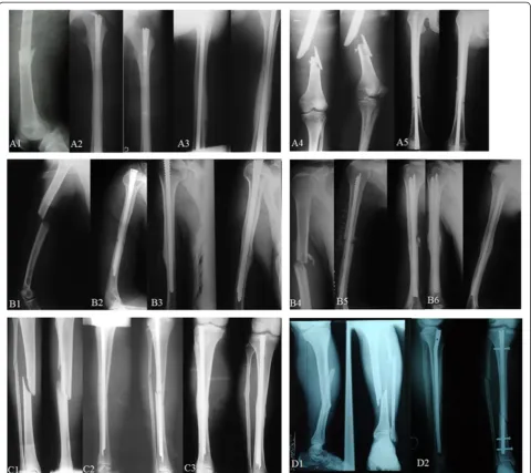
significant differences in the excellent and good rate of shoulder, elbow, knee, and ankle joint functional recov-ery between RSIN group and IIN group (97.7% vs. 89.6%, shoulder and 97.7% vs. 97.7%, elbow joint for hu-meral fractures; 93.2% vs. 92.3%, knee joint for femoral fractures; 95.2% vs. 94.4%, knee and 95.2% vs. 94.4%, ankle joint for tibial fractures,p> 0.05) (Table 2). Typical cases undergoing RSIN or IIN treatment are shown in Figure 5.
Discussions
In the present study, we demonstrated that our self-designed RSIN may be a reliable and practical alternative method for the treatment of long bone fractures because the RSIN group exhibited significantly shorter operative time, less intraoperative blood loss, higher healing rate, and excellent and good rate of shoulder, elbow, knee, and ankle joint functional recovery and few complica-tions compared with IIN group.
The superior results may be attributed to the specific design of our RSIN:
(1) By making only one small incision, the main nail is rotated into the medullary cavity via the cancellous bone screw thread at both of its ends, thus reducing the need for excessive reaming. It is reported that the excessive reaming of the medullary cavity causes an increase in intramedullary pressure, which provokes intravasation of the bone marrow and fat into the venous blood system, leading to intravascular thrombosis and adult respiratory distress syndrome [13]. Moreover, excessive cortical reaming also generates significant heat and induces cor-tical thermal necrosis, influencing bone healing [14,15], while limited reaming may improve blood flow in the surrounding soft tissues and promote bone formation [16]. As expected, the healing rate of our study was sig-nificantly higher in RSIN group.
study (65 ± 15 min vs. 95 ± 25 min for humeral fractures; 70 ± 20 min vs. 100 ± 30 min for femoral fractures; 55 ± 15 min vs. 85 ± 25 min for tibial fractures; allp< 0.001).
(3) The different screw thread pitch of main nail at proximal and distal segment plays a limited compression role. The proximal segment of locking tag is embedded
Table 2 Intraoperative and postoperative conditions of patients with humeral, femoral or tibial fracture in two groups
Fractures Operative parameters RSIN IIN pvalue
Humerus Operative time (min) 65 ± 15 95 ± 25 <0.0001
Intraoperative blood loss (ml) 80 ± 30 125 ± 30 <0.0001
Healing rate in patients with 1 year follow-up (%) 95.4% (83/87) 92.0% (80/87) 0.535
Complications (n) 0.046
Radial nerve injury 1 8
Incision infection 1 2
Neer score (n) 0.092
Excellent 79 73
Good 6 5
Poor 2 9
Aitken and Rombeck's score (n) 0.707
Excellent 74 73
Good 11 12
Poor 2 4
Femur Operative time (min) 70 ± 20 100 ± 30 <0.0001
Intraoperative blood loss (ml) 150 ± 50 190 ± 60 <0.0001
Healing rate in patients with one year follow up (%) 98.1% (102/104) 88.5% (92/104) 0.010
Complications (n) 0.016
Incision infection 0 1
Nail breakage or loosening 0 7
Lysholm score (n) 0.992
Excellent 84 83
Good 13 13
Fair 5 6
Poor 2 2
Tibia Operative time (min) 55 ± 15 85 ± 25 <0.0001
Intraoperative blood loss (ml) 10 ± 5 15 ± 10 <0.0001
Healing rate in patients with 1 year follow up 92.1% (116/126) 81.2% (103/126) 0.024
Complications (n) 0.000
Nail breakage or loosening 0 16
Lysholm score (n) 0.986
Excellent 110 109
Good 10 10
Fair 3 4
Poor 3 3
AOFAS score (n) 0.990
Excellent 113 112
Good 7 7
Fair 4 5
Poor 2 2
into the proximal bone of the fracture and its distal seg-ment bifurcates laterally along the groove to be embedded into the distal cortex of the fracture. This longitudinal, filled, locking fixation realizes bidirectional self-locking to maintain the roles of anti-rotation and anti-contraction [18]. The advantage of this fixation model is a relatively static fixation at early stage but relatively dynamic fix-ation at the middle and later periods due to local bone absorption and muscle contraction, which avoids stress
concentration to induce nail breakage [19]. In line with the above theory, no nail breakage was observed in our study.
radial nerve is one of the most commonly involved. The radial nerve is located at the lateral intermuscular septum within the distal third of the humerus, where the range of motion is small. Surgical dissection in this re-gion will cause tractional damage to this nerve [20]. Re-cent studies report that the rate of iatrogenic radial nerve injury in operatively treated humeral fractures is 4%–8% [21,22]. However, only one patient developed ra-dial nerve injury in RSIN group, and he was spontan-eously restored at 3 months after the operation. Thus, further attention should be paid during RSIN fixation.
Surgical indications for our RSIN are as follows: (1) humeral fractures: this procedure is applicable to shaft fracture located at 3 cm below the greater tuberosity of the humerus and 5 cm over the olecranon fossa but not the serious comminuted fractures or fractures with the osteoporosis at proximal end; (2) femoral fractures: this procedure is applicable to fractures located below the greater trochanter to 8 cm over the supracondylar seg-ment but not the serious comminuted fractures; (3) tib-ial fractures: this procedure is applicable to fractures at the upper 1/3 of tibia and 8 cm over the supracondylar segment.
In conclusion, our self-designed RSIN may be an ef-fective and safe approach for treating long bone frac-tures compared with IIN. However, further studies with large sample size and longer follow-up are still needed to comprehensively evaluate our RSIN.
Competing interests
The authors declare that they have no competing interests.
Authors’contributions
BLL and YX conceived and designed the research; HD, SG, FJ, and QHL analyzed the data; DXW contributed reagents/materials/analysis tools; XWG and WL wrote the paper. All authors read and approved the final manuscript.
Authors’information
Bailian Liu and Ying Xiong joint first authors.
Author details
1Department of Orthopaedics, Yan’an Hospital, No. 245 Renmin East Road,
Kunming, Yunnan 650051, China.2Emergency Department, Yan’an Hospital,
Kunming, Yunnan 650051, China.
Received: 28 September 2013 Accepted: 3 June 2014 Published: 21 July 2014
References
1. Barata I, Spencer R, Suppiah A, Raio C, Ward MF, Sama A:Emergency ultrasound in the detection of pediatric long-bone fractures.Pediatr Emerg Care2012,28:1154–1157.
2. Tyllianakis M, Megas P, Giannikas D, Lambiris E:Interlocking intramedullary nailing in distal tibial fractures.Orthopedics2000,23:805–808.
3. Haque MA, Hossain M, Kabir M, Akanda N, Hossain M:Interlocking intramedullary nailing in fracture shaft of the femur.Mymensingh Med J 2009,18:159–164.
4. Cho CH, Song KS, Kim SK:Antegrade interlocking intramedullary nailing in humeral shaft fractures.J Korean Shoulder Elbow Soc2010,13:1–6. 5. Levin PE, Schoen R, Browner B:Radiation exposure to the surgeon during
closed interlocking intramedullary nailing.J Bone Joint Surg1987,
69:761–766.
6. Roux A, Bronsard N, Blanchet N, de Peretti F:Can fluoroscopy radiation exposure be measured in minimally invasive trauma surgery?Orthop Traumatol Surg Res2011,97:662–667.
7. Chaudhary P, Karn N, Shrestha B, Khanal G, Rijal R, Maharjan R, Kalawar R:
Randomized controlled trial comparing dynamic compression plate versus intramedullary interlocking nail for management of humeral shaft fractures.Health Renaissance2011,9:61–66.
8. Investigators StPERINiPwTF:Randomized trial of reamed and unreamed intramedullary nailing of tibial shaft fractures.J Bone Joint Surg Am2008,
90:2567.
9. Kang S, Chung PH, Chae DJ, Kim JP, Kim JH, Park SP, Park JS:Nail breakage after femoral interlocking intramedullary nailing.J Korean Society Fractures 2002,15:363–370.
10. Neer CS 2nd:Displaced proximal humeral fractures. II. Treatment of three-part and four-part displacement.J Bone Joint Surg Am1970,
52:1090–1103.
11. Aitken GK, Rorabeck CH:Distal humeral fractures in the adult.Clin Orthop Relat Res1986,1986:191–197.
12. Lysholm J, Gillquist J:Evaluation of knee ligament surgery results with special emphasis on use of a scoring scale.Am J Sports Med1982,
10:150–154.
13. Duan X, Li T, Mohammed A-Q, Xiang Z:Reamed intramedullary nailing versus unreamed intramedullary nailing for shaft fracture of femur: a systematic literature review.Arch Orthop Trauma Surg2011,
131:1445–1452.
14. Frölke JPM, Peters R, Boshuizen K, Patka P, Bakker FC, Haarman HJTM:
The assessment of cortical heat during intramedullary reaming of long bones.Injury2001,32:683–688.
15. Giannoudis P, Snowden S, Matthews S, Smye S, Smith R:Temperature rise during reamed tibial nailing.Clin Orthop Relat Res2002,395:255–261. 16. Kuzyk PR, Li R, Zdero R, Davies JE, Schemitsch EH:The effect of
intramedullary reaming on a diaphyseal bone defect of the tibia.
J Trauma Acute Care Surg2011,70:1248–1256.
17. Babis GC, Benetos IS, Zoubos AB, Soucacos PN:The effectiveness of the external distal aiming device in intramedullary fixation of tibial shaft fractures.Arch Orthop Trauma Surg2007,127:905–908.
18. Huang ZL, Yang HL, Xu JK, Xia X, Wang XJ, Song JX, Hu J:Rotary self-locking intramedullary nail for long tubular bone fractures.Chin Med J 2013,126:3874–3878.
19. Shih KS, Hsu CC, Hsu TP:A biomechanical investigation of the effects of static fixation and dynamization after interlocking femoral nailing: a finite element study.J Trauma Acute Care Surg2012,72:E46–E53. 20. Bono CM, Grossman MG, Hochwald N, Tornetta P III:Radial and axillary
nerves: anatomic considerations for humeral fixation.Clin Orthop Relat Res2000,373:259–264.
21. Joiner ER, Skaggs DL, Arkader A, Andras LM, Lightdale-Miric NR, Pace JL, Ryan DD:Iatrogenic nerve injuries in the treatment of supracondylar humerus fractures: are we really just missing nerve injuries on preoperative examination?J Pediatr Orthop2014,34(4):388–392. 22. Wang J-P, Shen W-J, Chen W-M, Huang C-K, Shen Y-S, Chen T-H:
Iatrogenic radial nerve palsy after operative management of humeral shaft fractures.J Trauma Acute Care Surg2009,66:800–803.
doi:10.1186/1749-799X-9-47