Memory Dysfunction and Pathology
in Children with Temporal Lobe
Epilepsy
Stephen J. Wood
Institute o f Child Health
University College London Medical School
ProQuest Number: U644099
All rights reserved
INFORMATION TO ALL USERS
The quality of this reproduction is dependent upon the quality of the copy submitted.
In the unlikely event that the author did not send a complete manuscript and there are missing pages, these will be noted. Also, if material had to be removed,
a note will indicate the deletion.
uest.
ProQuest U644099
Published by ProQuest LLC(2016). Copyright of the Dissertation is held by the Author.
All rights reserved.
This work is protected against unauthorized copying under Title 17, United States Code. Microform Edition © ProQuest LLC.
ProQuest LLC
789 East Eisenhower Parkway P.O. Box 1346
Abstract
Temporal lobe epilepsy is known to affect cognitive functions in both
adults and children, with memory being particularly seriously affected.
Traditionally, clinical and neurophysiological data have provided information
regarding seizure lateralization and this has been related to memory deficits.
This technique, which has demonstrated the material-specificity of temporal
lobe function, is unable to control for the high degree of bilateral temporal lobe
pathology that is often seen in patients with temporal lobe epilepsy.
Modem imaging technology has enabled the non-invasive investigation of
the brain and the quantification of temporal lobe pathology. In this thesis, three
quantitative magnetic resonance techniques were used to evaluate the degree of
temporal lobe damage in children with temporal lobe epilepsy. These were
hippocampal volumetry, T2 relaxometry and proton magnetic resonance
spectroscopy. The first two techniques both measure pathology in the
hippocampus, but in different ways. Specifically, volumetry assesses how much
tissue has been lost, whilst T2 assesses the integrity of the remaining tissue.
Spectroscopy, by contrast, investigates the biochemistry of the medial temporal
lobe, providing information that is not available from standard magnetic
resonance imaging.
This information about medial temporal lobe pathology was related to
memory performance. Measures of left temporal, and particularly left
hippocampal, pathology were found to be associated with scores on a number of
associated with any test of memory, even those thought to be dependent on the
right temporal lobe.
The response of the epileptic brain to surgery was investigated using the
magnetic resonance techniques and neuropsychological measures. The results
suggested that outcome differed depending on the type of surgical procedure. In
particular, the contralateral hippocampus appeared to show a volume increase
follovring a temporal lobectomy, but an increase in T2 relaxation time following
a temporal lesionectomy. Spectroscopic data showed a trend towards more
normal biochemistry in both groups. Immediate memory function was found to
improve in patients who had a lesionectomy, but worsen following temporal
lobectomy. The relationship between these findings is discussed.
This thesis demonstrates the use of quantitative magnetic resonance
techniques in the investigation of temporal lobe epilepsy of the relationship
Table of Contents
ABSTRACT... 2
TABLE OF CONTENTS... 4
LIST OF TABLES... 8
LIST OF FIGURES... 10
ACKNOWLEDGMENTS... 12
ABBREVIATIONS... 14
CHAPTER 1. INTRODUCTION...17
1.1 The Investigationof Memory... 18
1.2 The Structure OF THE Hippocampal Sy st e m... 25
1.3 Epilepsy...29
1.3.1 A dult Epilepsy... 29
1.3.2 Childhood Epilepsy... 31
1.4 Temporal Lobectomyin Childhood... 32
1.4.1 Surgical Intervention... 32
1.4.2 Pathology o f the Resected Temporal Lobe... 36
1.5 Epilepsyand Researchinto Mem o r y... 38
1.6 Magnetic Resonance Imagingand Spectroscopy... 42
1.6.1 Hippocampal Volumetry...43
1.6.2 T2 Relaxometry... i... 49
1.6.3 Proton Magnetic Resonance Spectroscopy... 52
1.7 Aims OF THE St u d y... 54
CHAPTER 2. METHODOLOGY... 58
2.1 Su bje c t s...58
2.1.1 Patients... 58
2.1.2 Patient evaiuation...59
2.1.3 Control subjects...59
2.2 Magneticresonanceimagingandspectroscopy...61
2.2.1 The basic principles o f magnetic resonance... 61
2.2.2 Standard ciinicalprotocoi...62
2.2.3 Quantitative magnetic resonance: T2 M apping... 62
2.2.4 Quantitative magnetic resonance: H E voiumetry.... 64
2.2.4.1 Correcting for intracranial volume... 66
2.2.4.2 HFV Distribution Graphs...70
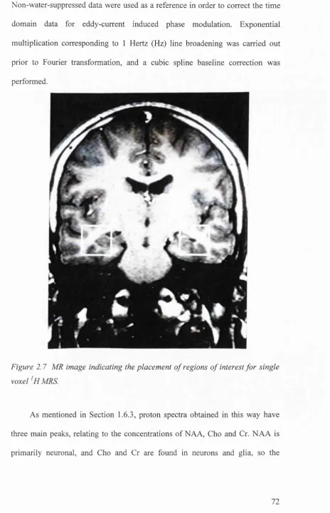
2.2.5 Quantitative magnetic resonance: Single volume MRS.... 71
2.3 Neuropsychological Examination...74
2.3.1 Tests o f Intelligence... 76
2.3.2 Tests o f M em ory... 78
2.3.3 Tests o f Language Function...88
2.3.4 Tests o f Visuo-perception... 91
2.3.5 Tests o f Executive Function...94
2.3.6 Other Tests...95
2.3.7 Academic Attainments... 97
CHAPTER 3. THE RELATIONSHIP BETWEEN MRS. HIPPOCAMPAL
VOLUMETRY AND T2 RELAXOMETRY IN PATIENTS WITH TEMPORAL LOBE
EPILEPSY... 100
3.1 Abstract...100
3.2 Introduction... 101
3.3 Methods...105
3.3.1 Subjects...105
3.3.2 Magnetic Resonance.... 105
3.3.3 Statistics...105
3.4 Results...106
3.4.1 Bilateral pathoiogy... 106
3.4.2 Correlations between pathology scores... 106
3.5 Discussion... 109
CHAPTER 4. A COMPARISON BETWEEN PATIENTS WITH LEFT AND RIGHT TEMPORAL LOBE EPILEPSY AND NORMAL CONTROLS USING THE W lS C -H l^ INTELLIGENCE TEST AND THE WECHSLER MEMORY SCALE...113
4.1 Abstract...113
4.2 Introduction... 114
4.3 Methods...116
4.3.1 Subjects... 116
4.3.2 Neuropsychological evaluation... 117
4.4 Results...117
4.4.1 Comparison o f IQ scores... 117
4.4.2 Comparison o f memory scores... 121
4.5 Discussion... 124
CHAPTER 5. THE RELATIONSHIP BETWEEN PRE-OPERATIVE COGNITIVE FUNCTION AND TEMPORAL LOBE PATHOLOGY...129
5.1 Abstract...129
5.2 Introduction... 130
5.3 Methods...133
5.3.1 Subjects...133
5.3.2 Magnetic Resonance... 134
5.3.3 Neuropsychology...134
5.3.4 Statistics...134
5.4 Results...137
5.4.1 Analyses o f variance using hippocampal pathology...138
5.4.2 Analyses o f variance using the NAA/(Cho+Cr) measure ofpath ology... 142
5.5 Regression An alysis... 144
5.5.1 IQ Scores... 145
5.5.2 Scores from the Wechsler Memory S cale... 151
5.5.3 Scores fro m the CAVLT-2...153
5.5.4 Negative fin dings...156
5.6 Discussion...157
5.6.1 IQ Scores...159
5.6.2 Logical m em ory... 162
5.6.3 Verbal Paired-Associate Learning...163
5.6.4 C \ C Score and Percent Retention o f C... 165
5.6.5 CAVLT-2...166
5.6.6 General Points and Negative Findings... 172
5.6.6.1 Right temporal lobe function...172
5.6.6 2 Recognition memory...174
5.6.6 3 Methods of analysis... 176
CHAPTER 6. AN ANALYSIS OF CHANGE IN OUANTITATIVE MR AND NEUROPSYCHOLOGY MEASURES IN TEMPORAL LOBECTOMY PATIENTS 178 6.1 Abstract...178
6.3.1 Subjects... 186
6.3.2 Magnetic Resonance....187
6.3.3 Neuropsychology... 187
6.4 Results...188
6.4.1 ^HMRS... 188
6.4.2 T2 relaxometry...190
6.4.3 Hippocampal Volumes...191
6.4.4 IQ Scores... 192
6.4.5 Memory S cores... 194
6.4.6 Other tests... 196
6.5 Discussion... 198
6.5.1 MR measures...199
6.5.1.1 T2 Relaxometry... 200
6.5.1.2 ‘H M R S ... 201
6.5.1.3 Hippocampal Volumetry... 203
6.5.2 Neuropsychological measures...206
CHAPTER 7. THE RELATIONSHIP BETWEEN CHILDREN WITH TLE AND THEIR UNAFFECTED SIBLINGS ON MEASURES OF TEMPORAL LOBE PATHOLOGY.. 211
7.1 Abstract...211
7.2 Introduction... 212
7.3 Methods...215
7.3.1 Subjects... 215
7.3.2 Magnetic Resonance.... 216
7.3.3 Statistics...217
7.4 Results... 217
7.4.1 T2 Relaxometry... 217
7.4.2 ^HM RS...218
7.4.3 Hippocampal Volumes... 219
7.5 Discussion... 221
CHAPTER 8. GENERAL DISCUSSION AND DIRECTIONS FOR FURTHER RESEARCH... 225
REFERENCES...243
APPENDIX I. PATIENT POPULATION... 263
APPENDIX H COMPLETE ANALYSES FOR CHAPTERS 5 AND 6 ... 268
Hi Analysisofpre-operativeneuropsychologicalmeasureswithhippocampal PATHOLOGY...268
H.Ll Scores from the Wechsler Intelligence S cale...268
H.Lll Scores from the Wechsler Memory Scale... 269
H.Llll Scores from other memory tests... 270
II.Llv Scores from non-memory tests... 271
II.li Analysisofpre-operativeneuropsychologicalmeasureswithdiffuse TEMPORAL LOBE PATHOLOGY...272
H.lLl Scores from the Wechsler Intelligence Scale...272
H.lLll Scores from the Wechsler Memory Scale...273
H.lLlll Scores from other memory tests... 274
ILlLlv Scores from non-memory tests... 275
II.HI Additionalgraphsandanalysesfrom Chapter 5 ... 276
Il.iv Analysisofpre- andpost-operativeneuropsychologicalmeasuresnot PRESENTED IN CHAPTER 6 ... 278
H.lv.l Subtests o f the Wechsler Intelligence Scale....278
H.lv.ll Scores from the Wechsler Memory Scale...279
H.lv.lll Scores from other memory tests...279
APPENDIX III. NORMATIVE DATA FOR THE COUGHLAN DESIGN LEARNING TEST AND THE PERFORMANCE OF CHILDREN WITH TEMPORAL LOBE
EPILEPSY... 282
III.1 Abstr a c t... 282
m . n In t r o d u c t i o n...283
m j i i Me t h o d s...285
IILiiLi Subjects...285
IIIMLii Procedure...285
IHMLiii Statistics...28 7 ni.iv R e s u l t s ... 288
Ill.iv.i Results from the WlSC-Ilf^^ Vocabulary Subtest...28 8 III.iv.ii Results from the CDLT....288
in.v DISCUSSION... 299
List of Tables
Ta b l e 2.1 Th et y p e so fm e m o r ya s s e s s e db yt h en e u r o p s y c h o l o g ic a lt e s t sint h e
ADMINISTERED PROTOCOL... 75 Ta b l e 2.2 Te s ta s s e s s in gl a n g u a g e, v is u a lp e r c e p t io na n de x e c u t iv ef u n c t io nint h e
ADMINISTERED PROTOCOL... 75 Ta b l e 3.1 Sp e a r m a n’sr a n k-o r d e rc o r r e l a t io nd a t af o rg r o u p sw it ha n dw it h o u t
MASS LESIONS... 107 Ta b l e 4.1 Me a n IQ s c o r e s (± s t a n d a r dd e v ia t io n s) f o rt h et h r e es u b je c tg r o u p s... 117 Ta b l e 4 .2 Me a nv e r b a l IQ s u b t e s ts c o r e s (± s t a n d a r dd e v ia t io n s) f o rt h et h r e e
SUBJECT GROUPS...119 Ta b l e 4 .2 Me a np e r f o r m a n c e IQ s u b t e s ts c o r e s (± s t a n d a r dd e v ia t io n s) f o rt h et h r e e
SUBJECT GROUPS...120 Ta b l e 4 .4 Me a nr a w M Q , Lo g ic a l Me m o r ya n d Vis u a l Re p r o d u c t io ns c o r e s (±
STANDARD DEVIATIONS) AND MEANS ADJUSTED FOR V IQ FOR THE THREE SUBJECT GROUPS... 122 Ta b l e 4.5 Me a nr a w V P A L a n d Co m p o s it es c o r e s (± s t a n d a r dd e v ia t io n s) a n dm e a n s
ADJUSTED FOR V IQ FOR THE THREE SUBJECT GROUPS... 123 Ta b l e 5.1 Dis t r ib u t io no fn o r m a l, u n il a t e r a la n db il a t e r a lp a t h o l o g ie so ne a c ho f
THE THREE QUANTITATIVE M R MEASURES... 138 Ta b l e 5.2 IQ s c o r e s (Mea n s ± s t a n d a r dd e v ia t io n s) f o rt h ef iv es u b je c tg r o u p s
DIVIDED BY HIPPOCAMPAL PATHOLOGY...139 Ta b l e 5.3 Vo c a b u l a r ya n d Bl o c k De s ig ns u b t e s ts c o r e s (m e a n s ± s t a n d a r d
DEVIATIONS) FOR THE FIVE SUBJECT GROUPS DIVIDED BY HIPPOCAMPAL PATHOLOGY... 140 Ta b l e 5 .4 C % a n d V PA De ls c o r e s (m e a n s ± s t a n d a r dd e v ia t io n s) f o rt h ef iv es u b je c t
GROUPS DIVIDED BY HIPPOCAMPAL PATHOLOGY... 141 Ta b l e 5.5 Dig ita n d Bl o c k Sp a ns c o r e s (m e a n s ± s t a n d a r dd e v ia t io n s) f o rt h ef iv e
SUBJECT GROUPS DIVIDED BY HIPPOCAMPAL PATHOLOGY... 142 Ta b l e 5 .6 IQ s c o r e s (Mea n s ± s t a n d a r dd e v ia t io n s) f o rt h ef iv es u b je c tg r o u p s
DIVIDED BY N A A /(C H 0+C R ) RATIO...143 Ta b l e 5.7 Re s u l t so ft h el in e a rm u l t iv a r ia t er e g r e s s io na n a l y s e sf o rt h e W ISC -111^146 Ta b l e 5.8 Re s u l t so fl in e a rm u l t iv a r ia t er e g r e s s io na n a l y s e sf o r Lo g ic a l Me m o r y
AND THE Co m p o s it e Sc o r ef r o mt h e W M S ...152 Ta b l e 5.9 Re s u l t so ft h el in e a rm u l t iv a r ia t er e g r e s s io na n a l y s e sf o rt h e C A V L T -2 154 Ta b l e 6.3 Pr e- a n dp o s t-o p e r a t iv em e a nc o n t r a l a t e r a l N A A /(Ch o+ Cr) r a t io s (±
STANDARD DEVIATIONS) FOR THE TWO TEMPORAL LOBE SURGERY GROUPS...188 Ta b l e 6.3 Pr e- a n dp o s t-o p e r a t iv em e a nc o n t r a l a t e r a l T 2 sc o r e s (± s t a n d a r d
DEVIATIONS) FOR THE TWO TEMPORAL LOBE SURGERY GROUPS... 190 Ta b l e 6.3 Pr e- a n dp o s t-o p e r a t iv em e a nc o n t r a l a t e r a l H FV s c o r e s (± s t a n d a r d
DEVIATIONS) FOR THE TWO TEMPORAL LOBE SURGERY GROUPS... 191 Ta b l e 6.4 Pr e- a n dp o s t-o p e r a t iv em e a n IQ s c o r e sf o rt h et w ol e f tt e m p o r a ll o b e
SURGERY GROUPS... 192 Ta b l e 6.5 Pr e- a n dp o s t-o p e r a t iv em e a n Ar it h m e t ica n d Co d in gs c o r e sf o rt h et w o
LEFT TEMPORAL LOBE SURGERY GROUPS... 194 Ta b l e 6.6 Pr e- a n dp o s t-o p e r a t iv em e a n L M -% a n d M Q s c o r e sf o rt h et w ol e f t
TEMPORAL LOBE SURGERY GROUPS... 195 Ta b l e 6.7 Pr e- a n dp o s t-o p e r a t iv em e a ns co resf o rt h e L M -I, D ’ a n d V PA Sc o r e
MEASURES FOR THE TWO LEFT TEMPORAL LOBE SURGERY GROUPS...195 Ta b l e 6.8 Pr e- a n dp o s t-o p e r a t iv em e a ns c o r e so nt h ed ig its p a n, C A V L T -2 a n d Do t
Lo c a t io nt e s t sf o rt h el e f tt e m p o r a ll o b es u r g e r yg r o u p s... 196 Ta b l e 6.9 Pr e- a n dp o s t-o p e r a t iv em e a nsc o reso n Wo r d Fl u e n c ya n dt h e W C S T f o r
BOTH LEFT TEMPORAL LOBE SURGERY GROUPS... 197 Ta b l e 7.1 Pr e-o p e r a t iv ec o n t r a l a t e r a l T 2 o fe ig h t T L E p a t ie n t sa n dt h e T 2 o fe ig h t
NORMAL SIBLINGS...217 Ta b l e 7.2 Pr e-o p e r a t iv ec o n t r a l a t e r a l N A A /(Ch o+ Cr) r a t ioo f T LB p a t ie n t sa n dt h e
Ta b l e 7.3. Me a n sa n ds t a n d a r dd e v ia t io n sf o rt h eh ip p o c a m p a lv o l u m e so ft h esix
SUBJECT GROUPS...221
Ta b l e II.i Re g r e s s io na n a l y s isf o r V PA Ha r d... 277
Ta b l e III.i Me a n sa n ds t a n d a r dd e v ia t io n sf o r Sc a l e ds c o r e so nt h e Vo c a b u l a r y SUBTEST FOR ALL AGE GROUPS...288
T a b l e III.ii M e a n s a n d s t a n d a r d d e v i a t i o n s f o r t h e DLT s c o r e f r o m t h e CDLT... 289
T a b l e III.iii M e a n s a n d s t a n d a r d d e v i a t i o n s f o r t h e DLI s c o r e f r o m t h e CDLT... 291
T a b l e lll.rv M e a n s a n d s t a n d a r d d e v i a t i o n s f o r t h e b s c o r e f r o m t h e CDLT...293
Ta b l e III.v Me a n sa n ds t a n d a r dd e v ia t io n sf o rt h e Im m e d ia t e Le a r n in gs c o r ef r o m THE CDLT...295
T a b l e III.v i M e a n s a n d s t a n d a r d d e v i a t i o n s f o r t h e A 6 s c o r e f r o m t h e CDLT... 296
List of Figures
F ig u r e 1.1 T h e c l a s s i f i c a t i o n o f lo n g - t e r m m em o ry (f r o mSq u ir e & Kn o w l t o n, 1996)...19
Figure 1.2 A stylizedstructureoftheh ippo ca m pu s... 26
Figure 2.1 M R imagesshow ingthedecayof T2-w eightedsignalandth ed eca ycu rv e. ...63
Figure 2.2 Thepositioningoftheslicefor T2 relaxom etryandofth eregionof in terestfo rm ea surem en t...64
Fig u re 2.3 Reform attingprocedureofthestandardsagittally-acquired M PRAGE INTO TILTED CORONAL SLICES (FROM VANPAESSCHEN, 1997)... 65
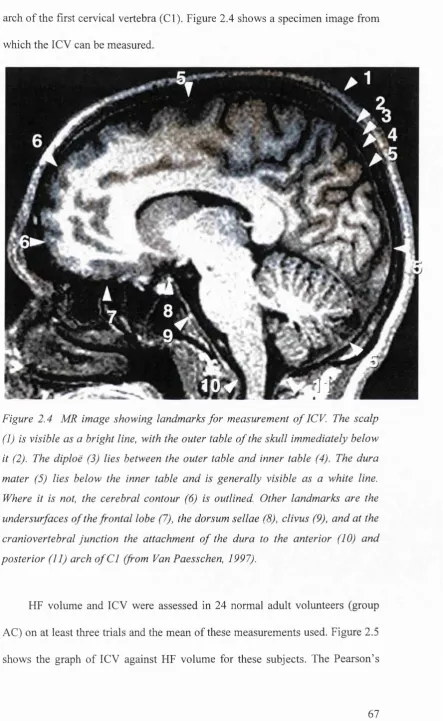
F ig u r e 2.4 M R im ag e s h o w in g l a n d m a r k s f o r m e a s u re m e n t o f ICV (f r o m Va n Pa e s s c h e n, 1997)...67
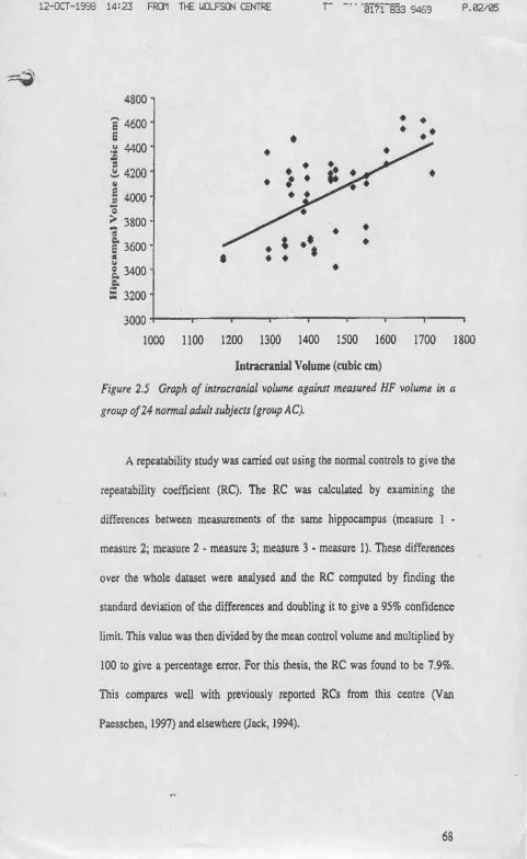
Fig u re 2.5 Graphofintracranialvolum eagainstm easured HF volum einagrou pof 24 NORMAL ADULT SUBJECTS...69
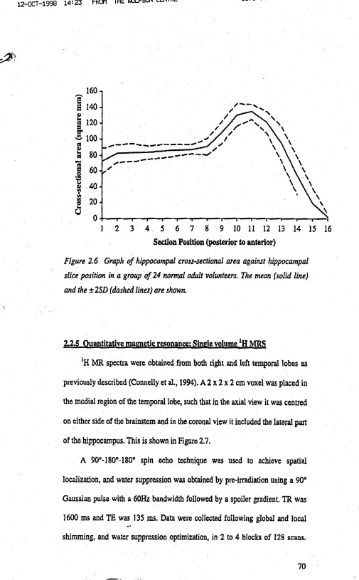
Fig u r e 2.6 Graphofhippocam palcross-sectionalareaagainsthippocam palslice POSITION IN 24 NORMAL ADULT VOLUNTEERS...71
Fig u re 2.7 M R im ageindicatingtheplacem entofregionsofinterestfo rsingle VOXEL M R S... 72
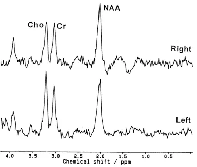
Fig u re 2.8 Norm alandabnorm al ‘h MRS spec t r a...73
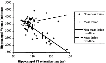
Fig u re 3.1 Graphshow ing HFV against T2 forpatientsw ithandw ithoutm a ss -LESIONS...108
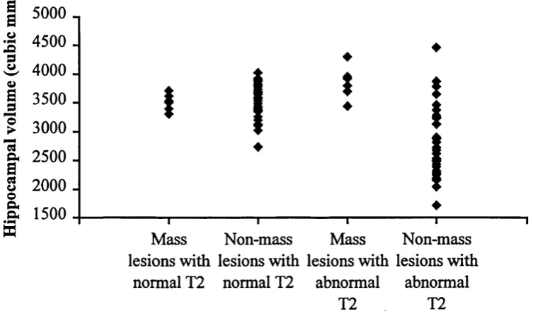
Fig u re 3.2 Graphshow ingth e HFV forbothpathologygrou ps, thegroupsbein g DIVIDED INTO THOSE WITH NORMAL AND THOSE WITH ABNORMAL T2 VALUES... 109
Fig u re 5.1 Scattergraphofleft NAA/(Cho+Cr) ratioagainst VIQ fo rbo th TLE PATIENTS AND SIBLING CONTROLS... 147
Fig u re 5.2 Scattergrapho fleft NAA/(Cho+Cr) ratioagainst Vocabularyscorefo r BOTH TLE PATIENTS AND SIBLING CONTROLS... 148
Fig u re 5.3 Scattergrapho fleft NAA/(Cho+Cr) ratioagainst Sim ilaritiesscorefo r BOTH TLE PATIENTS AND SIBLING CONTROLS... 149
Fig u re 5.4 Sca ttergraphofleft NAA/(Cho+Cr) ratioagainst Inform a tio nscorefor BOTH TLE PATIENTS AND SIBLING CONTROLS...150
Fig u re 5.5 Scattergraphofleft T2 against Comprehensionscoreforbo th TLE PATIENTS AND SIBLING CONTROLS... 150
Figure 5.6 Sca ttergraphofleft T2 againstthe C% scoreforboth TLE patientsand SIBLING CONTROLS...153
Fig u re 5.7 Scattergraphofleft T2 againstthe Delayed Recallscorefro mthe CAVLT-2 FOR BOTH TLE PATIENTS AND SIBLING CONTROLS... 155
Figu re 5.8 Scattergraphofleft T2 againstthetotaln u m berof In trusionsonthe CAVLT-2 FOR BOTH TLE PATIENTS AND SIBLING CONTROLS... 156
Figure 7.1 Chartshow ingth e HFVsforallsubjectsinea chofthesixsubjectgroups .220 Fig u re II.i Graphof VIQ againstleft NAA/(Cho+Cr) ratioinenlargedd a taset (n = 4 3 ) ... 276
Fig u re II.ii Graphof PIQ againstright NAA/(Cho+Cr) ratioinenlargedd ataset (n = 4 3 ) ... 276
Figure ILin Graphofleft T2 against VPA Ha r d...277
Fig u re III.I Design 1 ofth e Coughlan Design Learning Te s t...286
Fig u re III.ii Graphshow ingm ean DLT scoreagainstageg r o u p...290
Fig u re III.ui Graphofmean DLI scoreagainstageg r o u p... 292
Fig ure Ill.rv Graphshow ingmeanB scoreagainstageg ro u p... 294
Fig ure III.v Graphshow ingmean Im mediate Learningscoreagainstageg r o u p... 295
Fig ure III.vi Graphshow ingmean A6 (Immediate Recall) scoreagainstageg r o u p... 297
Fig u re III.vii Gra phshow ingmean Delayed Recallscorea gainsta geg r o u p...299
Fig u re r v .i Gra phshow ingmean Copyerrorscore (± standarddeviations) ofn orm a l con tro lsforth e Dot Locationt e s t... 301
Fig u r e IV .iii Gr a p hs h o w in gm e a nd e l a y e d Re c a l le r r o rs c o r e (± s t a n d a r d
AcknowleHgmenfs
This thesis was produced under the supervision of Professor David Gadian,
Professor of Biophysics, and Dr Faraneh Vargha-Khadem, Reader in Cognitive
Neuroscience, at the Institute of Child Health, University College London
Medical School, University of London. Their assistance and advice has been
instrumental in the production of this thesis.
I would like to thank the Wellcome Trust for their financial support.
A number of people have helped to obtain the data which I am presenting
here. Firstly, I would like to thank the research radiographers, Cheryl Johnson
and Clare Marshall, for scanning both the patients and their siblings and also for
giving me the MRS and T2 data when I asked for them. In addition, their
encouragement and support has been wonderful.
Secondly, thanks must go to Dr Elizabeth Isaacs, Kate Watkins, Marie-
Claude Jones and Dr Shalini Gupta, all of whom have performed
neuropsychological assessments on some of the patients presented here. Without
them, there would not be enough data to work with. Kate and Elizabeth, in
particular, have given me support and fiiendship and help with statistics since
my first day at the Wolfson Centre.
Thirdly, big thankyous to Drs. Helen Cross and Rod Scott, and Professor
Brian Neville, for allowing me access to their patients. Dr. Cross in particular
I would also like to thank Dr Alan Connelly, for providing such effective
advice and assistance; Dr Martin King, for helping with some of the statistics;
Celia Ajuba, for collecting some of the control data for the CDLT; and Dr Wim
Van Paesschen for teaching me the technique of hippocampal volumetry so well,
and allowing me the use of some of his figures.
I must also take this opportunity to recognize the contribution of the
patients I have studied, and the co-operation of their families. Without them
there wouldn’t be a thesis.
A big thankyou also to my friends and family, who have without exception
provided food and/or alcohol when it was required.
Last but not least, thank you to Amanda, who for the last months has put
up with my grumbling and whinging with a good humour it did not always
Abbreviations
3D AM AMIPB ANOVA BPVS Cl C’ C C% CA CAVLT-2 CDLT Cho CHQ cm cm^ CPS Cr CSF CT D ’ D D% DLI DLT DNET EEG FSIQ GD GFAP HF HFV Three-dimensional Amygdaloid bodyAdult Memory and Information Processing Battery
Analysis of variance
British Picture Vocabulary Scale First cranial vertebra
Immediate Composite score Delayed Composite score
Percentage o f the Composite score retained Cornu ammonis
Childrens Auditory-Verbal Learning Test (2nd edition)
Coughlan Design Learning Test Choline-containing compounds Child Health Questionnaire Centimetre
Cubic centimetre Complex partial seizure Creatine and phosphocreatine Cerebrospinal fluid
X-ray computed tomography Immediate visual reproduction score Delayed visual reproduction score
Percentage o f the visual reproduction score retained
Design Learning Intrusions Design Learning Total
Dysembryoplastic neuroepithelial tumour Electroencephalography
Full Scale Intelligence Quotient Glial density
Hz
Tukey’s h-s-d ICV
IQ
L M - D L M - I LM% m mm mm^ MNI MP-RAGE MQ MR MRI MRS ms MTS NAA ND PET PHA-L PHG PIQ RC SD SPECT SPS T TE TI TLE TR TROG VIQ VPA Del Hertz
Tukey’s honestly-significant-difference test Intracranial volume
Intelligence Quotient
Delayed Logical Memory score Immediate Logical Memory score Percentage o f the Logical Memory score retained
Month Millimetre Cubic millimetre
Montreal Neurological Institute
Magnetization-prepared rapid acquisition gradient echo
Memory Quotient Magnetic resonance
Magnetic resonance imaging Magnetic resonance spectroscopy Millisecond
Medial temporal sclerosis N-acetylaspartate
Neuronal density
Positron emission tomography Phaseolus vulgaris leucoagglutinin Parahippocampal gyrus
Performance Intelligence Quotient Repeatability coefficient
Standard deviation
Single photon emission computerised tomography
Simple partial seizure Tesla
Echo time Inversion time
Temporal lobe epilepsy Recovery time
Test for reception o f grammar Verbal Intelligence Quotient
VPA Easy
VPA Hard
VPAL VPA Score
vSRT WAIS WCST Wise
WMS WOND WORD WRMT y
Total number o f easy pairs correctly recalled in the learning trial o f the verbal paired-associate learning test
Total number o f hard pairs correctly recalled in the learning trial o f the verbal paired-associate learning test
Verbal paired-associate learning test
Weighted learning score for the verbal paired-associate learning test
Verbal selective reminding test Wechsler adult intelligence scale Wisconsin card sorting test
Wechsler intelligence scale for children Wechsler memory scale
Chapter 1. Introduction
“If our brains were simple enough to understand, we wouldn’t be able to understand them.” Anonymous graffiti
Memory has been considered and discussed for many centuries, at least as
far back as Plato, who described memory as a wax tablet on which perceptions
or ideas might be imprinted and retained (Parkin, 1987). The analogies used to
describe memory have changed over the years; in medieval times it was
regarded as a complex hydraulic system, in the seventeenth century as
clockwork, in the nineteenth century as electricity. Today we still use analogies -
the brain as a computer being the most common. Despite this very long-standing
interest in memory as a concept, its scientific investigation has only a century of
work behind it. In this time ideas have moved from the simple analogy to today's
complex multicomponent view of the memory system.
In recent years, new neuroimaging techniques have vastly increased our
ability to investigate and understand functions of the brain, and in particular to
examine specific regions and relate their integrity to cognitive functions such as
memory. The advent of non-invasive, non-ionizing imaging techniques has
resulted in an explosion of research studies, both in normal humans and in those
suffering from a wide variety of neurological disorders. Traditional
neuropsychological techniques, which related cognitive deficits to circumscribed
brain lesions were only definitively identified at autopsy, they can now be
visualised, quantified, and their progression followed in time fi-om the outset.
This chapter introduces the topics which are explored in this thesis -
cognitive impairments (particularly impairments of memory), epilepsy and the
pathology associated with it, and magnetic resonance imaging and its use in
quantifying such pathology. These three subjects are discussed with reference to
the way in which they are interrelated. Firstly though, some of the recent history
of the investigation of memory function is explored.
1.1 The Investigation of Memory
It is widely accepted that memory is not a unitary function but is a system
composed of several subcomponents (Squire & Knowlton, 1996). The study of
both animals with experimental brain lesions and human amnesic patients has
shown that cognitive (fact-and-event) memory can be dissociated from non-
cognitive (behavioural) memory (Figure 1.1). The two types of memory are
different not only in the kinds of memoranda retained, but also in the way in
which the information is stored. Cognitive memory, such as the memory for
personal events, can store information after just a single trial and this
information can readily be applied in new situations (Squire & Knowlton, 1996).
Non-cognitive memory, by contrast, is normally unconscious, acquired across
many trials and is not generally accessible outside the learning situation. This
Memory
Declarative (Explicit) Nondeclarative (Implicit)
Facts Events Skills Priming Simple Nonassociative
and classical learning
Habits conditioning
Figure 1.1 The classification o f long-term memory (from Squire & Knowlton,
1996)
On the basis of data obtained from amnesic patients, cognitive memory
can in turn be divided into different components. In general, these patients do
not have difficulty remembering information that has just been given to them,
provided that they are allowed to rehearse it subvocally. An example of this kind
of information would be a telephone number, which can be remembered for as
long as it is recited. Distraction, however, results in the loss of this information.
This implies a distinction between short- and long-term memory (Baddeley &
Warrington, 1970), with short-term being a period measured in seconds.
Presumably, the memory system that supports short-term memory is physically
separate from that which supports longer-term memories.
However, the system subserving long-term cognitive memories may not be
required indefinitely. The phenomenon of retrograde amnesia, in which the
patient forgets memories which were learnt before the onset of amnesia, is
frequently temporally graded (Squire & Knowlton, 1996), in that more recent
been challenged, however (Nadel & Moscovitch, 1997). In the model proposed
by Nadel and Moscovitch, each subsequent activation of a memory trace occurs
in a different experiential context, and as such is laid down as a new trace (the
trace is ‘re - membered’). A remote memory, therefore, will have multiple traces
and will be easier to recall since there will be a greater number of access routes
to it, although a memory recovered in such a fashion by an amnesic might not
have the full complexity of an autobiographical episode. Recent memories, by
the same token, would be more susceptible to even minimal damage (Nadel &
Moscovitch, 1997). This implies that the extent of retrograde amnesia and its
temporal gradient are entirely dependent on the size of the lesion to the memory
system.
But where is this memory system? The study of patients with lesions to
various regions of the brain has revealed the importance of the medial temporal
lobes in normal cognitive memory function (Squire & Zola-Morgan, 1991). The
main regions of the medial temporal lobe memory system are the hippocampus,
the amygdala, the entorhinal and perirhinal cortices, and the parahippocampal
cortex. Damage to any or all of these structures results in a significant memory
impairment, and this was dramatically demonstrated when Scoville and Milner
(1957) described a profound anterograde amnesia following bilateral medial
temporal lobe resection in the patient HM. He has been shown to have lost
(bilaterally) the medial temporal pole, most of the amygdala, most or all of the
entorhinal cortex and at least half of the hippocampus (Corkin et al., 1997). His
long term memory deficit is such that although his IQ and immediate memory
abilities are normal, he does not know where he lives, nor what he ate for
of stimuli, even after short delays (Corkin, 1984). He does, though, seem to have
gained some memory for facts since his surgery (so called semantic memory),
such as the fact that a man called Kennedy was assassinated and that Skylab was
“a docking place in space”.
More limited pathology restricted to a small area of the medial temporal
lobe bilaterally (such as a subfield of the hippocampus) has a reportedly lesser,
though still grave, effect (Zola-Morgan et al., 1986; Squire & Knowlton, 1996).
This indicates that the adjacent cortical regions also make important
contributions to memory function. This received recent support from a study of
three patients who became amnesic during early childhood (Vargha-Khadem et
al., 1997). The only pathology common to all three patients is bilaterally
damaged hippocampi. Their episodic memory is very impaired, but all three
have acquired levels of semantic knowledge and literacy skills commensurate
with their VIQs. One patient in particular has learnt who Martin Luther King
was and what ‘encumber’ means, yet has been amnesic since at least the age of
four. The authors suggest that the preserved semantic memory of these patients,
when considered together with results from work with experimentally lesioned
monkeys, indicates a role for the cortex underlying the hippocampus in context-
free semantic memories. Context-rich episodic memories, however, require the
integrity of the hippocampi.
Unilateral damage has a less catastrophic effect. Studies of patients with
unilateral temporal lobectomy have shown that this surgery does not result in
amnesia, but instead produces material-specific memory deficits which are
dependent on the side of operation (Milner, 1975). For example, left unilateral
information (by whatever modality) (Milner, 1968a) whilst right-sided removal
disrupts learning and retention of nonverbal memoranda (Corkin, 1965; Milner,
1968b). This latter finding does not occur as consistently as the former (Rausch,
1991; Loring et al., 1991).
These material-specific deficits have been shown to be greater with more
extensive and posterior resections. Most experimental support for this has come
from studies at the Montreal Neurological Institute (MNI), reported notably in
Philip Corsi’s doctoral thesis (Corsi, 1972) and in a number of other papers (e.g.
Smith & Milner, 1981; Jones-Gotman, 1986; see Jones-Gotman, 1987 for a
review). Corsi used four memory tasks; two designed to test verbal memory and
two to test non-verbal memory. The verbal tasks involved the recall of
consonant trigrams using a Brown-Peterson distractor technique, and the Hebb
recurring digit task, a test of incidental learning (Hebb, 1961). The non-verbal
tasks were an experimental analogue of Hebb’s recurring digits using block
tapping, and a test in which the patient was required to remember the position of
a circle on a horizontal line, with and without distraction (Posner & Konick,
1966). Not only did Corsi find that patients with left temporal lobe resection did
more poorly on the verbal memory tests than those whose right temporal lobe
was removed, he also found that the degree of impairment was dependent on the
extent of hippocampal removal. The converse was true for the non-verbal
memory tasks, with patients who had undergone right temporal lobectomy
showing deficits proportional to the extent of hippocampal removal. This
implies two things: one, that the tasks used are critically dependent on the
hippocampus, and two, that the degree of post-operative memory impairment is
The paradigm, however, is not immune to criticism. Corsi had larger
sample sizes on the side of the expected material-specific memory deficit, so
that more right than left temporal lobectomy patients were tested on the non
verbal memory tasks. This gives greater statistical power to data on the side of
the expected deficit. In addition, a large minority of cases (44%) were tested
during the initial three weeks following surgery, with no reassurance that they
were equally distributed between groups. Since the magnitude of memory deficit
is much greater at three weeks than at one year post-resection (Milner, 1975), it
is perfectly plausible that the increased deficit for patients with extensive
resections is due to an over-representation of such patients in the ‘early-tested’
group. Furthermore, even if there were an equal distribution, reliable inferences
could not be made due to the combination of relatively acute and stable lesions.
In addition to these concerns, Corsi included a few cases of adult head injury.
The onset of temporal lobe epilepsy following head injury is common in
childhood, but much less frequent in adults. This means that there could be
widespread brain damage in these adult cases, affecting many cognitive abilities
in addition to memory (Loring et al., 1991).
In addition, attempts to replicate Corsi’s work in similar populations have
not been particularly successful, either using his memory tests (Rausch & Ary,
1990) or ones not used by Corsi but which had previously shown clinical
sensitivity (Loring et al., 1991). Rausch and Ary (1990) administered the
recurring digits and block-tapping task to patients following left or right
temporal lobectomy. They found that patients with left resections performed
normally on both measures, but patients with right temporal lobe resections
task. The patients did show selective memory deficits, since patients with left
temporal lobe resections were markedly impaired on the recall of a prose
passage and the learning of novel word-pairs, whilst patients with right-sided
removals were not. They concluded that these differences between the two
studies were due to greater epileptogenic involvement of lateral neocortex
beyond the resection line in Corsi’s patients, since patients at MNI who had
large resections had presurgical evidence of a more posterior seizure onset.
In addition, the conclusion that post-operative memory impairment is
dependent on the extent of resection has not survived closer analysis. More
recent work (Chelune, 1995) has instead suggested that post-operative memory
impairment is related to the pre-operative level of function. Those patients with
relatively preserved memory abilities prior to temporal lobectomy are those who
suffer the greatest drop in memory fimction following surgery. A retrospective
analysis of the data from 383 temporal lobectomy patients seen at MNI between
1960 and 1981 also indicated that extent of resection did not explain the
performance of these patients on tests of memory fimction (Leonard, 1991).
In summary, it is widely accepted that left temporal lobectomy results in a
verbal memory impairment, whilst non-verbal memory is impaired following
right-sided removal, when compared to normal controls. However, this latter
finding does not occur consistently, and concern has been voiced as to its
continued orthodoxy (Baxendale et al., 1997). In addition, the finding of
increased impairment with greater hippocampal resection, though generally
robust in the hands of the group at MNI, does not appear to have been replicated
outside that centre. This is probably due to differences in the patient populations,
as opposed to hippocampal pathology, and the age at onset of the patients’
epilepsy.
1.2 The Structure o f the Hippocampal System
As has been discussed above, the integrity of the hippocampal system is of
great importance for normal cognitive memory functions. In order to understand
the way in which damage to this region can result in memory impairments, it is
helpful to have an understanding of its structure.
The hippocampus (the word is from the Greek for seahorse, which it is
said to resemble) is a cylindrical structure which forms a semicircle around the
thalamus. It is located in the medial temporal lobe, medial to the temporal horn
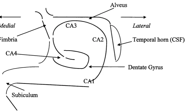
and beneath the anterior cistern (Figure 1.2), and can be divided into three main
segments. From posterior to anterior these are the tail, the body, and the head,
and these three regions are different in terms of their orientation as well as their
shape.
When a frontal section (i.e. perpendicular to the antero-posterior axis) is
examined under the microscope, it can be seen that the hippocampus is formed
of two cortical laminae that are embedded into each other. These are the cornu
ammonis (from the Latin for ram’s horn) and the dentate gyrus. The cornu
ammonis, along with the dentate gyrus, is a simple cortex (known as allocortex)
(Duvemoy, 1988). The comu ammonis is connected to the rest of the cortex
Alveus
Medial CA3 Lateral
Fimbria CA2 Temporal horn (CSF)
CA4-Dentate Gyrus
Subiculum
Figure 1.2 A stylized structure o f the hippocampus showing the subfields o f the
cornu ammonis (CAl - 4), the dentate gyrus, the alveus and fimbria, the
temporal horn and the subiculum.
Frontal sections of the hippocampus also reveal a heterogeneous structure
to the comu ammonis that can be divided into four subfields. These were named
by Lorento de No in 1934 (Duvemoy, 1988) and are known as CAl, CA2, CA3
and CA4. CAl is linked to the subiculum, and with CA3 makes up most of the
hippocampus proper. CAl is selectively vulnerable to hypoxia and is sometimes
known as the ‘vulnerable sector’ (Duvemoy, 1988). CA2 is smaller, and in some
species is so indistinct as to be ignored (Brown & Zador, 1990) - however, it is
clear in both humans and simians (Amaral, Insausti & Cowan, 1984). CA3 is
found at the curve of the comu ammonis where it enters the concavity of the
dentate gyms. CA2 and CA3 are similar in appearance, but there are fine
unmyelinated fibres running through CA3, which are mossy fibres from the
dentate gyms (Duvemoy, 1988). CA4 is in close contact with the dentate gyms
matter called the alveus. This joins the fimbria which extends backwards into
the fomix.
The dentate gyrus is concave and encloses the hilus. It is narrow and
composed mainly of small neurons called granule cells, and, like the comu
ammonis and the subiculum, constitutes a fimctional unit (Duvemoy, 1988).
Together, these three constitute the hippocampus proper.
Inputs to the hippocampus are numerous and arise mainly from the
entorhinal cortex, which receives information from a wide variety of sources
through the perirhinal and parahippocampal cortices. In the macaque monkey,
the perirhinal and parahippocampal cortices provide as much as two-thirds of
the cortical input to the entorhinal cortex (Insausti, Amaral & Cowan, 1987).
These stmctures in tum receive input from widespread areas of neocortex, with
the major perirhinal afferents being from visual areas, insular cortex and
orbitofrontal areas (Suzuki & Amaral, 1994b). In contrast, the parahippocampal
cortex receives input from polymodal association cortices, as well as a variety of
frontal sites and the posterior parietal lobe (Suzuki & Amaral, 1994b). This
indicates the position of these cortical regions as recipients of convergent
sensory information (Jones & Powell, 1970). In addition, the projections fi-om
these regions to the entorhinal cortex are topographically organised, such that
perirhinal cortex projects to the rostral two-thirds of entorhinal cortex, whilst the
parahippocampal cortex projects to the caudal two-thirds (Suzuki & Amaral,
1994a).
The entorhinal cortex is also arranged topographically, such that lateral
entorhinal cortex projects to caudal regions of the hippocampus, whilst more
1989). These inputs travel to the hippocampus via the perforant pathway, with
two possible endings. The fibres either terminate in CAl, CA2 and CA3, or in
the granule cell layer of the dentate gyrus. This latter target is the more densely
innervated by the perforant path fibres, which have an excitatory action on the
hippocampus. The dentate gyrus then sends projections to the pyramidal cells of
CA4 and especially CA3 via the mossy fibres. These in tum send outputs via the
alveus and the fimbria to the fomix, but also project on to CAl via the Schaffer
collaterals. Axons from CAl also traverse the alveus and the fimbria, but there
is a supplementary link to the subiculum which provides the major input to the
fomix (Duvemoy, 1988).
Because of the fimctional link between these stmctures, the entorhinal
cortex, dentate gyms, comu ammonis and subiculum are frequently known
collectively as the hippocampal formation (although see Section 1.6.1 for an
altemative definition). The major efferent pathway of this unit is via the fimbria
to the fomix at the posterior end of the hippocampus, and from there to the
anterior thalamus, either directly or via the mamillary bodies. There are also
projections to cortical association areas via the entorhinal cortex (Duvemoy,
1988). The reciprocal connections between the entorhinal cortex and the
perirhinal and parahippocampal cortices are strong and indicate a feedback
system within these stmctures (Suzuki & Amaral, 1994a). In addition, the two
hippocampi are joined by the hippocampal commissure. However, in primates
these fibres are few and do not extend through much of the hippocampus
(Amaral et al., 1984).
In the early 1970s, Anderson et al. (1971) suggested a lamellar hypothesis
‘the four pathways successively activated when a stimulus is delivered to the entorhinal area, i.e. the perforant path, the mossy fibres, the Schaffer collaterals, and finally the alvear fibres of CAl, are all oriented in the same direction, namely nearly transversely to the longitudinal axis.’
This implies that the hippocampus is organised in parallel lamellae, and
that these small strips could function independently, albeit with some lateral
inhibition or excitation from neighbouring lamellae. However, this hypothesis
has been shown to be incomplete, in that there are far more longitudinal fibres
cormecting the lamellae than previously thought (Amaral & Witter, 1989).
Studies using the anterograde tracer Phaseolus vulgaris leucoagglutinin
(PHA-L) have shown that the projections in the longitudinal axis of the hippocampus
are at least as extensive as those in the transverse. Indeed, some of these
projections appear to be specifically organised to integrate distant regions of the
hippocampus (Amaral & Witter, 1989). This means that when trying to
understand the structure and function of the hippocampus it is highly important
to realise its three-dimensional nature. This also applies to the interpretation of
data fi*om in vitro hippocampal slice experiments.
1.3 Epilepsy
1.3.1 Adult Epilepsy
Epileptic seizures are not uncommon - as many as one person in twenty
will experience at least one seizure during their lifetime (Kolb & Whishaw,
1996) - but the occurrence of multiple seizures is more rare. Epilepsy is a
The causes of these abnormal discharges are not fully understood, but they fall
into three general categories. Firstly, seizures may occur in a normal brain with
a transient change in brain chemistry such as hypoglycaemia. Secondly, the
brain may appear structurally normal but nonetheless have a permanent
tendency to seizures owing to some unknown biochemical or genetic
abnormality. Finally, a definite structural abnormality can be the precipitating
factor (Jackson, 1993).
It is this last category which has proved most informative for the
investigation of memory function. Given the importance of the medial temporal
lobes for memory function, any patient with seizures originating from these
areas would be likely to suffer from memory problems. Patients with temporal
lobe epilepsy (TLE) make up about a quarter of all epilepsies (Hauser et al.,
1991; Hauser, 1992).
The pathology in TLE can vary considerably, but is most often associated
with damage to the medial temporal lobe. Although this pathology primarily
affects the hippocampus, the amygdala and surrounding regions are also
frequently involved (Wieser et al., 1993). The histopathologic damage is
characterized by a loss of pyramidal cells from the CAl and (to a much lesser
extent) CA3 regions, and also by a loss of intemeurons from the hilus (CA4)
(Gloor, 1991). When damage to these areas results in the loss of more than 50%
of the neurons, the pathology is described as medial temporal sclerosis (MTS)
(Babb & Brown, 1987). This neuronal loss is commonly associated with gliosis,
which is predominantly an increase in astrocytes (Meldrum & Corsellis, 1984),
and axonal sprouting by the surviving neurons (Sutula et al., 1989). The
possibility is that MTS is a reaction to injury that results in neurons undergoing
apoptosis (McNamara, 1992) and as a consequence there is lesion-induced
axonal sprouting (McKinney et al., 1997). However, it has been shown (in rats)
that MTS is not a progressive disease, and that although rats with
experimentally-induced hippocampal epilepsy do have neuronal damage in
CAl, this is no worse in animals who have had many more seizures (Bertram et
al., 1990).
1.3.2 Childhood Epilepsy
Epilepsy is the most common brain disease of childhood, with a
prevalence of between 3 and 6 per 1000 and an incidence of between 43 to 152
per 100,000 (Forsgren, 1996). About 20 to 25% of childhood epilepsies will
involve partial seizures (such as temporal lobe epilepsy), which can either be
complex (CPS) in which consciousness is either altered or lost, or simple (SPS)
in which this does not happen. Both types of partial seizure generally involve
psychic phenomena such as déjà vu coupled with motor phenomena such as lip
smacking, chewing or swallowing (Brett & Neville, 1997; Aicardi, 1992).
Automatisms such as repetitive fumbling with buttons or carrying out tasks such
as sweeping or cleaning may also be seen.
The causes of TLE are not always clear, although complex febrile
convulsions in infancy are an important contributory factor (Ounsted et al.,
1966; Ounsted et al., 1987). There seems to be a strong genetic factor related to
TLE following complex febrile convulsions (Brett & Neville, 1997). This is
shown by the increased incidence of seizures in siblings of TLE patients who
of TLE patients with brain insults such as birth injury or meningitis (9-10%) or
no obvious aetiology (2%). The genetic factor appears to be an inherited
tendency towards febrile convulsions. This factor also plays a part in the
prognosis of TLE patients. Other factors such as an IQ below 90 or an onset of
seizures before the age of about two-and-a-half are associated with a poor
prognosis, but if a history of febrile convulsions in a first-degree relative is also
present, this is not the case (Lindsay et al., 1979).
Treatment for TLE is generally anti-epileptic drug therapy, either in
combination or as monotherapy. However, when Kotagal and colleagues
(Kotogal et al., 1987) studied a group of childhood TLE patients for five years,
it was found that the seizures of only 18% of patients were fully controlled, and
none were seizure-free off drugs. This suggests that childhood TLE is frequently
intractable to medication. As a result, it is now accepted that surgery for relief
from seizures should be considered at an earlier age, certainly before
adolescence (Brett & Neville, 1997).
1.4 Temporal Lobectomy in Childhood
1.4.1 Surgical Intervention
It has been recognized for more than 20 years that, in marked contrast to
the bleak outlook for those children who do not have surgery, temporal
lobectomy carried out on children with medically refiractory epilepsy can
provide an effective treatment. Davidson and Falconer (1975) published a study
study involved a group of 40 patients with drug-resistant epilepsy, who were
followed for between one and 24 years after surgery. More than half of the
patients had a diagnosis of MTS, and of these, 58% became seizure-free post-
operatively. In addition, 80% of those with a hamartoma or small astrocytoma (a
further quarter of the series) were seizure-free at follow-up. Falconer (1972)
commented on the excellent outcome in paediatric temporal lobectomy and
concluded that it was more successful than in adult patients. It has been shown,
however, that those patients who do not have surgery and whose epilepsy
continues into late adolescence, and beyond, have a significantly poorer outlook
(Lindsay et al., 1984). Lindsay and colleagues (1984) found that a number of
patients in their series deteriorated significantly between the ages of five and
fifteen. They recommended that in cases such as these surgery should not be
delayed; indeed, temporal lobectomy should not be considered only as a last
resort but as a sensible altemative to dmg therapy in cases where there is
obvious dmg resistance. In support of this, the authors report
‘When operation was performed in this series, and in other children we have studied, we saw a quite remarkable reversal o f social, intellectual and characterological {sic) handicap.’
They concluded that all children still suffering from TLE at school-leaving
age should be reviewed as candidates for surgery. As with adult epilepsy
surgery, however, there has yet to be a controlled, randomized trial comparing
surgical treatment with optimal medical and psychosocial intervention.
There is still little information, however, about surgical outcome in
patients below the age of 12 (Davidson & Falconer, 1975; Whittle et al., 1981).
However, Duchowny and colleagues (Duchowny et al., 1992), in a series of
concur with the suggestion that surgery in childhood is ultimately more
successful than in adults, and pointed to a study whose only conclusion was that
psychosocial rehabilitation was better in children than adults (Jensen, 1976).
However, another study of 50 patients below the age of 18, some of whom were
operated on as young as seven (Meyer et al., 1986), demonstrated clear benefits
gained by early surgical intervention in terms of social outcome. This study also
showed a potential for improvement in some components of the Wechsler
Intelligence Scale, provided the operation took place within a few years of the
onset of seizures. However, it was not stated which components were improved.
Despite this encouraging trend, there are also clear disadvantages of
temporal lobectomy. This is not only due to the possibility of physical side-
effects such as hemiparesis or visual field deficits, but also the increased chance
of cognitive or memory deterioration. The effects of right and left temporal
lobectomy on memory in adults have been outlined in Section 1.1 (Milner,
1968a; Milner, 1968b; Novelly et al., 1984; but see Rausch, 1991), but this has
only been in comparison to normal subjects. Temporal lobectomy can also result
in a decrease in memory function from pre-operative levels (Chelune, 1995).
Those patients most at risk for this memory decrement following surgery are the
patients with the greatest pre-operative memory abilities. Patients who have
significant memory impairments before surgery do not appear to suffer much
further loss as a result of temporal lobe removal (Chelune, 1995).
The picture is less clear in children for a number of reasons. As well as the
small number of such research studies (Fedio & Mirsky, 1969; Meyer et al.,
1986; Adams et al., 1990; Szabô, Wyllie, Stanford et al., 1998), patients who
those with extra-temporal resections, and children are frequently not
differentiated from adolescents. The range of neuropsychological tests has also
been limited, concerned mainly with IQ changes with little attention given to
memory, despite the importance of the medial temporal lobe structures in
subserving memory functions. Even when memory has been assessed, the test of
choice has most often been the Wechsler Memory Scale (WMS) which has a
number of limitations when used with patients, not least of which being its
inability to clearly differentiate between verbal and non-verbal memory deficits.
This inability may be due not to the insensitivity of the test, but to the unequal
distribution of verbal and non-verbal subtests. However, a recent multicentre
study in adults suggested that the non-verbal subtest of the WMS was not
helpful in lateralizing non-verbal memory function (Barr et al., 1997). In
addition, in the test’s original form there were no measures of delayed recall,
though all centres have now built in delayed recall scores (Milner, 1975; Parkin
& Leng, 1993). It is also difficult to assess memory function in children owing
to the paucity of suitable tests which are both developmentally sensitive and
adequately standardized for different ages.
One exception to this is the study by Adams and colleagues (Adams et al.,
1990) which examined 44 children treated by temporal lobectomy before their
sixteenth birthdays. They measured verbal memory, using the Verbal Paired
Associate Learning Test (VPAL) and the Story Recall test (also known as
Logical Memory) of the WMS, both immediately and after a one hour delay. In
addition, they used the Rey-Osterrieth figure to assess non-verbal memory. In
both domains adequate normative data were either collected or already
for IQ scores (in contrast to the improvement seen in adults [Milner, 1975]) or
non-verbal memory, and verbal memory declined after surgery only in the left
temporal lobectomy group. As a result they concluded that although there was
clearly a risk of verbal memory deficit following left temporal lobectomy, this
had to be set against the effects of continuing seizures on cognition and memory.
However, their patient population was not made up entirely of TLE patients.
There were nine (20%) who had some other form of pathology such as tuberous
sclerosis or cortical dysplasia which may have resulted in damage outside the
temporal lobe. Therefore their data need careful interpretation and should not be
taken as being definitive.
Psychosocial outcome variables have only rarely been studied. In one such
study, health-related quality of life was assessed using the Child Health
Questionnaire (CHQ), which is a valid and reliable testing instrument (Gilliam
et al., 1997). Whilst the parents of the children studied were generally satisfied
with the outcome of surgery (85% satisfied), the children themselves reported
significantly lower scale scores than age-matched controls in the domains of
physical function, general behaviour, general health, self-esteem, emotional
impact on parents, and time impact on parents. However, it is unknown whether
this represents a change in the quality of life following surgery, because the
questionnaire was not given pre-operatively.
1.4.2 Pathology of the Resected Temporal Lobe
As mentioned previously, the most frequently found lesion in pathological
surveys of resections in all age groups is MTS, and this often presents as the
various series (Babb & Brown, 1987; Falconer, 1970) and in Bruton’s review
was the only pathology in 43% of cases. However, it has been suggested that in
children it is more usual to see MTS in conjunction with some other lesion (Jay
et al., 1993), the so-called ‘dual pathology group’ (Levesque et al., 1991).
Adams and colleagues (Adams et al., 1990), however, found MTS as a lesion
secondary to other pathology in only three patients (7%) of their series of 44.
The difference between these studies is likely to be patient selection, since
patients with definitively identified MTS may be preferred for surgery - MTS
will therefore be found more frequently in the post-surgical specimen. In
addition, children have been shown to exhibit a higher proportion of neoplastic
and malformative lesions than do adults (Jay et al., 1993).
As mentioned above, early febrile convulsions have a high association
with TLE, and a history of these seizures is almost invariably associated vdth
MTS in the resected specimen (Sagar & Oxbury, 1987). Of course, this does not
imply that all febrile convulsions result in MTS. In a long-term follow-up study
of 154 children admitted to hospital because of febrile convulsions, only
nineteen developed epilepsy subsequently (Tsai & Hung, 1995). There must
therefore be other factors involved in the aetiology of TLE following a febrile
convulsion.
Also of interest are dysembryoplastic neuroepithelial tumours (DNET)
which have only been described relatively recently (Daumas-Duport et al., 1988)
although they may have been identified earlier by Cavanagh (1958), who
referred to them as “certain small tumours of the temporal lobe”. These are
epilepsy and tend to have a good prognosis with no evidence of recurrence (Jay
et al., 1993).
1.5 Epilepsy and Research into Memory
The relationship between epilepsy and memory dysfunction has been of
long-standing scientific interest. It is now well known that the areas of the brain
which are most epileptogenic are those which are integral to normal memory
function, i.e. the medial temporal lobe region including the hippocampus
(Temple, 1997). Patients with TLE are particularly suitable subjects for memory
research because their epilepsy is frequently well lateralized to one temporal
lobe or the other, as determined by electroencephalographic (EEG) and
neurological techniques. This often results in material-specific deficits in
memory performance, such that left temporal lobe damage results in a verbal
memory deficit, whilst damage to the right temporal lobe disrupts learning and
retention of nonverbal memoranda (Delaney et al., 1980; Loring et al., 1988a).
However, this latter result is not always found (Rausch, 1991; Loring et al.,
1991; Barr et al., 1997).
Another reason for the suitability of patients with TLE for memory
research is that a common treatment for intractable seizures (i.e. those which are
persistent and not alleviated by anti-epileptic drugs) is unilateral temporal
lobectomy. This also produces memory deficits as outlined above which are
generally more clear-cut than those described pre-operatively, but it is not
memory problems. It is known, however, that for a period following surgery of
about six months (and longer if seizures continue), the operation exacerbates a
pre-existing memory problem. In contrast, however, post-operative
improvements in memory abilities have been noted (Milner, 1975; Rausch &
Crandall, 1982).
Memory research in children Avith TLE is rare, but at least one study has
reported memory deficits similar to those demonstrated in adults (Jambaqué et
al., 1993). This study investigated the cognitive performance of 60 epileptic
children (mean age 10.9 years, mean age at onset of epilepsy 7.1 years) and 60
normal subjects with the Signoret Memory Battery. Of the 42 epileptic patients
who had partial epilepsy, in only 18 was it localized to one of the temporal lobes
(six right and twelve left). Memory testing consisted of the recall of a short
passage of prose and of a complex geometric figure, as well as of a list of words
and a list of abstract designs. There were also verbal and figurai recognition
tests, and paired-associate tasks for both words and figures. It was found that the
mean score of patients with epilepsy was significantly lower than that of the
controls on all the memory tasks, but there was also a difference between the
right and left TLE patients. When all the visual tasks were grouped together to
give one score, those with right-sided TLE did significantly worse than those
whose seizures originated from the left. This was reversed for a ‘verbal
memory’ score. However, no difference was found between the two groups for
intelligence. This indicates hemispheric specialization for memory functions,
which is comparable with that previously found in adults.
However, neither pre- nor post-surgical patients can be studied without