Tiwari et al. World Journal of Engineering Research and Technology
UNSUPERVISED LEARNING TECHNIQUES USING SOFT
COMPUTING APPROACH WITH MRI BRAIN IMAGE-A PROCESS
Sudha Tiwari* and S. M. Ghosh
CSE, Dept., CSE, Dept., Cvru, Bilaspur, India.
Article Received on 30/10/2017 Article Revised on 20/11/2017 Article Accepted on 11/12/2017
ABSTRACT
Image Segmentation is used for analysis, identification and extracting feature of image. It has been developed for detecting and classifying the brain tumor MRI images. The objective of this paper is to compare the different techniques of segmentation that is K-means and Fuzzy C- means clustering and thresholding techniques. This paper is based on soft computing tool system for detection of brain tumor tissue with accuracy. Soft computing tool comparing all segmentation techniques simultaneously this work will perform by job scheduler, job scheduler run process and give result at a time. It reduces the time analysis for detection of brain tumor image and shows different execution time and compare for their better performance.
KEYWORDS: Brain tumor detection Algorithm, segmentation techniques, Soft Computing tool model.
I. INTRODUCTION
Image processing is a technique developed by computer and Information technology. There are several types of images light intensity (visual) image, range image (depth image) and nuclear resonance image (MRI). The focus of this concept is the use of image processing in tumor detection from the brain by Magnetic Resonance Imaging (MRI), For the brain tumor detection, Computed Tomography (CT) and Magnetic Resonance Imaging (MRI) are the prominent imaging techniques, but most of the experts prefer MRI over CT. MRI is a very
wjert, 2018, Vol. 4, Issue 1, 191-199.
World Journal of Engineering Research and Technology
WJERT
www.wjert.org
ISSN 2454-695X Review Article
SJIF Impact Factor: 4.326
*Corresponding Author
Sudha Tiwari
CSE, Dept., CSE, Dept.,
Tiwari et al. World Journal of Engineering Research and Technology
popular technique which is used in radiology to analyze internal structure of the body such as brain, kidney etc.
The traditional method of tumor detection in MRI images is a manual inspection which provides variations in the results when analyzed by different experts, therefore, in view of the limitations of the manual analysis of MRI, there is a need for an automated system using soft computing technique that can produce globally acceptable and give accurate results.
II. REVIEW OF LITERATURE
There is enough amount of published literature available for the process of MRI images with the digital computer system using image processing techniques. Most of the researcher used various image processing technique to detect and segmenting tumor from MRI images. A variety of algorithms were developed for segmentation of MRI images by using different tools and techniques.
Fazli Wahid et al. (2016) he techniques proposed so far in the literature encompass shortcomings that affect the execution time and accuracy of abnormality detection. Our analysis in this paper found that there is severe need for more improvements in segmentation, performance, and accuracy for MRI images processing techniques. One of the way improvements in the performance may be achieved is by using Principle Component analysis (PCA), a very commonly known statistical technique used in data analysis.
Deepa et al. (2016) proposed work of detection and identification of Brain tumor image. It gives the brief comparisons of the MRI and CT scan techniques of the image processing.
Shah et al. (2016) This paper presents a comprehensive review of the state of the art methods for analysis of MRI images and methods for detection tumor from it. The review focuses, specifically, on important phases of MRI image analysis like feature extraction, segmentation and classification techniques. The challenges while processing brain MRI images as well as merits and demerits of existing methods for tumor analysis have been discussed.
Tiwari et al. World Journal of Engineering Research and Technology
Neeta et al. (2016) They have proposed brain tumor detection system based on MRI image as input. Pre-processing is performed on this MRI scanned image. Pre-processing includes 2 phases i.e. RGB to Grey scale image conversion and second one is use of median filter.
Ruchita et al. (2016) The prime objective of this paper is to compare the different technique which is used for the segmentation. This paper compares K-means and Fuzzy C means clustering image segmentation algorithm. The time required of FCM is greater than K-means. Thus FCM is more suitable for application where accuracy is more important than timing as in the medical diagnosis. Through FCM have greater accuracy but it is still less. So, to increase this segmentation accuracy we can use the optimization technique.
Neha et al. (2016) The objective of this paper is to study various segmented method implemented using MATLAB and to compare accuracy of each. Statistical Analysis/Findings: Preprocessing is required for better segmentation, as it removes noise and makes images having equal attribute so that accuracy to segment can be increased.
Riyazul Haque et al. (2016) This paper presents an analysis of various proposed methods for segmenting an MRI image which relatively take lesser time than manual process to detect and extract the brain tumor and detecting the particular boundary of the region containing a distinguished brain tumor that is a complex difficulty and must be addressed since it applies to many medical modalities and tumor categories.
Samriti et al. (2016) The main objective of this paper is to delay using watershed and contrast technique. In this paper we are using Image Segmentation method. We have used a hybrid of two different techniques, i.e. Watershed and Contrast Technique. This technique is well suited for detection of tumor in the image. This segmentation method gives high accuracy as compare to other methods.
Tiwari et al. World Journal of Engineering Research and Technology
III.RATIONALE STUDY
In this work propose for segmentation of MRI image. I will be use unsupervised segmentation techniques namely image segmentation through k-means and c-means clustering algorithms and segmentation using histogram technique. The problem of unsupervised learning for gray scale image and color image has been studied using several different methods. It makes the MRI-scan images an ideal source for detecting; identifying and classifying the right infected regions of the brain. K-Means clustering and fuzzy C-means clustering techniques are used for the purpose of segmentation of brain tissue classes which is considered efficient and effective for the segmentation of an image. Thresholding with morphological operations method allows the segmentation (detection) of tumor tissue with high level accuracy and reproducibility comparable to clustering segmentation methods. Most of the current conventional diagnosis techniques are based on human experience in interpreting the MRI-scan for judgment; certainly this increases the possibility to false detection and identification of the brain tumor. The segmentation of brain tissue in the magnetic resonance imaging is also very important for detecting the existence and outlines of tumors. But, the overlapping intensity distributions of healthy tissue, tumor, and surrounding edema makes the tumor segmentation become a kind of work full of challenge. On the other hand, applying digital image processing ensures the quick and better detection of the tumor with the help of unsupervised segmentation techniques using soft computing. In which automated comparative study is run and gives result at same and different execution time.
IV.OBJECTIVES
One of the most effective techniques to extract information from complex medical images that has wide application in medical field to detection of noise from image through the segmentation process.
Tiwari et al. World Journal of Engineering Research and Technology
algorithms that solve the well known clustering problems. The procedure follows a simple and easy way to classify a given data set through a given no. of clusters. K mean is the unsupervised algorithms that solve clustering problem. K-Means clustering forms a specific number of flat, disjoint clusters. The procedure for k-means clustering algorithm is simple and easy way to segment the image using basic knowledge of cluster value.
The fuzzy c- means (FCM) clustering algorithm has also been used in image segmentation. The fuzzy c- means (FCM) clustering algorithm uses an iterative optimization of an objective function based on a weighed similarity measure between the pixels in the image and each of the c-cluster centers. The use of a clustering analysis is to split a given objects into a cluster or set of data, which shows a group or subsets.
V. METHODOLOGY
There are different types of method to achieve the goal of image processing. Digital Image Processing is the use of computer algorithms to perform image processing on digital image.
In machine learning techniques, where computer is used to segment desired part of an image from its background, there can be broadly two types of algorithm. These are unsupervised and supervised algorithms.
Unsupervised Algorithms are such algorithms where no information is previously available to generate membership function of any class in image. Clustering is unsupervised learning which refers to finding some hidden structure in image. Such unsupervised learning can be implemented by methods like clustering (e.g., k-means, fuzzy c-means clustering and hierarchical clustering) and neural network models.
Supervised algorithm is task of deducing a function from training data. Such training data are input images and a desired output. Here training data are analyzed through some functions and are compared with images which are testing images. The inferred function calculates the correct value for any valid input image.
Tiwari et al. World Journal of Engineering Research and Technology
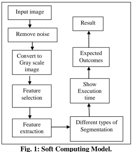
Fig. 1: Soft Computing Model.
There are some major steps in the proposed approach. Input image, noise filter, Feature selection, Feature extraction and Classification.
Step 1: Input Image.
Step 2: It is must to remove the noise from image. Each image is first applied with denoising technique.
Step 3: The proposed algorithm starts by reading the input image, converting it to grey scale image.
Step 4: Feature selection
Step 5: Feature extraction, the tumor is extracted from the MR image, its exact position and some features also determined.
Step 6: Then applying image segmentation techniques for extracting the Region of Interest (ROI) with (soft computing) parallel computing approaches. Soft computing tool run all technique simultaneously. Soft computing tool compare performance of clustering techniques, that is Fuzzy c-means and Fuzzy k-means. The accuracy of Fuzzy clustering algorithm is higher than k-means. It is the combined approaches namely K-means clustering, fuzzy C-means clustering. It is simple and fast algorithm. This algorithm is more robust to noise and provides better segmentation quality.
Step 7: Calculating execution time.
Step 8: Expected outcomes, compare all execution time at the end of the process.
Input image
Remove noise
Feature selection
Expected Outcomes
Result
Convert to Gray scale image
Show Execution
time
Feature extraction
Tiwari et al. World Journal of Engineering Research and Technology
A. UNSUPERVISED IMAGE SEGMENTATION TECHNIQUES
As discussed in last section unsupervised image segmentation technique is used to derive some hidden data from image, there has been lots of methods derived so far to segment image into non-overlapping clusters. Some of these techniques are discussed below:
There has been significant research attempt intended for on the way to methods for unsupervised brain tumor segmentation in MR images that do not utilize an anatomic idea determine. Rather than dividing the image along anatomically significant features these techniques divide images into homogeneous areas using image based characteristics such as intensities and/or textures and clustering is one process to do this. These techniques will not be enclosed in huge feature since there are major difficulties to this kind of approach. These comprise the realities that (1) number of areas often requires to be pre-specified, (2) tumors can be separated into multiple areas, and (3) tumors may not have evidently distinct intensity or textural boundaries.
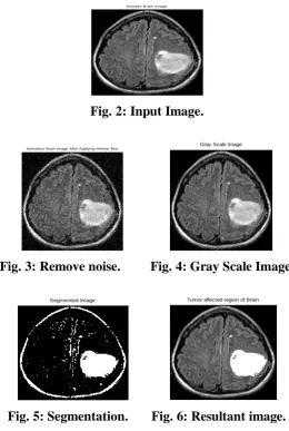
Sample Brain Image
Fig. 2: Input Image.
Noiseless Brain Image After Applying Wiener filter
Gray Scale Image
Fig. 3: Remove noise. Fig. 4: Gray Scale Image.
Segmented Image
Tumor affected region of Brain
Tiwari et al. World Journal of Engineering Research and Technology
VI.EXPECTED OUTCOMES
Unsupervised learning is an effective method of partitioning image and gives better result in the brain than supervised learning. Brain tumor detection using region base and with unsupervised learning detection technique is proposed. Co-processing is done by soft computing, it reduce the execution time to enhance the input MRI scan image. Brain tumor area is detected by using region growing segmentation and Brain tumor boundary is detected by using edge based segmentation. Segmentation detection will give exact and clear boundaries or edges. Working with risk analysis to identify area of tumor with the basis of it’s intensity value with thresholding technique. On the basis of literature survey, it is found that manual brain image analysis for brain tumor detection is a time taking task, so need to reduce the execution time with co-process computing.
VII.CONCLUSION
In this work, three different unsupervised image segmentation methods for detecting tumor region of brain MRI images has been designed and implemented with the help of soft computing in MATLAB Tool. It is important to detect and identification of that cell has been injured in human body and remove that problem, so improve the quality of MRI images, to achieve better quality image from co-process technique and accuracy will obtained in the final outcome depends on handing out of each step. For each step, there are numerous methods available. I investigated all these techniques to identify a method of soft computing that can provide superior accuracy and determine the best medical images and capability of improve the diagnosis. However, this paper presents a comprehensive review of the methods and techniques used to detect brain tumor through MRI image segmentation and capability of improve the error free image and less time required for accurate result.
VIII.REFERENCES
1. Fazli Wahid, Muhammad Fayaz and Abdul Salam Shah, “An Evaluation of Automated Tumor Detection Techniques of Brain Magnetic Resonance Imaging (MRI)”, International Journal of Bio-Science and Bio-Technology, 2016; 8(2): 265-278.
2. Deepa P Malashree and Dr. Bindu A. Thomas, “Brain Tumor Detection by MRI and CT Scan Images”, IJCTER, e-ISSN 2455–1392, June 2016; 2(6): 550–555.
Tiwari et al. World Journal of Engineering Research and Technology
4. E. T. Merlin Sathia Raj and Dr. M. Kumaresan, “Boundary Detection Algorithm For Brain Tumor Position And Area Detection Using OPENCV”, IJAER, SSN 0973-4562, 2016; 11(7): 5326-5331.
5. Neeta S. Shirsat, Vishal D. Garad, Mahesh V. Kamble, Yogesh S.Bane, Sunil B Gadkari, “Brain Tumor MRI Image Segmentation and Detection Using Fuzzy Clustering”, IJARCSSE, January 2016; 6(1): ISSN: 2277 128X.
6. Ruchita A. Banchpalliwar, Dr. Suresh S. Salankar, “A Review on B rain MRI Image Segmentation Clustering Algorithm”, (IOSR-JECE) e-ISSN: 2834,p-ISSN: 2278-8735, Jan.-Feb .2016; 11(1): Ver. III PP 80-84.
7. Neha Baraiya and Hardik Modi, “Comparative Study of Different Methods for Brain Tumor Extraction from MRI Images using Image Processing”, IJST, 9(4): DOI: 10.17485/ijst/2016/v9i4/85624, January 2016, ISSN (Print) : 0974-6846, ISSN (Online) : 0974-5645.
8. Riyazul Haque and Dayashankar Pandey, “A Modern Survey: On Various Existing Methods Based on MR Images and Tumor Detection”, July 2016; 6(7): ISSN: 2277 128X.
9. Samriti and Mr. Paramveer Singh, “Brain Tumor Detection Using Image Segmentation”, IJEDR, 2016; 4(2): ISSN: 2321-9939.
10.Kirna Rani, “A Study Of Various Brain Tumor Detection Techniques”, Int.J.Computer Technology & Applications, May-Jun, 2015; 6(3): 459-467.
11.Riddhi S. Kapse, Dr. S.S. Salankar and Madhuri Babar, “Literature Survey on Detection of Brain Tumor From MRI Images”, (IOSR-JECE) e-ISSN: 2834, p-ISSN: 2278-8735, Jan-Feb. 2015; 10(1): Ver. II, 80-86.