Manoj et al. World Journal of Pharmaceutical and Medical Research
A SIGNIFICANT METHOD TO EXPLORE DNA DAMAGE IN LYMPHOCYTES
Manoj A.*1, K. Ramachndra Rao1, B. Vishnu Bhat2, C. Venkatesh2 and Z. Bobby3 Department of Anatomy1, Paediatrics2 Biochemistry3
Jawaharlal Institute of Post Graduate Medical Education and Research (An Institution of National Importance -Govt. of India Ministry of Health and Family Welfare), Pondicherry, India.
Article Received on 29/03/2019 Article Revised on 19/04/2019 Article Accepted on 09/05/2019
INTRODUCTION
The DNA is a stable and well protected molecule composed of two chains of that coil around each other to form a double helix for carrying genetic instructions used in growth, development, functioning and reproduction of all living things.[1] Reactive Oxygen species can interact with it and cause several types of damage like modification of DNA bases, single and double strand breaks, loss of purines and pyramidine (apurinic and apyramidine sites), damage to deoxyribose sugar, DNA protein cross-linkage and damage to the DNA repair system . The .OH radical abstract hydrogen atom from deoxyribose sugar which gets fragmented in various ways. Reactions of deoxyribose derived radicals can lead to the release of purine and pyramidine bases from the DNA producing abasic site and strand breaks. Some altered sugars that remain attached to the DNA can split under alkaline condition leading to alkaline labile sites. The DNA damage due to OH radical formation is explained by the fact that H2O2 which crosses biological
membrane easily, can penetrate to the nucleus and react with ions of iron or copper to form hydroxyl radical (OH-).The OH- radical react with DNA and produces multiplicity of modifications like strand breakage, DNA modification, deoxyribose fragmentation and apoptosis.[2]
Single cell gel Electrophoresis (SCGE) or Comet assay is a rapid and sensitive procedure to detect, quantify and analyse DNA Damage in all eukaryotic cell introduced by Ostling and Johansson in 1984.[3] LaterSingh et al modified this technique in 1988, by Alkaline Lysis and Electrophoresis buffer for 1 hour and 20 min respectively in which the fragmented DNA migrates to the anode which attributes an image resembles a "Comet" with a distinct head and tail. The head is composed of intact DNA, while the tail consists of damaged (single-strand or double-strand breaks) or broken pieces of DNA depends upon the severity of the condition.[4] The applications of the Comet Assay have been to study animal eukaryotes, there have been reports of successful application in the study of plant cells.[5] Individual cells are embedded in a thin agarose gel on a microscope slide. All cellular proteins are then removed from the cells by lysing solution which is fresh during the treatment not to exceeds one hour. The DNA is allowed to unwind under alkaline electrophoresis buffer. Following the unwinding, the DNA undergoes electrophoresis, allowing the broken DNA fragments or damaged DNA to migrate away from the nucleus towards the anode of power pack. The dried slides have been staining with non-flouresent Silver nitrate dye. The extent of DNA liberated from the head of the comet is directly proportional to the amount of DNA damage.[6]
ISSN 2455-3301
WJPMR
AND MEDICAL RESEARCH
www.wjpmr.com
*Corresponding Author: Manoj A.
Department of Anatomy, Govt.Medical College Thrissur-680596, Kerala, India.
ABSTRACT
Deoxyribo Nucleic Acid (DNA) is the genetic material providing instructions for growth, development, functioning and reproduction of all living things. When it exposed to exogenous and endogenous causative factors leads to damage and alteration of the chemical structure, break in a strand and base missing from backbone. DNA damage can be explored by Single Cell Gel Electrophoresis (SCGE) or Comet Assay is a simple and sensitive technique used to measure Single Strand Break (SSB) and Double Strand Break (DSB) of DNA leucocytes. It includes the following steps Sample collection, Preparation of slides in normal (0.75%) and low (0.50%) melting point agarose, Lysis in fresh solution for one hour inorder expose the nucleus and dissolving the cell membrane and cytoplasm. DNA unwinding and detection of Alkali labile sites by staying 20 minutes in submarine gel buffers, by applying Electrophoresis in order to migrate the damage DNA towards the anode, Neutralization for 5 minutes, Fixing for 1 hour to an overnight, Drying, Staining with silver nitrate, Scanning of Comet in Fluorescent Microscope and Comet Quantification.
The Comet Assay can be used to detect DNA damage caused by single strand breaks (SSB), double strand breaks (DSB), alkali labile sites (ALS), oxidative base damage (OBD), and DNA cross-linking with DNA or protein (CLD). The slides have also stained with DNA-specific fluorescent dye such as Ethidium bromide in which the gel is read for amount of fluorescence in head and tail and length of tail.[3,7] The Comet Assay is also used to monitor or follow up cases of DNA repair by living cells after treatment.[10]
MATERIALS AND METHODS
The materials required for methodology of Comet Assay or Single cell gel electrophoresis has been depicted in the Table-1. The preparation of Stock solution and working solution of Phosphate Buffered Saline (PBS), 1-1.5%
NMA, 0.5-1% LMA, Lysis Buffer, Alkaline
Electrophoresis Buffer, Neutralizing Buffer, Fixatives, Staining solution A&B has to make according to the direction provided in the Table -1.
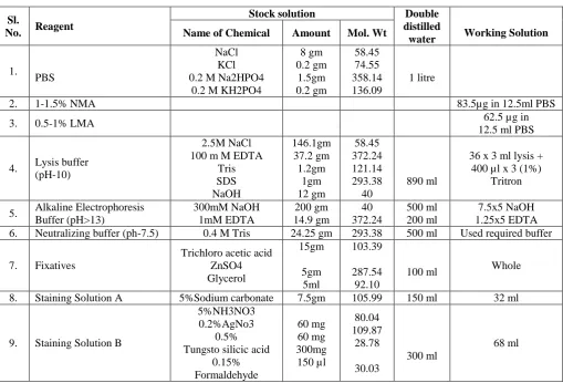
Table 1: Showing requirements of Stock solution and Working solution for Protocol of Day 1&2.
Sl.
No. Reagent
Stock solution Double
distilled
water Working Solution Name of Chemical Amount Mol. Wt
1.
PBS
NaCl KCl 0.2 M Na2HPO4
0.2 M KH2PO4
8 gm 0.2 gm 1.5gm 0.2 gm 58.45 74.55 358.14 136.09 1 litre
2. 1-1.5% NMA 83.5µg in 12.5ml PBS
3. 0.5-1% LMA 62.5 µg in
12.5 ml PBS
4. Lysis buffer (pH-10)
2.5M NaCl 100 m M EDTA
Tris SDS NaOH 146.1gm 37.2 gm 1.2gm 1gm 12 gm 58.45 372.24 121.14 293.38 40 890 ml
36 x 3 ml lysis + 400 µl x 3 (1%)
Tritron
5. Alkaline Electrophoresis Buffer (pH>13) 300mM NaOH 1mM EDTA 200 gm 14.9 gm 40 372.24 500 ml 200 ml 7.5x5 NaOH 1.25x5 EDTA
6. Neutralizing buffer (ph-7.5) 0.4 M Tris 24.25 gm 293.38 500 ml Used required buffer
7. Fixatives
Trichloro acetic acid ZnSO4 Glycerol 15gm 5gm 5ml 103.39 287.54 92.10
100 ml Whole
8. Staining Solution A 5%Sodium carbonate 7.5gm 105.99 150 ml 32 ml
9. Staining Solution B
5%NH3NO3 0.2%AgNo3
0.5% Tungsto silicic acid
0.15% Formaldehyde 60 mg 60 mg 300mg 150 µl 80.04 109.87 28.78 30.03 300 ml 68 ml
Methodology has to be done by two days procedures for which Protocol for Day-1 consists of Sample collection, Isolation of Leucocytes, Preparation of Slides, Lysis. Protocol of Day-2 involves Electrophoresis, Neutralization, Fixation, Staining, Scanning of Comets and Comet Scoring.
Protocol of Day-1 Sample Collection
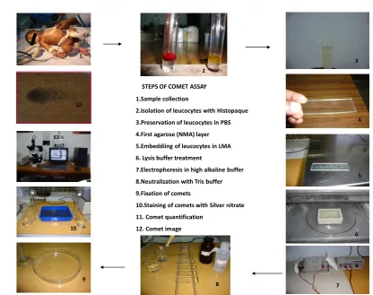
Blood samples have to be collected from peripheral vein into heparinised tubes (1-2ml) and processed within one hour (Fig1.1).
Isolation of Leucocytes
The mononuclear leucocytes isolation for the comet assay have to be performed by carefully layering heparinized whole blood over equal volume of
Histopaque 1077 (Sigma) .It have to be centrifuged at 1000 rpm for 30 mts at room temperature (Fig1.2). The buffy coat of cell pallets has to be aspirated with pasteur pipette and re-suspended in phosphate buffered saline (PBS). The aliquots of lymphocytes have to be preserved in centrifuge tubes containing PBS stored in refrigerator (Fig1.3).
Preparation of Slides
Plain grease free, clean microscope slides were used for layering of gels. Three slides were prepared for every single sample and code has to be marked at one end.
First layer- Agarose Pre-coating
45 º. The gel has to be allowed to solidify at room temperature for 30 minutes or by placing it on a slide warmer maintained at 40º to 50ºC for 10 minutes. The first layer offers increased gel bonding / stability and less slippage (Fig1.4).
Second Layer
Agarose cell mixture - The layering of cell suspension over the NMA.
The second layer consists of embedding of leucocytes. 10 µL of cells suspension mixed with 50 µL of 0.5-1% low melting point agarose (LMA). The mixture of cells and LMA dropped gently on the first layer and spread out evenly using cover slip. The slides have to be kept in the refrigerator at -4ºC for 15 minutes to allow solidification of the agarose. The cover slip has to be removed after 15 minutes (Fig1.5).
Third Layer
Agarose for residual holes filling- Layering of LMA over cell suspension. The third layer has to be made by addition of 50 micro litre of LMA dropped and flattened with approximately sized cover slip and kept in the refrigerator at -4ºC for 15 minutes .Thus the cellular layer was made sandwiched between the first and third layer to prevent the escape of sample. After 15 minutes, the cover slip has to be removed.
Cell Lysis
The slides with agarose have to be gently immersed in freshly prepared chilled lysing solution for 1 hour at 4º c which being the most significant part of comet assay standardised at Cytogentic division of JIPMER-Pondicherry. Depends upon the fresh quality of Lysing solution, the technique will be more effective for electrophoresis treatment. This treatment eliminates cell membrane, soluble cell constituents as well as histones, leaving the DNA of the nucleus unaffected (Fig1.6).
Alkali unwinding of DNA
The slides have then removed from the lysing solution and placed on a horizontal submarine gel electrophoresis unit. The indexed end of the slide was kept pointing towards the anode electrode and the gel layer faced upwards. The unit has to be filled with specified volume of electrophoretic buffer (high alkaline pH>13) (Protocol-5) to a level approximately 2-3 mm above the agarose .The slides have to be allowed to stay in the buffer for 20 minutes for unwinding of DNA(Fig1.7).
Electrophoresis
The submarine electrophoretic tank/chamber has to be connected to a power pack with 300 m Amp of 25 volt. The process was carried out for 25 minutes (Singh et al).The damaged loops or fragments of DNA migrated towards the anode and forms the tail and undamaged DNA retained in the nucleus as the head. The extension of the migration of DNA as the tail of the comet was
indicator of the level of damaged DNA (Heartman et al) (Fig1.7)
Neutralization
The slides have to be rinsed three times gently with chilled neutralization buffer for 5 minutes to remove traces of alkali and detergents, if any; of electrophoresis buffer which would otherwise interfere with staining process. Rinsing is essential as it provides good background for comet scoring. After neutralization, the slideshas to be washed with double distilled water thrice and air dried completely (Fig1.8).
Fixation
The air dried slides have to be immersed in fixing solution for 10 minutes. After fixation the slides have to be flooded 3 times with double distilled water for 5minutes each.The washed slides have to be allowed to air dry for 1 hour or to an overnight before staining(Fig1.9).
Protocol of Day-2 Staining
The fixed air dried slides have to be stained by non fluorescent dye/ Silver nitrate as per method of Silvina et al. The staining has to be done by 32 ml of staining solution A and 68 ml of staining solution B was simultaneously poured, thus mixing both parts of stain and spread onto the tray without disturbing the slides. After frequent gentle agitation with staining solution for three or four times until a greyish colour developed. Then the slides have to be transferred into a trey containing stopping solution for 5minutes until a grey colour developed. The slides have to be washed with distilled water and allowed to air dry completely in an inclined position at room temperature(Fig1.10).
Scanning of Comet
The slides have to seen under bright field microscopy- Trinocular Research microscope- CCD camera (BX 51 Olympus- Japan). The slides have to be scanned using magnification 20X. The images of comets have to be analysed and scored with Comet Score Software and the microscope connected to an IBM 15” PC (Fig1.11).
Comet scoring
The quantification of comet was done by, Comet scoreTM software TriTek Corp, USA (Fig1.11). The images of minimum 50 comets per slide has to
be captured randomly and analysed using the software coupled with a CCD camera.
For quantification of the comet parameters, Comet score software file was executed and the captured images were imported into it.
The entire area of the comet and the centre of the comet head of every single comet seen in the image was individually marked with the tool-box provided with the software.
length (C.L), head diameter (HD), % DNA in head (DH), tail length( T.L), % DNA in tail (DT), Tail Migration, Tail movement (Fig1.12).
The comet metrics thus quantified were recorded in a third party spread sheet and later transferred to Microsoft excel sheet for further analysis.
The results were tabulated and analysed using Graph Pad 2 (InStat, San Diego, USA)
STEPS OF COMET ASSAY
1
2
3
4
5
6
7
8
9
10 11
1.Sample collection
2.Isolation of leucocytes with Histopaque
3.Preservation of leucocytes in PBS
4.First agarose (NMA) layer
5.Embeddiing of leucocytes in LMA
6. Lysis buffer treatment
7.Electrophoresis in high alkaline buffer
8.Neutralization with Tris buffer
9.Fixation of comets
10.Staining of comets with Silver nitrate
11. Comet quantification
12. Comet image
9 1
7 12
Figure 1: Exhibiting the Methodology of Comet Assay in Clock-wise direction with No.1 to 12. Observation and Parameters
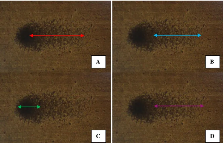
Tail length: Migration of DNA from centre of the head to smallest detectable fragment of Comet (Fig.3A).
Percentage of DNA in Tail: Amount of DNA fragments in the tail of Comet (Fig.3B).
Percentage of DNA in Head: The percentage of undamaged DNA in head (Fig.3C).
Tail Migration: DNA migration from the edge of the head to smallest detectable fragment (Fig.3D). Tail Movement is the product of percentage of DNA
in tail multiplied by tail length.[8]
Figure 2: Showing Silver Nitrate stained Comet images A.Damaged cell; B. Apoptotic cell.
Figure 3: Exhibiting Captures of Silver Nitrate stained Comet Image with marked Parameters A. Tail Length (Red); B. % DNA in Tail (Blue); C. % DNA in Head (Green) ; D. Tail Migration (Pink).
DISCUSSION
Single Cell Gel Electrophoresis is been a significant method to explore the DNA damage in eukaryotic cells for which migration of damaged DNA fragments to the Anode and the percentage of DNA in tail and length of tail of comet depends upon the degree of DNA damage whether it is single or double strand breakage. This method had been standardised during the Doctoral Research studies of the Author in the year 2008 at the division of Genetics of Department of Anatomy JIPMER-Pondicherry. Singh et al introduced single cell microgel electrophoresis technique under alkaline conditions which appears to be sensitive and useful for detecting DNA damage and its repair in single cells.[4] In their study they immersed the agarose coated solidified slide in Lysing solution for One Hour inorder to lyse the cells and permit for unfolding the DNA. Hartmann et al followed the same procedure of Singh et al for lysis treatment of lymphocytes.[5] The current study agrees with methodology of Singh et al for which the agarose coated solidified slides being submersed in fresh alkaline lysis solution for One Hour at 4°C.[6] If the alkaline lysis treatment step is been beyond one hour which leads to disintegration of not only cell membrane and cytoplasm but DNA resulting distortion of the Comet. However some researchers are been using to immerse the agarose coated slides in old lysis buffer or not in fresh solution for one overnight or 12 hours at 4°C, even if they are been getting comet but results being negative and it would not been based on the rules of Single Cell Gel Electrophoresis. The sensitiveness of SCGE is been based on the quality of the working solution which would be reflected in the quantification of Comet.[4,5,6] After lysis treatment the unwinded DNA has to be subjected for electrophoresis for 20 minutes in which the fragments of damaged DNA migrates to the Anode
which is the basic principle of Comet Assay.[5] Ostling and Johanson et al had underwent lysis treatment in neutral detergent solution for 1 hour at 0°C for detection of DSB of DNA in which the migration could more pronounced in irradiated than in control cells.[3] Since the SCGE is sensitive method and more reproducibility to quantify the DNA damage in leucocytes we could follow the alkaline method rather than neutral method.[6] Olive et al had evaluated the double strand breaks of DNA in S-phase cell than G1 cells by ionizing radiation inducing agents by using alkaline method at 50°C in which the tail moment is the product of percentage of DNA in the tail multiplied by the tail length.[8] Our study could not follow Ostling and Johanson method due to the fact that it could not detect SSB. Nelms et al stated that eukaryotic cells have intrinsic ability to self destruction once they severely damaged referred as Apoptosis or programmed cell death in which the DNA of apoptotic cells exits the cell body and migrates through the gel forming pronounced comet tails in which direction of migration in these images is left to right and the tail moments for apoptotic cells are extremely high due to the large fraction of DNA in the tail (Fig.2B) and the pronounced distance of migration manifests itself as the "hump" in the distribution at greater tail moments.[9] Tice et al reported that single cell gel (SCG) /comet assay as a tool for the biomonitoring of individuals accidently, environmentally or occupationally exposed to ionizing radiation and detects single-strand DNA breaks, alkali-labile damage, incomplete excision repair sites, DNA cross linking at the level of the individual cell. The advantages of this technique include data are collected at the level of the individual cell, providing information on the intercellular distribution of damage and repair; only small numbers of cells are required, virtually any eukaryotic cell population can be used and the assay is
C D
relatively sensitive.[7] Our study agrees with suggestions of previous report of Tice et al. Collins et al reported that endogenous damage to DNA, caused by reactive oxygen species or free radicals may be significant in the aetiology of cancer which can be reduced by dietary factors to protect against cancer in which a diet rich in fruit and vegetables commonly attributed to the dietary content of antioxidants such as Vitamin C, Vitamin E and various Carotenoids. DNA strand breaks and oxidized pyrimidines had been measured in lymphocytes using the comet assay which detects strand breaks in DNA and it allows supercoiled loops of DNA to relax and move out to form a tail and the fraction of DNA in the tail reflects the frequency of breaks and detection of oxidized bases.[10] Our study agrees with above reports in which we aimed to evaluate the Oxidative stress induced DNA damage in Perinatal Asphyxia which proves that Single Cell Gel Electrophoresis is been Significant method to Explore DNA damage.[6]
CONCLUSION
Single Cell Gel Electrophoresis/ Comet Assay is a sensitive technique to detects SSB, DSB, alkali-labile sites (ALS) damage, incomplete excision repair sites , DNA cross linking (DCL) at the level of the individual cell and repair.
REFERENCE
1. JD Watson, FHC Crick. Molecular structure of Nucleic Acid, A Stricture for Deoxyribonucleic Acid. Nature No.4356; April 25, 1953, 737-738. 2. Kaufmann WK, Richard S, Paules. DNA damage
and cell cycle and checkpoints. FASEB journal USA, 1996; 283: 238-47.
3. Ostling and Johanson. Microeleectrophoretic study of Radition Induced DNA damage in Induvidual Mammalian Cells. Biophysical and Biochemical Research Communication, 1983; 123(1); 291-298. 4. Singh NP, McCoy MT, Tice RR, Schneider EL: A
simple technique for quantification of low levels of DNA damage in individual cells. Experimental Cell Research, 1988; 175: 184-191.
5. Hartman A, Agurell E, Beevers C, Brendler S, Tice RR, Collins AR :. Recommendations for conducting the invivo alkaline comet assay. Mutagenesis, 2003; 8: 45-51.
6. Manoj A, Rao RK, Bhat VB, Venkatesh C, Bobby Z. Oxidative stress induced DNA damage in Perinatal asphyxia. Curr Ped. Res, 2011; 15(1): 19-23.
7. Tice RR, Agurell E, Anderson D, Burlinson, B, Hartman A, Kobayashi H. Single Cell Gel/ Comet assay: Guidelines for invitro and invivo genetic toxicology testing. Environmental and Molecular Mutogenesis, 2000; 35: 206-221
8. Peggy Olive, D Wlodek, JP Banath. DNA Double-Strand Breaks Measured in Individual Cells Subjected to Gel Electrophoresis. Cancer Research 51, 4671-4676. September 1, 1991.
9. B E Nelms. Measuring Apoptosis in Individual Cells with the Comet Assay Promega Notes Number 64, 1997; 13.