Available online on 15.3.2018 athttp://ujpr.org
Universal Journal of Pharmaceutical Research
An International Peer Reviewed Journal Open access to Pharmaceutical research
This is an open access article distributed under the terms of the Creative Commons Attribution-Non Commercial Share Alike 4.0 License which permits unrestricted non
commercial use, provided the original work is properly cited
Volume 3, Issue 1, 2018
REVIEW ARTICLE
MOST IMPORTANT CELLULAR CHANGES INVOLVED IN RENAL
ISCHEMIA REPERFUSION INJURY AND THE CONSEQUENT IMPACT ON
SELECTED REMOTE ORGANS
Asmaa A. Khalifa1 , Mai El-Sayed Ghoneim2
1
Department of Pharmacology and therapeutics, Faculty of Pharmacy, Pharos University, Alexandria, Egypt. 2Department of pharmacology and toxicology, Faculty of Pharmacy, Tanta University, Tanta, Egypt.
ABSTRACT
Because of the high rate of baseline oxygen use by renal cells, kidney is highly influenced by obstruction of arterial blood inflow and subsequent shortage of the received oxygen, this condition is known as Ischemic injury. There are many clinical settings associated with unavoidable ischemic state such as kidney transplantation, partial nephrectomy or suprarenal procedures of the aorta. During ischemia many cellular changes occur including vascular congestion and adhesion of inflammatory cells to the endothelium with subsequent infiltration into the kidney tissue. Following ischemia, a phase known as Reperfusion begins and involves a return of blood and oxygen supply to micro vessels. Reperfusion was expected to restore the damage occurred during the ischemic phase, paradoxically, reperfusion leads to more congestion, red cells trapping and excessive generation of reactive oxygen species (ROS), which can oxidatively modify significantly every type of biomolecule, thereby inducing cell dysfunction and induce reperfusion injury. Ischemia reperfusion injury (IRI) is also related to a phenomenon called Remote Organ Injury (ROI) in which the damaging effect induced by I/R is not only restricted to the tissue that undergoing the initial ischemia but also it leads to injury to remote organs such as the liver, lung, gut. ROI usually occurs by the same mechanisms seen in the local injury induced by I/R including the generation of ROS, leukocytes, and inflammatory mediators (e.g; TNF-α). These substances are directly released from the primary injured tissue or indirectly from activated leukocytes or other inflammatory cells causing organ dysfunctions in distant organs.
Keywords: Inflammatory response, remote organ injury, renal ischemia, reperfusion injury reactive oxygen species (ROS).
Article Info: Received 3 March 2018; Revised 8 March; Accepted 11 March, Available online 15 March 2018
Cite this
article-Khalifa AA, El-Sayed GM. Most important cellular changes involved in renal ischemia reperfusion injury and the consequent impact on selected remote organs. Universal Journal of Pharmaceutical Research 2018; 3(1): 78-86.
DOI: http://doi.org/10.22270/ujpr.v3i1.RW1
Address for Correspondence:
Asmaa A. Khalifa, Department of Pharmacology and therapeutics, Faculty of Pharmacy, Pharos University, Alexandria, Egypt. Tel: +201093771967, E-mail: [email protected]
INTRODUCTION
The functions of the kidney are vital to life and are regulated by the endocrine system by hormones such as
antidiuretic hormone (ADH), aldosterone, and
parathyroid hormone (PTH). The important functions
that the kidneys serve including1:
1. Filtration and excretion of metabolic waste products
2. Regulation of necessary electrolytes, fluid, and
acid-base balance
3. Controlling reabsorption of water and maintaining
intravascular volume, also kidneys reabsorb glucose, amino acids
4. Stimulation of red blood cell (RBC) production.
5. Regulation of blood pressure via the
renin-angiotensin-aldosterone system,
6. Hormonal functions via erythropoietin, calcitriol,
and vitamin D activation.
The kidney is considered as the most important organ for the excretion of water soluble drugs and/or their
metabolites in to the urine2.
Nephrons are urine-producing functional structures of
the kidney1 which are distributed at the cortex and
medulla. A normal human kidney contains 800,000 to
1.5 million nephrons3. Each nephron is composed of:
The renal corpuscle (Bowman capsule): containing
the glomerulus.
The Proximal convoluted tubule (PCT),
located in the renal cortex.
LOOP of Henle (LOH): descending limb and
ascending limb located in renal medulla
The distal convoluted tubule.
Cortical nephrons have their loop of Henle in the renal medulla near its junction with the renal cortex.
Juxtamedullary nephrons have their loop of
Henle deep in the renal medulla4.
RENAL BLOOD SUPPLY
Normally, the kidneys receive 1,000 to 1,250 ml/min of blood in the adult person which is about 25% of the
cardiac output (COP)5. This amount far exceeds that
needed to provide the kidney's intrinsic oxygen requirement but ensures optimal clearance of all wastes and drugs from the body. Essentially, all blood passes through glomeruli, and about 10 % of renal blood flow is filtered (a glomerular filtration rate GFR of 125 mL/min in the normal adult). The basal normal blood flow is 3 to 5 ml/min/g of tissue, greater than in most other organs.
The vascular structure of the renal cortex is complex. The renal artery enters the kidney at the hilum, where it divides into five interlobar arteries, each an end artery. The afferent arterioles, which arise from the interlobular arteries, divide within the cortical tissue to form the glomerular capillary network. The capillaries then reunite to form the efferent arterioles. Vessels from the efferent arterioles supply the proximal and distal tubules and portions of the loops of Henle and the collecting ducts. The juxta glomerular apparatus is between the afferent and efferent arterioles and the macula densa, a specialized group of cells which are located in the distal convoluted tubule. The point at which the afferent arterioles enter the glomerulus and the efferent arteriole leaves it, the tubule of nephron return back to touch the arterioles of the glomerulus of the same nephron from which it exists. At this position, thick ascending limb of loop of Henle, there is a specific modified region of tubular epithelium called the Macula densa.
Renal ischemia/reperfusion
Simply the term ischemia means that there is a deficient blood supply to tissues due to obstruction of arterial blood inflow. The body is able to adapt to a reduction in blood flow to a certain level, but when delivery of oxygen and nutrient substrates becomes
inadequate, cellular injury leads to organ dysfunction6.
Kidney is considered as one of the most susceptible body organs to ischemia. Renal parenchymal oxygenation is graded with the highest oxygen levels noted in the cortex, medium levels in the outer medulla, and the lowest levels in the papillae. As a consequence, cortical cells are the most sensitive to ischemia, while cells in the outer medulla can shift to oxygen-independent metabolism making them less sensitive to a hypoxic environment. Inner medullary and papillae cells use predominantly glucose to generate ATP via anaerobic glycolysis. Thus, these regions demonstrate a reduced sensitivity to ischemia. Reperfusion could paradoxically induce and exacerbate
tissue injury and necrosis7. Renal ischemia/reperfusion
injury (IRI) results from a generalized or localized impairment of oxygen and nutrient delivery to, and
waste product removal from, cells of the kidney8-10.
There is a mismatch of local tissue oxygen supply and
demand and accumulation of waste products of metabolism. As a result of this imbalance, the tubular epithelial cells undergo injury and, if it is severe, death by apoptosis and necrosis (acute tubular necrosis [ATN]), with organ functional impairment of water and electrolyte homeostasis and reduced excretion of waste
products of metabolism10. There are major clinical
settings or medication use which may lead to
deposition of ischemia reperfusion injury6,11.
Acute renal failure caused by medications for the
treatment of hypertension, especially with
angiotensin converting enzyme inhibitors (ACEIs)
Progressive azotemia
Acute pulmonary edema
Renal transplantation.
Medication use: Vasoconstrictive drugs.
Cyclosporine use
Tacrolimus use
Overuse of NSAIDs and Radiocontrast agents
Hypotension linked to sepsis or blood loss after
surgery and trauma.
Renal vascular diseases
In the following lines we will discuss the most important cellular changes involved in the ischemia and reperfusion injury. Also the remote organ injury that occurs in the liver following renal ischemia reperfusion will be mentioned.
Cellular changes during ischemia:
One of the most important changes in ischemia are that
occur in the endothelium12. Lately these changes lead
to endothelial dysfunction, these changes include a). Changes in the Vascular tone:
Nitric oxide (NO) one of the autacoids that is acting on vascular smooth muscle cells to induce vasodilatation NO is generated by the enzymatic transformation reaction illustrated below and is catalyzed by an
enzyme called nitric oxide synthase (NOS)13.
L- arginine + O2
NOS
L-citrulline + NO NOS exists in two different isoforms which both are
found in the kidney;14
The first isoform is endothelial NOS (eNOS):
found in vasa recta, inner medullary collecting
duct and glomeruli15.
The second inducible NOS (iNOS) can be
expressed by vascular smooth muscle cells and
immune cells such as monocytes, macrophages,
neutrophilsin the kidney16,17.
NO which is derived from the enzymatic activity of
(iNOS) appears to participate in vascular dysfunction18
and leading to tissue damage19. There are two main
pathways involved in the tissue damage produced by NO derived from iNOS:
i. Peroxynitrite (ONOO-) generation (oxidant and
nitrating agent). Due to its oxidizing properties, peroxynitrite can damage a wide array of molecules in cells, including DNA and proteins leading to
endothelial dysfunction and tissue damage20.
ii. Secondary to endothelial dysfunction and damage,
increase susceptibility to microvascular thrombosis
which leading to further tissue damage0.
One of the future approaches is to examine the effect of iNOS inhibitors on the protective effects against
ischemia22.
b). Changes in the microvascular Permeability The increased microvascular permeability observed in ischemia is likely to be caused by a combination of factors, most of them is due to the activation of matrix
metalloproteinase-2 (MMP-2) or matrix
metalloproteinase-9 (MMP-9) which leading to Severe alterations in the integrity of the adherent junctions of
the renal microvasculature23-28.
c). Changes in the Coagulation process
The interaction between Endothelial cells through their interaction with protein C and thrombomodulin. Protein C is considered as one of the natural anticoagulants while thrombomodulin is a protein cofactor expressed on endothelial cell surfaces that modifies the substrate specificity of thrombin. Under the physiological condition, the interaction between thrombin and thrombomodulin leads to the formation of thrombin-thrombomodulin complex which in turn activates protein C. The activated form of protein C (APC) plays an important role in regulating blood clotting, inflammation, cell death and maintaining the permeability of blood vessel walls in humans and other
animals29.
During an inflammatory response such as in ischemia, decreases in the anticoagulant and anti-inflammatory effects of the protein C pathway occur .that is due to:
The degradation or decreased production of
protein C.
Down regulation of endothelial protein C receptors
EPCR.
Decreased thrombomodulin expression.
The microvascular function is compromised, resulting in spreading intravascular coagulation and thrombosis, the local tissue perfusion is decreased, and finally
organ dysfunction is developed30.
d). Acute epithelial cell injury
It worth to mention that during ischemic injury, all segments of the nephron can be affected, but the most commonly injured epithelial cell is the proximal tubular cell. There are many reasons that make proximal tubular cells are particularly susceptible for ischemic injury. Proximal tubular cells have a high metabolic rate and a limited capacity to undergo anaerobic glycolysis. Owing to the unique blood flow in the outer stripe of the S3 segment of the nephron, there is marked microvascular hypoperfusion and congestion in this region after injury, which persists and mediates continued ischemia even when cortical blood flow might have returned to near-normal levels. According to the extent of injury, epithelial cells undergoing sub-lethal or less severe injury will have the capability of functional and structural recovery if the insult is interrupted. While cells that suffer a more-severe or lethal injury will undergo apoptosis or necrosis, leading to cell death. Moreover, following a reduction in effective kidney perfusion, epithelial cells cannot maintain the adequate intracellular ATP for the essential processes made by the cells. In case of sever
reduction in the renal perfusion, cell death by necrosis or apoptosis may occur.
e). Role of Inflammation
Following ischemic injury, a number of potent mediators are generated by the injured epithelial proximal tubular cell, including pro inflammatory cytokines, such as tumor necrosis factor (TNF),
interleukin (IL) -6, and IL-1β andIL-831. Early
inflammation is characterized by margination of leukocytes to the activated vascular endothelium via interactions between selectins and ligands that enable. Leucocytes interact with the vascular endothelium via a series of distinct steps characterized by leukocyte „rolling‟ on the endothelium, firm adherence of leucocytes to the endothelium and endothelial
transmigration32. Upon reaching the extravascular
compartment, activated leucocytes release toxic ROS, proteases and elastases, resulting in increased microvascular permeability, edema, thrombosis and
parenchymal cell death33.In many experimental studies
it was shown that the level of both TNF-α and MPO is increased following the ischemic attack so the following points will to illustrate their role in ischemic injury34.
Tumor necrosis factor- alpha (TNFα)- is a protein hormone produced by systemic leukocytes (primarily by activated macrophages). It has been implicated as a systemic mediator in the development of septic shock and other pathologic conditions. Serum TNF-alpha has also been detected in a variety of cardiac disease states
and after ischemia-reperfusion injury35.
Neutrophils- are the first cells to accumulate at the site of ischemic injury. Blockade of neutrophil function or neutrophil depletion provides only partial protection against injury, indicating that other leukocytes also mediate injury. These inflammatory mediators include
macrophages, B cells, and T cells36. These cells
mediate tubular injury at various phases of the process, and there are synergistic interactions between different cell types33. Neutrophils are the inflammatory cells that abundantly produce ROS during IR injury.
Myeloperoxidase (MPO)-a heme-containing protein which is found mainly in the azurophilic granules of neutrophils and to a lesser extentin the lysosomes of monocytes in humans; MPO has an important role in the oxidative stress process through catalyzing the formation of hypochlorous acid (HOCl), a toxic agent
to cellular components, that initiates oxidative injury37.
MPO is considered as one of the marker of oxidative
stress during ischemic conditions34. Oxidative stress is
defined as imbalance between reactive oxygen species (ROS) production and the internal antioxidant system. MPO or hypochlorite (or hypochlorous acid HOCl) may further mediate oxidative modification of lipids, proteins and DNA which in turn leading to cell injury
and dysfunction38. Before ending this section of the
article it worth to mention that Cyclooxygenase-2 enzyme has an important role during ischemia. In some experimental animals including: mice, rats, rabbits, and dogs; it was shown that COX-2 expression in kidney cortex has been localized to the macula densa/cortical
thick ascending limb of Henle (cTALH)39. There was a
human kidney but studies in humans >60 years of age
have demonstratedCOX-2 in macula densa and have
documented increased macula densa COX-2 in patients with Bartter syndrome (a rare inherited defect in the
thick ascending limb of the loop of Henle)40. It has
been suggested that the increased macula densa COX-2 seen in elderly humans may be secondary to decreased
basal renin production associated with aging41. In
general, COX-1 functions in the control of renal hemodynamics and the glomerular filtration rate (GFR); COX-2 functions affect salt and water
excretion, although there is some overlap42. In a person
with normal renal hemodynamic parameters,
prostaglandins (PGs) do not play a dominant physiologic role in maintaining renal blood flow and
GFR43. However, prostaglandins role become of high
importance in a person with compromised renal hemodynamics. In such conditions, vasodilating prostaglandins are synthesized by kidney as an autoregulatory response to offset vasocontrictin
gautacoids and to maintain renal perfusion and GER44.
Only PGs derived from COX-1 are involved in normal renal function while COX-2-derived PGs will have
different role45. Up regulation in COX-2 expression
during the inflammatory situation associated with the
renal ischemia, COX-2 induction has been
demonstrated in several phagocytic cells due to the effect of many proinflammatory cytokines such as IL 1β, TNFα, platelet activating factor PAF. Induction of COX-2 in macrophages involves reactive oxygen intermediates and an increase in prostanoids synthesis which are potent inflammatory mediators that can
exaggerate the inflammatory condition46.The blockade
of COX-2 effect can prevent the subsequent inflammatory cascade. So the use of COX-2 inhibitors is considered one of the treatment approaches for the clinical situation associated with unavoidable ischemic
state such as kidney transplantation, partial
nephrectomy or suprarenal procedures of the aorta34.
Cellular changes during reperfusion injury
Following ischemia, reperfusion is unequivocally essential for the survival of ischemic tissues as the reestablishment of blood flow as well as the recovery of tissue oxygenation in the affected area bring indispensable nutrients to tissue repair. Paradoxically, reperfusion of the acutely ischemic tissue may lead to local and systemic complications. Reperfusion of previously viable ischemic tissues may augment tissue injury in excess of that produced by ischemia alone so
it is called “oxygen paradox” phenomenon47.
The Reperfusion injury following ischemia can be mediated by several mechanisms that will be discussed below:
a). Free radical role in reperfusion injury:
Low levels of oxygen radicals and oxidants are normally formed in cells and play important roles in cellular homeostasis, mitosis, differentiation, and
signaling. Although mammalian cells express
endogenous free radical scavenging enzymes, such as superoxide dismutase (SOD), catalase and glutathione
peroxidase, these antioxidative defenses are
overwhelmed or consumed after ischemia and
reperfusion period48. During cellular ischemia ATP is
degraded to form hypoxanthine. Under normal physiological conditions, hypoxanthine is oxidized by xanthine oxidase (XO) to xanthine using oxygen; therefore during ischemia (a state of oxygen deprivation) it is unable to catalyze the conversion of hypoxanthine to xanthine, resulting in a build-up of excess tissue levels of hypoxanthine. When oxygen is reintroduced during reperfusion, the conversion of accumulated hypoxanthine by xanthine oxidase (XO) results in the formation of toxic ROS (reactive oxygen
species) including peroxide anions (O2−), hydroxyl
radicals (OH−), hypochlorous acid (HOCl). Owing to
their highly reactive nature, ROS generated upon reperfusion can oxidatively modify every type of
biomolecule found in cells affecting their function49.
Another free radical type is also formed called reactive nitrogen species (RNS), which refers to radical molecules derived from NO. The produced free radicals ROS and RNS may interact together and produce more aggressive product called reactive nitrogen oxide species (RNOS), such as strong pro-oxidant peroxynitrite. Free radical production can be described as a nonstop cascade process the eventually
lead to cellular injury7.
b). pH paradox phenomenon
In the ischemic cells, changes in metabolism occur which include anaerobic glycolysis and the hydrolysis of adenosine triphosphate. These metabolism changes lead to intracellular pH falls. If ischemic cells are reperfused at acidotic pH, cell killing is abrogated. In contrast, the rapid rise in intracellular pH during reperfusion provokes cell killing, this phenomenon is called pH paradox. Reperfusion exacerbates this damage by triggering an inflammatory reaction and
disrupts the microcirculation50.
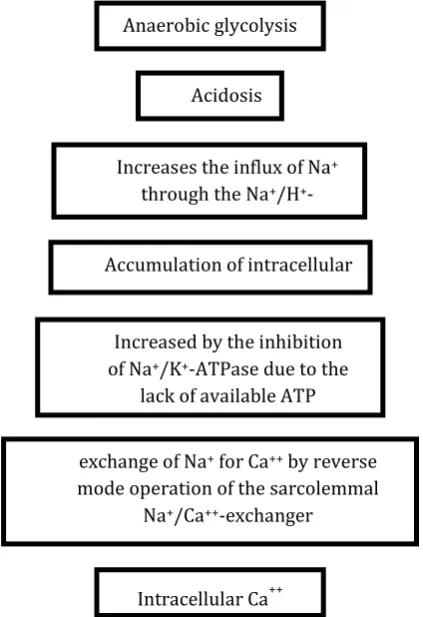
c). Calcium overloads
During ischemia as we mentioned above the cells become dependent on anaerobic glycolysis to maintain ATP level in the absence of oxygen supply. Hence, accumulation of lactate and protonscauses a fall in cytosolic pH. In an attempt to reestablish normal pH,
the cell releases H+ ions out of the cell in exchange for
Na+ via the Na+/H+ exchanger (NHE). Then Na+ ions
are, in turn, exchanged for Ca2+ by Na+/Ca2+
exchanger. This increase in cytosolic Ca2+ is greatly
exacerbated upon reperfusion due to the rapid pH
increase and removal of extracellular H+ ions further
increases the proton gradient across the plasmalemma,
thereby accelerating NHE exchanger function7. In
addition Ca2+ reuptake into the calcium stores
(endoplasmic/sarcoplasmic reticulum) ER/SR is
impaired by I/R. Hence, we reached a state in which there is an increase in the calcium level with inability for the excess calcium amount to be properly stored
which leads to lethal elevations in intracellular Ca2+ or
calcium overload as illustrated in Figure 1. Here is a question, what are the consequences of calcium
overload; these massive alterations in Ca2+ activate a
variety of systems, all of which can contribute to cell death following I/R:
Lethal increase in Ca2+ is to take it up into the
mitochondria via the mitochondrial Ca2+ uniporter.
excessive, they can trigger the mitochondrial permeability transporter MPT response. This leads to mitochondrial swelling and cell death in another
word the high cytosolic concentrations of Na+ and
Ca++ result in intracellular edema.
Activation of Ca2+/calmodulin-dependent protein
kinases (CaMKs), which also contribute to cell
death and organ dysfunction following ischemia51.
Figure 1: Steps that lead to calcium overload during ischemic injury
d). The no-reflow phenomenon
Simply from its words the no reflow phenomenon can describe the capillaries of organs through which the blood did not flow properly after reperfusion. In another words, the no-reflow phenomenon refers to the clinical observation that blood flow to an ischemic organ is often not fully restored following the release of a vascular occlusion. So, no matter now the blood flow is efficiently or rapidly restored to the blood deprived capillaries if microvascular obstruction still
exists51. During reperfusion a large number of
capillaries fail to adequately re-perfuse which lead to the evolution of the no reflow phenomenon. Activated neutrophils play an important role in the development of no-reflow phenomenon. Activated neutrophils are arrested in the capillaries due to the decrease in the driving flow pressure during ischemia and the large size of neutrophils both reasons allow the blockade of the capillaries. Furthermore, as mentioned previously the acidic environment associated with ischemia increases the stiffness of these white cells thereby increasing the likelihood for leukocyte plugging in capillaries. Endothelial barrier disruption associated with I/R leads to trans microvascular fluid filtration and protein efflux in turn edema forms. When the blood supply is reestablished in reperfusion, restoration of luminal pressures occur and hence the edema
formation rate is increased. The fluid accumulates in the ischemic tissues leading to increased interstitial pressure surrounding blood vessels, as a result collapse of the micro vessels occur and produce extravascular compression. This leading to inability of the blood to pass through theses microcapillaries during reperfusion
so, the no-reflow phenomenon exists52. This
extravascular compression mechanism is especially important in tissues that cannot expand during edematous state because they are surrounded by structures that limit expansion such as the brain, many skeletal muscles and the kidney. Continued organ dysfunction in the post-reperfusion period, failure of a transplanted graft or increased infarct size is all clinical settings that may be explained to an extent by the no reflow phenomenon.
Remote organ injury following renal I/R injury Untoward effects of I/R are not necessarily restricted to the specific tissue undergoing the initial ischemia. That is, a frequent consequence induced by reperfusion after localized tissue ischemia is injury to other organ systems, so-called distant or remote organ injury (ROI). The ultimate expression of ROI is multiple organ dysfunction syndromes. As known, renal ischemia reperfusion (IR) is one of the most pivotal causative mechanisms of acute kidney injury (AKI) which is deemed a pan-organ problem that exerts Acidosis
Increases the influx of Na+ through the Na+/H+
-exchanger
Accumulation of intracellular Na+
Increased by the inhibition of Na+/K+-ATPase due to the
lack of available ATP
exchange of Na+ for Ca++ by reverse mode operation of the sarcolemmal
Na+/Ca++-exchanger
negative impact on many organs of the body53. The hypothesis of distant organ injury (lung, heart, brain, liver, etc.) has emerged over the last decade and may demonstrate the reason for the potential negative
influence of AKI on outcome53-55. High mortality rate
during AKI is largely due to this multiple organ dysfunction. Animal studies obviously indicate that AKI simulates remote organ dysfunction through different particular pathways including apoptosis,
inflammatory cascades, differential molecular
expression, and induction of remote oxidative stress56.
The Proposed underlying mechanism of remote organ injury consequences after AKI could be categorized into four following mechanisms:
1. Classical manner of acute uremic case which affects all metabolic and endocrine pathways, causes disruption of volume and electrolyte homeostasis, and further proximate agents have a profound influence on
immune-competence57,58.
2. Inflammatory nature of the injured kidneys whichmay produces clearly higher inflammatory
chemokines expression and renal fibrosis59 as well as
oxidative stress by disturbing systemic iron
homeostasis60. This inflammatory process may
eventually transform into systemic inflammatory
reaction mediating remote organ injury61.
3. A great modulating effect on the remote organ injury would be induced by the disturbance of cytokine/chemokine homeostasis in AKI, which may be attributable to the decreased renal clearance and/or
increased production of these cytokine/
chemokine59,60,62.
4. Healthcare impediment of renal replacement therapy (RRT) support is considered as essential for
AKI patients with fluid overload63. However, RRT is
proven to carry dramatic risks for adverse patient outcome leading to the reactive oxygen species as well as, hemodynamic instability and nutrients loss during
RRT and inflammatory reaction64,65. Depend on the
mechanisms mentioned before; several complex pathways are involved in remote organs injury during AKI including pulmonary, cardiovascular,
gastroin-testinal, hepatobiliary, and neuromuscular56,66-69.
Remote impact on the heart
Acute kidney injury (AKI) may result in acute cardiac disorder via some mechanisms including:
1. Myocardial damage due to neutrophil trafficking, myocyte apoptosis, endothelial dysfunction, as well as elevated level of inflammatory cytokines (IL-1, IL-6, and TNFα resulting from increased production and impaired clearance.
2. Increased preload secondary to AKI-induced salt
and water retention70,71. To illustrate the association
between acute kidney injury and cardiovascular risk, Ko et al. revealed that mortality and major adverse cardiovascular and cerebrovascular events significantly correlated with the severity of AKI, and the severity of AKI influences strongly patient outcomes, so it has to be recognized immediately and treated aggressively
when possible72. Furthermore, the association between
AKI and subsequent risk for cardiovascular disorder
were identified in other studies73,74. A research study
conducted by Kelly70 showed an increased level of
TNF-α and IL-1 in the heart in the 48 first hours after renal ischemia reperfusion. This was accompanied by rise in myeloperoxidase activity in the heart. Furthermore, it is also observed increases in left ventricular end systolic diameter, left ventricular end diastolic diameter, and decreased fractional shortening by echocardiography after renal ischemia reperfusion. Remote impact on the liver
The underlying mechanisms between acute kidney
injury and liver remains to be understood75. Evidence
showed that AKI has significant effect on liver inflammatory response and drug as well as other
nutrient metabolism, and even patient outcomes67.
Other experimental studies showed that AKI cause
increased vascular permeability, T-lymphocyte
infiltration, neutrophil in the liver66. Moreover, AKI
invigorates oxidative stress, up regulate the expression
of injury-promoting molecules and decreases
antioxidants level leading to tissue damage of
hepatocytes68,54. In study conducted in Wister Male
Rats, hepatic levels of TNF-α and Malondialdehyde increased significantly after renal ischemia reperfusion, while total glutathione decreased, suggesting the activation of oxidative stress). Hepatocytes apoptosis increased in 24 h after nephrectomy. In addition to that, Authors found histological of hepatocyte injury
following AKI54. Another study conducted on mice
showed rapid hepatocyte necrosis, neutrophil
infiltration, pro inflammatory mRNA up regulation,
and vacuolization76.
Remote impact on the brain
Acute kidney injury has neurological complications including attention deficits, dizziness, seizure, tremor,
delirium, altered mental status, and even death56 soluble
and cellular inflammatory cytokines and uremic toxins contribute to the neurological complications. Animal studies using mice showed that AKI may result in augmentation of vascular permeability, increased cerebral pro inflammatory cytokines (6, 1β, IL-12, and glial fibrillary acidic protein), disruption in the blood brain barrier, and microgliosis (up-regulation of
brain macrophages)56,77. In addition to that, posterior
reversible encephalopathy syndrome and myopathy have been presented in AKI patients with and without
hypertension78,79.
Remote impact on the lung
Respiratory outcomes are the most clinically connected to Remote organ injury in AKI seen in patients with
pulmonary inflammation and mechanical
ventilation56,68. Acute kidney injury changes peripheral
vascular responses by increasing oxidative stress80.
Several experimental studies revealed that AKI results in pulmonary injury via following pathways:
1. Increased production of chemokines and cytokines related to impaired renal clearance.
2. Lung edema resulted from increased lung vascular permeability.
3. Increased leukocyte and mononuclear phagocyte production.
Additionally, AKI may express modulatory effects that
vary with the severity of pulmonary injury56,81.
Brøchneret et al., compared 5 mice (C57BL/6) groups
that Myeloperoxydase production in the lung significantly increased in the groups with acute kidney
injury than in limb ischemia and sham groups82.
Additionally, interleukin (IL)-6 and IL-10 blood levels significantly increased in the AKI groups compared to sham group, suggesting the role of ischemia
reperfusion to the systemic inflammatory response82.
Remote impact on the Gut
Gut is a new organ which is remotely injured during AKI. The hypervolemia and inflammatory response related to AKI change the permeability of mesenteric vascular membrane and stimulate the formation of
intestinal edema leading to sepsis83. The underlying
mechanisms including: disruption of mucosal integrity, liberation of pro-inflammatory mediators, increased intestinal permeability, and translocation of intestinal
microorganisms84.
CONCLUSION
Renal ischemia reperfusion injuries have been demonstrated in many clinical settings, such as kidney transplantation, partial nephrectomy or suprarenal procedures of the aorta where ischemia cannot be avoided. Several mechanisms are involved in the induction of Renal I/R injury. The most important mechanisms are related to generation of the reactive oxygen species (ROS) and infiltration of inflammatory mediators such as cytokines (tumor necrosis factor alpha (TNF-α)) and interleukins which eventually leading to cell death and loss of cellular functions. Moreover the local injury may spread to other distant organs (heart, brain, liver, lung and gut) and cause multiple organ injury. Each mechanism can be target of therapeutic intervention to protect the kidney and the distant organ from the expected damage occurred as a result of the ischemia and reperfusion injury.
REFERENCES
1. Sampaio FJ. Anatomical background for nephron-sparing surgery in renal cell carcinoma. J Urol 1992; 147(4):999-1005. https://doi.org/10.1016/S0022-5347(17)37445-1
2. Masereeuw R, Russel FG. Mechanisms and clinical implications of renal drug excretion. Drug Metab Rev 2001; 33:299-351. https://doi.org/10.1081/dmr-120000654
3. Guyton AC, Hall JE. Urine Formation by the Kidneys: I. Glomerular Filtration, Renal Blood Flow, and Their Control. In: Guyton AC, Hall JE (eds). Textbook of Medical Physiology. 11th ed. Philadelphia: Elsevier Saunders 2006; 310-26.
4. Lindeman RD. Overview: Renal Physiology and Pathophysiology of Aging. Am J Kidney Diseases 1990; 16(4):275–82.https://doi.org/10.1016/S0272-6386(12)80002-3
5. Barger AC, Herd JA. Renal vascular anatomy and distribution of blood flow. In: Orlaff J, Berliner RW (eds): Handbook of Physiology, section 8. Baltimore: Williams and Wilkins; 1973. 249.
6. Preston RA, Epstein M. Ischemic renal disease: an emerging cause of chronic renal failure and end-stage renal disease. J Hypertens 1997; 15(12 Pt 1):1365-77.
https://doi.org/10.1097/00004872-199715120-00001
7. Kalogeris T, Christopher P, Krenz M, Ronald J. Cell Biology of Ischemia/ Reperfusion Injury. Int Rev Cell Mol Biol 2012; 298:229-317.
https://doi.org/10.1016/B978-0-12-394309-5.00006-7
8. Bell PD, Navar LG. Cytoplasmic calcium in the mediation of macula densatubuloglomerular feedback responses. Science 1982; 215(4533):670-3.
https://doi.org/10.1126/science.6800034
9. Brenner BM, Lawler EV, Mackenzie HS. The hyperfiltration theory: a paradigm shift in nephrology. Kidney Int 1996; 49(6):1774-7.
https://doi.org/10.1038/ki.1996.265
10. Pratt RE, Flynn JA, Hobart PM, Paul M, Dzau VJ. Different secretary pathways of renin from mouse cells transfected with the human renin gene. J Biol Chem 1988; 263(7):3137-41.https://doi.org/10.1152/ajprenal.00710.2012
11. Thurman JM. Triggers of inflammation after renal ischemia/reperfusion. Clin Immunol 2007; 123(1):7-13.
https://doi.org/10.1016/j.clim.2006.09.008
12. Molitoris BA. Actin cytoskeleton in ischemic acute renal failure. Kidney Int 2004; 66:871-83.
https://doi.org/10.1111/j.1523-1755.2004.00818.x
13. Pallone TL, Silldorff EP. Pericyte regulation of renal medullary blood flow. Exp Nephrol 2001; 9(3):165-70.
https://doi.org/10.1159/000052608
14. Kone BC, Baylis C. Biosynthesis and homeostatic roles of nitric oxide in the normal kidney. Am J Physiol 1997; 272:F561-78.
https://doi.org/10.1152/ajprenal.1997.272.5.F561
15. Wu F, Park F, Cowley AW Jr, Mattson DL. Quantification of nitric oxide synthase activity in microdissected segments of the rat kidney. Am J Physiol 1999; 276:F874-81.
https://doi.org/10.1152/ajprenal.1999.276.6.f874
16. Johannes T, Mik EG, Ince C. Non resuscitated endotoxemia induces microcirculatory hypoxia areas in the renal cortex in the rat. Shock 2009; 31(1):97-103.
https://doi.org/10.1097/SHK.0b013e31817c02a5
17. Buttery LD, Evans TJ, Springall DR, Carpenter A, Cohen J, Polak JM. Immunochemical localization of inducible nitric oxide synthase in endotoxin treated rats. Lab Invest 1994; 71(5):755-64. https://doi.org/10.1172/JCI118948
18. Gunnett CA, Lund DD, McDowell AK, Faraci FM, Heistad DD. Mechanisms of inducible nitric oxide synthase mediated vascular dysfunction. Arterioscler Thromb Vasc Biol 2005; 25(8):1617-22.
https://doi.org/10.1161/01.ATV.0000172626.00296.ba
19. Guan Z, Gobe G, Willgoss D, Endre ZH. Renal endothelial dysfunction and impaired autoregulation after ischemia reperfusion injury result from excess nitric oxide. Am J Physiol Renal Physiol 2006; 291(3):F619-28.
https://doi.org/10.1152/ajprenal.00302.2005
20. Schild L, Reinheckel T, Reiser M, Horn TF, Wolf G, Augustin W. Nitric oxide produced in rat liver mitochondria causes oxidative stress and impairment of respiration after transient hypoxia. FASEB J 2003; 17(15)21:2194-201.
https://doi.org/10.1096/fj.02-1170com
21. Goligorsky MS, Brodsky SV, Noiri E. NO bioavailability, endothelial dysfunction, and acute renal failure: new insights into pathophysiology. Semin Nephrol 2004; 24:316-23.https://doi.org/10.1016/j.semnephrol.2004.04.003
22. Heemskerk S, Masereeuw R, Russel FG, Pickkers P. Selective iNOS inhibition for the treatment of sepsis-induced acute kidney injury. Nat Rev Nephrol 2009; 5(11):629-40. https://doi.org/10.1016/j.jcrc.2008.11.011
23. Sutton TA, Henry EM, Silvia BC, Ruben MS, Mervin CY, Bruce AM. Injury of the renal microvascular endothelium alters barrier function after ischemia. Am J Physiol Renal Physiol 2003; 285:F191-8.
https://doi.org/10.1152/ajprenal.00042.2003
24. Kelly KJ, Williams WW Jr, Colvin RB, Meehan SM, Springer TA,Gutierrez-Ramos JC, et al. Intercellular adhesion molecule-1-deficient mice are protected againstischemic renal injury. J Clin Ivest 1996; 97(4):1056-63. https://doi.org/10.1172/JCI118498
25. Singbartl K, Green SA, Ley K. Blocking P-selectin protects from ischemia/reperfusion-induced acute renal failure. FASEB J 2000; 14(1):48-54.
https://doi.org/10.1096/fasebj.14.1.48
injury and neutrophil adhesion. Am J Physiol 2000; 279(5):F809-18.
https://doi.org/10.1152/ajprenal.2000.279.5.F809
27. Sutton TA, Kelly KJ, Mang HE, Plotkin Z, Sandoval RM, Dagher PC. Minocycline reduces renal microvascular leakage in a rat model of ischemic renal injury. Am J Physiol Renal Physiol 2005; 288: F91-7.
https://doi.org/10.1152/ajprenal.00051.2004
28. Molitoris BA, Sutton TA. Endothelial injury and dysfunction: role in the extension phase of acute renal failure. Kidney Int 2004; 66:496-9.
https://doi.org/10.1111/j.1523-1755.2004.761_5.x
29. Sadler JE, Sadler JE. Thrombomodulin structure and function. Thromb Haemost 1997; 78(1):392-5. PMID: 9198185
30. Sharfuddin AA, Sandoval RM, Berg DT, McDougal GE, Campos SB, Phillips CL, Jones BE, Gupta A, Grinnell BW, Molitoris BA. Soluble thrombomodulin protects ischemic kidneys. J Am Soc Nephrol 2009; 20:524-34.
https://doi.org/10.1016/j.yexcr.2020.112007
31. Akcay A, Nguyen Q, Edelstein CL. Mediators of inflammation in acute kidney injury. Mediators Inflamm 2009; 13:70-2. https://doi.org/10.1155/2009/137072
32. DeVries B, Köhl J, Leclercq WK, Wolfs TG, VanBijnen AA, Heeringa P, et al. Complement factor C5a mediates renal ischemia-reperfusion injury independent from neutrophils. J Immunol 2003; 170:3883-9.
https://doi.org/10.4049/jimmunol.170.7.3883
33. Burne-Taney MJ, Ascon DB, Daniels F, Racusen L, Baldwin W, Rabb H. B cell deficiency confers protection from renal ischemia reperfusion injury. J Immunol 2003; 171:3210–5.https://doi.org/10.4049/jimmunol.171.6.3210
34. Farag MM, Khalifa AA, Elhadidy WF, Rashad RM. Hepatorenal protection in renal ischemia/reperfusion by celecoxib and pentoxifylline. J Surg Res 2016; 204(1):183-91. https://doi.org/10.1016/j.jss.2016.04.064
35. Gurevitch J, Frolkis I, Yuhas Y, Paz Y, Matsa M, Mohr R, Yakirevich V.Tumor necrosis factor-alpha is released from the isolated heart undergoing ischemia and reperfusion. J Am CollCardiol 1996; 28(1):247-52.
https://doi.org/10.1016/0735-1097(96)00105-2
36. Burne-Taney MJ, Rabb H. The role of adhesion molecules and T cells in ischemic renal injury. Curr Opin Nephrol Hypertens 2003; 12:85-90.
https://doi.org/10.1097/01.mnh.0000049806.69874.d5
37. Klebanoff SJ, Kettle AJ, Rosen H, Winterbourn CC, Nauseef WM. Myeloperoxidase: a front-line defender against phagocytosed microorganisms. J Leukoc Biol 2013; 93(2):185-98. https://doi.org/10.1189/jlb.0712349
38. Wu CC, Chen JS, Wu WM, Liao TN, Chu P, Lin SH, Chuang CH, Lin YF. Myeloperoxidase serves as a marker of oxidative stress during single haemodialysis session using two different biocompatible dialysis membranes. Nephrol Dial Transplant 2005; 20(6):1134-9.
https://doi.org/10.1093/ndt/gfh764
39. Khan KN, Venturini CM, Bunch RT, Brassard JA, Koki AT, Morris DL, Trump BF, Maziasz TJ, Alden CL. Interspecies differences in renal localization of cyclooxygenase isoforms: implications in non-steroidal anti-inflammatory drug-related nephrotoxicity. Toxicol Pathol 1998; 26(5):612–20.
https://doi.org/10.1177/019262339802600504
40. Adegboyega PA, Ololade O. Immuno histochemical expression of cyclooxygenase-2 in normal kidneys. ApplImmuno histochem Mol Morphol 2004; 12:71–4.
https://doi.org/10.1016/j.jcpa.2014.03.008
41. Harris RC. COX-2 and the Kidney. J Cardiovasc Pharmacol 2006; 47:S37-42.
42. Schnermann J, Briggs JP. The macula densa is worth its salt. J Clin Invest 1999; 104:1007–9.
https://doi.org/10.1172/JCI8539
43. Weir MR, Froch L. Weighing the renal effects of NSAIDs and COX-2 inhibitors. Clin Dilemmas 2000; 1:3–12.
https://doi.org/10.1159/000046212
44. Patrono C, Dunn MJ. The clinical significance of inhibition of renal prostaglandin synthesis. Kidney Int 1987; 32:1–12.
https://doi.org/10.1038/ki.1987.164
45. Ricciotti E, Fitz Gerald GA. Prostaglandins and Inflammation Arterioscler Thromb Vasc Biol 2011; 31(5):986–1000.
https://doi.org/10.1161/ATVBAHA.110.207449
46. Dinchuk JE, Car BD, Focht RJ, Johnston JJ, Jaffee BD, Covington MB, Contel NR, Eng VM, Collins RJ, Czerniak
PM, et al. Renal abnormalities and an altered inflammatory
response in mice lacking cyclooxygenase II. Nature 1995; 378:406-9.https://doi.org/10.1038/378406a0
47. Parks DA, Granger DN. Contributions of ischemia and reperfusion to mucosal lesion formation. Am J Physiol 1986; 250:G749-53.
https://doi.org/10.1152/ajpgi.1986.250.6.G749
48. Dhalla NS, Elmoselhi AB, HataT, Makino N. Status of myocardial antioxidants in ischemia/reperfusion injury. Cardiovasc Res. 2000; 47:446–56.
https://doi.org/10.1016/S0008-6363(00)00078-X
49. Berry CE, Hare JM. Xanthine oxido reductase and cardiovascular disease: molecular mechanisms and pathophysiological implications. J Physiol 2004; 555:589– 606.https://doi.org/10.1113/jphysiol.2003.055913
50. Massberg S, Messmer K. The nature of ischemia/reperfusion injury. Transplant Proc 1998; 30: 4217–23.https://doi.org/10.5525/gla.researchdata.646
51. Ibáñez B, Heusch G, Ovize M, Van de Werf F. Evolving therapies for myocardial ischemia/reperfusion injury. J Am Coll Cardiol 2015; 65(14):1454-71.
https://doi.org/10.1016/j.jacc.2015.02.032
52. Arendshorst WJ, Finn WF, Gottschalk CW. Pathogenesis of acute renal failure following temporary renal ischemia in the rat. Circ Res 1975; 37:558.
https://doi.org/10.1161/01.RES.37.5.558
53. Hassoun HT, Grigoryev DN, Lie ML, Liu M, Cheadle C, Tuder RM, Rabb H. Ischemic acute kidney injury induces a distant organ functional and genomic response distinguishable from bilateral nephrectomy. Am J Physiol Renal Physiol 2007; 293(1):F30-40.
https://doi.org/10.1152/ajprenal.00023.2007
54. Golab F, Kadkhodaee M, Zahmatkesh M, Hedayati M, Arab H, Schuster R, Zahedi K, Lentsch AB, Soleimani M. Ischemic and non-ischemic acute kidney injury cause hepatic damage. Kidney Int 2009; 75(8):783-92.
https://doi.org/10.1038/ki.2008.683
55. Dépret F, Prud'homme M, Legrand M. A role of remote organs effect in acute kidney injury outcome. Nephron 2017; 137(4): 273-6.
https://doi.org/10.1186/s40560-016-0146-3
56. Grams ME, Rabb H. The distant organ effects of acute kidney injury. Kidney Int 2012; 81: 942–8.
https://doi.org/10.1038/ki.2011.241
57. Vaara ST, Korhonen AM, Kaukonen KM, Nisula S, Inkinen O, Hoppu S, Laurila JJ, Mildh L, Reinikainen M, Lund V,
et al. Fluid overload is associated with an increased risk for
90-day mortality in critically ill patients with renal replacement therapy: data from the prospective FINNAKI study. Crit Care 2012; 16(5):R197.
https://doi.org/10.1186/cc11682
58. Silva RC, Landgraf MA, Correa-Costa M, Semedo P, Cenedeze MA, Pacheco-Silva A, Landgraf RG, Câmara NO. Acute kidney injury reduces phagocytic and microbicidal capacities of alveolar macrophages. Cell Physiol Biochem.2013; 31(2-3):179–88.
https://doi.org/10.1159/000343359
59. Bolisetty S, Zarjou A, Hull TD, Traylor AM, Perianayagam A, Joseph R, Kamal AI, Arosio P, Soares MP, Jeney V, et al. Macrophage and epithelial cell H-ferritin expression regulates renal inflammation. Kidney Int 2015; 88(1):95– 108.
https://doi.org/10.1038/ki.2015.102
renal ischemia-reperfusion injury by modulating systemic iron homeostasis. J Am Soc Nephrol 2015; 26(11):2800–14.
https://doi.org/10.1681/ASN.2014101037
61. Grigoryev DN, Liu M, Hassoun HT, Cheadle C, Barnes KC, Rabb H. The local and systemic inflammatory transcriptome after acute kidney injury. J Am Soc Nephrol 2008; 19(3):547–58. https://doi.org/10.1681/ASN.2007040469
62. Hoke TS, Douglas IS, et al. Acute renal failure after bilateral nephrectomy is associated with cytokine-mediated pulmonary injury. J Am Soc Nephrol 2007; 18(1):155–64.
https://doi.org/10.1681/ASN.2006050494
63. Bellomo R, Kellum JA, Ronco C. Acute kidney injury. Lancet 2012; 380(9843):756–66.
64. Elseviers MM, Lins RL, Van der Niepen P, Hoste E, Malbrain ML, Damas P, Devriendt J. Renal replacement therapy is an independent risk factor for mortality in critically ill patients with acute kidney injury. Crit Care 2010; 14(6):R221. https://doi.org/10.1186/cc9355
65. Oudemans-van Straaten HM, Kellum JA, Bellomo R. Clinical review: anticoagulation for continuous renal replacement therapy–heparin or citrate? Crit Care 2011; 15(1):202. https://doi.org/10.1186/cc9358
66. Ologunde R, Zhao H, Lu K, Ma D. Organ cross talk and remote organ damage following acute kidney injury. Int Urol Nephrol 2014; 46(12):2337–45.
https://doi.org/10.1007/s11255-014-0766-2
67. Lane K, Dixon JJ, MacPhee IA, Philips BJ. Renohepatic crosstalk: does acute kidney injury cause liver dysfunction? Nephrol Dial Transplant 2013; 28(7):1634–47.
https://doi.org/10.1093/ndt/gft091
68. Druml W. Systemic consequences of acute kidney injury. Curr Opin Crit Care. 2014; 20(6):613–9.
https://doi.org/10.1097/MCC.0000000000000150
69. Yap SC, Lee HT. Acute kidney injury and extrarenal organ dysfunction: new concepts and experimental evidence. Anesthesiology 2012; 116(5):1139–48.
https://doi.org/10.1097/ALN.0b013e31824f951b
70. Kelly KJ. Distant effects of experimental renal ischemia/reperfusion injury. J Am Soc Nephrol 2003; 14(6):1549–58.
https://doi.org/10.1097/01.asn.0000064946.94590.46
71. Bhalodia YS, Sheth NR, Vaghasiya JD, Jivani NP. Homocysteine-dependent endothelial dysfunction induced by renal ischemia/reperfusion injury. J Nephrol 2011; 24(5):631–5. https://doi.org/10.5301/JN.2011.6245
72. Ko T, Higashitani M, Sato A, et al. Impact of acute kidney injury on early to long-term outcomes in patients who underwent surgery for type a acute aortic dissection. Am J Cardiol 2015; 116(3):463–8.
https://doi.org/10.1016/j.amjcard.2015.04.043
73. Mitchell AM, Kline JA, Jones AE, Tumlin JA. Major adverse events one year after acute kidney injury after contrast-enhanced computed tomography. Ann Emerg Med 2015; 66(3):267–74.
https://doi.org/10.1016/j.annemergmed.2015.04.028
74. Wu VC, Wu CH, Huang TM, Wang CY, Lai CF, Shiao CC, Chang CH, Lin SL, Chen YY, Chen YM, et al. Long-term risk of coronary events after AKI. J Am Soc Nephrol 2014; 25(3):595–605.
https://doi.org/10.1681/ASN.2013060610
75. Francoz C, Glotz D, Moreau R, Durand F. The evaluation of renal function and disease in patients with cirrhosis. J Hepatol 2010; 52(4):605–13.
https://doi.org/10.1097/MCG.0000000000001325
76. Park SW, Chen SW, Kim M, Brown KM, Kolls JK, D‟Agati VD, Lee HT. Cytokines induce small intestine and liver injury after renal ischemia or nephrectomy. Lab Invest 2011; 91:63–84.
https://doi.org/10.1038/labinvest.2010.151
77. Liu M, Liang Y, Chigurupati S, Lathia JD, Pletnikov M, Sun Z, Crow M, Ross CA, Mattson MP, Rabb H. Acute kidney injury leads to inflammation and functional changes in the brain. J Am Soc Nephrol 2008; 19(7):1360–70.
https://doi.org/10.1681/ASN.2007080901
78. Loh HH, Tan CH. Acute renal failure and posterior reversible encephalopathy syndrome following multiple wasp stings: a case report. Med J Malaysia 2012; 67(1):133–5. https://doi.org/10.3389/fneur.2019.01420
79. Kim SM, Choi H, Kim Y, Shin J, Jang HR, Lee JE, Huh W, Kim DJ, Oh HY, Kim YG. Posterior reversible encephalopathy syndrome during recovery from acute kidney injury after hepatitis a infection. Case Rep Nephrol Urol 2012; 2(1):33–7. https://doi.org/10.1159/000339253
80. Phillips SA, Pechman KR, Leonard EC, Friedrich JL, Bian JT, Beal AG, Basile DP. Increased ANG II sensitivity following recovery from acute kidney injury: role of oxidant stress in skeletal muscle resistance arteries. Am J PhysiolRegulIntegr Comp Physiol 2010; 298(6):R1682–91.
https://doi.org/10.1152/ajpregu.00448.2009
81. Andres-Hernando A, Dursun B, Altmann C, Ahuja N, He Z, Bhargava R, Edelstein CE, Jani A, Hoke TS, Klein C, et al.
Cytokine production increases and cytokine clearance decreases in mice with bilateral nephrectomy. Nephrol Dial Transplant 2012; 27(12):4339–47.
https://doi.org/10.1093/ndt/gfs256
82. Brøchner AC, Dagnaes-Hansen F, Højberg- Holm J, Toft P. The inflammatory response in blood and in remote organs following acute kidney injury. APMIS. 2014; 122:399–404.
https://doi.org/10.1111/apm.12157
83. Lautenschlager I, Dombrowsky H, Frerichs I, Kuchenbecker SC, Bade S, Schultz H, Zabel P, Scholz J, Weiler N, Uhlig S. A model of the isolated perfused rat small intestine. Am J Physiol Gastrointest Liver Physiol 2010; 298(2):G304–13.
https://doi.org/10.1152/ajpgi.00313.2009