Quantitative pathological changes in the cerebellum of multiple
system atrophy
Richard A. Armstrong
Vision Sciences, Aston University, Birmingham, UK
Folia Neuropathol 2015; 53 (3): 193-202 DOI: 10.5114/fn.2015.54420
A b s t r a c t
Multiple system atrophy (MSA) is a rare neurodegenerative disorder associated with parkinsonism, ataxia, and auto-nomic dysfunction. Its pathology is primarily subcortical comprising vacuolation, neuronal loss, gliosis, and α-synuclein- immunoreactive glial cytoplasmic inclusions (GCI). To quantify cerebellar pathology in MSA, the density and spatial pattern of the pathological changes were studied in α-synuclein-immunolabelled sections of the cerebellar hemisphere in 10 MSA and 10 control cases. In MSA, densities of Purkinje cells (PC) were decreased and vacuoles in the granule cell layer (GL) increased compared with controls. In six MSA cases, GCI were present in cerebellar white matter. In the molec-ular layer (ML) and GL of MSA, vacuoles were clustered, the clusters exhibiting a regmolec-ular distribution parallel to the edge of the folia. Purkinje cells were randomly or regularly distributed with large gaps between surviving cells. Densities of glial cells and surviving neurons in the ML and surviving cells and vacuoles in the GL were negatively correlated consis-tent with gliosis and vacuolation in response to neuronal loss. Principal components analysis (PCA) suggested vacuole densities in the ML and vacuole density and cell losses in the GL were the main source of neuropathological variation among cases. The data suggest that: (1) cell losses and vacuolation of the GCL and loss of PC were the most significant pathological changes in the cases studied, (2) pathological changes were topographically distributed, and (3) cerebellar pathology could influence cerebral function in MSA via the cerebello-dentato-thalamic tract.
Key words: multiple system atrophy, cerebellum, vacuolation, α-synuclein, spatial pattern.
Introduction
Multiple system atrophy (MSA) is a rare, largely sporadic neurodegenerative disorder, associated with varying degrees of parkinsonism, ataxia, and auto nomic dysfunction [25]. The average annual incidence of the disorder is 3.0/100,000 of the population and median survival time is 8.5 years [12]. Symptoms of MSA begin early in the fifth decade and the dis order is slightly more common in males than in fe males (male : female 1.3 : 1) [37]. Two main subtypes
of the disease are recognised: the cerebellar subtype (MSAC) and the parkinsonian subtype (MSAC) [19 21]. A third subtype, viz. ShyDrager syndrome, in which the symptoms are primarily autonomic, is not currently included as a subtype of MSA [21].
The neuropathology of MSA largely affects sub cortical grey matter including the substantia nigra, striatum, inferior olivary nucleus, pontine nuclei, and cerebellum [8,9,17,25]. In some cases, there is a pro gressive cerebral atrophy affecting the frontal lobes [22] and the motor/premotor areas [36]. Histological
Communicating author:
ly, MSA is characterised by selective neuronal loss, gliosis, and myelin pathology [25], the ‘signature’ pathological lesion being the glial cytoplasmic inclu sion (GCI) found mainly in oligodendrocytes [28]. The GCI are composed of argyrophilic 1015 nm diam eter coated filaments immunoreactive for ubiquitin and αsynuclein, but glial fibrillar acid protein (GFAP) reactivity is absent [34]. αSynucleinimmunoreac tive neuronal cytoplasmic inclusions (NCI) have also been observed in MSA but at significantly lower densities [8,16,17,29]. αSynuclein is a small presyn aptic protein that regulates the normal functioning of dopamine transporter and tyrosine hydroxylase [24]. It normally exists in a relatively unfolded state and is highly soluble, but in synucleinopathies such as MSA it, undergoes a conformational change to insol uble amyloid fibrils that form a major component of the GCI.
Cerebellar pathology has been reported in previ ous studies of MSA [18,20,27] including loss of Pur kinje cells (PC) [26] and the presence of αsynuclein immunoreactive cellular inclusions in the molecular layer (ML) [31,33]. Cerebellar pathology could influ ence a variety of brain functions in MSA including motor function, the fine timing of events, sensory analysis, feeding behaviour, the modulation of cog nition, and the regulation of emotion [22]. Hence, to quantify cerebellar pathology in MSA and iden tify the anatomical pathways likely to be affected, the density and spatial pattern of vacuoles, surviv ing neurons, glial cell nuclei, and glial cytoplasmic inclusions (GCI) were studied in αsynucleinimmu
nolabelled sections of the cerebellar hemisphere in 10 MSA and 10 control cases. The specific objec tives were: (1) to quantify and compare pathological changes in the cerebellar hemisphere in MSA and cognitively normal brain, (2) to determine the spa tial topography of the pathological changes within each layer, (3) to examine the spatial correlations between the vacuoles, glial cell nuclei, and GCI both within and between layers, (4) to investigate patho logical differences among cases, and (5) to consider how cerebellar pathology might affect cerebral func tion in MSA.
Material and methods
Cases
Ten cases of MSA (details in Table I) and 10 con trol cases (5080 years of age) were obtained from the Brain Bank, Department of Neuropathology, Institute of Psychiatry, King’s College London, UK. Control cases had no neurological or psychiatric his tories and were matched as closely as possible for gender and age to the MSA cases. Multiple system atrophy cases were diagnosed according to the Min neapolis Consensus Criteria [1921] and subsequent ly neuropathologically verified. All cases had GCI in subcortical grey matter, including the striatum, sub stantia nigra, pontine nuclei, and medulla [8]. The major clinical features of the 10 cases are shown in Table II. Four cases were diagnosed as the MSAC subtype and two as the MSAP subtype. Four cas es had a more complex pathology, exhibiting both
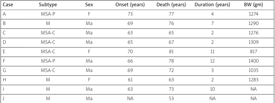
Table I. Subtypes, demographic data, and brain weights (BW) of the multiple system atrophy (MSA) cases studied
Case Subtype Sex Onset (years) Death (years) Duration (years) BW (gm)
A MSA-P F 73 77 4 1274
B M Ma 69 76 7 1290
C MSA-C Ma 63 65 2 1276
D MSA-C Ma 65 67 2 1309
E MSA-C F 70 81 11 817
F MSA-P Ma 66 78 12 1400
G MSA-C Ma 69 72 3 1035
H M F 61 63 2 1283
I M Ma 63 73 10 NA
J M Ma NA 53 NA NA
parkinsonism and cerebellar clinical signs, and could not easily be assigned to either the MSAC or MSAP subtypes. Hence, the two MSAP cases exhibited significant parkinsonism but no cerebellar ataxia, the four MSAC cases showed significant cerebellar ataxia but with minimum parkinsonism, while the four ‘mixed’ cases exhibited a combination of cer ebellar and parkinsonian symptoms. Only cases of the MSAC subtype exhibited evidence of cognitive impairment including memory impairment and con fusion.
Histological methods
After death, consent of the next of kin was obtained for brain removal following local Ethical Committee procedure and the 1964 Declaration of Helsinki (as revised in Edinburgh, 2000). A block of the right cerebellar cortex was taken from each case at the level of the superior cerebellar peduncle. Tissue was fixed in 10% phosphatebuffered for malsaline and embedded in paraffin wax. For quan titative analysis, sequential coronal 7µm sections were stained with haematoxylin and eosin (H/E) or immunohistochemistry (IHC) was performed using a nonphosphorylated polyclonal rabbit antibody (a116), after formic acid pretreatment, and at a dilu tion 1/3000, against the 116131 amino acid sequence of αsynuclein (kindly supplied by Dr D. Hanger). This type of antibody is regarded as one of the most effi cient available, especially for revealing the GCI, and is particularly recommended for diagnostic use [15].
The secondary antibody was biotinylated antirab bit antibody (DAKO diagnostics, Germany), used at a concentration of 1/200, which binds to the avidin peroxidase complex. Chromogen 3,3diaminoben zidene tetrahydrochloride was used to reveal the GCI. Immunolabelled sections were also stained with haematoxylin.
Morphometric methods
Variations in density of histological features were measured parallel to the edge of randomly selected Table II. Clinical features of the multiple system atrophy cases studied
Case Clinical features
A Rigidity and akinesia. No cerebellar ataxia. Nocturnal sweating, salivation, and frequent urination. Cognition preserved. Perseveration of speech
B Minimal parkinsonism. Balance problems. Impotence, urination and swallowing difficulties, impaired blinking. Cognition preserved. Depression
C Minimal parkinsonism. Restricted mobility and falls. Cerebellar ataxia. Urination difficulty. Cognition preserved. Dysarthria
D Some tremor of hands. Significant cerebellar ataxia. Dysarthria
E Minimal parkinsonism. Significant cerebellar ataxia affecting limbs and trunk. Memory impairment
F Significant parkinsonism with tremor and cogwheel rigidity. Urination and swallowing difficulties. Cognition preserved G Minimal parkinsonism. Significant cerebellar ataxia. Confusion
H Significant parkinsonism and cerebellar ataxia. Cognition preserved
I Some rigidity. Significant cerebellar ataxia. Swallowing difficulties. Cognition preserved. Dysarthria
J Significant parkinsonism and cerebellar ataxia. Dysphagia. Cognition preserved.
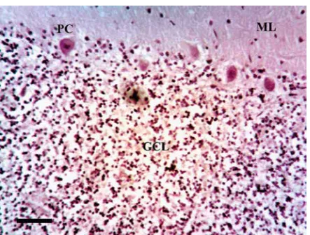
folia within each case (Fig. 1). Within each folium, a strip of cerebellar cortex 3200 to 4800 µm in length, starting at a randomly determined location, was studied with 6496, 50 × 250 µm sample fields arranged contiguously (Fig. 2) [3]. First, the sample field was positioned with the shorter dimension aligned along the upper edge of the GL at the base of the PC layer to quantify the density of PC and the pathology of the inner region of the ML. In each sam
ple field, the number of PC, distinct vacuoles great er than 5 µm in diameter, neurons, glial cell nuclei, and αsynucleinimmunoreactive inclusions were counted. Second, at the same position, the field was moved to sample the outer region of the GL, the short edge of the field aligned with the edge of the granule cells (Fig. 2). It was not possible in these preparations to differentiate between different cell types in the GL, e.g. granule cells, Golgi type II cells, glia, and a single count of cell density was made. Third, at the same location, the number of vacuoles, glial cell nuclei, and GCI (Fig. 3 and 4) were count ed in sample fields arranged along the white mat ter, the upper short edge of the sample field being aligned with the lower edge of the GL (Fig. 2).
Data analysis
Data analysis was carried out using STATISTICA software (Statsoft Inc., 2300 East 14th St, Tulsa, Ok, 74104, USA). First, densities of histological features in the ML, GL, and white matter were compared in MSA and control subjects using a ‘t’ test. Second, the spatial pattern of a histological feature, i.e. whether the feature was distributed randomly, regularly, or in clusters, was determined using the variance/mean (V/M) method described previously [1,2,5,6]. Third, spatial correlations between histological features along the folia were tested in each case using Pear son’s correlation coefficient (‘r’) [4]. Fourth, to study pathological variation among cases, the data were Fig. 2. Quantitative method of sampling the cer
ebellum hemisphere in multiple system atrophy (MSA) showing the 50 × 250 mm plots. (αsy nuclein immunohistochemistry, haematoxylin; Magnification bar = 50 mm). Arrow indicates surviving Purkinje cell. GCL – granule cell layer, ML – molecular layer, PC – Purkinje cell layer.
Fig. 3. Vacuolation in a case of multiple system atrophy (MSA) (αsynuclein immunohistochem istry, haematoxylin; magnification bar = 20 mm). GCL – granule cell layer, ML – molecular layer, PC – Purkinje cell layer.
analysed using principal components analysis (PCA) [11]. The result of a PCA is a plot of the ten MSA cas es in relation to the extracted PC in which distance between cases reflects their pathological similarity or dissimilarity. To correlate the location of a case on a PC axis with the numerical density of a specific histological feature, correlations (Pearson’s ‘r’) were calculated between the densities of each histologi cal feature and the factor loadings of cases on PC1 and PC2. Clinical features were also plotted onto the PCA to determine if cases were segregated accord ing to clinical symptoms in relation to PC1 and PC2.
Results
Pathological features observed in MSA included: (1) modest vacuolation of the ML in some cases and more extensive vacuolation of the GL, (2) loss of PC, and (3) GCI in white matter (Fig. 14). No αsynucle inimmunoreactive GCI or NCI were observed in the ML or GL. Mean density of vacuoles in the GL was significantly increased (t = 2.57, p < 0.05) and PC was decreased (t = 7.65, p < 0.001), in MSA compared with controls (Fig. 5). In addition, the mean density of vacuoles was significantly greater in the GCL com pared with the ML (t = 3.52, p < 0.01) but was similar to vacuole density in adjacent white matter (t = 2.01, p > 0.05).
Examples of the spatial patterns of histological features along the folia are shown in Fig. 6. The V/M of the PC was not significantly different to unity at any field size, suggesting a random distribution. The V/M of the vacuolation in the ML, however, revealed significant peaks at field sizes 100 mm and 400 mm, suggesting clustering at two scales in the tissue, i.e. vacuoles were clustered, the mean dimen sion of the clusters being equal to 100 mm, and they were regularly distributed along the folium, the smaller clusters being aggregated into larger clusters, 400 mm in diameter.
The spatial patterns of all histological features in each case are shown in Table III. Vacuoles were clustered in the ML in the majority of cases, regular spaced clustering of vacuoles along the folia being present in 4/10 (40%) cases. Similarly, neurons in the ML were clustered, a regular distribution of clusters being present in 6/10 (60%) cases. By contrast, gli al cell nuclei in the ML were randomly or regularly distributed. In the majority of cases, PCs were ran domly or regularly distributed and there were large gaps between surviving cells. In the GL, the vacuoles
and cell nuclei were clustered, a regular distribution of clusters being present. In the white matter, large clusters of vacuoles were present and the GCI and glial cell nuclei exhibited a regular distribution of clusters along the folia. The frequency of the differ ent types of spatial pattern was similar in the differ ent layers (c2 = 5.28, 6DF, p > 0.05).
Fig. 5. Mean densities (50 × 250 mm field, stan dard error of mean in parentheses) of histolog ical features (Vac – vacuolation, PC – Purkinje cells, GCI – glial cytoplasmic inclusions) in vari ous layers of the cerebellar cortex (ML – molecu lar layer, PC – Purkinje cell layer, GL – granule cell layer, WM – white matter) in ten cases of multi ple system atrophy (MSA) and ten control cases. 24
22 20 18 16 14 12 10 8 6 4 2 0
ML-Vac ML-N ML-Glia PC GL-Vac GL-Cells WM-Vac WM-GCI Histological feature
Control MSA
Mean density (50 × 250 µm field)
Fig. 6. Examples of the topographical patterns of the vacuolation and glial cell nuclei in the cerebellum in a case of multiple system atro phy (MSA) (Case A). *Significant variance/mean peaks.
4.0
3.5
3.0
2.5
2.0
1.5
1.0
0.5
0 200 400 600 800 1000 1200 1400 1600 1800 Field size (µm)
Molecular layer vacuolation Purkinje cells
V
Spatial correlations among histological features within and between layers are summarised in Table IV. The most notable correlations were: (1) in the ML of 5 cases, a negative spatial correlation between gli al cell nuclei and neurons, (2) in the GL of 5 cases,
a negative spatial correlation between cells and vac uolation, and (3) in the PC layer of 3 cases, a negative spatial correlation between PC and vacuoles. Histo logical features in different layers of the cerebellar cortex were not spatially correlated.
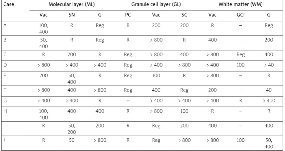
Table III. Spatial patterns of histological features in various layers of the cerebellar cortex in 10 cases of multiple system atrophy
Case Molecular layer (ML) Granule cell layer (GL) White matter (WM)
Vac SN G PC Vac SC Vac GCI G
A 100,
400
R Reg R 200 200 R – Reg
B 50,
400
R Reg R > 800 R 400 – 200
C R 200 R Reg > 800 400 > 800 Reg 400
D > 800 > 400 > 400 Reg > 400 > 800 > 400 100 > 40
E 200 50,
400
R Reg 100 R > 800 – R
F > 800 400 > 800 Reg 400 Reg 200 – 40
G > 400 > 400 R – > 400 > 400 > 400 R > 400
H 100,
400
400 400 R > 800 100 R – R
I R 50,
200
200 R Reg 200 400 – 400
J R 50 > 800 R Reg > 800 > 800 100 50,
400 Comparison of spatial patterns (c2 contingency table): ML vs. GL vs. WM, c2 = 5.28 (6DF, p > 0.05)
Vac – vacuolation, SN – surviving neurons, G – glial cell nuclei, PC – Purkinje cells, SC – surviving cells, GCI – glial cytoplasmic inclusions
Table IV. Frequency of correlations (Pearson’s ‘r’) between histological features within and between layers of the cerebellum in multiple system atrophy. Figures indicate the number of cases in which a positive (+) or negative (–) correlation was recorded
X variable
Y MLV MLN MLG PC GLV GLC WMV WMGCI WMG
MLV – 2(+) – 3(–) 1(–) – – – 1(+)
MLN – 5(–) 1(+) 1(+) – 1(+) 1(+) 1(+)
MLG – 1(–) – – 1(+) – –
PC – 1(+) – – – 1(+)
GLV – 5(–) – 1(+) 2(+)
GLC – 1(+)2(–) – –
WMV – – 1(+)2(–)
WMGCI – 1(+)
WMG –
A PCA of the data resulted in the extraction of two PC accounting in total for 87% of the total vari ance (PC1 = 72%, PC2 = 15%). A plot of the 10 cases in relation to PC1 and PC2 is shown in Fig. 7. MSAC cases were located at the upper right of the plot and the MSAP and cases of mixed pathology to the left of the plot. In addition: (1) PC1 was negative ly correlated with the density of vacuoles in the ML (r = –0.66, p < 0.05) and (2) PC2 was negatively cor related with the density of cells in the GL (r = –0.81, p < 0.05) and positively correlated with the density of vacuoles in the GL (r = 0.82, p < 0.001). In addition, cases to the right of the plot exhibited significant cerebellar ataxia but with minimum parkinsonism while those to the left of the plot exhibited a greater degree of parkinsonism, i.e. rigidity, akinesia, tremor, and less cerebellar ataxia.
Discussion
In the 10 MSA cases studied, a significant loss of PC and vacuolation of the GL were the most con sistent pathological changes compared with controls [26,30,37]. Some vacuolation was also evident in the ML and white matter but at significantly lower levels than the GCL, and it did not differ quantita tively from controls. No αsynucleinimmunoreactive inclusions were observed in control cases or in the grey matter of any MSA case, but such structures have been reported previously in the ML located into GFAPimmunoreactive radial processes of Bergmann glia [30,32]. αSynucleinimmunoreactive GCI were present in white matter, but not in all cases. GCI have been observed in other subcortical white mat ter tracts in MSA, including the external and internal capsules and central tegmental tract [9].
The vacuoles and surviving neurons were fre quently clustered, and in some cases the clusters were regularly distributed relative to the edge of the folia, a pattern evident in both ML and GL. In addi tion, significant gaps were observed between PC perikarya, surviving PC often being regularly distrib uted, which suggests loss of clusters of PC. These results are consistent with a topographic pattern of the cerebellar pathology in MSA, which has also been observed in the cerebellum in the sporadic [7] and variant subtypes of CreutzfeldtJakob disease (CJD) [10]. A topographic loss of PC may also occur in NiemannPick type C disease, in which there is a complex pattern of cell loss in the cerebellum, with surviving PC being aligned in strips [32]. Further loss
of PC then occurs as the disease develops, resulting in large gaps between surviving cells similar to those observed in MSA.
There was a negative correlation between cells and vacuoles in the GL suggesting that vacuolation replaces lost neurons. Furthermore, there was a neg ative correlation between the densities of neuronal perikarya in the ML and glial cell nuclei consistent with gliosis. A negative spatial correlation was also observed between individual PCs and clusters of vac uoles in the ML of three MSAC cases, which could represent a more specific cerebellar pathology in MSA. These vacuoles may have developed in relation to the dendritic trees of the PC, which branch in a plane per pendicular to that of the section, the climbing fibres that ramify over individual PC, or the parallel fibres that ramify in the plane of the section and which are in contact with many adjacent PCs [10].
Although the number of cases of MSA is small, the PCA suggested some variations in quantitative pathology among cases. First, PC1 was negatively correlated with the density of vacuoles in the ML. Although there was no significantly increased vacu ole density overall in the MSA cases, the vacuolation in the ML did vary among cases with more significant
Fig. 7. Principal components analysis (PCA) of ten cases of multiple system atrophy (MSA) based on the densities of all histological fea tures in the cerebellum. A plot of the cases in relation to PC1 and PC2 (MSAC – cerebellar sub type, MSAP – Parkinsonian subtype, M – mixed pathology). Arrows PC1 was negatively correlat ed with the density of vacuoles in the molecu lar layer (ML Vac) and PC2 negatively correlated with the density of cells in the granule cell layer (GL Cells –) and positively correlated with the density of vacuoles in the GL (GL Vac +). 0.8
0.6
0.4
0.2
0
–0.2
–0.4
–0.6
–0.98 –0.94 –0.90 –0.86 –0.82 –0.78 –0.74 –0.70 PC1
PC2
M
M
M M
MSA-P
MSA-P MSA-C
MSA-C
MSA-C
vacuolation in cases that exhibited more significant parkinsonism compared with those with cerebellar ataxia. Second, PC2 was negatively correlated with cell density and positively correlated with vacuole density in the GL. This result suggests that increased vacuolation and cell loss in the GL may be a more significant feature of the MSAC subtype.
The cerebellum receives input from several sourc es (Fig. 8): (1) the spinal cord (posterior spinocere bellar tract), reticular formation nuclei (reticulocer ebellar tract), and pontine nuclei (pontocerebellar tract), which relay signals to the cerebellum, via the inferior and superior cerebellar peduncles, to the large diameter, rapidly conducting mossy fibres, the synaptic endings terminating in complex glomeruli; (2) climbing fibres that originate in the inferior olive (olivocerebellar tract) and which synapse directly on to the PC; and (3) fibres from the white matter
which enter the GL and course parallel to the pia mater before synapsing with the PC [14]. αSynu cleinimmunoreactive GCIs have been observed in white and grey matter regions, which provide these inputs to the cerebellum in MSA, e.g. the pontocer ebellar and reticulocerebellar tracts [13], and in the present study they were also observed in cerebellar white matter. This pathology has also been observed in motor tracts providing both the input and output pathways of the cerebellum, e.g., the corticopontine, cortical bulbar, corticospinal, and spinoreticular tracts. In addition, significant densities of inclusions have been observed in precerebellar nuclei such as the inferior olivary nucleus [8], lateral reticular nucle us, interfascicular nucleus, and the nucleus of Roller in MSA [13].
Hence, αsynuclein pathology spreading via cer ebellar connections [35] could result in: (1) cell loss
Cerebral cortex/Limbic system
Ventrolateral thalamus Pons
PF ML
PC
GL
MCP
PST LVT RCT
ICP CVT OCF
DN
AST
SCP
CDThT
CDRT to red nucleus FBT to medulla GM
GC
PCT
(+) (–)
(+) CF
SC
BC (–)
es and vacuolation in the GL, (2) loss of parallel and climbing fibres, (3) a reduction in the degree of facil itation of surviving PC, (4) a reduction in the degree of inhibitory control by PC of the dentate nucleus (DN), and (5) a reduction of fine tuning of the cere bral output via the cerebellodentatothalamic tract, which leaves the cerebellum as the superior cerebel lar peduncle and connects the cerebellum to various regions such as the red nucleus (cerebellodenta torubral tract), medulla (fastigiobulbar tract), and the cerebral cortex/limbic system, the latter via the ventrolateral thalamus. This pathology could poten tially influence a variety of clinical symptoms report ed in MSA, including dysfunction of motor activity, the fine timing of events, sensory analysis, feeding behaviour, the modulation of cognition, and in the regulation of emotions [22].
Conclusions
Cerebellar pathology in MSA may affect all layers of the cerebellar hemisphere, but cell losses and vac uolation in the GL and loss of PC were the most sig nificant pathological changes in the cases studied. There was evidence of a topographic distribution of pathological change, which could reflect the spread of αsynuclein pathology via anatomical connec tions. Cerebellar pathology may ultimately influence a variety of clinical symptoms in MSA, especially in the MSAC subtype. Nevertheless, only 10 cases of this rare disorder were studied quantitatively, and these observations should be repeated on a larger series of wellcharacterised MSA cases.
Acknowledgements
Dr Diane Hanger is thanked for the generous donation of αsynuclein antibody, and Heidi Barnes and Mavis Kibble for their excellent technical assis tance.
Disclosure
Authors report no conflict of interest.
References
1. Armstrong RA. The usefulness of spatial pattern analysis in understanding the pathogenesis of neurodegenerative disor-ders, with particular reference to plaque formation in Alzhei-mer’s disease. Neurodegeneration 1993; 2: 73-80.
2. Armstrong RA. Analysis of spatial patterns in histological sec-tions of brain tissue. J Neurosci Meth 1997; 73: 141-147.
3. Armstrong RA. Quantifying the pathology of neurodegenera-tive disorders: quantitaneurodegenera-tive measurements, sampling strate-gies and data analysis. Histopathology 2003; 42: 521-529. 4. Armstrong RA. Measuring the degree of spatial correlation
between histological features in thin sections of brain tissue. Neuropathology 2003; 23: 245-253.
5. Armstrong RA. Methods of studying the planar distribution of objects in histological sections of brain tissue. J Microsc (Oxf) 2006; 221: 153-158.
6. Armstrong RA. Measuring the spatial arrangement patterns of pathological lesions in histological sections of brain tissue. Folia Neuropathol 2007; 44: 229-237.
7. Armstrong RA, Cairns NJ. Spatial patterns of the pathological changes in the cerebellar cortex in sporadic Creutzfeldt-Jakob disease (sCJD). Folia Neuropathol 2003; 41: 183-189.
8. Armstrong RA, Lantos PL, Cairns NJ. A quantitative study of the pathological changes in ten patients with multiple system atro-phy (MSA). J Neural Transm 2004; 111: 485-495.
9. Armstrong RA, Cairns NJ, Lantos PL. A quantitative study of the pathological changes in white matter in multiple system atro-phy. Neuropathology 2007; 27: 221-227.
10. Armstrong RA, Ironside JW, Lantos PL, Cairns NJ. A quantitative study of the pathological changes in the cerebellum in 15 cases of variant Creutzfeldt-Jakob disease (vCJD). Neuropathol Appl Neurobiol 2009; 35: 36-45.
11. Armstrong RA, Ellis W, Hamilton RL, Mackenzie IRA, Hedreen J, Gearing M, Montine T, Vonsattel J-P, Head E, Lieberman AP, Cairns NJ. Neuropathological heterogeneity in frontotemporal lobar degeneration with TDP-43 proteinopathy: a quantitative study of 94 cases using principal components analysis. J Neural Transm 2010; 117: 227-239.
12. Bower JH, Maraganore DM, McDonnell K, Rocca WA. Incidence of progressive supranuclear palsy and multiple system atrophy in Olmstead County, Minnesota, 1976-1990. Neurology 1997; 49: 1284-1288.
13. Braak H, Rub U, Del Tredici K. Involvement of pre-cerebellar nuclei in multiple system atrophy. Neuropathol Appl Neurobiol 2003; 29: 60-76.
14. Brodal A. Neurological anatomy. 3rd ed. Oxford University Press, New York, Oxford 1981.
15. Croisier E, Mres DE, Deprez K, Goldring K, dexter DT, Pearce RKB, Graeber MB, Roncaroli F. Comparative study of commercially available anti-α-synuclein antibodies. Neuropath Appl Neuro-biol 2006; 32: 351-356.
16. Dickson DW, Liu WL, Liu WK, Yen SH. Multiple system atrophy: a sporadic synucleinopathy. Brain Pathol 1995; 9: 721-732. 17. Dickson DW, Liu WK, Hardy J, Farrar M, Mehta N, Uitti R, Mark M,
Zimmerman T, Golbe L, Sage J, Sima A, d’Amato C, Albin R, Gil-man S, Yen SH. Widespread alterations of alpha-synuclein in multiple system atrophy. Am J Pathol 1999; 155: 1241-1251. 18. Ehot V, Brieger P, Broich K, Marneros A. Psychotic symptoms as
initial manifestation of a multiple system atrophy. Fortshritte der Neurol Psych 1999; 67: 104-107.
Con-sensus statement on the diagnosis of multiple system atrophy. J Auto Nerv Syst 1998; 74: 189-192.
20. Gilman S, Low PA, Quinn N, Albanese A, Ben-Schlomo Y, Fowl-er CJ, Kaufman H, KlockgethFowl-er T, Lang AE, Lantos PL, Litvan I, Mathias CJ, Oliver E, Robertson D, Schatz I, Wenning GK. Con-sensus statement on the diagnosis of multiple system atrophy. J Neurol Sci 1999; 163: 94-98.
21. Gilman S, Wenning GJK, Low PA, Brooks DJ, Mattias CJ, Troja now-ski JQ, Wood NW, Colosima C, Durr A, Fowler CJ, Kaufmann H, Klockgether T, Lees A, Poese W, Quinn N, Revesz T, Robertson D, Sandroni T, Seppi K, Vidailhet M. Second consensus statement on the diagnosis of multiple system atrophy. Neurology 2008; 71: 670-676.
22. Ioannides AA, Fenwick PBC. Imaging cerebellum activity in real time with magnetoencephalographic data. In: Creating Coordi-nation in the Cerebellum. Prog in Brain Res 2005; 148: 139-150. 23. Konogaya M, Sakai M, Matsuoka Y. Konogaya Y, Hashzume Y.
Multiple system atrophy with remarkable frontal lobe atrophy. Acta Neuropathol 1999; 97: 423-428.
24. Kovacs GG, Milenkovic IJ, Preusser M, Budka H. Nigral burden of alpha-synuclein correlates with striatal dopamine deficit. Move Disord 2008; 23: 1608-1612.
25. Lantos PL. Cellular pathology of multiple system atrophy: a re- view. J Neurol Neurosurg Psychiatr 1994; 57: 129-133.
26. Mori F, Piao YS, Hayashi S, Fujiwara H, Hasegawa M, Yoshimo- to M, Iwatsubo T, Takahashi H, Wakabayashi K. Alpha-synuclein accumulates in Purkinje cells in Lewy body disease but not in multiple system atrophy. J Neuropathol Exp Neurol 2003; 62: 812-819.
27. Oertel WH, Bandmann O. Multiple system atrophy. J Neural Transm (Suppl) 1999; 56: 155-164.
28. Papp MI, Kahn JE, Lantos PL. Glial cytoplasmic inclusions in the CNS of patients with multiple system atrophy (striatonigral degeneration, olivopontocerebellar atrophy and Shy-Drager syndrome). J Neurol Sci 1989; 94: 79-100.
29. Papp MI, Lantos PL. The distribution of oligodendroglial inclu-sions in multiple system atrophy and its relevance to clinical symptomology. Brain 1994; 117: 235-243.
30. Park SH, Becker-Catania S, Gatti RA, Crandall BF, Emelin JK, Vinters HV. Congenital olivopontocerebellar atrophy: report of two siblings with paleo and neocerebellar atrophy. Acta Neuro-pathol 1998; 96: 315-321.
31. Piao YS, Mori F, Hayashi S, Tanji K, Yoshimoto M, Kakita A, Waka-bayashi K, Takahashi H. Alpha-synuclein pathology affecting Bergmann glia of the cerebellum in patients with alpha-synu-cleinopathies. Acta Neuropathol 2003; 105: 403-409.
32. Sarna JR, Larouche M, Marzban H, Sillitoe RV, Rancourt DE, Hawkes R. Patterned Purkinje cell degeneration in mouse mod-els of Niemann-Pick type C disease. J Comp Neurol 2003; 456: 279-291.
33. Sebeo J, Hof PR, Perl DP. Occurrence of alpha-synuclein patholo-gy in the cerebellum of Guamanian patients with parkinsonism dementia complex. Acta Neuropathol 2004; 107: 497-503. 34. Spillantini MG, Crowther RA, Jakes R, Cairns NJ, Lantos PL,
Goedert M. Filamentous α-synuclein inclusions link multiple system atrophy with Parkinson’s disease and dementia with Lewy bodies. Neurosci Lett 1998; 251: 205-208.
35. Steiner JA, Angot E, Brunden P. A deadly spread: cellular me ch-anisms of α-synuclein transfer. Cell Death and Differ 2011; 18: 1425-1433.