RESEARCH
Toe-to-room temperature gradient
correlates with tissue perfusion and predicts
outcome in selected critically ill patients
with severe infections
Simon Bourcier
1,2, Claire Pichereau
1,2, Pierre‑Yves Boelle
3, Safaa Nemlaghi
1,4, Vincent Dubée
1,2, Gabriel Lejour
1,
Jean‑Luc Baudel
1, Arnaud Galbois
1,4, Jean‑Rémi Lavillegrand
1, Naïke Bigé
1, Jalel Tahiri
1, Guillaume Leblanc
1,5,
Eric Maury
1,2,6, Bertrand Guidet
1,2,6and Hafid Ait‑Oufella
1,2,7*Abstract
Background: Microcirculatory disorders leading to tissue hypoperfusion play a central role in the pathophysiology of organ failure in severe sepsis and septic shock. As microcirculatory disorders have been identified as strong predictive factors of unfavourable outcome, there is a need to develop accurate parameters at the bedside to evaluate tissue perfusion. We evaluated whether different body temperature gradients could relate to sepsis severity and could pre‑ dict outcome in critically ill patients with severe sepsis and septic shock.
Method: We conducted a prospective observational study in a tertiary teaching hospital in France. During a 10‑month period, all consecutive adult patients with severe sepsis or septic shock who required ICU admission were included. Six hours after initial resuscitation (H6), we recorded the hemodynamic parameters and four temperature gradients: central‑to‑toe, central‑to‑knee, toe‑to‑room and knee‑to‑room.
Results: We evaluated 40 patients with severe sepsis (40/103, 39 %) and 63 patients with septic shock (63/103, 61 %). In patients with septic shock, central‑to‑toe temperature gradient was significantly higher (12.5 [9.2; 13.8] vs 6.9 [3.4; 12.0] °C, P < 0.001) and toe‑to‑room temperature gradient significantly lower (1.2 [−0.3; 5.2] vs 6.0 [0.6; 9.5] °C,
P < 0.001) than in patients with severe sepsis. Overall ICU mortality rate due to multiple organ failure (MOF) was 21 %. After initial resuscitation, toe‑to‑room temperature gradient was significantly lower in patients dead from MOF than in the survivors (−0.2 [−1.1; +1.3] °C vs +3.9 [+0.5; +7.2] °C, P < 0.001) and the difference in gradients increased during the first 24 h. Furthermore, toe‑to‑room temperature gradient was related to tissue perfusion parameters such as arterial lactate level (r=−0.54, P < 0.0001), urine output (r= 0.37, P= 0.0002), knee capillary refill time (r=−0.42,
P < 0.0001) and mottling score (P= 0.001).
Conclusions: Toe‑to‑room temperature gradient reflects tissue perfusion at the bedside and is a strong prognosis factor in critically ill patients with severe infections.
Keywords: Sepsis, Shock, Microcirculation, Outcome, Temperature gradient
© 2016 The Author(s). This article is distributed under the terms of the Creative Commons Attribution 4.0 International License (http://creativecommons.org/licenses/by/4.0/), which permits unrestricted use, distribution, and reproduction in any medium, provided you give appropriate credit to the original author(s) and the source, provide a link to the Creative Commons license, and indicate if changes were made.
Open Access
*Correspondence: [email protected]
1 Service de réanimation médicale, Hôpital Saint‑Antoine, Assistance Publique‑Hôpitaux de Paris (AP‑HP), 184 rue du Faubourg Saint‑Antoine, 75571 Paris Cedex 12, France
Background
During the last decade, fundamental research and intra-vital microscopy in human helped to understand the unique pathophysiology of severe sepsis and septic shock compared with other types of shock. Those researches highlighted two key points. First, it demonstrated the heterogeneity and the complexity of microcirculatory disorders in response to bacterial invasion, such as modi-fications in vasomotor tone, activation of the coagulation cascade and increased platelet–leucocyte interactions [1]. Second, it demonstrated the discrepancy between global hemodynamic parameters (such as arterial blood pressure) and microcirculatory blood flow [2, 3]. Studies also have reported a decrease in microcirculatory blood flow in patients with septic shock [4], even when global hemodynamic parameters seemed adequate [5]. Moreo-ver, microcirculatory alterations have been considered to be a stronger determinant of outcome than global hemo-dynamic parameters [6]. These accumulating pathophysi-ological and epidemipathophysi-ological evidences explained why the last conference of European experts excluded arterial blood pressure from the definition of shock but stressed the identification of microvascular alterations through the detection of tissue hypoperfusion [7].
As microcirculatory disorders have been identified as strong predictive factors of unfavourable outcome, there is a need to develop accurate parameters at the bedside to evaluate tissue perfusion [8]. We previously reported that the mottling extension [9, 10] and the capillary refill time (CRT) [11] are strongly related to other tissue perfusion parameters (i.e. arterial lactate level and urine output) and strongly predictive of ICU mortality in patients with septic shock. However, their use is limited in patients with dark skin. On the contrary, central temperature and temperature gradients between different body compart-ments can be measured in all patients no matter the col-our of their skin and have been proposed as parameters to evaluate peripheral perfusion in critically ill patients. In a cohort of patients who required ICU admission for trauma, sepsis and surgical complications, Kaplan et al. [12] reported that patients with cold extremities, evalu-ated in a subjective manner, had a higher arterial lac-tate level and a lower SVO2 than patients with warm
extremities. Fifty years ago, Joly et al. [13] studied toe-to-room temperature gradient in a mixed ICU popula-tion (patients with cardiogenic, hypovolemic or septic shock and shock due to poisoning). They reported a significantly lower toe-to-room temperature gradient in ICU non-survivors than in ICU survivors. However, tem-perature gradient has never been specifically and objec-tively quantified during septic shock, a distributive acute circulatory failure where microcirculatory alterations and endothelial dysregulation predominate. The aim of
this study was to evaluate the relationship between perature gradients and sepsis severity and whether tem-perature gradients could predict outcome in critically ill patients with severe infections.
Methods
We conducted a prospective observational study in an 18-bed ICU at a tertiary teaching hospital in France. Dur-ing a 10-month period, we included all adult patients (≥18 years of age) who required ICU admission for a severe sepsis or septic shock (according to the 2001 SCCM/ESICM/ACCP/ATS/SIS International Sepsis Definitions Conference) from any causes [14]. Patients could be admitted from the emergency department or the medical wards. Patients with hypothermia (defined as central temperature <35 °C) were excluded. Patients with severe sepsis were included at ICU admission, and patients with septic shock were included when vasopres-sors were required (within 24 h of admission). In septic shock patients, vasopressor initiation was defined as H0.
Protocol for the management of patients
Management of patients with severe sepsis and sep-tic shock was guided by our local protocol, adapted from international guidelines [15]. In patients with sep-tic shock, intravenous volume expansion was provided to achieve predefined endpoints: pulse pressure varia-tion <13 % [16], no response to passive leg raising [17] or no respiratory variations of the inferior vena cava diam-eter [18]. Norepinephrine was used in a stepwise manner to achieve predefined endpoints: mean arterial pressure (MAP) ≥65 mmHg and urine output ≥0.5 mL/kg/h. All patients were investigated with transthoracic echocar-diography (Vivid 7 Dimension’06, GE, Healthcare®). When a cardiac dysfunction (left ejection fraction <30 % by Simpson’s biplane methodology) was identified, an inotropic therapy was introduced and/or epinephrine replaced norepinephrine. Ventilation support was pro-vided when needed. If required, patients were sedated with propofol and/or midazolam and analgesia pro-vided with sufentanil. Use of low doses hydrocortisone (200 mg/day) was considered when there was persistence of vasopressors requirement despite a perceived ade-quate intravascular volume. Glycemic control and venous thrombosis prophylaxis were provided according to Sur-viving Sepsis Campaign Guidelines [15].
Data collection
[20], primary site of infection, mode of ventilation and vasopressors use. We collected global hemodynamic parameters (MAP, heart rate (HR) and cardiac index) and microcirculatory dysfunction and organ perfusion parameters (arterial lactate level, urine output, mottling score and knee CRT) at study inclusion and at 6, 12 and 24 h following inclusion. Evaluation of mottling score and knee CRT was not done in patients with dark skin.
Toe and knee skin temperature was measured using a skin temperature sensor (STS-400 Level1®, Smiths Medi-cal, Rockland, MA, USA) applied at patient inclusion and remained for the next 24 h. Central body tempera-ture was measured with an electronic rectal thermometer (SureTemp® Plus 692 Electronic Thermometer, Welch Allyn®), and room temperature was also recorded. The four temperature gradients (°C) were calculated as fol-lows: central-to-toe (central T–toe T), central-to-knee (central T–knee T), toe-to-room (toe T–room T) and knee-to-room (knee T–room T). Outcome at ICU dis-charge was recorded by the physician at bedside who was blinded to the temperature gradients. Patients were classified as survivors, dead from multiple organ failure (deaths from MOF group) or dead from secondary com-plications or do-not-resuscitate orders (DNR) (late deaths group). The MOF deaths group consists in patients who had not recovered from MOF secondary from their severe sepsis or septic shock and died following the bac-terial injury/invasion. The late deaths group was charac-terized by hemodynamic improvement and vasopressors weaning, but these patients ultimately died from second-ary complications or do-not-resuscitate orders (DNR).
Statistical analysis
Patient characteristics were expressed as median (25–75th percentiles) or percentages as appropri-ate. Differences among groups were assessed using the Kruskal–Wallis test with post hoc Mann–Whitney analy-sis with adjustment for multiple comparisons. Associa-tion analyses were performed using the Chi-squared test. Correlation analyses were performed using the Spearman test. When correlation was significant, a linear regression model was fit to the data. The toe-to-room temperature gradient compared with mottling score was tested using the Kruskal–Wallis test. Toe-to-room temperature gra-dient was analysed as a function of time using analysis of variance for repeated measures. The model included terms for the outcome (survivors, deaths from MOF and late deaths), the time (continuous) and an interac-tion term between time and outcome. Receiver operat-ing characteristics (ROC) curves were constructed to compare the accuracy of gradient temperature in the prediction of death from MOF. We used logistic regres-sion to model MOF death as a function of patient’s
characteristics. All tests were computed with the R soft-ware. Significance was defined as a two-sided P < 0.05.
Temperature gradients were recorded blindly by an independent physician who did not participate in patient’s care. The protocol was approved by our institu-tion’s ethical committee Comité de Protection des Person-nes (CPP Saint-Louis, Paris, France).
Results
During a 10-month period, 103 consecutive adult patients who required ICU admission for severe infec-tions were included in the study (Table 1). The median time between ICU admission and inclusion was 2 [0; 5] h. There were 40 patients with severe sepsis (40/103, 39 %) and 63 patients with septic shock (63/103, 61 %). The most prevalent primary sites of infection were the lungs (43 %) and the abdomen (21 %). Most of the patients had comorbidities such as cirrhosis, malignancy or dia-betes. Patients with septic shock had higher SAPS II and SOFA scores and required more frequently support
Table 1 Characteristics of patients
SAPS II was recorded at H24, Simplified Acute Physiology Score. Data are expressed as number, percentage or median and interquartiles (IQRs)
Characteristics
of patients at H6 Severe sepsis (n = 40) Septic shock (n = 63) P value
N 40 63 –
Age (years), median
and IQRs 65 [56; 73] 68 [60; 83] NS Male gender, (%) 57 66 NS
Comorbidities (%)
Diabetes 25 20 NS
Cirrhosis 5 21 <0.05 Vascular disease 25 32 NS Solid malignancies 22 22 NS Haematologic
malignancies 22 13 NS
Primary site of infection (%)
Lung 43 43 NS
Abdomen 25 19 NS
Urinary tract 7 14 NS Soft tissue 15 13 NS
Other 10 11 NS
SAPS II, median and
IQRs 37 [28; 46] 60 [46; 69] <0.001 Mechanical ventila‑
tion (%) 28 75 <0.001
Norepinephrine
n − 58 −
Dose [μg/kg/min] − 0.5 [0.2; 0.9]
Epinephrine
n − 1 −
therapies (such as mechanical ventilation) than patients with severe sepsis. All patients with septic shock received vasopressor therapy, but four of them were weaned in the first 6 h after inclusion. The vasopressor mainly used was norepinephrine (at H6, 58/59 patients, median dose 0.5 [0.2; 0.9] µg/kg/min) and one patient received epineph-rine (at H6, 0.9 µg/kg/min). Thirteen patients had dark skin leaving 90 patients for the evaluation of mottling score and knee CRT.
Of the 63 patients with septic shock, 20 patients (20/63, 32 %) died within the first days from multiple organ fail-ure and shock (deaths from MOF group). Forty-three patients (43/63, 68 %) survived the episode of initial sep-tic shock and were weaned from vasopressors, but 11 of them died later from various causes (11/63, 17 %, late deaths group, Additional file 1: Figure S1). Those patients died from acute myocardial infarction (n = 2), stroke (n = 1), bleeding (n = 2), pulmonary aspiration (n = 1), secondary infections (n = 2) and following a withdrawal of life-sustaining therapies (n = 3). Thirty-three patients were discharged alive from ICU (33/63, 52 %). In the severe sepsis group, two patients developed a shock after 24 h of inclusion and died from multiple organ fail-ure. Six patients died later (28 [18; 44] days) from others causes (Additional file 1: Figure S1).
Hemodynamic parameters assessment according to sepsis severity
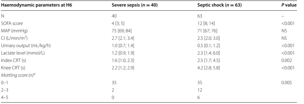
Hemodynamic parameters at 6 h according to sepsis severity are presented in Table 2. After initial resuscita-tion, mean arterial blood pressure and cardiac index were not different between the severe sepsis and the sep-tic shock groups. However, tissue perfusion parameters were significantly different between groups (Table 2).
Compared to patients with severe sepsis, patients with septic shock had a significantly lower urine output, higher arterial lactate level, higher knee CRT and a larger mottling score. Central-to-knee and knee-to-room tem-perature gradients were not different between groups. However, central-to-toe temperature gradient was sig-nificantly higher (12.5 [9.2; 13.8] vs 6.9 [3.4; 12.0] °C,
P < 0.001) and toe-to-room significantly lower (1.2 [−0.3; 5.2] vs 6.0 [0.6; 9.5] °C, P < 0.001) in patients with septic shock (Fig. 1).
Hemodynamic parameters assessment according to ICU outcome
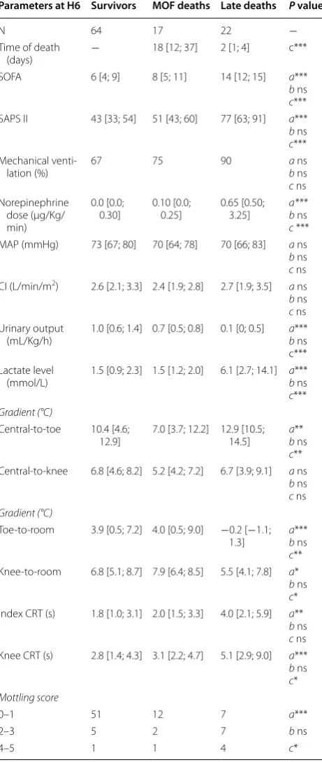
Hemodynamic parameters at H6 according to ICU out-come (survivors, deaths from MOF group and late deaths group) are presented in Table 3. Patients in the survivors and late deaths groups were similar in terms of severity, organ failures and vasopressor use. However, patients in the MOF deaths group were different than patients in the survivors and late deaths groups. Patients who died from an early MOF had a more severe disease as illustrated by a higher SOFA score (14 [12, 15] vs 6 [4, 9], P < 0.001), a higher SAPS II (77 [63; 91] vs 43 [33; 54], P < 0.001) and a higher need in vasopressors in comparison with sur-vivors (Table 3). Mean arterial blood pressure and car-diac index were not different according to ICU outcome. Regarding tissue perfusion parameters and temperature gradients, there was no difference between the survivors and late deaths groups. However, when compared with survivors, patients in the deaths from MOF group had a significantly lower urine output (0.1 [0; 0.5] vs 1.0 [0.6; 1.4] mL/kg/h, P < 0.001), higher arterial lactate level (6.1 [2.7; 14.1] vs 1.5 [0.9; 2.3] mmol/L, P < 0.001), higher knee CRT (5.1 [2.9; 9.0] vs 2.8 [1.4; 4.3] s, P < 0.001), higher
Table 2 Haemodynamic parameters of patients with severe sepsis and septic shock
Data are expressed as number or median and IQRs
SOFA Sequential Organ Failure Assessment, MAP mean arterial pressure, CI cardiac index, CRT capillary refill time a Patients with dark skin were excluded
Haemodynamic parameters at H6 Severe sepsis (n = 40) Septic shock (n = 63) P value
N 40 63 –
SOFA score 4 [3; 5] 12 [8; 14] <0.001
MAP (mmHg) 75 [69; 84] 71 [67; 76] NS
CI (L/min/m2) 2.7 [2.1; 3.4] 2.5 [2.0; 3.0] NS
Urinary output (mL/kg/h) 1.0 [0.7; 1.4] 0.5 [0.1; 1.2] <0.001 Lactate level (mmol/L) 1.2 [0.9; 1.9] 2.3 [1.4; 6.0] <0.001 Index CRT (s) 1.6 [1.0; 2.3] 2.5 [1.7; 4.5] 0.002 Knee CRT (s) 2.2 [1.2; 2.9] 4.2 [2.8; 5.8] <0.001
Mottling score (n)a
0–1 35 35 0.005
2–3 2 12
index CRT (4.0 [2.1;5.9] vs 1.8 [1.0; 3.1] s, P = 0.002) and larger mottling score (P < 0.001).
Central-to-knee temperature gradient was not statisti-cally different according to the ICU outcome (Table 3). However, central-to-toe temperature gradient was sig-nificantly larger and knee-to-room and toe-to-room tem-perature gradients were significantly lower in the deaths from MOF group in comparison with survivors. The dif-ference was more pronounced for toe-to-room gradient (−0.2 [−1.1; +1.3] for death from MOF group and +3.9 [+0.5; +7.2] °C for survivors, P < 0.001). In a multivari-able regression, we found that toe-to-room temperature gradient remained associated with death from MOF (OR 0.7 [0.5, 0.9], P < 0.001) after adjustment on acute severity, measured by the SOFA score (OR 1.5 [1.2, 1.8],
P < 0.001), and after adjustment on morbidity, as meas-ured by the presence of malignancy (OR 4.3 [1.1, 16.7],
P = 0.03) (Table 4).
In the analysis of variance, the difference in toe-to-room gradient between the three groups was maintained over time, with a gradient decreasing by an average of 0.04 ± 0.05 °C/h in the deaths from MOF group but increasing by 0.08 ± 0.05 °C/h (P < 0.03) in the survivors (vs deaths from MOF, P = 0.001) (Fig. 2) and increasing by 0.06 ± 0.07 °C/h in the late deaths group (vs deaths from MOF, P = 0.006). The toe-to-room temperature gradient was predictive of death due to MOF at H6 with an area under the curve (AUC) of 0.76 [0.65; 0.86]. Pre-dictive value increased over time, at H12 the AUC was 0.83 [0.71; 0.95] and at H24 it reached 0.84 [0.74; 0.94]. At H24, a threshold of toe-to-room temperature gradient of 1.75 °C was predictive of death from MOF with a sen-sitivity of 75 % (CI 95 %, 53; 98) and a specificity of 75 % (CI 95 %, 62; 85).
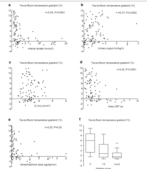
We also analysed correlations between toe-to-room temperature gradient and other hemodynamic variables
both measured at H6 after initial resuscitation (Fig. 3) in a pooled analysis of patients with severe sepsis and patients with septic shock. The toe-to-room temperature gradient did not correlate with cardiac index but correlated weakly with vasopressor doses (r = −0.20, P = 0.05). In contrast, we observed a significant relationship between toe-to-room temperature gradient and tissue perfusion variables such as arterial lactate level (r = −0.54, P < 0.0001), urine output (r = 0.37, P = 0.0002), knee CRT (r = −0.42,
P < 0.0001) and mottling score (P = 0.001) (Fig. 2). When analysing only patients with septic shock, we found the same correlation between toe-to-room temperature gra-dient and arterial lactate level, urine output, knee CRT and mottling score. However, the correlation between toe-to-room temperature gradient and vasopressor doses was not significant (data not shown).
Discussion
In this prospective observational study of 103 critically ill patients with severe infections, central-to-toe and toe-to-room temperature gradients were statistically differ-ent between patidiffer-ents with severe sepsis and patidiffer-ents with septic shock. After initial resuscitation, central-to-toe, toe-to-room and knee-to-room temperature gradients were associated with death from MOF. The toe-to-room temperature gradient showed the largest difference between survivors and patients dead from MOF. This difference increased during the first 24 h of ICU admis-sion. Finally, the toe-to-room temperature gradient sig-nificantly correlated with tissue perfusion markers such as urine output, arterial lactate level, knee CRT and mot-tling score.
Temperature gradients are available clinical tools, but their use had not yet been standardized. This is the first study reporting an objective evaluation of tem-perature gradients in a selected population of patients
with severe infections. Kaplan et al. [12] subjectively described in a mixed surgical ICU population that patients with cold extremities had higher arterial lactate levels. Lima et al. [21] proposed a subjective combined approach of tissue perfusion and defined an abnormal peripheral perfusion if the examined extremity had an increase in index CRT or if it was cool to the examiner hands. In another study, patients with abnormal perfu-sion had a higher forearm-to-fingertip temperature gra-dient and a higher central-to-toe temperature difference (10 ± 4.1 vs 6.5 ± 3.4 °C, P < 0.001). Central-to-toe tem-perature differences that we observed were in the same range than data reported in the Lima’s study with a median central-to-toe temperature gradient of 12.5 [9.2; 13.8] °C in the septic shock group and 6.9 [3.4; 12.0] °C in patients with severe sepsis. Joly et al. [13] quantified objectively toe-to-room temperature gradient with a
Table 3 Hemodynamic parameters of patients with severe sepsis or septic shock according to ICU outcome
Data are expressed as number, percentage or median and IQRs
SOFA Sequential Organ Failure Assessment, SAPS II Simplified Acute Physiology Score, MAP mean arterial pressure, CI cardiac index, CRT capillary refill time Statistical analysis, a survivors vs MOF deaths (multi-organ failure), b survivors vs late deaths, c deaths from MOF vs late deaths
* P < 0.05, ** P < 0.01, *** P < 0.001
Parameters at H6 Survivors MOF deaths Late deaths P value
N 64 17 22 −
Time of death
(days) − 18 [12; 37] 2 [1; 4] c*** SOFA 6 [4; 9] 8 [5; 11] 14 [12; 15] a***
b ns
c*** SAPS II 43 [33; 54] 51 [43; 60] 77 [63; 91] a***
b ns
c*** Mechanical venti‑
lation (%) 67 75 90
a ns
b ns
c ns Norepinephrine
dose (μg/Kg/ min)
0.0 [0.0;
0.30] 0.10 [0.0; 0.25] 0.65 [0.50; 3.25] a***b ns
c *** MAP (mmHg) 73 [67; 80] 70 [64; 78] 70 [66; 83] a ns
b ns
c ns CI (L/min/m2) 2.6 [2.1; 3.3] 2.4 [1.9; 2.8] 2.7 [1.9; 3.5] a ns
b ns
c ns Urinary output
(mL/Kg/h) 1.0 [0.6; 1.4] 0.7 [0.5; 0.8] 0.1 [0; 0.5] ab*** ns c*** Lactate level
(mmol/L) 1.5 [0.9; 2.3] 1.5 [1.2; 2.0] 6.1 [2.7; 14.1] ab*** ns
c***
Gradient (°C)
Central‑to‑toe 10.4 [4.6;
12.9] 7.0 [3.7; 12.2] 12.9 [10.5; 14.5]
a**
b ns
c** Central‑to‑knee 6.8 [4.6; 8.2] 5.2 [4.2; 7.2] 6.7 [3.9; 9.1] a ns
b ns
c ns
Gradient (°C)
Toe‑to‑room 3.9 [0.5; 7.2] 4.0 [0.5; 9.0] −0.2 [−1.1; 1.3] ab*** ns
c** Knee‑to‑room 6.8 [5.1; 8.7] 7.9 [6.4; 8.5] 5.5 [4.1; 7.8] a*
b ns
c* Index CRT (s) 1.8 [1.0; 3.1] 2.0 [1.5; 3.3] 4.0 [2.1; 5.9] a**
b ns
c ns Knee CRT (s) 2.8 [1.4; 4.3] 3.1 [2.2; 4.7] 5.1 [2.9; 9.0] a***
b ns
c*
Mottling score
0–1 51 12 7 a***
2–3 5 2 7 b ns
4–5 1 1 4 c*
Table 4 Multivariable logistic regression analysis of risk factors measured at H6 for MOF death
Crude OR
(95 % CI) Adjusted OR (95 % CI) P
Toe-to-room temperature gradient
Per additional
degree Celsius 0.7 [0.6, 0.9] 0.7 [0.5, 0.9] <0.001
SOFA score
Per additional point 1.4 [1.2, 1.6] 1.5 [1.2, 1.8] <0.001
Malignancy
Yes versus no 2.4 [0.9, 6.3] 4.3 [1.1, 16.7] 0.03
skin probe on the ventrum of the great toe and reported a significant lower gradient in non-ICU survivors. How-ever, the authors included a mixed ICU population.
Several clinical studies have highlighted that changes in tissue perfusion parameters following resuscitation were related to prognosis. We reported that survival increased
with mottling improvement in patients with septic shock [9]. In a small population of patients with severe sepsis and septic shock, Hernandez et al. recently described that an early recovery of both CRT and central-to-toe temperature gradient at H6 was predictive of lactate nor-malization at H24 [22]. Here, we found that the variations of toe-to-room temperature gradients were different according to outcome and the cause of death. The tem-perature gradient decreased in patients dead from MOF during the first 24 h of management, whereas it increased in survivors and patients in the late death group. In non-selected ICU patients with acute circulatory failure, Hen-ning et al. [23] also observed an increase in toe-to-room temperature gradient to more than 4 °C in survivors, whereas this gradient did not reach 3 °C over an interval of 12 h in non-survivors.
We measured skin temperature on the knee because mottling, a prognosis factor during septic shock, is pre-sent mainly in this area [24]. In addition, we have recently reported that CRT measured on the knee area was a better prognosis parameter than CRT measured on the finger tip [11]. We found that central-to-knee and knee-to-room temperature gradients were not related to sep-sis severity, but knee-to-room temperature gradient was related to ICU outcome.
We analysed the relationship between the temperature gradients and the hemodynamic parameters. The toe-to-room temperature gradient showed the largest difference between survivors and late dead patients. There was no relationship between toe-to-room temperature gradient and cardiac index. In paediatric ICU patients, the asso-ciation between the core/peripheral temperature gradi-ent and the cardiac index has been reported to be either weak [25] or non-significant [26]. Joly et al. [13] found a significant correlation (r = 0.73, P < 0.01) between the temperature gradient and cardiac output in a mixed ICU population, but another group did not. Vincent et al. [27] reported a good correlation in patients with cardiogenic shock but not in patients with septic shock. We identified a significant correlation between toe-to-room tempera-ture gradient and tissue perfusion variables such as urine output, arterial lactate level, knee CRT and mottling score. This suggests that room-to-toe temperature gra-dient reflects more the peripheral tissue perfusion than the global hemodynamic status and also suggests that it could replace mottling score or CRT in patients with dark skin.
We assessed whether confounding factors could affect temperature gradients. We observed a significant but weak correlation between toe-to-room temperature gradient and vasopressor doses in a pooled analysis of patient with severe sepsis and septic shock. However, when analysing only patients with septic shock, this
relationship was not significant. The association between MOF-related mortality and toe-to-room gradient tem-perature measured at H6 was unaffected by stratification on known arterial disease (defined as a previous vascular event, symptomatic or requiring therapeutic interven-tion) or by stratification on room temperature [28]. It is noteworthy that we applied no exclusion criteria in order to be as close as possible to the “real life”, and to iden-tify a parameter that could be widely used in critically ill patients. We excluded two patients that had low central temperature at admission (33.1 and 34 °C). Some of the patients included suffered cardiovascular disease or cir-rhosis, both conditions known to impair vascular reac-tivity and peripheral perfusion [29]. The heterogeneity of the included population might explain the statistically significant but mild relationship between toe-to-room temperature gradient and tissue perfusion parameters.
Our study is observational, and the next step should be to evaluate whether a therapeutic approach based on clinical evaluation of tissue hypoperfusion including toe-to-room temperature gradient could improve the prognosis of critically ill patients with severe infections. Two recent preliminary studies support this hypothesis. Lima et al. [30] described that nitroglycerin infusion improved forearm-to-fingertip temperature gradient in few patients with acute circulatory failure from 3.3 ± 0.7 to 0.7 ± 0.6 °C (P < 0.05). Van Genderen et al. [31], in a proof-of-concept study including 30 septic shock patients, reported that a therapeutic strategy based on peripheral perfusion evaluation, including temperature gradient, led to a trend towards less fluid infusion com-pared with conventional regimen.
In this study, we investigated the predictive value of temperature gradients, but we did not analyse the under-lying mechanisms leading to gradient changes according to outcome. In the context of severe infection, sympa-thetic activation and endothelial dysfunction could both participate to impairment of distal blood flow and in fine to extremities temperature changes.
Conclusions
In a prospective observational study of critically ill patients with severe infections, toe-to-room tempera-ture gradient reflected disease severity and correlated with tissue perfusion parameters. Toe-to-room tempera-ture gradient and its variations following resuscitation are independent predictors of mortality due to MOF in patients with septic shock.
Additional file
Abbreviations
MOF: multi‑organ failure; ICU: intensive care unit; MAP: mean arterial pressure; OR: odds ratio; SOFA: Sequential Organ Failure Assessment; SAPS II: Simplified Acute Physiologic Score II; HR: heart rate; ROC: receiver operating characteris‑ tics; CRT: capillary refill time.
Authors’ contributions
All authors were involved in study concept and design. SB, CP, SN, VD, NB, AG, GL, JLB, JT and HAO were involved in acquisitions of data. SB, HAO, GL, EM and BG drafted the manuscript. All authors were involved in critical revision of manuscript. SB, PYB and HAO were involved in statistical analysis. All authors read and approved the final manuscript.
Author details
1 Service de réanimation médicale, Hôpital Saint‑Antoine, Assistance Publique‑ Hôpitaux de Paris (AP‑HP), 184 rue du Faubourg Saint‑Antoine, 75571 Paris Cedex 12, France. 2 Université Pierre et Marie Curie‑Paris 6, Paris, France. 3 Ser‑ vice de santé publique, AP‑HP, Hôpital Saint‑Antoine, 75571 Paris Cedex 12, France. 4 Service de Réanimation polyvalente Quincy‑sous‑Sénart, Générale de Santé, Hôpital Privé Claude Galien, Quincy‑Sous‑Sénart, France. 5 Department of Anesthesiology and Critical Care Medicine, Faculty of Medicine, Université Laval, Québec, QC, Canada. 6 Inserm U1136, 75012 Paris, France. 7 Inserm U970, Centre de recherche cardiovasculaire de Paris (PARCC), Paris, France.
Competing interests
The authors declare that they have no competing interests.
Received: 16 November 2015 Accepted: 27 June 2016
References
1. Ait‑Oufella H, Maury E, Lehoux S, Guidet B, Offenstadt G. The endothe‑ lium: physiological functions and role in microcirculatory failure during severe sepsis. Intensive Care Med. 2010;36(8):1286–98. doi:10.1007/ s00134‑010‑1893‑6.
2. Ince C. The microcirculation is the motor of sepsis. Crit Care. 2005;9(Suppl 4):S13–9.
3. Ait‑Oufella H, Bourcier S, Lehoux S, Guidet B. Microcirculatory disorders during septic shock. Curr Opin Crit Care. 2015;21(4):271–5. doi:10.1097/ MCC.0000000000000217.
4. De Backer D, Donadello K, Sakr Y, Ospina‑Tascon G, Salgado D, Scolletta S, et al. Microcirculatory alterations in patients with severe sepsis: impact of time of assessment and relationship with outcome. Crit Care Med. 2013;41(3):791–9. doi:10.1097/CCM.0b013e3182742e8b.
5. De Backer D, Creteur J, Dubois MJ, Sakr Y, Koch M, Verdant C, et al. The effects of dobutamine on microcirculatory alterations in patients with septic shock are independent of its systemic effects. Crit Care Med. 2006;34(2):403–8.
6. Sakr Y, Dubois MJ, De Backer D, Creteur J, Vincent JL. Persistent micro‑ circulatory alterations are associated with organ failure and death in patients with septic shock. Crit Care Med. 2004;32(9):1825–31.
7. Cecconi M, De Backer D, Antonelli M, Beale R, Bakker J, Hofer C, et al. Con‑ sensus on circulatory shock and hemodynamic monitoring. Task force of the European Society of Intensive Care Medicine. Intensive Care Med. 2014;40(12):1795–815. doi:10.1007/s00134‑014‑3525‑z.
8. Ait‑Oufella H, Bakker J. Understanding clinical signs of poor tissue perfusion during septic shock. Intensive Care Med. 2016. doi:10.1007/ s00134‑016‑4250‑6.
9. Ait‑Oufella H, Lemoinne S, Boelle PY, Galbois A, Baudel JL, Lemant J, et al. Mottling score predicts survival in septic shock. Intensive Care Med. 2011;37(5):801–7. doi:10.1007/s00134‑011‑2163‑y.
10. Ait‑Oufella H, Joffre J, Boelle PY, Galbois A, Bourcier S, Baudel JL, et al. Knee area tissue oxygen saturation is predictive of 14‑day mortality in septic shock. Intensive Care Med. 2012;38(6):976–83. doi:10.1007/ s00134‑012‑2555‑7.
11. Ait‑Oufella H, Bige N, Boelle PY, Pichereau C, Alves M, Bertinchamp R, et al. Capillary refill time exploration during septic shock. Intensive Care Med. 2014;40(7):958–64. doi:10.1007/s00134‑014‑3326‑4.
12. Kaplan LJ, McPartland K, Santora TA, Trooskin SZ. Start with a subjective assessment of skin temperature to identify hypoperfusion in intensive care unit patients. J Trauma. 2001;50(4):620–7.
13. Joly HR, Weil MH. Temperature of the great toe as an indication of the severity of shock. Circulation. 1969;39(1):131–8.
14. Levy MM, Fink MP, Marshall JC, Abraham E, Angus D, Cook D, et al. 2001 SCCM/ESICM/ACCP/ATS/SIS international sepsis definitions conference. Crit Care Med. 2003;31(4):1250–6. doi:10.1097/01. CCM.0000050454.01978.3B.
15. Dellinger RP, Levy MM, Rhodes A, Annane D, Gerlach H, Opal SM, et al. Surviving Sepsis Campaign: international guidelines for management of severe sepsis and septic shock, 2012. Intensive Care Med. 2013;39(2):165– 228. doi:10.1007/s00134‑012‑2769‑8.
16. Michard F, Boussat S, Chemla D, Anguel N, Mercat A, Lecarpentier Y, et al. Relation between respiratory changes in arterial pulse pres‑ sure and fluid responsiveness in septic patients with acute circulatory failure. Am J Respir Crit Care Med. 2000;162(1):134–8. doi:10.1164/ ajrccm.162.1.9903035.
17. Monnet X, Rienzo M, Osman D, Anguel N, Richard C, Pinsky MR, et al. Pas‑ sive leg raising predicts fluid responsiveness in the critically ill. Crit Care Med. 2006;34(5):1402–7. doi:10.1097/01.CCM.0000215453.11735.06. 18. Feissel M, Michard F, Faller JP, Teboul JL. The respiratory variation in infe‑
rior vena cava diameter as a guide to fluid therapy. Intensive Care Med. 2004;30(9):1834–7. doi:10.1007/s00134‑004‑2233‑5.
19. Moreno R, Vincent JL, Matos R, Mendonca A, Cantraine F, Thijs L, et al. The use of maximum SOFA score to quantify organ dysfunction/failure in intensive care. Results of a prospective, multicentre study. Working Group on sepsis related problems of the ESICM. Intensive Care Med. 1999;25(7):686–96.
20. Le Gall JR, Lemeshow S, Saulnier F. A new simplified acute physiology score (SAPS II) based on a European/North American multicenter study. JAMA. 1993;270(24):2957–63.
21. Lima A, Jansen TC, van Bommel J, Ince C, Bakker J. The prognostic value of the subjective assessment of peripheral perfusion in critically ill patients. Crit Care Med. 2009;37(3):934–8. doi:10.1097/CCM.0b013e31819869db. 22. Hernandez G, Pedreros C, Veas E, Bruhn A, Romero C, Rovegno M, et al.
Evolution of peripheral vs metabolic perfusion parameters during septic shock resuscitation. A clinical‑physiologic study. J Crit Care. 2012;27(3):283–8. doi:10.1016/j.jcrc.2011.05.024.
23. Henning RJ, Wiener F, Valdes S, Weil MH. Measurement of toe tempera‑ ture for assessing the severity of acute circulatory failure. Surg Gynecol Obstet. 1979;149(1):1–7.
24. Ait‑Oufella H, Bourcier S, Alves M, Galbois A, Baudel JL, Margetis D, et al. Alteration of skin perfusion in mottling area during septic shock. Ann Intensive Care. 2013;3(1):31. doi:10.1186/2110‑5820‑3‑31.
25. Ryan CA, Soder CM. Relationship between core/peripheral temperature gradient and central hemodynamics in children after open heart surgery. Crit Care Med. 1989;17(7):638–40.
26. Tibby SM, Hatherill M, Murdoch IA. Capillary refill and core‑peripheral temperature gap as indicators of haemodynamic status in paediatric intensive care patients. Arch Dis Child. 1999;80(2):163–6.
27. Vincent JL, Moraine JJ, van der Linden P. Toe temperature versus trans‑ cutaneous oxygen tension monitoring during acute circulatory failure. Intensive Care Med. 1988;14(1):64–8.
28. Lima A, Bakker J. Clinical assessment of peripheral circulation. Curr Opin Crit Care. 2015;21(3):226–31. doi:10.1097/MCC.0000000000000194. 29. Galbois A, Bige N, Pichereau C, Boelle PY, Baudel JL, Bourcier S, et al.
Exploration of skin perfusion in cirrhotic patients with septic shock. J Hepatol. 2015;62(3):549–55. doi:10.1016/j.jhep.2014.10.012. 30. Lima A, van Genderen ME, van Bommel J, Klijn E, Jansem T, Bakker J.
Nitroglycerin reverts clinical manifestations of poor peripheral perfusion in patients with circulatory shock. Crit Care. 2014;18(3):R126. doi:10.1186/ cc13932.