153906-8787-IJMME-IJENS © December 2015 IJENS I J E N S
The Effect of Using PLA-HA Coating on
Uncemented Tibia Prosthesis to Decrease Aseptic
Loosening and Stress Shielding
N. Fouda
*, B. Eltlhawy
**, T. El-Midany
**** Asst. Prof. of Prod. Eng., Faculty of Engineering, Mansoura University **B.Sc. Prod. Eng., Mansoura University, [email protected] ***Prof. of Prod. Eng., Faculty of Engineering, Mansoura University
Abstract-- Many factors affect total knee replacement TKR surgery success such as: fixation technique, geometry and material type of the implant. The implant material influences on the stresses developed in the surrounding bones of the artificial knee prosthesis and then will influence the bone stress shielding. It also causes high stress concentration at the stem tip region which leads to patient's pain. Mechanical stress shielding in the bone causes aseptic loosening of the tibial component and may lead to revision surgery. The bone/implant interface micro-movement is the main risk of aseptic loosening which means the implant loses its stability. These problems are a conflict that face implant design field. So this study investigates the effect of using uncemented titanium tibial tray with a thin polylactic acid hydroxyapatite PLA-HA coating material around the stem as a fixationtechnique to relieve this conflict. The results indicate a trend toward using thicker PLA-HA coating. It achieves an improvement for the stress shielding on cancellous bone and the stress concentration of the stem tip compared to the initial model. These outcomes indicate to reduce revision surgery need. This means low aseptic loosening as a result of a minimal micro-motion of the implant. It increases the implant performance and effectiveness. Also coating the implant has the ability to bond with bone tissue and that improves the implant stability with a long life.
Index Term-- TKR Total knee replacement, finite element analysis, stress shielding, aseptic loosening, stress concentration, PLA-HA coating material.
1. INTRODUCTION
Low stability of fixation is produced by aseptic loosening of the tibial components of uncemented knee implants. Sufficient stability after surgery provides a minimal relative motion between the implant and bone interfaces which allow osseointegation to occur by providing a strong implant to bone biological attachment [1, 2]. Simon et al. studied the affect of the implant material on stress distribution and micro-motion resulted from interface shear stress of bone/implant. Implant with a modulus closer to the surrounding bone provides a more uniform stress distribution with low micro-motion at the interface of bone/implant. That was proven by sheep animal model [3].
Although biocompatible metallic prosthesis are strong, their bonding ability to bone tissue is very low, so coatings have drawn consideration as a technique to enhance their adherence. Coating the implant has many advantages such as:
reducing pain and revision surgery, eliminating the need for cemented implants and shorter recovery time. Coating provides integration with the bone which reduces loosening and improves implant stability. Hydroxyapatite (HA) coatings are commonly used as a ceramic biomaterial, applied by plasma spray. Thin layer of HA coating on the implant surface is an improvement of titanium surfaces. HA provides a way for the near bone to grow into the prosthesis itself, bonding the implant to the surrounding bone (osseointegration) without cement. It reduces the healing time necessary after surgery as it provides a rapid bone adaption. Also HA coating strengthens the implant and increases the effectiveness and the stability which increases the life of the implant [17, 18]. Gautier et al. showed that, plating of intact bones leads to a reduction of bone tissue strain which is highest in the region underneath the implant. This reduction depends on the rigidity of the used implant as t h e modulus of elasticity and the geometrical properties of the plate can influence the amount of bone unloading. Through this investigation, there are no remarkable differences in strain reduction for plates of different materials. Titanium offers no valuable advantage compared with steel in a plate-bone construct, but it has biological advantages such as infection resistance and tissue compatibility [4]. Cossetto et al. investigated uncemented tibial model with porocoat and HA coating, with a smooth stem and coated pegs. It provides a stable fixation with excellent clinical outcomes [5].
Mattila et al. focuses on bone bonding behavior for three types of cylindrical implants. These implants are: fibre-reinforced composites FRC with a porous surface, solid polymethyl methacrylate PMMA and titanium implants. They were tested in a transverse direction of femur and tibia bones of rabbits. Porous FRC implants have mechanical properties which introduce uniform interface shear stress distribution and reduce the stress shielding [6].
153906-8787-IJMME-IJENS © December 2015 IJENS I J E N S stress concentrations in the bone at the stem tip of the
prosthesis which relieve the stem tip pain. The stress reduction is dependent on plate location, material and fixation method [9]. Completo et al. introduced a new press fit stem to minimize strain changes in the stem tip at the distal end to reduce the stem pain end. This stem consists of a binary material, where the main body is titanium, and the stem tip in contact with cortex is of a polyethylene material [10, 11]. N. Fouda et al. studied 2D axisymmetric of tibia and tibial prosthesis to get the optimal functionally graded material (FGM) and direction. The results proved that the optimal direction is vertically graded direction from the hydroxyapatite material at the end of tibia stem. This reduces the stress shielding in cancellous bone compared to initial uncemented titanium tibial tray with reduction in the interface shear stress of cancellous diaphyseal bone [12]. Enab et al. also investigated 2D finite element models of artificial knee and the surrounding bone to find the optimal design of a FGM tibia tray. The elastic modulus gradation is changed vertically and horizontally to determine the suitable gradation direction. CoCrMo tibia tray which has the highest stiffness increases bone stress shielding and may lead to aseptic loosening and then total knee replacement failure. Changing the elastic modulus of tibia tray gradually from 40 to 110 GPa in vertical direction downwardly FGMVertUD will reduce the developed stresses. FGMVertUD transfers the stresses from the tibial tray to the bone via cement. FGMVertUD tibia tray will reduce the stress shielding compared with the other tibia tray materials. FGMs increase the biocompatibility of the tibia tray, performance, life of the implant and that will improve the TKR design [13].
2. MATERIAL AND METHOD
2.1 Model geometry a. Natural bone
This study is based on using a three-dimensional (3D) solid model of real left human tibia bone without any ligaments and muscles. This anatomy is taken from GRABCAD website as a SolidWork 3D model [16]. After that, this model is divided into four sections; cortical diaphyseal, cortical metaphyseal, cancellous epiphyseal, and cancellous diaphyseal bones. Their positions have been considered theoretically, as shown in Fig. (1a, 1b).
b. Initial artificial tibial model
The uncemented tibial model is composed of the natural tibial bone and the tibial tray component, as shown in Fig. (1c, 1d). The upper cortical diaphyseal part of the natural tibia was removed. A hole was produced in the cancellous diaphyseal part to insert the implant. The tibial component has two parts; titanium alloy tibial tray and ultra high molecular weight polyethylene UHMWPE insert [14, 15].
The tibia has a medial–lateral width equals 74 mm, the UHMWPE insert height equals 8mm. The initial shape of the metal tibial tray is 4mm stem metal height (H) with 2mm inner groove to house the UHMWPE insert and the stem
length (L) is 40mm and 12mm diameter (D), as shown in Fig. (2a) [14, 15].
c. Optimal shape model
The optimal shape of uncemented titanium tibial model is similar to the initial artificial tibial model except the length of the metal tibial tray height to house the polymer UHMWPE insert. The optimal shape is to be a shorter cylindrical stem equals 40mm with a smaller diameter equals 12mm at the distal stem tip and a longer metal tibial tray height equals 6mm, as shown in Fig. (2b). These dimensions obtained from the optimization technique carried out using ANSYS 14.5 program [19].
d. Polylactic acid hydroxyapatite PLA-HA coating model
PLA-HA coating model is uncemented titanium cylindrical stem equals 40mm with a thin PLA-HA coating around it and a metal tibial tray height equals 6mm to house the polymer insert, as shown in Fig. (1e).
2.2 Loads and Materials
The applied load of this model corresponds to a three times body weight of 70 kg, where a 2000 N vertical force is applied to the femoral component. In which the joint reaction force is distributed to 40% on the lateral condyle (800 N) and 60% on the medial condyle (1200 N). This load distribution is for a stance phase during normal level walking, as shown in Fig. (3). The distal end of the tibia was fixed in all directions [7].
Linear elastic behavior, isotropic, and heterogeneous bone were assumed in this analysis. These assumptions were made to make it easy to get the natural bone and the uncemented tibial models. The material properties of the natural bone and the artificial tibial models were illustrated in Table (1) [14, 15].
2.3 Finite Element Model
The 3D artificial tibia model was meshed with 2mm element size and patch-conforming tetrahedral elements in ANSYS 14.5 program. The 3D model was consisted of approximately 1400962 elements and 2138971 nodes for all model components.
2.4 Selection Technique a. Objective Functions
The aim of this analysis is to relieve the aseptic loosening that appeared after knee replacement operation and causes implant instability which leads to revision surgery at the end. So the objective of this analysis is to minimize the maximum interface shear stress value on the diaphyseal cancellous bone/implant region.
b. Constraints
153906-8787-IJMME-IJENS © December 2015 IJENS I J E N S value of von-mises stress value at the same part of the
initial model σ1i. σ1i ≤ σ1 ≤ σ1n
2. The maximum von-mises stress value σ2 on the stem tip cancellous diaphyseal bone region equals or less than the maximum von-mises stress value using the initial titanium tibia model σ2i. However, its minimum value equals or greater than the maximum value of von-mises stress at this part of natural tibia bone σ2n.
σ2n ≤ σ2 ≤ σ2i
3. RESULTS AND DISCUSSION
3.1 Optimal shape model
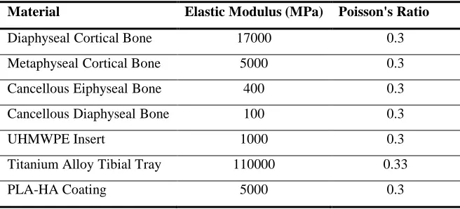
The optimal shape model increased the maximum von-mises stress value five times the initial model on medial cancellous bone region. This value is very close with that obtained using the natural bone with about 6.5% reduction in the maximum von-mises stress value on medial cancellous bone region, as shown in Fig.(4). Also the maximum von-mises stress value of the optimal shape is increased by 3% compared to the initial model on lateral cancellous bone region, as shown in Fig. (5) [19].
In addition, there's a 1% reduction in the stress concentration of the optimal shape model at the stem tip region compared to initial model, as shown in Fig.(6) [19].
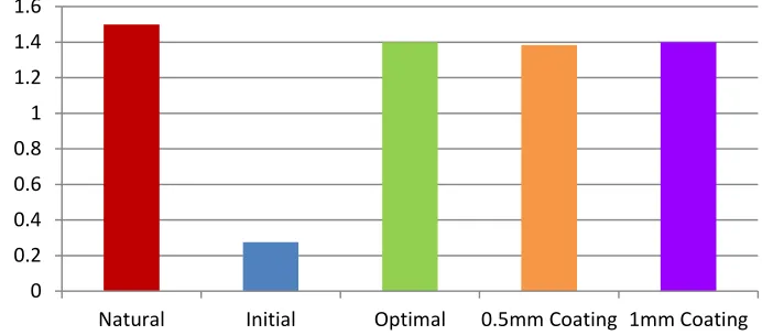
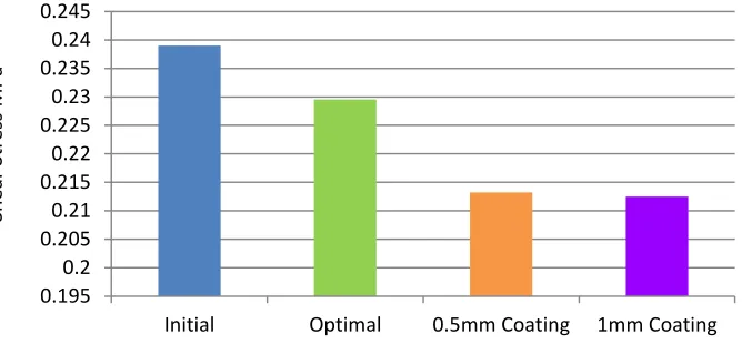
As well as, there are no noticeable differences in maximum interface shear stress values for medial cancellous region for both initial and optimal shape models, as shown in Fig.(7). However, the maximum interface shear stress value on lateral diaphyseal bone region for the optimal shape model is decreased by 4% compared to the initial model, as shown in Fig.(8) [19].
3.2 PLA-HA coating model
A comparison between a titanium stem and a titanium stem with a thin Polylactic acid hydroxyapatite PLA-HA coating, which has 5000MPa modulus of elasticity, around it is a trial to relieve the aseptic loosening of the tibia component. Titanium stem with a PLA-HA of 0.5mm and 1mm coating thickness were investigated, as shown in Figs. (4 to 8).
a. 0.5mm PLA-HA coating thickness around the tibia stem model
The maximum von-mises stress value of this model is increased on medial cancellous bone region five times the initial model, as shown in Fig.(4). This value is very close to the maximum von-mises stress of the natural bone on cancellous medial region by 8% reduction. However, this model increased the maximum von-mises stress values on lateral cancellous bone region by 6.5% compared to the initial model, as shown in Fig.(5). These results reduce the bone stress shielding which leads to minimal bone loss after TKR surgery.
In addition, the maximum von-mises stress value of this model is reduced by 2.5% compared to the initial stem at the
stem tip region, as shown in Fig. (6). This result relieves the stress concentration which leads to less patients' pain.
On the other hand, the maximum interface shear stress of this model is reduced by 2% on medial diaphyseal cancellous bone region compared to the initial model, as shown in Fig.(7). As well as, the maximum interface shear stress of this model is reduced by 11% on lateral diaphyseal cancellous bone region compared to the initial model, as shown in Fig.(8). This result reduces the micro-motion of the prosthesis which leads to reduce the aseptic loosening. This achieves minimal micro-motion of the implant which provides a more stable implant.
b. 1mm PLA-HA coating thickness around the tibia stem model
The maximum von-mises stress value of this model is increased on medial cancellous bone region five times the initial model. This value is very close to the maximum von-mises stress of the natural bone on cancellous medial region by 6.5% reduction, as shown in Fig.(4). However, this model increased the maximum von-mises stress values on lateral cancellous bone region by 6.5% compared to the initial model, as shown in Fig.(5). This improves the bone stress shielding which means less bone loss.
In addition, the maximum von-mises stress of this model is reduced by 12% in stress concentration at the stem tip region compared to the initial stem, as shown in Fig.(6). This result relieves the stress concentration which leads to less patients' pain.
On the other hand, the maximum interface shear stress of this model is reduced by 13.5% on medial diaphyseal cancellous bone region compared to the initial model, as shown in Fig.(7). As well as, the maximum interface shear stress of this model is reduced by 11% on lateral diaphyseal cancellous region bone compared to the initial model, as shown in Fig.(8). This result reduces the micro-motion of the prosthesis which leads to reduce the aseptic loosening.
4. CONCLUSION
153906-8787-IJMME-IJENS © December 2015 IJENS I J E N S Also HA coating reduces patient's pain and the need of
revision surgery.
REFERENCES
[1] D. Chong, U. Hansen, A. Amis, "Analysis of bone–prosthesis interface micromotion for cementless tibial prosthesis fixation and the influence of loading conditions", Journal of Biomechanics, doi: 10.1016/j.jbiomech.2009.12.006. Epub 2010 Mar 1, (2010).
[2] T. Enab and N. Bondok, "Material selection in the design of the tibia tray component of cemented artificial knee using finite element method", Materials and Design, doi: 10.1016/j.matdes.2012.08.017, (2012). [3] U. Simon, P. Augat, A. Ignatius, L. Claes, "Influence of the stiffness of
bone defect implants on the mechanical conditions at the interface: a finite element analysis with contact", Journal of Biomechanics, doi: 10.1016/S0021-9290(03)00114-3, (2003).
[4] E. Gautier, S. Perren, J. Cordey, "Strain distribution in plated and unplated sheep tibia: An in vivo experiment", Elsevier Ltd., doi: 10.1016/S0020-1383(00)80030-3, (2000).
[5] D. Cossetto, A. Gouda, "Uncemented tibial fixation total knee arthroplasty", The Journal of Arthroplasty, doi: 10.1016/j.arth.2009.12.008. Epub 2010 Mar 23, (2010).
[6] R. Mattila, P. Laurila, J. Rekola, J. Gunn, L. Lassila, T. Mäntylä, A. Aho, P. Vallittu, "Bone attachment to glass-fibre-reinforced composite implant with porous surface", Acta Biomaterialia, doi: 10.1016/j.actbio.2009.01.020. Epub 2009 Jan 29, (2009).
[7] A. Completo, P. Talaia, F. Fonseca, J. Simões, "Relationship of design features of stemmed tibial knee prosthesis with stress shielding and end of stem pain", Materials and Design, doi: 10.1016/j.matdes.2008.06.071, (2008).
[8] S. Darwish and A. Al-Samhan, "The effect of cement stiffness and tibia tray material on the stresses developed in artifacial knee", International
Journal of Adhesion and Adhesives, doi: 10.1016/j.ijadhadh.2007.05.003, (2007).
[9] C. Kimpton, A. Crocombe, W. Bradley, B. Owen, "Analysis of stem tip pain in revision total knee arthroplasty", The Journal of Arthroplasty, doi: 10.1016/j.arth.2012.10.007. Epub 2013 Mar 22, (2013).
[10] N. Fouda, "The effect of shape optimization and bimaterial stem on increasing the performance of a cemented tibia", International Journal of Mechaninal and Mechatronics Engineering, (2014).
[11] A. Completo, F. Fonseca, J. Simões, A. Ramos, C. Relvas, "A new press-fit stem concept to reduce the risk of end-of-stem pain at revision TKA: A pre-clinical study", The Knee, doi: 10.1016/j.knee.2011.12.008. Epub 2012 Jan 26, (2012).
[12] N. Fouda and H. Hedia, "Improve stress shielding on a cementless tibia tray using functionally graded material", Carl Hanser Verlag, doi: 10.3139/120.110507, (2013).
[13] T. Enab, "A comparative study of the performance of metallic and FGM tibia tray components in total knee replacement joints", Computational Materials Science, doi: 10.1016/j.commatsci.2011.09.032, (2011). [14] S. Machan, "Finite element analysis of a total knee replacement", The
University of Sydney.
[15] "Structural analysis of replacement knee design", Depuy, Ajohnson & Johnson Company.
[16] www.grabcad.com
[17] R. Kossowsky, N. Kossovsky, "Advances in Materials Science and Implant Orthopedic Surgery", united state military academy, New York, doi: 10.1007/978-94-011-0157-8, (1995).
[18] D. Alfaiate, T. Escobar, P. Oliveira, I. Almeida, D. Rosas, B. Borges, "Hydroxyapatite-coated implants: clinical advantages", Clinical Oral Implants Research, (2014).
153906-8787-IJMME-IJENS © December 2015 IJENS I J E N S
[a]
[b]
Fig. 2. The dimensions of the tibial models: [a] initial model [b] optimal shape model
[a]
[b]
Fig. 3. The loading conditions applied at: [a] the natural tibia model [b] artificial tibia prosthesis
Table I
The mechanical properties of the natural bone and the prosthesis components
Material Elastic Modulus (MPa) Poisson's Ratio
Diaphyseal Cortical Bone 17000 0.3
Metaphyseal Cortical Bone 5000 0.3
Cancellous Eiphyseal Bone 400 0.3
Cancellous Diaphyseal Bone 100 0.3
UHMWPE Insert 1000 0.3
Titanium Alloy Tibial Tray 110000 0.33
153906-8787-IJMME-IJENS © December 2015 IJENS I J E N S
Fig. 4. A comparison between von-mises stresses for natural bone, initial, optimal shape, and PLA-HA of 0.5mm and 1mm coating thickness models for cancellous medial region
Fig. 5. A comparison between von-mises stresses for natural bone, initial, optimal shape, and PLA-HA of 0.5mm and 1mm coating thickness models for cancellous lateral region
Fig. 6. A comparison between von-mises stresses on the stem tip region for initial, optimal shape, and PLA-HA of 0.5mm and 1mm coating thickness models
0 0.2 0.4 0.6 0.8 1 1.2 1.4 1.6
Natural Initial Optimal 0.5mm Coating 1mm Coating
Von
-Mis
es
Stres
s
MPa
0 0.05 0.1 0.15 0.2 0.25
Natural Initial Optimal 0.5mm Coating 1mm Coating
Von
-Mis
es
Stres
s
MPa
0.42 0.44 0.46 0.48 0.5 0.52 0.54
Initial Optimal 0.5mm Coating 1mm Coating
Von
-Mi
se
s
Stre
ss
153906-8787-IJMME-IJENS © December 2015 IJENS I J E N S
Fig. 7. A comparison between interface shear stresses for initial, optimal shape, and PLA-HA of 0.5mm and 1mm coating thickness models for cancellous medial region
Fig. 8. A comparison between interface shear stresses for initial, optimal shape, and PLA-HA of 0.5mm and 1mm coating thickness models for cancellous lateral region
0.23 0.24 0.25 0.26 0.27 0.28 0.29 0.3 0.31
Initial Optimal 0.5mm Coating 1mm Coating
Sh
ear
St
re
ss
MPa
0.195 0.2 0.205 0.21 0.215 0.22 0.225 0.23 0.235 0.24 0.245
Initial Optimal 0.5mm Coating 1mm Coating
Sh
ear
St
re
ss
![Fig .1. Model geometry: [a] A coronal plane section in human body [b] A 3D model for a natural tibia bone [c] A 3D artificial tibial prosthesis model [d] A coronal section of a 3D artificial tibial prosthesis model](https://thumb-us.123doks.com/thumbv2/123dok_us/1359279.1645006/5.612.47.524.65.708/geometry-coronal-section-artificial-prosthesis-section-artificial-prosthesis.webp)