PHD THESIS DANISH MEDICAL JOURNAL
This review has been accepted as a thesis together with four previously published papers by University of Copenhagen, October 2013 and defended on March 5th 2014
Tutor(s): Mads Hald Andersen
Official opponents: Marianne Hokland and Viktor Umansky
Correspondence: Center for Cancer Immune Therapy, Department of Hematology, University Hospital Herlev, 2730 Herlev, Denmark
E-mail: [email protected]
Dan Med J 2016;63(1)B5188
THE THESIS IS BASED ON THE FOLLOWING TWO PAPERS AND TWO APPENDICES
Larsen SK, Munir S, Woetmann A, Frøsig TM, Ødum N, Svane IM, Becker JC, Andersen MH. Functional characterization of Foxp3-specific spontaneous immune responses. Leukemia 2013, Juli 1.
Larsen SK, Ahmad SM, Idorn M, Met Ö, Svane IM, thor Straten P, Andersen MH. Spontaneous presence of Foxo3-specific T cells in cancer patients. Oncoimmunology 2014, November 14;3(8)
Larsen SK, Svane IM, thor Straten P, Andersen MH. Therapeu-tic cancer vaccines. Current Cancer Therapy Reviews 2010(6)2.
Larsen SK, Munir S, Iversen TZ, Donia M, Klausen TW, Svane IM, thor Straten P, Andersen MH. Natural CD4+ T cell responses against idoleamine 2,3-dioxygenase. PloS One 2012(7)4.
OBJECTIVES
This thesis describes the existence of cellular immune re-sponses towards regulatory cells.
The objective of this thesis has been to investigate antigen-specific immune responses towards cells that regulate local sup-pression of the immune system in the tumor microenvironment. We have used the reverse immunology approach to search for antigens expressed not only by the cancer cells but also in the tumor microenvironment by cells that support tumor growth. The focus of these investigations has been on two transcrip-tion factors that play prominent roles in immune suppression of anti-tumor immunity, namely Forkhead box p3 (Foxp3) and Fork-head box o3 (Foxo3). In addition to establishing that
antigen-specific responses towards these proteins exist, the aims of the research have been to characterize these immune responses in respect to functionality and particularly their lytic capabilities. The main hypothesis was that Foxp3 and Foxo3 could be targets for specific T cells, and that recognition of Foxp3- and Foxo3-derived epitopes could lead to cytolysis of cells expressing these tran-scription factors. Particularly, we wanted to investigate CTL-specific recognition of Foxp3-expressing Tregs and Foxo3-expressing tolerogenic DCs.
The broader aims of this thesis have been to explore the pos-sibility to harness the immune system to target Foxp3- and Foxo3-expressing cells in order to improve cancer immunotherapy. BACKGROUND
Every time a cell replicates, each of the 3 billion basepairs in the chromosomes must be copied accurately. Considering the amount of cells in a human body and the number of replications during the lifespan of a human this is a remarkable task. Cancer arises when several mutations have occurred in the same cell and enabled this cell to become a malignant growth. This requires the mutations to occur in genes that alter proteins which are regulat-ing crucial aspects of cell behaviour. These aspects have been termed “the hallmarks of cancer” [1] and encompass six basic abilities that all cancer cells possess; sustaining proliferative sig-nalling, evading growth suppressors, activating invasion and metastasis, enabling replicative immortality, inducing angiogene-sis and reangiogene-sisting cell death. Recently the cancer hallmarks have been revised and four new hallmarks have been suggested, two enabling hallmarks and two emerging hallmarks – interestingly one of each has to do with the immune system [2]. The enabling hallmarks that have been included are genome instability and tumor-promoting inflammation. The hallmarks that are emerging through recent year’s scientific efforts are deregulation of cellular energetics and avoidance of immune destruction. Hence, the impact of the immune system on cancer biology is now well-established. However, the remarkable facts point to a dual role for the immune system in cancer – it generates tumor-promoting inflammation and at the same time is capable of tumor destruc-tion.
CANCER IMMUNOEDITING
The protective role of the immune system against tumor de-velopment has been heavily debated for more than a century. Nobel prize winner and one of the founders of immunology Paul Ehrlich suggested in 1908 that cancers would be more prevalent in human if not for the protection of the immune system (re-viewed in [3]). The same idea was revisited in the 1950ies by Burnet and Thomas [4], fueled by advances in experiments with inbred mice and the discovery of tumor antigens [5] they
pro-Cellular Immune Responses Towards Regulatory
Cells
posed the idea of cancer immunosurveillance. This idea was largely rejected by the scientific community but four decades later was taken up again and revised to encompass cancer immu-noediting [6].
Cancer immunoediting is believed to comprise three phases; elimination, equilibrium and escape. In the elimination phase newly transformed cells are detected and destroyed by the im-mune system. When the elimination is not flawless the equilibri-um phase begins, in which the cancer cells are held in check by the immune system; as new cancer cells arise, others are killed in the same pace. In this phase the immunogenicity of the cancer cells are sculpted by the immune system. In the attempt of pro-tecting the host, the immune system generates a Darwinian selec-tion pressure that eventually foster cancer cells able to avoid immune destruction. Finally, in the escape phase, these tumor cell escape variants progress to become clinically relevant tumors that eventually lead to life-threatening disease.
Several lines of evidence support the theory of cancer immu-noediting. First, immune cells are present in cancers of different origin and the presence of certain subtypes of cells has been correlated to increased survival. Hence, the infiltration of CD3+ T cells in colorectal cancer [7], ovarian cancer [8] and melanoma [9] predicts patient survival or regression of the tumor. The same is true for several other cancer types including head and neck, breast, bladder, esophageal, prostate and pancreatic cancer as well as Merkel cell and urothelial cell carcinoma, as reviewed recently [10]. Second, spontaneous immune responses against cancer [11] as well as spontaneous regression of cancer has been demonstrated [12]. In a few cases spontaneously regressing mel-anomas accompanied by clonal expansion of T cells have provided strong evidence of the elimination phase in vivo [13], [14]. Finally, immunodeficient individuals have been reported to have an increased incidence of cancer. However, the prevalent cancers among AIDS patients belong to the group of virus-induced cancers raising the question of whether this is due to lack of viral control or lack of control of malignant transformation. None the less, some studies have demonstrated an increased risk of non-viral cancers in immunodeficient individuals [15], [16]. Recently, sup-port for the cancer immunoediting theory using mice models was published in Nature, both emphasizing the importance of cytotox-ic CD8 T cells in shaping the immunogencytotox-icity of tumors [17], [18]. CYTOTOXIC T CELLS
The immune system is an intricate network of cells and mole-cules developed to protect vertebrates from invading pathogens. The key function of the immune system is therefore to recognize and appropriately react to these pathogens, i.e. to react when something does not belong in the healthy body. The immune system has unspecific (innate immunity) and specific (adaptive immunity) means of doing this. Adaptive immunity consists of a humoral and a cellular response. Cytotoxic T lymphocytes (CTL) direct the effector functions of the cellular response.
T cells develop in the bone marrow and migrate to the thy-mus for maturation. During the maturation process random rear-rangement of V(D)J gene segments of the α and β chains provides the T cell with a unique T cell receptor (TCR) [19]. Also during maturation T cells commit to express either of the co-receptors CD4 or CD8, defining the T helper cell subset or the cytotoxic T cell subset, respectively. Another important aspect of thymic maturation is the concept of central tolerance. In the thymic medulla T cells with a strong affinity for self-antigens are nega-tively selected and deleted, to avoid release of potential auto-reactive T cell in the body [20].
Activation and regulation
Even after maturation in the thymus the T cell is still consid-ered a naïve T cell and becomes functional only upon antigen stimulation through its TCR. Naïve T cells monitor the cells of the body and when encountering their antigen they are stimulated through their TCR to proliferate and differentiate. Hence, clonal proliferation generates large numbers of armed effector T cells with identical TCR. Once licensed, effector CTL that encounter their antigen effectively lyse and kill the presenting cell. When the infection is cleared and antigen is no longer present most effector CTLs die by apoptosis in a subtraction phase, leaving a few central memory and effector memory T cells, should the infection be encountered again [21].
Activation of naïve T cells into effector CTLs that are licensed killers is a tightly regulated process, which requires three distinct signals; antigen recognition by the TCR, co-stimulation and signal-ing through the interleukin-2 (IL-2) receptor. The TCR binds the antigen, consisting of a peptide-HLA complex and the binding is strengthened by co-receptor CD8 engagement of the HLA class I molecule. Signaling through the TCR is mediated via the intracel-lular CD3 complex. The best understood co-stimulatory signal is mediated via the CD28 co-receptor on the CTL when binding to the ligands CD80 or CD86 on the antigen presenting cells (APC). Importantly, activation of CTLs prompts expression of the inhibi-tory receptor cytotoxic T lymphocyte antigen 4 (CTLA-4) with a higher affinity for CD80 and CD86, thereby ensuring termination of the responses and T cell homeostasis [22]. In addition, reverse signaling by CTLA-4 blocks expression of CD80 and CD86 on the DC, which, in turn become unable to activate T cells [23]. Pro-grammed death-1 (PD-1) is another important co-inhibitory re-ceptor, which is also expressed on activated T cells. Ligation of PD-1 by its ligands PD-L1 (B7-H1) or PD-L2 in the vicinity of the TCR, recruits the phosphatase SHP2 which dephosphorylates the TCR signaling complex and thereby inhibit signaling [24]. The balance between co-stimulatory and co-inhibitory interactions determines the strength of T cell activation. The inhibition of full blown activation is pivotal in minimizing tissue damage and pre-vention of auto-immunity, however it is detrimental in anti-tumor immunity. The third signal is through stimulation of the IL-2 re-ceptor, which is upregulated on the surface of activated CTLs [25]. Activated CTLs produce small amounts of IL-2 that aid in their own activation, usually however CD4 T helper cells produce the IL-2 necessary for full activation of the CTLs.
Effector functions
The effector function of CTLs is the cell mediated lysis, which can be orchestrated by at least three different mechanisms (re-viewed in [25]). CTLs can release granules containing pre-synthesized lytic mediators, perforin and granzymes, directly onto the cell surface of the target cell triggering caspase-dependent and independent apoptosis of the target cell. Alternatively, CTLs are able to induce apoptosis in target cells via ligation of Fas death-receptors on target cells by their Fas-ligand. A third option, which does not require direct cell-to-cell contact, is via release of cytokines. Tumor necrosis factor α (TNFα) binds to its receptor on the target cells and activates the caspase-cascade resulting in apoptosis. Interferon γ (IFNγ) leads to upregulation of HLA and antigen-processing proteins as well as Fas, leading to enhanced antigen presentation or Fas ligation.
HLA restriction and antigen presentation
The cognate antigen required for stimulation of a TCR consist of a peptide bound to a major histocompatibility molecule (MHC), in human called human leucocyte antigen (HLA) [26]. CD8 CTLs are stimulated by HLA class I molecules, which are expressed on
all nucleated cells. These are highly polymorphic proteins, whose function is to display peptides derived from cellular proteins on the surface of the cell. This function allows T cells to monitor all cells of the body for inappropriate alterations in protein synthesis, i.e. during viral replication or malignant transformation. CD4 helper T cells are stimulated by HLA class II molecules, which are expressed only on professional antigen-presenting cells (APC). These are equally polymorphic molecules; however their function is to present extracellularly derived peptides in the context of proper co-stimulatory signals in order to activate the naïve T cells. The most important APCs are dendritic cells (DC), which are able to cross-present extracellularly derived peptides in HLA class I molecules, in order to activate naïve CD8 T cells (reviewed in [27]).
TUMOR ASSOCIATED ANTIGENS
The genetic instability of cancer cells leads to an altered pro-tein repertoire, parts of which are displayed in HLA molecules on the cell surface. These changes enable T cells to distinguish be-tween normal “self” and malignantly transformed cells and are called tumor associated antigens. The first human tumor associ-ated antigen was identified in 1991 using a cosmid DNA library to search for the gene that rendered tumor cells susceptible to CTL recognition [28]. Since then more than 400 tumor associated antigens have been discovered [29]. The changes in protein syn-thesis in malignant cells give rise to different types of tumor associated antigens. Point mutations that lead to altered proteins give rise to neo-antigens. These can be patient specific or tumor specific. Mutations that lead to expression of proteins not nor-mally expressed give rise to other types of antigens; differentia-tion antigens and cancer/testis antigens. Finally, deregulated protein synthesis can lead to over-expression of normal proteins resulting in over-expression antigens (reviewed in [30] - appendix A).
The most frequently used method to identify new tumor as-sociated antigens is the reverse immunology approach, in which a target protein is screened for potential HLA-binding sequences using online prediction databases [31]. After selection of candi-date peptides, these are used to raise peptide-specific CTL, which are finally used to provide evidence that the peptide is processed and presented by cancer cells.
THE TUMOR MICROENVIRONMENT
In recent years it has become evident that tumors are more than a mass of cancer cells. Research has long focused on malig-nant cells but in addition to cancer cells tumors consist of host cells that have evolved together with the growing malignancy to form the tumor microenvironment. The tumor microenvironment comprises the supportive framework for the cancer cells formed by stromal cells and extracellular matrix proteins and by infiltrat-ing immune cells of all types [32]. The tumor microenvironment also contains signaling molecules that orchestrate the creation of an environment suited to the requirements of the growing tumor. This includes factors that actively create immune suppression in order for the tumor to escape immune attack. Several suppres-sive mechanisms have been well-described i.e. expression of PD-L1 by tumor cells as well as expression of indoleamine 2,3-dioxygenase (IDO) by tumor cells or DC (reviewed in [33], [34]). In addition, the infiltration of immune cells with a suppressive func-tion has been revealed. This includes the infiltrafunc-tion of myeloid derived suppressor cells (MDSC) and regulatory T cells (Treg) as well as the presence of functionally impaired DCs resulting in
tolerized or anergic CTL [2]. Interestingly, it was recently demon-strated that the induction of several suppressive mechanisms in the tumor environment are orchestrated not by factors expressed by the tumor cells, but by factors expressed by infiltrating CD8 effector cells [35]. This emphasizes the role of these suppressive mechanisms as negative feedback loops which are necessary in all biological systems.
PD-L1 AND IDO
As mentioned, PD-1 is a co-inhibitory receptor, expressed on activated T cells. Tumor cells and Tregs have been shown to ex-press the ligand PD-L1, thus terminating the immune response by inhibiting signaling via the TCR and inducing apoptosis of the CTL [36].
Indoleamine 2,3-dioxygenase (IDO) is a rate-limiting enzyme in the degradation of the essential amino acid tryptophan. Up-regulation of IDO has been shown to suppress immune responses by depletion of tryptophan, as well as by suppressive effects of tryptophan catabolites [37]. In addition, IDO expressing DCs have also been shown to induce Tregs [38]–[40].
MYELOID-DERIVED SUPPRESSOR CELLS
MDSC are a heterogeneous group of immature myeloid cells that have failed to differentiate to macrophages, DCs or granulo-cytes. They are characterized by the ability to suppress T cell activation and they are induced by chronic inflammation, which is often present at tumor sites [41]. MDSCs suppress anti-tumor immunity through various pathways. They express arginase which catabolizes the amino acid arginine and thereby deplete their surroundings of arginine. T cells that are deprived of this amino acid are deficient for the CD3ζ chain important for signaling through the TCR. MDSCs also deprive surroundings of the amino acid cysteine and thereby suppress T cell proliferation. In addition they produce high amounts of reactive oxygen and nitrogen species which induce nitration of the TCR and thus abrogate epitope recognition or induce apoptosis. They also disrupt T cell migration by down-regulation of the homing-receptor L-selectin on the T cells [42]. Recently, it was demonstrated that MDSCs express IDO and exert IDO-mediated suppression of T cells [43]. Interestingly, it has also been demonstrated in vitro that MDSCs from patients with hepatocellular carcinoma induces Foxp3+ Tregs [44].
REGULATORY T CELLS
In the 1970ies Gershon and Kondo described a subset of T cells that was able to induce tolerance in mice, they named these suppressor T cells [45]. However, contradictory evidence and skepticism in the immunological field resulted in this important finding to be disregarded for decades. Until, in 1995 Sakaguchi and colleagues reported a subset of CD4 T cells expressing the IL-2 receptor α-chain CDIL-25 that maintained self-tolerance and whose removal resulted in autoimmune disease in mice [46]. These cells were termed regulatory T cells (Treg). This discovery was followed by the identification of a similar subset in human [47]–[51].
Tregs constitute about 10% of CD4 T cells in normal humans and are required for maintenance of immune homeostasis, self-tolerance and prevention of autoimmune disease. The function of Tregs in the healthy body is to control activation and expansion of self-reactive and non-self-reactive T cells, thereby ensuring pro-tection from autoimmune disease, fetal tolerance and allergy [52]. At least two subsets of Tregs exist, natural Tregs (nTreg) and induced Tregs (iTreg). The natural Tregs exit the thymus as Tregs, whereas the iTregs are CD4 T cells that are induced in the periph-ery to become Tregs. nTregs are believed to have an autoreactive TCR with an affinity for self-antigens in between normal CD4 T
cells and those that are negatively selected, i.e. deleted in the thymus [53]. Several factors in the periphery are believed to induce Tregs, including suboptimal antigen stimulation and stimu-lation by TGFβ [54].
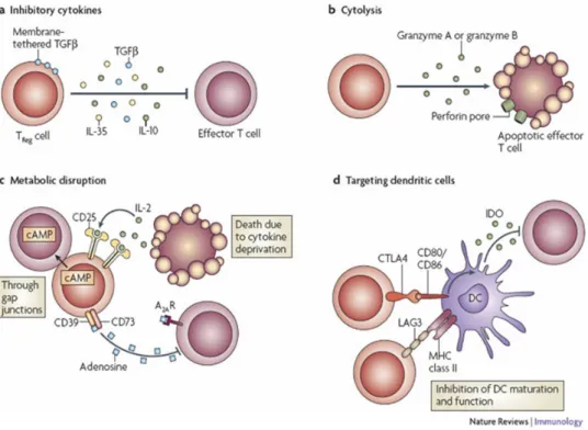
Tregs exert their suppression of immune responses through various mechanisms (Figure 1) and are able to suppress CD4 T cells, CD8 T cells, B cells, NK cells and DC (reviewed in [55]). Tregs suppress immune responses via secretion of anti-inflammatory cytokines TGFβ, IL-10 and possibly IL-35. In addition they are able to kill effector T cells as well as APC through granzyme and perfor-in dependent pathways. The expression of the high affperfor-inity IL-2 receptor enables the Tregs to deprive surrounding cells of IL-2, resulting in cell death. Expression of CD39 and possibly CD73 renders Tregs able to degrade extracellular ATP and thereby inhibit ATP-dependent DC maturation, secretion of
pro-inflammatory cytokines and production of granzymes and perfor-in. In addition Tregs increase cAMP levels in target cells through gap junctions which inhibit IL-2 production, survival and prolifera-tion [56]. Constitutive expression of the co-inhibitory receptor CTLA-4 downregulates expression of CD80 and CD86 on DC, thereby creating sub-optimal antigen-presentation [23]. Similarly, expression of PD-L1 on Tregs modulate DC function by binding to PD-1 [57]. In addition, PD-1 is expressed on Tregs, and ligation to PD-L1 on DC inhibit cytokine production and maturation by the DC [24].
Foxp3
The crucial importance of Tregs in maintaining immune toler-ance to self is illustrated by the disease immunodysregulation polyendocrinopathy enteropathy X-linked syndrome (IPEX) [59]. IPEX is caused by mutations that result in a complete deficiency of Tregs and the resulting syndrome is a multi-organ autoimmune and inflammatry condition. The severity of this syndrome is evi-denced by the fact that it is lethal in infancy. The mutations caus-ing IPEX and the mouse equivalent scurfy was found to be located in the gene Forkhead box p3 (Foxp3) [60], [61] which is now the best known marker for Tregs
Foxp3 belongs to a superfamily of transcriptional regulators that share a common DNA-binding domain termed the forkhead box or winged helix domain. More than 40 genes in this familiy have been identified in human. Of these three belong to the P subfamiliy; Foxp1, Foxp2 and Foxp3 [62]. Foxp3 is essential for the development and function of Tregs and is expressed by nTregs as well as iTregs. In human there exist three isoforms of the protein, the longest of which is homologous to mouse Foxp3. Strong stimulation through CD3 and CD28 or stimulation via TGFβ is believed to induce expression of Foxp3, however the exact determinants of Foxp3 expression is still unknown. The CD3/CD28 mediated stimulation of nTregs in the thymus mediates a stable expression of Foxp3, characterized by demethylated CpG islets. In contrast, the TGFβ induced expression in iTregs in the periphery is characterized by methylated CpGs at the Foxp3 locus and a less stable expression [63]. In addition to the DNA-binding domain, Foxp3 contains a repressor domain, and is known to be a tran-scriptional repressor as well as a trantran-scriptional activator [64]. Foxp3 also contains two nuclear export signals (NES) which have
Figure 1: Regulatory T cell mechanisms of suppression.
Depiction of mechanisms exerted by Tregs to suppress immune responses centered around four basic modes of action. A) Inhibitory cytokines. B) Granzyme- and perforin mediated cytolysis. C) Metabolic disruption, including IL-2- and ATP-deprivation. D) Targeting dendritic cells. Vignali et al., Nature Reviews Immunolgy, 2008 [58]
been shown to determine the subcellular localization of the pro-tein and hence the transcriptional activity [65].
In human, unlike in mice, activated non-Treg T cells transient-ly express Foxp3 [66]. In fact, it was very recenttransient-ly demonstrated that the expression of Foxp3 in conventional T cells negatively regulates proliferation and cytokine secretion [67]. In addtition, Foxp3 has been shown to be expressed by malignant cells in several cancers including cutaneous T cell lymphoma [68], pan-creatic adenocarcinoma [69], breast cancer [70], [71], papillary thyroid cancer [72], adult T cell leukemia [73], melanoma [74] as well as ovarian cancer [75].
Accumulation of Tregs in the tumor microenvironment
Increased numbers of Tregs have been observed in the blood and at tumor sites of patients with cancers of various origins. This has been reported for tumor-infiltrating lymphocytes in ovarian cancer [76], [77], non-small cell lung cancer [76]–[78] peripheral blood and tumor in pancreatic and breast cancer [79], peripheral blood in lung, breast and colorectal cancer [80], peripheral blood and tumor infiltrating lymphocytes of Hodgkin lymphoma [81], gastric and esophageal cancer [82], peripheral blood of gastric carcinoma [83] and peripheral blood [84] and metastatic lymph nodes in melanoma [85].
Treg suppress immune responses in a dose-dependent man-ner [86] and the accumulation of Treg in the tumor microenvi-ronment is therefore considered a major obstacle for induction of effective tumor immunity. In this regard adoptive transfer exper-iments of Tregs has been shown to directly suppress effector T cell control of tumor growth [86]. Strikingly, abundant Treg infil-tration in ovarian tumors has been correlated with decreased survival [86]. This finding has been elaborated in a later study showing that a low ratio of CD8 T cells to Tregs predicts signifi-cantly decreased survival [87]. The negative impact of Treg accu-mulation in the tumor on disease outcome for the patient have since been described in several cancer types, including gastric [88], [89], esophageal [88], pancreatic [90], lung [91] and liver cancer [92], renal cell carcinoma [93] and breast cancer. In breast cancer patients however, Treg infiltrates surrounding the tumor, but not in the tumor, has been correlated to risk of relapse and death [94].
Furthermore, in clinical trials with adoptive cell transfer ther-apy of melanoma it has been shown that the patients that re-spond to treatment have lower numbers of Tregs in their blood, than the non-responders [95]. In contrast, Treg infiltration has been associated with a better prognosis in patients with certain lymphomas [96], [97], head and neck cancer [98] and colorectal cancer [99]. In colorectal cancer , however low CD3+/Treg ratio has also been correlated to reduced survival [100]. In conclusion, most studies indicate a negative influence of Tregs on patient survival, although contradictory evidence exists for some types of cancer.
The reason for accumulation of Treg in the tumor environ-ment is believed to derive from a combination of recruitenviron-ment, expansion and conversion of Tregs at the tumor site. In this re-gard, CCL22 secreted by tumor cells and tumor-infiltrating mac-rophages have been shown to attract Treg cells through their expression of CCR4 [86]. In mice it has been shown that intra-tumoral Tregs proliferate more extensively than non-Foxp3 CD4 T cells [101]. Tumor cells expressing IDO have been shown to con-vert CD4+/CD25- T cells to Foxp3+ suppressor cells [39], and the same has been shown for TCR stimulation in the presence of TGFβ [38].
TOLEROGENIC DC IN THE TUMOR MICROENVIRONMENT Another contributing factor to immune suppression in the tumor microenvironment is tolerogenic DCs. DCs are specialised antigen presenting cells; they scan peripheral tissue where they capture antigen which they present in MHC class II molecules and by cross-presentation in MHC class I molecules. When antigens are captured in conjunction with danger signals, such as pro-inflammatory cytokines and TLR ligands, the DCs mature and express co-stimulatory molecules. Thus, T cells that encounter their cognate antigen on mature, stimulatory DCs receive co-stimulatory signals and differentiate into effector T cells [102]. However, T cells that encounter their antigen on a DC in the absence of appropriate co-stimulatory signals become tolerized, and do not respond on subsequent encounters with the antigen. This mechanism helps to maintain peripheral tolerance [103].
However, the same mechanism is believed to contribute to immune suppression in tumors. Functionally impaired DCs in the tumor microenvironment are considered a major obstacle to induction of tumor immunity [104]. In this regard, DCs with de-creased stimulatory abilities have been identified in patients suffering from melanoma [105], breast cancer [106] and prostate cancer [107]. Furthermore human prostate cancer cell lines have shown to impede DC maturation in vitro [108]. Certain subsets of DCs have been associated with tumor tolerance; in this regard plasmacytoid DC (pDC) and IDO-expressing DC have been the focus of investigations. pDC infiltration of tumors have been correlated to reduced survival in breast cancer patients [109]. In patients with ovarian cancer accumulation of pDC have been shown to directly induce suppressive CD8 T cells [110], [111]. The presence of IDO expressing DCs in the tumor draining lymph nodes has been correlated to reduced survival in melanoma [112] and IDO expression has been suggested as a marker for tolerizing DCs [113]. Another catabolizing enzyme, arginase, which depletes L-arginine, has been found to be expressed in DCs in a breast cancer model and mediate suppression of CTL function [114].
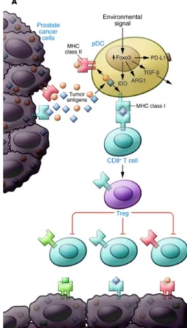
In an elegant study by Watkins and colleagues using a mouse model of prostate cancer, tumor associated DC were demonstrat-ed to be less stimulatory than DCs from non-cancerous tissue [107]. Furthermore, the tumor associated DCs prevented T cells from reacting to subsequent antigen challenge, thus de facto tolerizing the T cells. This tolerization proved to be contagious; CD8 T cells were not merely tolerized but had acquired suppres-sive abilities after encounter with the tolerizing DC. The tumor associated DCs were shown to express high levels of immunosup-pressive mediators IDO, arginase, PD-L1 and TGFβ (Figure 2). A similar tolerizing DC subset was identified in human prostate tumors. Common to mouse and human tolerizing DCs were ex-pression of the transcription factor Foxo3 [107].
Figure 2: Tolerogenic DC in the tumor microenvironment. Environmental signals drive the expression of Foxo3 in the nucleus, which in turn control the expression of immune-inhibitory molecules IDO, arginase, TGF-β and PD-L1. Presentation of antigen to CD8 T cells by the Foxo3+ DC tolerizes the CTLs and in addition, converts them to CD8 regulatory cells that suppress nearby CTL. Bronte, J Clin Invest, 2011 [115].
Foxo3
Foxo3 has been identified as an important transcription factor in determining the stimulatory or tolerizing capacity of DCs. It has been suggested as the responsible transcriptional control of the tolerogenic programme in DCs [116]. The expression of Foxo3 in tolerizing DCs isolated from human prostate tumors were shown to be 40-fold higher than in DCs from non-cancerous tissue, and knockdown of Foxo3 abrogated the tolerizing phenotype of the DC [107]. In addition, Foxo3-deficient mice have been demon-strated to exhibit enhanced T cell proliferation in response to viral infections, because of an improved stimulatory capacity of the Foxo3-deficient DCs [117]. Further support for the regulatory role of Foxo3 in immune cells was very recently published, showing that increased expression of Foxo3 in human monocytes leads to production of fewer pro-inflammatory cytokines and more IL-10 by these cells. Interestingly, the SNP that regulates this increased expression of Foxo3 was associated with a less severe course of the autoimmune diseases Crohn’s disease and rheumatoid arthri-tis [118].
Foxo3 belongs to the same superfamily of transcriptional reg-ulators as Foxp3. They share a common DNA-binding domain; the forkhead box or winged helix domain. In human, more than 40 genes have been identified in this family and three belong to the O subfamily; Foxo1, Foxo3 and Foxo4 [62]. The activity of Foxo3 is
regulated by post-translational modifications, which in turn de-termines the subcellular localization of the protein. Translocation from the nucleus to the cytoplasm is triggered by phosphorylation at three conserved sites that diminish DNA-binding. This phos-phorylation is performed by Akt activated by the growth factor-PI3K pathway and naturally abrogates the function of Foxo3 in the nucleus [119]. In contrast, stress-inducing signals that activate the MST1 kinase lead to phosphorylation at Ser207, translocation into the nucleus and target gene transcription [120].
Foxo3 are implicated in regulation of various cell cycle events including protection from oxidative stress [121], longevity [122], fertility [123] and induction of apoptosis [119]. It is therefore not surprising that the Foxo family members have been identified as tumor-suppressor genes [124] and upregulation of Foxo3 has been shown to suppress tumor growth [125]. In addition, nuclear localization of Foxo3, and thus active Foxo3, has been correlated to survival in breast cancer patients [126]. However, it has also been shown that Foxo3 is sequestered in the cytoplasm in a renal cancer cell line [127]. In addition, Foxo3 has been implicated in the regulation of Foxp3 and thus differentiation of Tregs [128]– [130]. Furthermore, Foxo3 has been shown to be expressed in placenta [131] and intestinal epithelial cells [132] in human. IMMUNE THERAPY OF CANCER
The common denominator for immune therapy of cancer is to use the most important defense system of the body to fight tu-mors; the immune system. This aim can be pursued in several ways, but all immune therapy intents to either amplify efficient immune responses or to block suppressive immune responses. Immune therapy falls into four treatments categories; cytokines, adoptive cell transfer, vaccination or antibodies. In addition to these traditional immune therapies, it has recently emerged that conventional chemotherapy also has an immune mediated effect. These five treatment modalities will be described in the following sections.
CYTOKINES
Cytokines are secreted signaling molecules of the immune system and have long been investigated for use as a cancer ther-apy. Only two cytokines are currently approved for treatment of cancer; Il-2 and IFNα. IL-2 is used for treatment of the immune sensitive tumors metastatic melanoma and renal cell carcinoma, because of the fundamental role it has in T cell proliferation [133]. IL-2 treatment is limited by its severe toxicity to the organ-ism, and a further disadvantage is the documented expansion of Tregs [134]. However, high dose IL-2 is used in adoptive cell ther-apy to support the transferred cells [135].
IFNα is used for treatment of malignant melanoma, hairy cell leukemia, chronic myeloid leukemia, multiple myeloma and fol-licular lymphoma [133].
A frequently used cytokine in experimental cancer immune therapy is granulocyte macrophage colony-stimulating factor (GM-SCF). GM-SCF is approved for treatment of hematological deficiencies owing to its actions as a hematopoietic growth factor and for that reason it is used in various clinical trials to boost tumor immunity [133].
VACCINATION
Therapeutic immunization for the treatment of cancer en-compasses various strategies whose common goal is to actively elicit an immune response in vivo directed at the cancer cells. There are two components in any vaccine; the antigen and an adjuvant. The adjuvant is the immune activating component, and the antigen defines the target. Adjuvants frequently used in
ther-apeutic immunization include GM-CSF, TLR agonists and DC (re-viewed in [30] – appendix A). The simplest way of targeting can-cer cells is through peptide vaccination with tumor associated antigens; these can be short CD8 epitopes or longer CD4 epitopes. Alternatively, whole protein, DNA encoding the protein target and whole tumor cells have been employed. Another ap-proach is DC vaccination, in which autologous DC are harvested and matured ex vivo in combination with peptides, whole pro-teins, tumor lysate, or transfected with DNA encoding the target protein. All of these strategies have been investigated intensively over the past decades. However clinical benefit has been scarce [136]. The lack of success of this treatment modality is widely believed to stem from the immune suppressive microenviron-ment hindering effective immunization in cancer patients [137]. The tumor has evolved a microenvironment around itself special-ized to avoid immune attack, rich in Tregs, MDCS and tolerizing DC. Current efforts in immunization have not been adequate to break these tolerizing barriers and mount an effective immune response.
Recently however, a DC based vaccine was approved after demonstrating 4 months prolonged survival in prostate cancer patients. This Sipuleucel-T vaccine is made of autologous periph-eral blood mononuclear cells (PBMC), including APC, activated ex-vivo with a recombinant fusion protein. The protein is a fusion of a prostate antigen, prostatic acid phosphatase and GM-SCF [138]. ADOPTIVE CELL TRANSFER
Adoptive cell transfer is based on the reintroduction of large amounts of activated lymphocytes into a preconditioned patient together with high-dose IL-2. The lymphocytes most often are isolated from tumor biopsies [135], but can also derive from PBMC transduced with tumor specific TCR or chimeric antigen receptors (CAR) [139]. CARs recognize tumor antigens with an antibody-derived complementarity-determining region, however signal through the TCR intracellular pathway and mediate lysis of the target cell.
Adoptive cell transfer has proven to be an effective treatment for malignant melanoma, since lymphodepleting conditioning of the patients were included [135]. In fact, response rate correlate with the degree of lymphodepletion, and an overall response rate of 72% have been achieved [140]. Other factors correlating with response rate is the phenotype of the infused cells, in particular their telomere length and IFNγ production in response to autolo-gous tumor [141]–[143]. Interestingly, isolation of CD8 T cells before infusion, does not improve response rate, even though cytotoxic T cells are believed to mediate the tumor regression [142]. In fact, adoptive transfer of CD4 T cells has been shown to induce complete remission of malignant melanoma [144]. Pre-sumably, the infused CD4 T cells generate a tumor microenviron-ment permissive of endogenous CD8 T cell activation.
The infusion of high dose IL-2 to support the proliferation of the infused cells causes severe toxicity, and has been associated with an increase in Tregs in the patients [134].
Two aspects of this treatment are particularly noteworthy; the ex vivo activation of lymphocytes and the conditioning of the patients prior to cell transfer. The isolation, expansion and activa-tion of lymphocytes ex vivo with IL-2, anti-CD3 antibodies and other growth factors enable the lymphocytes to overcome the suppressive influences that dominate in vivo [136]. Moreover, the conditioning of the patients with lymphodepleting chemotherapy such as cyclophosphamide and fludarabine aid in breaking the immunesuppression exerted by Tregs and MDSC in the tumor microenvironment. Lymphodepletion is also thought to reduce competition for essential cytokines [145].
ANTIBODIES
During the past 15 years several monoclonal antibodies have been approved for treatment of cancers of various origins. The antibodies can be divided into five main categories based on their mode of action. 1) Activation of the immune system against the cancer cells; i.e. rituximab, which is a CD20 mAb that targets tumor cells in lymphoma and activates antibody dependent cellu-lar cytotoxicity (ADCC). 2) Inhibition of cell-intrinsic signaling pathways in the cancer cell; i.e. cetuximab, which bind to the epidermal growth factor receptor (EGFR) in colorectal cancer and blocks the survival signals from this receptor. 3) Delivery of toxic compounds to the tumor; i.e. tositumomab targeting CD20 in lymphoma patients with radioactive iodide. 4) Interference with the interaction between tumor and tumor-stroma; i.e. bevaci-zumab, which block vascular endothelial growth factor (VEGF) and inhibits tumor growth by inhibiting angiogenesis in patients suffering from colorectal, breast, renal or lung cancer (reviewed in [146]).
5) More recently, blocking of immune checkpoints in the tu-mor microenvironment is being targeted by antibodies. In this regard, anti-CTLA-4 antibody (ipilimumap) has been approved for treatment of melanoma after showing improved overall survival in this group of patients [147]. The overall response rate is 10.9%, indicating that although effective, only a fraction of patients benefit from the treatment. Biomarker analysis points to a high total lymphocyte count after treatment to correlate with survival [148]. However, CTLA-4 therapy causes frequent immune-related adverse events, emphasizing the role of this regulatory pathway in controlling normal immune homeostasis as well as inhibiting anti-tumor immunity. Antibodies against PD-1 [149] and PD-L1 [150] have recently demonstrated induction of durable responses in patients suffering from melanoma, non-small cell lung cancer and renal cell cancer. Response rates were 20-25% for PD-1 and 6-17% for PD-L1, respectively, and the adverse events were less severe, however also immune-related. The CTLA-4 and the PD-1/PD-L1 pathway are both immune checkpoints that dampen T cell responses; CTLA-4 is important early in T cell activation, whereas PD-1 regulates the effector phase [151]. The main mech-anism of action of the antibodies is believed to be related to releasing the breaks on T cell activation, although depletion of Tregs, which express CTLA-4 and PD-1 on the surface, have also been suggested as a mechanism [24], [152].
Another antibody based approach to boost tumor immunity relies on antibodies directed at the IL-2 receptor CD25, which have been used to deplete Tregs. The CD25 antibody daclizumab is approved for prevention of acute kidney allograft rejection and has been used experimentally for depletion of Tregs [153]. CHEMOTHERAPY
Virtually all conventional antineoplastic agents have been se-lected for their ability to preferentially kill cells with an increased proliferation, as this is one of the earliest recognized hallmarks of cancer cells. However, this selection criterion has severe side effects including gastrointestinal toxicity and lymphopenia. There-fore, the dogma has long been that immune-suppressive chemo-therapy and immune chemo-therapy were incompatible. Recent data now indicate that several renowned chemotherapeutics have immunestimulatory effects, in addition to their cytotoxic effect [154]. Chemotherapy can promote tumor immunity in two ways; through the intended killing of cancer cells, however in a way that stimulate the immune system termed immunogenic cell death, or through unexpected modification of the cancer cells or the tumor microenvironment [155].
Immunogenic cell death enables dying cells to be taken up by DC and processed and presented to T cells concurrent with pro-duction of pro-inflammatory cytokines. This ability is due to ex-pression of calreticulin on the cell surface and release of high-mobility group box 1 (HMGB1) and ATP to the surroundings. These cellular effects are induced by gemcitabine, cyclophospha-mide, oxaliplatin, doxorubicin and paclitaxel [156].
Modifications of the cancer cells improve immunogenicity in several ways. Treatment with 5´-aza-2´deoxycytidine upregulate cancer antigens and HLA class I molecules resulting in improved antigen presentation [157], [158]. Improved antigen presentation is also mediated by treatment with paclitaxel and doxorubicin, which increase expression of proteins involved in antigen-processing [159]. Doxorubicin treatment has also been shown to downregulate PD-L1 on breast cancer cells, and in this way pre-vent infiltrating T cells from being shut down [160]. Treatment with 5-fluorouracil or dacarbazine has been demonstrated to sensitize melanoma cells to lysis by Fas-mediated or granzyme/perforin-mediated pathways [161].
Modulation of the tumor microenvironment with chemother-apy has also been demonstrated; by depletion of Tregs or MDCS (at least in mice) or by enhancing the function of DC [155]. Cyclo-phosphamide in metronomic doses have been shown by several groups to deplete Tregs, inhibit their suppressive function [162] and restore T cell function in effector T cells [163]. Interestingly, this unexpected effect of cyclophosphamide was first reported almost three decades ago [164]. Very recently, even low concen-trations of cyclophosphamide was reported to slightly decrease Tregs in the periphery with a more pronounced effect on Tregs at the tumor site [165]. Similar effects of paclitaxel [166] and flu-darabin [167] have also been observed in patients. Low dose in vitro treatment with vincristine, vinblastine, paclitaxel, 5’-aza-2´-deoxycytidine, methotrexate, azacytidine and mitomycin C have been shown to improve DC function by upregulation of co-stimulatory receptors on DC [168]. Similar DC enhancing effects have been shown in mice for cyclophosphamide [169].
THERAPEUTIC TARGETING OF REGULATORY CELLS
Due to their prominent role in inhibition of anti-tumor im-mune responses several strategies to deplete or suppress regula-tory cells in the tumor microenvironment are currently being investigated.
TARGETING CD25
As mentioned, targeting Tregs through their expression of CD25 is actively pursued [55]. CD25 is the α-chain of the IL-2 receptor, which has an enhanced affinity for IL-2 and is constitu-tively expressed on Tregs. Daclizumab is an anti-CD25-antibody that blocks IL-2 binding to CD25. It is approved for used in auto-immune disease and transplantation and have been tested in combination with cancer vaccination. In metastatic breast cancer patients daclizumab has been shown to reduce the numbers of circulating CD25high Foxp3+ Tregs [153]. Interestingly, a tendency towards increased immune responses to peptide vaccination with daclizumab was also demonstrated [153]. A similar study testing daclizumab in combination with a DC vaccine showed decreased Treg numbers, however fewer patients harboured vaccine-specific effector T cells [170].
Another strategy targeting CD25 is through the immunotoxin denileukin difitox. Denileukin difitox is a fusion protein of IL-2 and diphteria toxin assumed to be internalized upon binding to CD25. The internalization in endocytic vesicles leads to cleavage of the
protein and liberation of the toxin to the cytosol where it inhibits protein synthesis and leads to apoptosis. While early trials showed a lasting depletion of Tregs [171] more recent clinical trials have showed only a transient depletion of Tregs in patients [172] or no depletion [173], [174]. Furthermore, clinical benefit has not been demonstrated. In fact, recent data demonstrated that treatment with denileukin difitox prior to DC vaccination in melanoma patients impaired the development of vaccine-induce immune responses. This effect was demonstrated to be caused by the induction of a tolerogenic phenotype in DC and an increased survival advantage for resting Tregs [174].
The risks when targeting CD25 are that not all Tregs express CD25 and not all CD25-expressing cells are Tregs as activated effector T cells have also been shown to express CD25. A major hurdle in this regard is that lack of CD25 signalling may impede anticancer T cell responses, whether induced by therapy or spon-taneously occurring. Likewise, apoptosis of CD25-expressing effector cells could be detrimental to anticancer immunity [23]. CYCLOPHOSPHAMIDE
As mentioned, there is evidence that some chemotherapeu-tics exert some of their effects through an immune modulating function. In this regard, cyclophosphamide in low repetitive, so called metronomic, doses are being investigated for reducing the number of Tregs [175]. One important study has shown that cyclophosphamide in metronomic doses decreased the number of circulating Tregs in 9 out of 9 examined patients with advanced solid tumors [163]. In accordance with this finding peptide-vaccination of renal cell cancer patients recently demonstrated that a single dose of cyclophosphamide reduced Treg numbers and in particularly the number of proliferating Tregs [176]. Fur-thermore, this study showed that cyclophosphamide only pro-longed the survival of patiens that harboured vaccine-elicited immune responses. Another study of metronomic cyclophospha-mide as a single agent treatment in 12 breast cancer patients found the reduction of circulating Tregs to be transient, however resulting in induction of stable tumor-specific T cell responses [177]. Moreover, low concentration of cyclophosphamide has also been reported to transiently decrease Treg numbers in pe-ripheral blood of 12 breast cancer patients [165]. However, we have been unable to detect such a decrease after treatment with metronomic cyclophosphamide in combination with DC vaccina-tion and IL-2 in melanoma patients [178]. Paradoxically, cyclo-phosphamide treatment has been shown to increase the numbers of MDSCs in a study of 17 breast cancer patients treated with standard doxorubicin-cyclophosphamide [179]. This finding was supported recently in a study demonstrating that even low dose cyclophosphamide increase MDCS frequencies in mice [180]. The opposing effects of cyclophosphamide on Tregs and MDSCs might be an explanantion for a recent clinical trial that showed no bene-fit on induction of effector T cells or clinical efficacy by addition of cyclophosphamide [181].
INHIBITION OF TREG FUNCTION
Another way of targeting Tregs is through functional inhibi-tion of the pathways that Tregs employ. Molecules that are con-stitutively expressed by Tregs and have a functional role in the immune suppressive mechanisms are being targeted through antibody therapy. These include CTLA-4, GITR and anti-OX40. As described, anti-CTLA-4 therapy is emerging as a promis-ing treatment in certain cancers [147]. However, evidence indi-cates that anti-CTLA-4 treatment increases Treg numbers suggest-ing that CTLA-4 blockade act mainly through activation of effector T cells [182]. Anti-GITR and anti-OX40 antibodies are currently
being tested for inhibition of Treg function in murine models [183].
TYROSINE KINASE INHIBITORS
Immunosuppressive pathways in the tumor microenviron-ment are also targeted through tyrosine kinase inhibitors. Soraf-enib and sunitinib are small molecule inhibitors of tyrosine kinas-es involved in angiogenkinas-esis including vascular endothelial growth factor receptor (VEGF-R) and platelet-derived growth factor re-ceptor (PDGF-R). They have proved to possess immune enhancing effects in addition to their anti-angiogenic effects. Sunitinib treatment of patients with renal cell cancer have shown to de-crease the numbers of circulating Tregs [184]. Pioneering studies have shown that VEGF inhibits DC maturation [185] and since immature DC are able to induce Tregs this might contribute to the mechanism [183]. Similarly, sunitinib treatment have been shown to induce a significant reduction in circulating MDSCs in renal cell cancer patients [186]. Another PDGF-R inhibitor, imatinib that is used widely in the clinic was recently shown to decrease IDO-expression by the tumor cells and induce Treg apoptosis [187]. TARGETING TOLEROGENIC DCS
Recently, the immune checkpoint PD-1/PD-L1 is also being successfully targeted with monoclonal antibodies. As described, Tregs express both PD-1 and PD-L1 and antibodies blocking these pathways interferes with Treg suppression [188]. In addition, some DCs also express both receptor and ligand, and these are implicated in poor stimulatory capacity of the DCs [57], [189], [190]. Hence blocking the PD-1/PD-L1 axis might interfere with both Tregs and tolerogenic DCs in the tumor microenvironment and has proven to prolong survival for some patients [149], [150].
Tregs express various Toll-like receptors (TLR) and modulation of their activity might be possible through engagement of these receptors. In this regard, TLR8 activation has been demonstrated to inhibit Treg function and enhance tumor immunity in mice [191]. Furthermore, treatment of melanoma patients with TLR9 agonists has shown to decrease the frequency of Tregs in sentinel lymph nodes [192]. However, this effect is believed to be mediat-ed through increasmediat-ed activation of DCs via their expression of TLR9.
Tolerizing DCs are further targeted through inhibition of their suppressive mechanisms. The IDO inhibitor 1-MT in combination with vaccination in a mouse model has shown to slow the pro-gression of tumors [193]. This discovery has led to a great interest in IDO-inhibitors and currently one is in clinical phase II testing [194].
A wealth of efforts is put into targeting suppressive pathways in the tumor microenvironment. Especially, depletion of Tregs or inhibition of their suppressive actions is being intensely investi-gated although inhibition or conversion of tolerogenic DCs is also a major area of research. However, evident from the above para-graphs is the fact that several targeted mechanism affect both Tregs and tolerogenic DCs. It is especially interesting to note that some well-established treatment modalities possess immune modulating effects, in addition to their known mechanisms of action.
T-CELL RECOGNITION OF REGULATORY CELLS
An alternative way of targeting regulatory cells in the tumor microenvironment is by making the immune system attack these cells. The idea resembles that of making the immune system attack cancer cells. However, the recent awareness of immuno-suppressive cells in the tumors allows us to suggest that these cells, as well as cancer cells, could be targeted through immune
therapy. Hence, we sat out to determine if regulatory cells serve as targets for specific T cell responses. This was done by identify-ing and characterizidentify-ing the spontaneous presence of specific T cells in peripheral blood of patients suffering from cancers of different origins. In this regard, we have described T cells that recognize peptides derived from proteins that are expressed in regulatory cells. We have identified T cells that are specific for IDO and PD-L1 and - in the present thesis - Foxp3 and Foxo3.
We have described the existence of spontaneous T cell reac-tivity against IDO-derived peptides in the blood and tumor micro-environment of cancer patients, as well as in the blood of healthy individuals. We have shown that IDO-specific CD8 T cells can be detected in the blood and in tumor infiltrating lymphocytes in cancer patients, and that these cells are CTLs that efficiently lyse and kill IDO expressing tumor cells as well as IDO-expressing tolerizing DCs [195]. In addition, we have described that IDO-specific CTL can be detected in the blood of healthy donors and that the presence of such cells boosts immune responses against tumor associated antigens in vitro. The increase in TAA-specific immunity was accompanied by a reduction in the number of immunosuppressive Tregs. In addition, we demonstrated that IDO-specific CTLs were induced when IDO-expression was upregu-lated by IDO-inducing agents [196].
We have also described the presence of IDO-specific CD4 T cells in peripheral blood from cancer patients and healthy indi-viduals. These IDO-specific T helper cells were demonstrated to recognize a long IDO-derived peptide in the context of HLA class II molecules and to produce IFNγ, TNFα and IL-17 in response to stimulation. However, IL-10 producing IDO-specific CD4 T cells were also detected, indicating that a subset of IDO-specific CD4 T cells might be regulatory T cells [197] (Appendix B).
We believe that these results justified clinical testing to eval-uate the efficiency and safety of an IDO-based peptide vaccine. Hence, the HLA-A2 restricted IDO-derived epitope that we had identified has been clinically tested in a phase I trial
(www.clinicaltrials.gov NCT01219348). 15 HLA-A2 patients with stage III-IV non-small cell lung cancer have been vaccinated with the IDO-peptide in Montanide adjuvant and stimulated with Imiquimod ointment every second week [198]. The vaccine was safe and well-tolerated and led to a significant long-term survival advantage. Overall survival was >2 years and compared to the untreated group of HLA-A2 negative excluded patients this was a significantly improved overall survival (p=0.02). Immunological monitoring of peripheral blood from patients during treatment demonstrated that IDO-specific T cells were detectable; however, the vaccination did not seem to induce strong responses. Inter-estingly, there was a significant reduction in the Treg population detected after the 6th vaccine in all 15 patients (p=0.04).
Our group has also described the natural presence of PD-L1 specific CTL in peripheral blood of cancer patients and healthy donors. These specific CTL were able to lyse and kill expressing cancer cell lines and importantly, to lyse and kill PD-L1-expressing DCs in a PD-L1-dependent manner [199], [200]. In addition, we have described the existence of PD-L1-specific CD4 T-cells in peripheral blood of cancer patients and healthy individ-uals. These T helper cells recognized a long PD-L1 peptide in an HLA class II restricted fashion and secreted IFNγ, TNFα and IL-17 in response. However, some PD-L1-specific T cells were also de-scribed to produce IL-10, indicating that a subset of PD-L1-specific CD4 T cells might be of a regulatory phenotype [201].
The idea of targeting regulatory T cells in the tumor microen-vironment by specific T cells was pioneered by Nair and col-leagues. They demonstrated that Foxp3-specific CD8 T-cells can
be generated by vaccination of mice with transduced DCs ex-pressing Foxp3. Moreover, they showed that these Foxp3-specific T cells were CTLs and were able to kill Foxp3-expressing DCs and Tregs. More importantly, they demonstrated that simultaneous vaccination of mice against a tumor antigen and Foxp3 was supe-rior to vaccination against the tumor antigen alone in protecting the mice from tumor growth [202]. They were able to demon-strate that this enhanced tumor immunity was due to decreased numbers of Tregs in tumor. Strikingly, they also noted that Tregs in the periphery were not depleted. Very recently, a study from the same group has demonstrated in vitro generation of human Foxp3-specific CTL using mRNA transduced DCs as stimulators of PBMC. They used these specific CTL to lyse Foxp3-expressing breast cancer cell lines, emphasizing the role of Foxp3 as a possible target for tumor immunotherapy [71].
The work by Nair and colleagues prompted us to investigate whether spontaneous T cell reactivity against Foxp3 exists in humans. Having established that T cell responses directed at the transcription factor Foxp3 do occur the prominent role of Foxo3 in the development of tolerogenic DCs prompted us to investigate the presence of Foxo3-specific T cells.
RESULTS PAPER I
Using the reverse immunology approach previously described we have screened the Foxp3 full length protein sequence for HLA-A2 binding motifs and identified 15 short peptides as putative epitopes. We have screened PBMC from cancer patients and healthy donors in ELISpot assays and identified a peptide among these which elicit spontaneous IFNγ T cell responses. Notably, specific T cell responses against Foxp3 were detected in cancer patients as well as in healthy donors, although there was a signifi-cant difference between the frequencies of these responses. To further explore the prevalence of Foxp3-specific T cells in cancer patients, we stimulated PBMCs from two patients with peptide-pulsed DCs. After 2-3 rounds of stimulation Foxp3-specific T cells could be detected in both patients, though one of them had no detectable response prior to stimulation and the other only a weak response. In one of the patients the response could be visualized by MHC-tetramer staining of the Foxp3-specific T cells.
In order to investigate the functional capacity of such Foxp3-specific T cells we generated Foxp3-Foxp3-specific T cell cultures against this peptide in vitro. These cultures were used to confirm by flow cytometric analysis that MHC-tetramer specific cells secreted IFNγ in response to peptide stimulation. The cytotoxic abilities of Foxp3-specific T cells were demonstrated by the display of CD107a on the surface of MHC-tetramer specific cells in response to the peptide-antigen, as well as by release of granzyme B in an ELISpot assay. Finally, Foxp3-specific cultures were used to lyse peptide-pulsed T2 cells as well as Foxp3-expressing cancer cell lines in 51Cr-release assays. Peptide specificity was confirmed by using T2 cells pulsed with an irrelevant peptide, an HLA-A2 nega-tive, Foxp3-expressing cell line and an HLA-A2 posinega-tive, Foxp3 negative cell line as controls. To further explore the potential of Foxp3-specific T cells to react against Tregs, we isolated and expanded Foxp3-expressing Tregs and confirmed by ELISpot analysis that Foxp3-specific T cell cultures released granzyme B, TNFα and IFNγ in recognition of Tregs. In addition, directly ex vivo sorted Tregs elicited IFNγ release by the Foxp3-specific culture. Finally, we showed that DCs cultured with whole Foxp3 protein was able to process and cross-present the peptide that the Foxp3-specific T cell cultures recognized. These data demonstrate that
the identified Foxp3-derived peptide is naturally processed and presented on the surface of Foxp3-expressing cancer cells and on immunosuppressive Tregs.
PAPER II
Using the reverse immunology approach we have screened the Foxo3 protein sequence for HLA-A2 binding motifs and identi-fied the 2 peptides predicted to be the best binders. Using IFNγ ELISpot assays we have detected spontaneous T cell responses against Foxo3 in peripheral blood of renal cell carcinoma patients, malignant melanoma patients and breast cancer patients. Nota-bly, we could not detect any responses in healthy donors. Next, we investigated four responding patients for the presence of Foxo3-specific T cells in the blood directly ex vivo. In two renal cell carcinoma patients we detected such cells recognizing the peptide Foxo3118-126 in IFN ELISpot assays; however the cells could not be visualized with MHC-tetramer staining.
In order to investigate the functional capacity of the Foxo3-specific T cells we generated Foxo3-Foxo3-specific T cell cultures against this peptide in vitro. These cultures confirmed by intracellular cytokine staining that MHC-tetramer positive cells secreted IFNγ and TNFα in response to the peptide-antigen. The cytotoxic abili-ties of the Foxo3-specific T cell cultures were demonstrated by the display of CD107a on the surface of MHC-tetramer specific cells in response to peptide stimulation, as well as by cytolysis of peptide-pulsed T2 cells in 51Cr-release assays. The Foxo3-specific T cells were also able to lyse HLA-matched Foxo3-expressing tumor cell lines of different origins, including myeloid leukemia, melanoma, breast and colon cancer. Peptide specificity was con-firmed by using T2 cells pulsed with an irrelevant peptide and HLA-A2 negative leukemic and breast cancer cell lines as controls. We further demonstrated by flow cytometric analysis that Foxo3-specific T cells secrete IFNγ and TNFα in response to an HLA-A2 positive, Foxo3-expressing melanoma cell line. Interestingly, the same was true in response to in vitro generated autologous DC. Hence, autologous and allogeneic HLA-matched DCs were used in 51Cr-release assays and were lysed efficiently. Lastly, we exam-ined the ability of immune cells to process and cross-present extracellular Foxo3. To this end we synthesized a long peptide containing the Foxo3118-126 minimal epitope and demonstrated by ELISpot that a PBMC culture generated against the long pep-tide did in fact recognize Foxo3118-126. In addition, non-professional antigen-presenting cells incubated with the long peptide were specifically lysed in 51Cr-release assays by the Foxo3118-126-specific T cell culture. The data indicate that the identified Foxo3-derived epitope is naturally processed and pre-sented on a variety of Foxo3-expressing cancer cells and on im-munosuppressive DCs.
DISCUSSION
We have demonstrated that peripheral blood obtained from patients suffering from cancers of different origins contain CD8 T cells that recognize peptides derived from the transcription fac-tors Foxp3 and Foxo3. We have shown that the specific T cells are CTL that are able to lyse cancer cells expressing their target pro-tein in an HLA-A2 restricted fashion, and more importantly that they are able to recognize immune suppressive cell populations expressing their target. Hence, Tregs present the identified Foxp3-derived epitope in HLA-A2 molecules on their surface, and in vitro generated DC expressing Foxo3 present the identified Foxo3-derived epitope in HLA-A2 molecules on their surface. The spontaneous presence of CTLs specific for self-antigens derived
from the two transcription factors suggests a possible role for these cells in cancer immunoediting, immune homeostasis and therapeutic intervention in disease.
AUTO-REACTIVE CTLS AS A NEGATIVE FEEDBACK MECHANISM Our findings indicate that Foxp3 and Foxo3 specific T cells are more prevalent in cancer patients than in healthy donors. How-ever, as we have focused on a few HLA-A2-restricted epitopes our studies does not preclude the existence of Foxp3- and Foxo3-specific T cells in the non-responders among our study subjects. Hence, in patients and healthy donors without detectable re-sponses there might be T cells recognizing another epitope from the same protein, restricted to the same or another HLA mole-cule. That said, the statistical significant difference between the frequencies of Foxp3- and Foxo3-specific T cells in cancer patients and healthy donors, may indicate that the over-expression of these proteins occurring during cancer development induces the emergence of specific T cells. Therefore, these cells could play a role in cancer immunoediting. However, the fact that we did find Foxp3-specific T cells in one healthy donor implies that the pres-ence of these cells might not be induced by cancer. We therefore suggest that T cells specifically recognizing epitopes derived from proteins that regulate immune responses might play a role in normal immune homeostasis.
Foxp3 and Foxo3 are self-antigens expressed in the thymus by developing T cells [203], [204]. The lack of tolerance against these antigens could suggest that specific CD8 T cells recognizing Foxp3 and Foxo3 might play a role in regulation of immune responses. In this regard, we have previously shown that other proteins in-volved in immune regulation are targets of CD8 and CD4 T cells. As mentioned, we have described the spontaneous existence of T cells recognizing epitopes from IDO and PD-L1. These are both proteins that are similarly involved in regulating immune re-sponses. All of these proteins are induced in immune cells under different physiological conditions including inflammation, infec-tion and cancer.
Based on these findings the notion that there might exist an additional layer of immune regulation has formed. Once an im-mune response is initiated suppressive feedback mechanism are engaged whose function it is to terminate the response again [35]. For example upregulation of PD-L1 on activated T cells, expression of Foxo3 and IDO in tolerogenic DCs and accumulation of IDO-expressing MDSCs or Foxp3-expressing Tregs. However, the emergence of these molecules in immune cells and the presentation of peptides derived from them might stimulate specific T cells to proliferate. Hence, the regulatory molecules induce a counter-regulatory response that in effect postpones or abrogates the termination of the mounted immune response. In this regard, it is well known that normal development and home-ostasis of the immune system involves the elimination of certain cells [205].
All nucleated cells express HLA class I molecules. However, as described, naïve CD8 T cells are not able to respond to their anti-gen unless they receive proper co-stimulatory signals from an APC once they encounter the antigen. Foxp3 is not described to be expressed in DCs, which therefore needs to acquire exogenous protein for cross-presentation, before effector CTLs specific for these antigens can become functional. In this regard, we have shown that whole Foxp3 protein can be taken up, processed and cross-presented by professional and non-professional APCs. FOXP3- AND FOXO3-SPECIFIC CTLS A CANCER THERAPY
As described in this thesis, targeting of suppressive pathways in the tumor microenvironment is under intense investigation and is providing hope for more effective treatment of patients
suffer-ing from deadly malignancies. Especially blockade of immune checkpoints with antibodies against CTLA-4 and PD-1/PD-L1 as well as TIL therapy, whose success is also dependent on manipu-lation of the tumor microenvironment, are prolonging survival of cancer patients.
An alternative way of targeting the regulatory mechanism in the tumor microenvironment could be to directly target the sup-pressive cells. This is currently attempted in several clinical trials, for example through depletion of regulatory T cells via chemo-therapy or anti-CD25-antibodies. Our studies provide an alterna-tive approach of targeting suppressive cell subsets through anti-gen-specific cytotoxic T cells.
Foxp3 and Foxo3 are both intracellular proteins and thus can-not be targeted with antibodies. They are, however, targets of cytotoxic T cells. In this line of thinking, elimination of entire cell subsets that express the target protein is a fundamentally differ-ent approach than targeting a suppressive molecule and thereby blocking its function. Hence, the severity of side effects and pos-sible unleashing of autoimmunity must be critically evaluated.
In this regard, expression of the target proteins by cell popu-lations other than the suppressive subsets that we wish to target is naturally of great concern. As an example, the transient upregu-lation of Foxp3 on activated effector T cells might cause these to be the target for depletion as well. This could have profound effects on the development of an effective anti-cancer immune response. However, the known expression of CD25 by activated T cells has not prevented attempts on using anti-CD25-antibodies for depletion of Tregs. In this regard, the pioneering paper by Nair and colleagues demonstrated that anti-CD25-antibody treatment interfered with tumor immunity, whereas induction of Foxp3-specific T cells did not [202].
Similarly, the expression of Foxo3 in tissues other than tolero-genic DCs could be a reason of concern. Since Foxo3 is expressed in normal cells and regulate several biological functions induction of auto-immunity against this protein could be devastating. How-ever, the cancer patients in which we detected Foxo3-specific T cells did not show any signs of autoimmunity.
As mentioned, accumulation of Tregs is not always a bad prognostic factor. In cancer types associated with inflammation, such as certain lymphomas [96], [97], head and neck cancer [98] and colorectal cancer [99] Treg infiltration has been associated with a better prognosis. This may be caused by the ability of Tregs to counteract inflammation, in cancers where inflammation is linked to tumor progression [183]. The effect of depleting Tregs thus naturally depend on the aetiology of the cancer in question, and may not be applicable to all cancer types.
As demonstrated by the IPEX syndrome, Tregs are protective against autoimmune disease. Thus, targeting Tregs involves a risk of inducing severe auto-immunity. In this regard, using the vac-cination protocol against Foxp3 from Nair et al. aggravation of atherosclerosis has been demonstrated in a mouse model [206]. Tregs are protective in atherosclerosis, and by targeting them via vaccination with Foxp3-mRNA-transduced DCs disease symptoms worsened. However, it should be noted that the
Foxp3-vaccination of tumor-bearing mice which resulted in enhance-ment of tumor immunity did not cause autoimmune events [202]. Furthermore, the cancer patients and healthy donors in which we detected Foxp3-specific T cells did not show any signs of autoim-mune disease.
The relevance of including vaccination against Foxp3-expressing Tregs in current cancer vaccines was recently high-lighted by the observation that peptide vaccination in addition to