ORIGINAL ARTICLE
Allergens
Ara h 1 CD4
+
T cell epitope-based peptides: candidates for a peanut
allergy therapeutic
S. R. Prickett1,2, A. L. Voskamp1,2, T. Phan1,2, A. Dacumos-Hill1,2, S. I. Mannering3, J. M. Rolland1,2and R. E. O’Hehir1,2 1
Department of Immunology, Monash University, Melbourne, Vic., Australia,2Department of Allergy, Immunology and Respiratory Medicine, The Alfred
Hospital and Monash University, Melbourne, Vic., Australia and3Immunology and Diabetes, St Vincent’s Institute of Medical Research, Melbourne, Vic.,
Australia
Clinical
&
Experimental
Allergy
Correspondence:Professor Robyn O’Hehir, Department of Allergy, Immunology and Respiratory Medicine, The Alfred Hospital and Monash University, Commercial Road, Melbourne, Vic. 3004, Australia.
E-mail: [email protected] Cite this as:S. R. Prickett, A. L. Voskamp, T. Phan, A. Dacumos-Hill, S. I. Mannering, J. M. Rolland and R. E. O’Hehir,Clinical & Experimental Allergy, 2013 (43) 684–697.
Summary
Background Peanut allergy is a life-threatening condition; there is currently no cure. While whole allergen extracts are used for specific immunotherapy for many allergies, they can cause severe reactions and even fatalities in peanut allergy.
Objective To identify short, HLA-degenerate CD4+ T cell epitope-based peptides of the major peanut allergen Ara h 1 that target allergen-specific T cells without causing IgE-med-iated inflammatory cell activation, as candidates for safe peanut-specific immunotherapy.
Methods Ara h 1-specific CD4+ T cell lines (TCL) were generated from peripheral blood mononuclear cells (PBMC) of peanut-allergic subjects using CFSE-based methodology. T cell epitopes were identified using CFSE and thymidine-based proliferation assays. Epitope HLA-restriction was investigated using blocking antibodies, HLA-genotyping and epitope prediction algorithms. Functional peanut-specific IgE reactivity to peptides was assessed by basophil activation assay.
Results A total of 145 Ara h 1-specific TCL were generated from 18 HLA-diverse peanut-allergic subjects. The TCL recognized 20-mer peptides throughout Ara h 1. Nine 20-mers containing the most frequently recognized epitopes were selected and their recognition confirmed in 18 additional peanut-allergic subjects. Ten core epitopes were mapped within these 20-mers. These were HLA-DQ and/or HLA–DR restricted, with each presented on at least two different HLA-molecules. Seven short ( 20 aa) non-basophil-reactive peptides encompassing all core epitopes were designed and validated in peanut-allergic donor PBMC T cell assays.
Conclusions and Clinical Relevance Short CD4+T cell epitope-based Ara h 1 peptides were identified as novel candidates for a safe, T cell targeted peanut-specific immunotherapy for HLA-diverse populations.
Keywords Ara h 1, CD4+T cell, Immunotherapy, peanut allergy, peptide, T cell epitope
Submitted 11 October 2012; revised 25 February 2013; accepted 27 February 2013
Introduction
Peanut allergy is the leading cause of food-induced anaphylactic fatalities world-wide [1, 2]. It is a major health care problem affecting 1–2% of the population [2–4], with clinical symptoms ranging from mild oro-pharyngeal irritation to life-threatening anaphylaxis. Unlike egg and milk food allergy that is present in infants and typically resolves by school age, peanut allergy is lifelong in 80% of the cases. This significantly impairs the quality of life of afflicted individuals and their families [5–7], with further impact on the wider
community through efforts to manage this severe con-dition [2, 8]. There is currently no cure for peanut allergy. Avoidance is the only means of control, with epinephrine as emergency treatment for anaphylaxis. Even with diligent precautions, most peanut-allergic subjects have accidental exposures which can have severe or even fatal consequences [1, 4, 5, 9].
Whole allergen extracts are currently used for spe-cific immunotherapy for respiratory and insect venom allergies, but are unavailable in clinical practice for treatment of food allergy due to risks of severe side-effects or even death in the case of peanut allergy. The
limited studies on specific immunotherapy for peanut allergy provide encouragement that desensitization is feasible, but the observed adverse reactions highlight major safety concerns [10–18]. These risks are espe-cially pertinent for peanut allergy since peanut aller-gens may induce anaphylaxis at minute doses with little correlation between previous severity of reactions and a person’s first anaphylactic episode [11, 19]. Con-sequently, there is an urgent need to develop a safe, disease-modifying therapeutic for peanut-allergic indi-viduals.
Anaphylaxis results from the release of inflammatory cell mediators triggered by binding and cross-linking of cell-bound allergen-specific IgE by the relevant aller-gen. During specific immunotherapy, stimulation of appropriate T cell responses is considered essential for successful desensitization and the subsequent reduction and/or inhibition of allergen-specific IgE [20–22]. Although conventional immunotherapy administers whole allergen extracts, studies on cat allergy [23–27] and bee venom allergy [28, 29] clearly demonstrate that short T cell epitope-based peptides of major allergens are sufficient for effective desensitization without caus-ing adverse IgE-mediated reactions. Importantly, target-ing T cells specific for immunodominant epitopes of major allergens can alter responses to whole allergen extracts (linked suppression). Many studies reporting successful peptide immunotherapy in murine models of allergy demonstrated that administration of immuno-dominant T cell epitope peptides of major allergens induced tolerance not only to those peptides but also to purified allergen and whole allergen extracts [30–35]. More recently, clinical administration of Fel d 1 T cell epitope peptides in humans altered T cell responses to those peptides, other non-related Fel d 1 peptides and whole cat allergen extract [25].
We aimed to design peptides based on the most reliably recognized CD4+ T cell epitopes of major pea-nut allergens for a T cell-targeted immunotherapy for peanut allergy as a safe (non-IgE reactive) and effec-tive alternaeffec-tive to whole allergens. Of 11 peanut aller-gens identified (Ara h 1-11) [36], Ara h 1 and Ara h 2 are the two designated major allergens whose recog-nition is most consistently reported in > 50% of cohorts tested [4, 37, 38]. Although a number of stud-ies have indicated Ara h 2 to be the more potent of these two allergens [39–41], Ara h 1 also plays a major role in the pathogenesis of peanut allergy, with numerous studies reporting strong correlations between symptom severity and IgE reactivity to both Ara h 1 and Ara h 2 [42–47]. Ara h 1 is the most abundant major allergen in peanut, accounting for 12–16% of total peanut protein [48]. This is an impor-tant consideration for driving linked epitope suppres-sion in allergen immunotherapy, since inducing T cell
suppressor activity against abundant major allergens will undoubtedly facilitate reduced responses to whole allergen extracts. We recently designed a panel of T cell epitope-based Ara h 2 peptides for inclusion in a peptide therapeutic [49]. In a single report of sequences of T cell-reactive peptides from Ara h 1 using predictive tetramer-based epitope mapping [50], core epitopes were not determined and only 10 HLA-DR tetramers were used, preventing detection of epi-topes presented on other HLA-types. Here, we provide a comprehensive report of precise core T cell epitopes of Ara h 1 based on analysis of full T cell repertoires from a large cohort of HLA-diverse peanut-allergic subjects. We reveal novel HLA-DQ-restricted epitopes as well as epitopes within previously reported T cell-reactive Ara h 1 20-mers [50]. We also demonstrate presentation of the latter epitopes on additional HLA molecules to those previously reported [50]. Using these sequences, we designed a panel of HLA-degener-ate, T cell-reactive Ara h 1 peptides to combine with those we identified previously from Ara h 2 [49], pro-viding a broadly acting therapeutic to take forward for pre-clinical and clinical testing to treat HLA-diverse peanut-allergic populations.
Methods
Subjects
Peanut-allergic adult subjects were recruited from The Alfred Allergy Clinic, Melbourne, Australia (Table S1). All subjects had clinical symptoms of IgE-mediated peanut allergy and peanut-specific IgE CAP score 1 ( 0.49 kUA/l; Pharmacia CAP System
TM
, Pharmacia Diagnostics, Uppsala, Sweden). Subjects used for T cell line (TCL) generation were genotyped (HLADRB1, -DQB1 and -DPB1, exon 2) by the Victorian Transplan-tation and Immunogenetics Service (Table S2). The study was approved by The Alfred and Monash Univer-sity Ethics Committees and informed written consent was obtained from each subject.
Antigens
Crude peanut extract (CPE) was prepared from commer-cial unsalted, dry-roasted peanuts as described [49, 51]. Ara h 1 and Ara h 2 were enriched from CPE by liquid chromatography as described [49]. Endotoxin contents were 1.7, 4.0 and 78.0 EU/mg for CPE, Ara h 1 and Ara h 2 respectively (Endpoint Chromogenic LAL assay; Lonza, Walkersville, MD, USA). Ara h 1 peptides (Mi-motopes, Clayton, Victoria, Australia and GenScript USA Inc., Piscataway, New Jersey, USA; Table S3) were reconstituted at 2 mg/mL in 10% dimethyl sulfoxide/ PBS (20-mers and truncated peptide sets) or PBS alone
(custom-synthesized core epitope peptides). All antigens were confirmed to be neither mitogenic nor toxic as described [52].
Generation of Ara h 1-specific CD4+ TCL
Ara h 1-specific oligoclonal TCL were generated from peripheral blood mononuclear cells (PBMC) of peanut-allergic subjects using 5,6-carboxyfluorescein diacetate succinimidylester (CFSE)-based methodology [53] as described [49], with CPE (100 lg/mL), Ara h 1 (10 lg/ mL) or 20-mer peptides spanning the Ara h 1 sequence [11 amino acid (aa) overlap (17 aa overlap for the last peptide); Table S3; 10 lg/mL/peptide] as the driving antigens. All TCL were tested for specificity (prolifera-tion) to individual Ara h 1 20-mers (10 lg/mL) as well as CPE (100 lg/mL) and/or Ara h 1 (10lg/mL). Core epitope sequences were mapped within selected 20-mers using peptide sets truncated from the N- or C-terminus of the 20-mer as described [49].
T cell assays
All culturing was performed in RPMI-1640 containing 2 mM L-glutamine, 100 IU/mL penicillin-streptomycin
and 5% heat-inactivated human AB serum (Sigma-Aldrich, St Louis, MO, USA) (cRPMI). Antigen-induced TCL proliferation was assessed by 3H-thymidine (3 H-TdR) uptake assays as described [49]. A stimulation index (SI; cpm antigen-stimulated T cells/cpm unstimu-lated T cells) 2.5 was considered positive and all positive responses confirmed in 2 assays. HLA-restriction of epitope recognition by TCL was assessed using monoclonal antibodies (mAb) against HLA-DR (L243), HLA-DQ (SVP-L3) or HLA-DP (B7/21) to block epitope presentation as described [49]. To allow detec-tion of peptide-induced CD4+ T cell proliferation within whole PBMC, 7-day cultures of CFSE-labelled PBMC were set up as described for TCL generation [49]. At least 10 000 CD4+T cells were analysed per sample and SI calculated as percentage of CD4+CFSElo(proliferated) cells with antigen/percentage of CD4+CFSElocells with-out antigen (background). The detection threshold for a specific response in this assay was assessed by expand-ing peptide-specific TCL from proliferated CD4+ cells over a range of SI values for three subjects. Specific TCL could be generated from divided T cells with SI as low as 1.1 in all three subjects (data not shown) allow-ing designation of an SI 1.1 as positive.
Basophil activation test
Basophil activation was assessed by CD63 up-regulation detected by flow cytometry as described [54]. Positive controls were rabbit anti-human IgE antibody (7.5 lg/
mL; DAKO Corporation, Carpinteria, CA, USA), N -for-myl-methionine-leucine-phenylalanine (fMLP) (0.4 lg/ mL; Sigma) and CPE. CPE, Ara h 1 and peptides were tested over a 3-log concentration range (5, 0.5 and 0.05
lg/mL). Results
Selection of Ara h 1 20-mer peptides containing CD4+T cell epitopes most reliably recognized by peanut-allergic subjects
A total of 145 Ara h 1-specific TCL were generated from PBMC of 18 HLA-diverse peanut-allergic subjects (Tables S1 and S2) by isolating and expanding antigen-specific (proliferated) CD4+CFSElo T cells from 7-day CFSE-labelled PBMC cultures stimulated with CPE, Ara h 1 or pools of Ara h 1 20-mer peptides collectively spanning the Ara h 1 sequence (Table S3). The 20-mer peptide(s) recognized (SI 2.5) by each subject are shown in Table 1 and data summarized in Fig. 1. For some subjects, CPE or Ara h 1 stimulation generated most TCL whilst for others it was the peptide pools. Where TCL were generated from a given subject using different antigen preparations (CPE, Ara h or peptide pools), TCL 20-mer specificities were comparable. Over-all, there was no bias in the TCL 20-mer specificity gen-erated depending on antigen preparation.
The 145 TCL collectively recognized epitopes throughout the entire Ara h 1 sequence, with only four of the sixty-nine 20-mers failing to stimulate any TCL. Fourteen 20-mers (23, 24, 26, 38, 40, 44–51 and 57) were each recognized by four (22%) or more subjects, with peptides 50 and 51 having the most responders (six subjects; 33%; Fig. 1). Although dominant allergen epitopes are most simply defined as being those pep-tides/regions most frequently recognized within the respective allergen sequence [55–59], we considered a number of factors in addition to TCL responder fre-quencies to further refine our selection of epitopes for inclusion in a therapeutic. These included the magni-tude of TCL response, number of specific TCL per sub-ject, reproducibility of specific TCL response and ability to target specific T cells in PBMC. Based on these parameters, nine of the fourteen 20-mers (peptides 23, 24, 40, 46, 47, 49, 50, 51 and 57) were selected for sub-sequent analyses. These nine 20-mers were collectively recognized by 16 of the 18 subjects (89%) in this cohort, and typically induced strong and consistent pro-liferative responses in specific TCL, with the majority of SI over 5 and many considerably higher (Table 1). Fur-thermore, each of these 20-mers was recognized by multiple TCL from many responders reflecting a preva-lence of T cells specific for these peptides among the subjects’ T cell repertoires. To assess recognition in a
Table 1.Proliferative responses (thymidine uptake) of T cell lines to Ara h 1 20-mer peptides
wider cohort, PBMC from an additional 20 peanut-aller-gic subjects were screened by CFSE assay for CD4+ T cell proliferation in whole PBMC following 7 day stim-ulation with each peptide (Table 2, upper panel and Figure S1). This assay provided a sensitive and accurate screen for detecting the full repertoire of peptide-spe-cific CD4+ T cell proliferative responses within whole PBMC. All 20 subjects showed PBMC T cell proliferation to CPE or a combination of enriched Ara h 1 and Ara h 2. The 20-mers were collectively recognized by 18 (90%) of these subjects, with 40–79% responding to each 20-mer. Analysis of four subjects from the original cohort used for TCL generation confirmed that they also had T cells specific for other 20-mers in addition to those recognized by their TCL (Table 2, lower panel).
Combined totals for all 24 subjects tested with the CFSE assay showed 46–81% responded to each 20-mer. If a higher SI of 1.5 was used as a positive cut-off, the fre-quency of responders per 20-mer was only slightly reduced to 33–69%. Overall, T cell recognition of one or more of the selected panel of nine 20-mers was con-firmed in 35 (92%) of 38 subjects analyzed using either cut-off.
Mapping core T cell epitopes within selected Ara h 1 20-mer peptides
Minimal length peptides decrease the risk of cross-link-ing cell-bound IgE on inflammatory cells durcross-link-ing clini-cal administration and facilitate therapeutic production.
Table 1.Continued
TCL, T cell line. Only positive stimulation indices (SI 2.5) are shown. For subjects with multiple TCL specific for a given 20-mer, the highest SI is shown. SIs above 10 have been rounded to the nearest whole number. Dark grey, SI 2.5<5.0; Black, SI 5.0.
Fig. 1.Donor responder frequency profile for Ara h 1 20-mer peptides. Donor responder frequencies for TCL recognition of Ara h 1 20-mer pep-tides (n=18 peanut-allergic subjects).
Table 2. CFSE-based detection of peanut-allergic don or CD4 + T cell prolif eration in response to selected Ara h 1 20-me rs *Backg round prolif eration with no antigen , % CD 4 +CFSE lo T cells o f total CD4 + T cells. †A comb ination of enriched Ara h 1 and Ara h 2 (10 l g/mL of each) was use d instead of CPE for these subjects. Upper pane l show s new pean ut-allergic don or co hort; lower panel shows fo ur subj ects from peanu t-allergic don or co hort use d for TCL gener ation with comb ined to tals from upper and lower panels. CPE, crud e p eanut extr act; + ve, positive; nt, not tes ted (pepti de stoc ks not availabl e a t tim e o f tes ting); Gre y, stimul atio n in dices 1.1 < 2.5; Black, stimul ation indices 2.5.
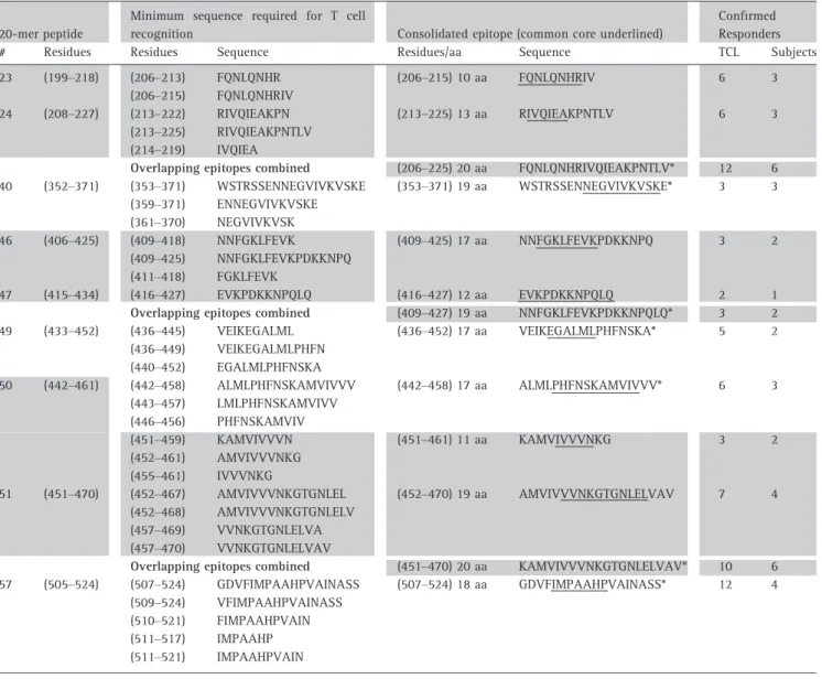
The minimum T cell stimulatory sequence (core epitope) within each selected 20-mer was determined by testing the proliferation of reactive TCL from different subjects to truncated peptide sets (e.g. Fig. 2 and Table 3). The number of residues required to induce maximal T cell proliferation varied from 6 to 19 aa between different TCL and/or subjects (Table 3), consistent with previous reports for CD4+ T cell epitopes [60, 61]. Due to varia-tion in the number of flanking residues required for optimal epitope recognition [61], TCL were considered to recognize the same epitope if peptides containing a common core sequence induced recognition. Based on this criterion, 10 distinct CD4+ T cell epitopes were identified (‘consolidated epitopes’, Table 3), with com-mon cores varying from 5 to 12 aa (underlined sequences, Table 3). ‘Consolidated epitope’ sequences were selected to encompass residues required for maxi-mal stimulation of all specific TCL tested to ensure broadest possible recognition.
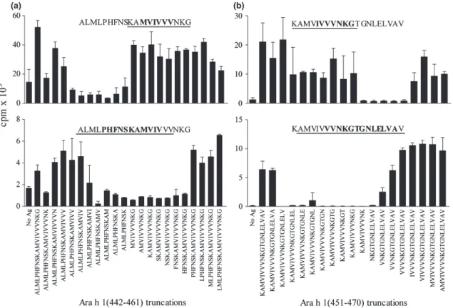
At least one epitope was found within each of the nine 20-mers, with 20-mers 50 and 51 each containing two distinct but overlapping T cell epitopes: one unique to each 20-mer (442–458 and 452–470), and the other within the overlap sequence (451–461, Table 3 and Fig. 2). No single TCL responded to both epitopes
within either 20-mer, further confirming the distinction of these epitopes (data not shown). HLA-epitope predic-tion algorithms [62, 63] also highlighted one or more strong HLA class II (HLA-II) binding motifs within each of our minimal-stimulatory sequences. Data are shown for the Propred [62] HLA-DR binding algorithm in Table S4. This algorithm did not predict HLA-DR epi-topes within peptide 40, but algorithms of the Immune Epitope Database (IEDB) and Analysis Resource [63] predicted epitopes within this peptide to bind most strongly to HLA-DP and/or -DQ molecules.
Finally, to avoid unnecessary sequence duplication and to minimize peptide numbers for a therapeutic, six of the consolidated epitopes (comprising three overlap-ping epitope pairs) were combined into three single peptides of 20 aa or less (206–225, 409–427 and 451– 470; grey shading, Table 3). The combined epitope peptides efficiently stimulated TCL specific for either epitope (data not shown) and together with the remaining four consolidated epitopes (353–371, 436– 452, 442–458 and 507–524), provided a panel of seven candidate peptides for further characterization (see asterisks, Table 3). CFSE-based screening of nine sub-jects from our cohorts confirmed that these peptides could each directly target detectable numbers of Ara h
(a) (b)
Fig. 2.Mapping core T cell epitopes within Ara h 1 20-mer peptides 50 and 51. 20-mer-specific TCL proliferation to truncated peptide sets. Repre-sentative TCL shown for peptides 50 (a) and 51 (b) (mean cpm replicate wells+SD). Upper panels indicate the epitope in overlap between the 20-mers (n=2; 3 TCL). Lower panels indicate epitopes unique to each 20-mer; (a)n=3; 6 TCL. (b)n=4; 7 TCL. Epitope sequences recognized by represented TCL are bolded and ‘consolidated epitopes’ recognized by all specific TCL are underlined.
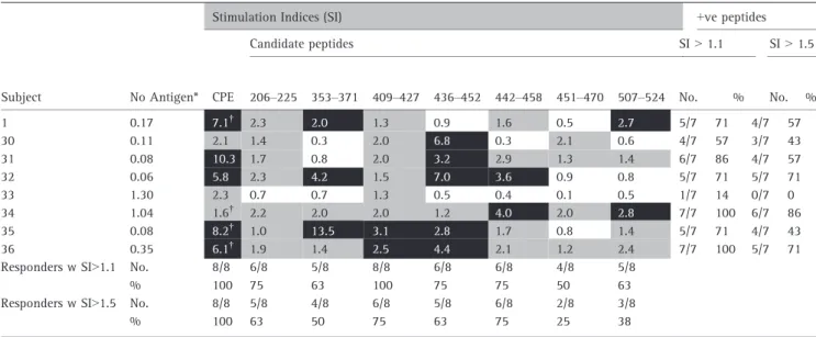
1-specific T cells among whole PBMC of peanut-aller-gic subjects (Table 4). In a few cases, the T cell response of a given subject to the original 20-mer and the corresponding candidate peptide differed. Where responses to candidates were reduced as compared to the 20-mer, flanking residue(s) required for optimal T cell recognition or HLA-binding may have been removed. It is well recognized that flanking residues can stabilize peptide binding to HLA class II molecules. In contrast, improved responses could reflect genera-tion of addigenera-tional new epitopes, improved epitope pur-ity [cores were synthesized at high purpur-ity (> 95%), whilst 20-mers were produced as peptide sets with a minimum estimated purity of 70%] or alteration of epitopes (or flanking residues) to enable better interac-tion with HLA and/or T cell receptor (TCR) molecules.
Indeed, this was considered the case where responses to candidate peptide (442–458) were much stronger than to 20-mer 50. As this region contains multiple adjacent hydrophobic residues, even single residue changes could significantly alter the charge and struc-ture of this peptide, thus affecting its biochemical properties and interactions with HLA and/or TCR mole-cules. Nonetheless, most responses to candidates were comparable or improved compared to responses to the original 20-mers.
Determining HLA class II restriction specificity of Ara h 1 T cell epitopes
There is no identified HLA-II association with peanut allergy [64], therefore peptides selected for therapy
Table 3.Core T cell epitope sequences mapped within selected Ara h 1 20-mers
*The seven candidate peptides proposed for a therapeutic.
must bind diverse HLA-II molecules for wide applicabil-ity. To determine the HLA-II type presenting each epitope, anti-HLA-DR, -DP or -DQ mAbs were used to block individual epitope presentation to T cells. For each TCL tested, epitope recognition was prevented by
one or more HLA-mAb in a dose-dependent manner (e.g. Figure S2) and the same mAb blocked recognition of CPE (data not shown), demonstrating consistency for presentation of naturally processed and synthetic epitope forms. At least two subjects and/or TCL were
Table 4.CFSE-based detection of peanut-allergic donor CD4+T cell proliferation in response to selected Ara h 1 candidate peptides
*Background proliferation with no antigen, % CD4+CFSEloT cells of total CD4+T cells.
†A combination of enriched Ara h 1 and Ara h 2 (10lg/mL of each) was used instead of CPE for these subjects. CPE, crude peanut extract;+ve, positive; Grey, stimulation indices 1.1<2.5; Black, stimulation indices 2.5.
Table 5.HLA class II restriction of core epitope peptides
nt, not tested (TCL not available); Grey shading indicates overlapping epitope pairs combined into single peptides for further analyses as outlined in the text.
tested per epitope (Table 5). Consistent with predictions of the HLA-II algorithms described above [62, 63], anti-HLA-DR blocked recognition of all but one epitope (353–371), which was blocked by anti-HLA-DQ in both subjects tested. For epitopes 436–452 and 507–524, rec-ognition was blocked by anti-HLA-DR for some TCL but by anti-HLA-DQ for others, confirming HLA-bind-ing degeneracy for these epitopes.
To assess HLA-binding degeneracy of epitopes whose recognition was blocked by a single HLA-mAb, the respective HLA-alleles of at least two subjects with TCL specific for that epitope were compared (Table S2 and Table 5). The absence of shared HLA-DRB1 or HLA-DQB1 alleles between subjects recognizing HLA-DR- or HLA-DQ-restricted epitopes, respectively, confirmed that each epitope was present on at least two different HLA-molecules. The HLA-binding algorithms further supported these data, with each epitope containing motifs predicted to bind multiple HLA molecules [62, 63] (e.g. Table S4).
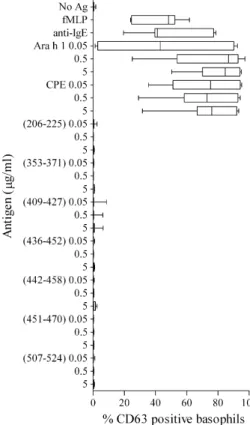
Testing candidate peptides for basophil activation
To provide a safe alternative to whole allergens, pep-tides must not bind and cross-link cell-bound IgE.
Basophil reactivity to peptides was assessed in fresh blood from seven of the peanut-allergic subjects recruited for this study (Fig. 3). All seven subjects showed high levels of basophil activation to CPE over a concentration range. Whilst responses to Ara h 1 varied between subjects at the lowest dose, the highest con-centration induced high activation in all subjects. How-ever, none of the candidate peptides induced activation at any concentration tested. One subject showed a very low response (8%) to peptide 409–427, but this was below the threshold of positive activation [65] and was negligible compared to the activation induced by Ara h 1 (80–90%) or CPE (74–76%) in this subject.
Discussion
An appropriately selected T cell-targeted peptide immu-notherapeutic will provide a safe treatment option for peanut-allergic individuals. Candidate peptides must comprise HLA-degenerate CD4+ T cell epitopes of the major peanut allergens recognized by HLA-diverse pea-nut-allergic individuals, without cross-linking cell-bound IgE and activating inflammatory cells. To maxi-mize the population coverage and efficacy of a thera-peutic, we designed a peptide set containing T cell epitopes from the two major allergens Ara h 1 and Ara h 2. Following on from our previous study of Ara h 2 [49], we now provide the first report of core sequences of CD4+ T cell epitopes of the most abundant major peanut allergen Ara h 1. We identified and character-ized 10 HLA-diverse CD4+ T cell epitopes of Ara h 1, and used these sequences to design candidate Ara h 1 peptides to combine with our candidate Ara h 2 pep-tides for therapeutic development.
To commence this study, we selected nine 20-mers of Ara h 1 as containing the most frequently recognized epitopes based on responses of 145 TCL from 18 HLA-diverse peanut-allergic subjects. The cohort HLA profile was typical of Caucasian populations [66] in countries where peanut allergy is prevalent [67]. We further vali-dated our 20-mer selection by demonstrating their col-lective recognition by PBMC T cells directly ex vivo
from an additional 18 peanut-allergic subjects (Table 2), resulting in a total responder frequency of 92% for the 38 subjects analysed. Although we could not confirm T cell recognition of these 20-mers in four subjects, it is possible that specific T cells went undetected for two of these subjects (5 and 7), as data were only obtained from three and four TCL respectively (Table 1).
The minimum T cell stimulatory sequences identified within our selected 20-mers varied from 6 to 19 aa (Table 3), consistent with reports of different peptides processed for HLA-II presentation and/or required for HLA- and/or TCR-binding both within and between sub-jects [60, 61]. As we used oligoclonal TCL, it is possible
Fig. 3.Basophil activation in response to candidate Ara h 1 peptides. Box-and-whiskers plot showing percentage of activated (CD63hi) ba-sophils (IgEhi) in response to Ara h 1 or candidate peptides for seven peanut-allergic subjects. Negative control was no antigen (unstimu-lated) and positive controls were anti-IgE, fMLP and CPE. Whiskers show minimum to maximum values.
that the longer sequences contained more than one epi-tope. Indeed, algorithms [62, 63] predicted up to three HLA-binding motifs within some of our consolidated epi-topes (Table S4). Seven of our epiepi-topes showed overlap with T cell-reactive Ara h 1 20-mers recently identified using HLA-DR tetramers [50], providing further support for recognition of these peptides in larger peanut-allergic populations. In addition, we confirmed the presentation of five of these peptides on additional HLA molecules to those used for the tetramer mapping [50]. However, epi-topes 353–371, 436–452 and 442–458 were unique to our study. Consistent with this observation, we showed these epitopes were either presented on HLA-DQ mole-cules (commonly observed for allergen T cell epitopes [68–72]), or HLA-DR types for which no tetramer-specific T cells were detected [50] (Table 5). Inclusion of HLA-DQ-restricted epitopes is particularly advantageous for therapeutics as these alleles are less variable and thus more prevalent in mixed populations than HLA-DR alleles [73]. However, we confirmed HLA-binding degen-eracy for all epitopes identified (both HLA-DR and -DQ-restricted) (Table 5), with further degeneracy predicted by algorithms [62, 63] (e.g. Table S4), emphasizing their collective suitability for targeting HLA-diverse peanut-allergic populations.
The main rationale for developing a peptide immuno-therapy for peanut allergy is to identify an effective allergen preparation that does not invoke the adverse effects seen with whole peanut extract [10, 12–14, 19, 37, 74]. Mapping the core sequences of T cell epitopes enables refined peptide design for a therapeutic, but selecting optimal peptide combinations is a balance between peptide length and number. Longer peptides will increase population coverage by encompassing more T cell epitopes, and being fewer in number will reduce the complexity of therapeutic standardization compared to using a greater number of shorter peptides. However, the main concern with longer peptides is the
increased potential for IgE binding and cross-linking, resulting in adverse reactions. We opted to combine overlapping epitopes into peptides up to 20 aa in length. Of over 23 linear IgE epitopes reported for Ara h 1 [75–77], only two minor epitopes (409–418 and 461–470) fell within our candidate peptides [75]. Most importantly, none of these peptides caused activation of reactive basophils in all of the seven peanut-allergic subjects tested (Fig. 3), emphasizing the poten-tial for these peptides to provide a safe alternative to whole allergen extract for immunotherapy.
In summary, we report the novel identification of 10 reliably recognized CD4+T cell epitopes of Ara h 1 that collectively show diverse HLA class II restriction. We incorporated these epitopes into a panel of seven short ( 20 aa), HLA-degenerate peptides that can target T cells within PBMC of HLA-diverse allergic individuals without causing activation of peanut-allergic donor ba-sophils. The combination of these Ara h 1 peptides with our three T cell epitope-based Ara h 2 peptides [49] provides strong candidates for a broad acting and safe peptide-therapeutic to treat peanut allergy.
Acknowledgements
We thank Dr Nicole Mifsud for helpful intellectual dis-cussions, Neeru Varese for technical assistance and Karen Symons and Kirsten Deckert for patient recruit-ment and blood collection. This study was supported by grants from the Ilhan Food Allergy Foundation and the National Health and Medical Research Council of Aus-tralia.
Conflicts of interest
The authors declare that they have no conflict of inter-est relating to this manuscript.
References
1 Bock SA, Munoz-Furlong A, Sampson HA. Further fatalities caused by ana-phylactic reactions to food, 2001– 2006. J Allergy Clin Immunol 2007;
119:1016–8.
2 Burks AW. Peanut allergy. Lancet
2008;371:1538–46.
3 Sicherer SH, Munoz-Furlong A, Godbold JH, Sampson HA. US prevalence of self-reported peanut, tree nut, and sesame allergy: 11-year follow-up. J Allergy
Clin Immunol2010;125:1322–6.
4 Husain Z, Schwartz RA. Peanut allergy: an increasingly common
life-threatening disorder. J Am Acad
Der-matol2012;66:136–43.
5 Kemp AS, Hu W. Food allergy and anaphylaxis - dealing with uncer-tainty.Med J Aust2008;188:503–4. 6 Busse PJ, Nowak-Wegrzyn AH, Noone
SA, Sampson HA, Sicherer SH. Recur-rent peanut allergy. N Engl J Med
2002;347:1535–6.
7 Yun J, Katelaris CH. Food allergy in adolescents and adults. Intern Med J
2009;39:475–8.
8 Avery NJ, King RM, Knight S, Hourih-ane JO. Assessment of quality of life in children with peanut allergy. Pediatr
Allergy Immunol2003;14:378–82.
9 Sampson MA, Munoz-Furlong A, Sich-erer SH. Risk-taking and coping strate-gies of adolescents and young adults with food allergy. J Allergy Clin
Immunol2006;117:1440–5.
10 Oppenheimer JJ, Nelson HS, Bock SA, Christensen F, Leung DY. Treatment of peanut allergy with rush immunother-apy. [see comment]. J Allergy Clin
Immunol1992;90:256–62.
11 Pumphrey R. Anaphylaxis: can we tell who is at risk of a fatal reaction?Curr
Opin Allergy Clin Immunol 2004;
4:285–90.
12 Varshney P, Steele PH, Vickery BPet al.
immunotherapy home dosing.J Allergy
Clin Immunol2009;124:1351–2.
13 Nelson HS, Lahr J, Rule R, Bock A, Leung D. Treatment of anaphylactic sensitivity to peanuts by immunother-apy with injections of aqueous peanut extract. J Allergy Clin Immunol 1997;
99(6 Pt 1):744–51.
14 Jones SM, Pons L, Roberts JL et al.
Clinical efficacy and immune regula-tion with peanut oral immunotherapy.
J Allergy Clin Immunol2009; 24:292–
300.
15 Varshney P, Jones SM, Scurlock AM
et al. A randomized controlled study
of peanut oral immunotherapy: clinical desensitization and modulation of the allergic response.J Allergy Clin Immu-nol2011;127:654–60.
16 Anagnostou K, Clark A, King Y, Islam S, Deighton J, Ewan P. Efficacy and safety of high-dose peanut oral immu-notherapy with factors predicting out-come.Clin Exp Allergy2011;41:1273– 81.
17 Allen KJ, O’Hehir RE. The evolution of oral immunotherapy for the treatment of peanut allergy. Clin Exp Allergy
2011;41:1172–4.
18 Thyagarajan A, Varshney P, Jones SM
et al. Peanut oral immunotherapy is
not ready for clinical use. J Allergy
Clin Immunol2010;126:31–2.
19 Hofmann AM, Scurlock AM, Jones SM
et al.Safety of a peanut oral
immuno-therapy protocol in children with pea-nut allergy. J Allergy Clin Immunol
2009;124:286–91.
20 Sabatos-Peyton CA, Verhagen J, Wraith DC. Antigen-specific immuno-therapy of autoimmune and allergic diseases. Curr Opin Immunol 2010;
22:609–15.
21 Rolland JM, Gardner LM. O’Hehir R E. Functional regulatory T cells and aller-gen immunotherapy.Curr Opin Allergy
Clin Immunol2010;10:559–66.
22 Akdis CA, Akdis M. Mechanisms of allergen-specific immunotherapy. J
Allergy Clin Immunol. 2011;127:18–
27; quiz 8-9.
23 Alexander C, Ying S. A BK, Larche M. Fel d 1-derived T cell peptide therapy induces recruitment of CD4+ CD25+; CD4+interferon-gamma+T helper type 1 cells to sites of allergen-induced late-phase skin reactions in cat-allergic subjects. Clin Exp Allergy 2005;
35:52–8.
24 Alexander C, Tarzi M, Larche M, Kay AB. The effect of Fel d 1-derived T-cell peptides on upper and lower air-way outcome measurements in cat-allergic subjects. Allergy 2005;
60:1269–74.
25 Campbell JD, Buckland KF, McMillan
SJ et al. Peptide immunotherapy in
allergic asthma generates IL-10-depen-dent immunological tolerance associ-ated with linked epitope suppression.J
Exp Med2009;206:1535–47.
26 Kay AB, Larche M. Allergen immuno-therapy with cat allergen peptides.
Springer Semin Immunopathol 2004;
25:391–9.
27 Oldfield WL, Larche M, Kay AB. Effect of T-cell peptides derived from Fel d 1 on allergic reactions and cytokine pro-duction in patients sensitive to cats: a randomised controlled trial. Lancet
2002;360:47–53.
28 Muller U, Akdis CA, Fricker M et al.
Successful immunotherapy with T-cell epitope peptides of bee venom phos-pholipase A2 induces specific T-cell anergy in patients allergic to bee venom. J Allergy Clin Immunol 1998;
101(6 Pt 1):747–54.
29 Kammerer R, Chvatchko Y, Kettner A, Dufour N, Corradin G, Spertini F. Mod-ulation of T-cell response to phospho-lipase A2 and phospholipase A2-derived peptides by conventional bee venom immunotherapy. J Allergy Clin
Immunol1997;100:96–103.
30 Yang M, Yang C, Mine Y. Multiple T cell epitope peptides suppress allergic responses in an egg allergy mouse model by the elicitation of forkhead box transcription factor 3- and trans-forming growth factor-beta-associated mechanisms. Clin Exp Allergy 2010;
40:668–78.
31 Yoshitomi T, Nakagami Y, Hirahara K, Taniguchi Y, Sakaguchi M, Ya-mashita M. Intraoral administration of a T-cell epitope peptide induces immunological tolerance in Cry j 2-sensitized mice. J Pept Sci 2007;
13:499–503.
32 Marazuela EG, Rodriguez R, Fernan-dez-Garcia H, Garcia MS, Villalba M, Batanero E. Intranasal immunization with a dominant T-cell epitope peptide of a major allergen of olive pollen pre-vents mice from sensitization to the whole allergen. Mol Immunol 2008;
45:438–45.
33 Rupa P, Mine Y. Oral immunotherapy with immunodominant T-cell epitope peptides alleviates allergic reactions in a Balb/c mouse model of egg allergy.
Allergy2012;67:74–82.
34 Hoyne GF, O’Hehir RE, Wraith DC, Thomas WR, Lamb JR. Inhibition of T cell and antibody responses to house dust mite allergen by inhalation of the dominant T cell epitope in naive and sensitized mice. J Exp Med 1993;
178:1783–8.
35 Hall G, Houghton CG, Rahbek JU, Lamb JR, Jarman ER. Suppression of allergen reactive Th2 mediated responses and pulmonary eosinophilia by intranasal administration of an immunodominant peptide is linked to IL-10 production. Vaccine 2003;
21:549–61.
36 Allergen Nomenclature, International Union of Immunological Societies [database on the Internet]. [cited 2010]. Available from: http://www.allergen. org/Allergen.aspx.
37 de Leon MP, Rolland JM, O’Hehir RE. The peanut allergy epidemic: allergen molecular characterisation and pros-pects for specific therapy. Expert Rev
Mol Med2007;9:1–18.
38 Palmer K, Burks W. Current develop-ments in peanut allergy. Curr
Opin Allergy Clin Immunol 2006;
6:202–6.
39 Blanc F, Adel-Patient K, Drumare MF, Paty E, Wal JM, Bernard H. Capacity of purified peanut allergens to induce degranulation in a functional in vitro assay: Ara h 2 and Ara h 6 are the most efficient elicitors. Clin Exp
Allergy2009;39:1277–85.
40 Koppelman SJ, Wensing M, Ertmann M, Knulst AC, Knol EF. Relevance of Ara h1, Ara h2 and Ara h3 in peanut-allergic patients, as determined by immunoglobulin E Western blotting, basophil-histamine release and intracu-taneous testing: Ara h2 is the most important peanut allergen. Clin Exp
Allergy2004;34:583–90.
41 Palmer GW, Dibbern DA Jr, Burks AW
et al.Comparative potency of Ara h 1
and Ara h 2 in immunochemical and functional assays of allergenicity.Clin
Immunol2005;115:302–12.
42 Glaumann S, Nopp A, Johansson SG, Rudengren M, Borres MP, Nilsson C. Basophil allergen threshold sensitivity, CD-sens, IgE-sensitization and DBPCFC
in peanut-sensitized children. Allergy
2012;67:242–7.
43 Chiang WC, Pons L, Kidon MI, Liew WK, Goh A, Wesley Burks A. Serologi-cal and cliniSerologi-cal characteristics of chil-dren with peanut sensitization in an Asian community. Pediatr Allergy
Immunol. 2009;21(2 Pt 2):e429–38.
44 Asarnoj A, Moverare R, Ostblom E
et al. IgE to peanut allergen
compo-nents: relation to peanut symptoms and pollen sensitization in 8-year-olds.
Allergy. 2010;65:1189–95.
45 Moverare R, Ahlstedt S, Bengtsson U
et al. Evaluation of IgE antibodies to
recombinant peanut allergens in patients with reported reactions to
pea-nut. Int Arch Allergy Immunol 2011;
156:282–90.
46 Lin YT, Charles Wu CT, Cheng JH, Huang JL, Yeh KW. Patterns of sensi-tization to peanut allergen compo-nents in Taiwanese Preschool children.
J Microbiol Immunol Infect 2012;
45:90–5.
47 Peeters KA, Koppelman SJ, van Hoffen
Eet al. Does skin prick test reactivity
to purified allergens correlate with clinical severity of peanut allergy?Clin
Exp Allergy2007;37:108–15.
48 Koppelman SJ, Vlooswijk RA, Knippels
LMet al.Quantification of major
pea-nut allergens Ara h 1 and Ara h 2 in the peanut varieties Runner, Spanish, Virginia, and Valencia, bred in differ-ent parts of the world. Allergy 2001;
56:132–7.
49 Prickett SR, Voskamp AL, Dacumos-Hill A, Symons K, Rolland JM, O’Hehir RE. Ara h 2 peptides containing dominant CD4+T-cell epitopes: candidates for a peanut allergy therapeutic. J Allergy
Clin Immunol2011;127:608–15; e1-5.
50 DeLong JH, Simpson KH, Wambre E, James EA, Robinson D, Kwok WW. Ara h 1-reactive T cells in individuals with peanut allergy.J Allergy Clin Immunol
2011;127:1211–8; e3.
51 de Leon MP, Glaspole IN, Drew AC, Rolland JM, O’Hehir RE, Suphioglu C. Immunological analysis of allergenic cross-reactivity between peanut and tree nuts. Clin Exp Allergy 2003;
33:1273–80.
52 Eusebius NP, Papalia L, Suphioglu C
et al.Oligoclonal analysis of the atopic
T cell response to the group 1 allergen of Cynodon dactylon (bermuda grass) pollen: pre- and post-allergen-specific
immunotherapy. Int Arch Allergy
Immunol2002;127:234–44.
53 Mannering SI, Dromey JA, Morris JS, Thearle DJ, Jensen KP, Harrison LC. An efficient method for cloning human autoantigen-specific T cells.J Immunol
Methods2005;298:83–92.
54 Drew AC, Eusebius NP, Kenins L et al.
Hypoallergenic variants of the major latex allergen Hev b 6.01 retaining human T lymphocyte reactivity. J
Immunol2004;173:5872–9.
55 Etto T, de Boer C, Prickett S et al.
Unique and cross-reactive T cell epi-tope peptides of the major Bahia grass pollen allergen, Pas n 1. Int Arch
Allergy Immunol2012;159:355–66.
56 Pascal M, Konstantinou GN, Masilama-ni M, Lieberman J, Sampson HA. In silico prediction of Ara h 2 T cell epi-topes in peanut-allergic children. Clin
Exp Allergy2013;43:116–27.
57 Cardaba B, Del Pozo V, Jurado Aet al.
Olive pollen allergy: searching for im-munodominant T-cell epitopes on the Ole e 1 molecule. Clin Exp Allergy
1998;28:413–22.
58 de Silva HD, Gardner LM, Drew AC, Beezhold DH, Rolland JM, O’Hehir RE. The hevein domain of the major latex-glove allergen Hev b 6.01 contains dominant T cell reactive sites.Clin Exp
Allergy2004;34:611–8.
59 Oseroff C, Sidney J, Kotturi MF et al.
Molecular determinants of T cell epi-tope recognition to the common Timo-thy grass allergen. J Immunol 2010;
185:943–55.
60 Hemmer B, Kondo T, Gran B et al.
Minimal peptide length requirements for CD4(+) T cell clones–implications for molecular mimicry and T cell survival. Int Immunol 2000; 12:375– 83.
61 Suri A, Lovitch SB, Unanue ER. The wide diversity and complexity of pep-tides bound to class II MHC molecules.
Curr Opin Immunol2006;18:70–7.
62 Singh H, Raghava GP. ProPred: predic-tion of HLA-DR binding sites.
Bioinfor-matics2001;17:1236–7.
63 Vita R, Zarebski L, Greenbaum JA
et al. The immune epitope database
2.0. Nucleic Acids Res 2010; 38
(Data-base issue): D854–62.
64 Shreffler WG, Charlop-Powers Z, Sich-erer SH. Lack of association of HLA class II alleles with peanut allergy.[see comment].Ann Allergy Asthma Immu-nol2006;96:865–9.
65 Boumiza R, Debard AL, Monneret G. The basophil activation test by flow cytometry: recent developments in clinical studies, standardization and emerging perspectives. Clin Mol
Allergy2005;3:9.
66 Middleton D, Menchaca L, Rood H, Ko-merofsky R. New allele frequency data-base: http://www.allelefrequencies.net.
Tissue Antigens. 2003;61:403–7.
67 Shek LP, Cabrera-Morales EA, Soh SE
et al. A population-based
question-naire survey on the prevalence of pea-nut, tree pea-nut, and shellfish allergy in 2 Asian populations. J Allergy Clin
Immunol2010;126:324–31; e7.
68 Bateman EA, Ardern-Jones MR, Ogg GS. Identification of an immunodomi-nant region of Fel d 1 and character-ization of constituent epitopes.[see comment]. Clin Exp Allergy 2008;
38:1760–8.
69 Verhoef A, Higgins JA, Thorpe CJet al.
Clonal analysis of the atopic immune response to the group 2 allergen of Der-matophagoides spp.: identification of HLA-DR and -DQ restricted T cell epi-topes.Int Immunol1993;5:1589–97. 70 Higgins JA, Thorpe CJ, Hayball JD,
O’Hehir RE, Lamb JR. Overlapping T-cell epitopes in the group I allergen of Dermatophagoides species restricted by HLA-DP and HLA-DR class II mole-cules. J Allergy Clin Immunol 1994;
93:891–9.
71 Ruiter B, Rozemuller EH, van Dijk AJ
et al.Role of human leucocyte antigen
DQ in the presentation of T cell epi-topes in the major cow’s milk allergen alphas1-casein. Int Arch Allergy
Immunol2007;143:119–26.
72 van Neerven RJ, van de Pol MM, van Milligen FJ, Jansen HM, Aalberse RC, Kapsenberg ML. Characterization of cat dander-specific T lymphocytes from atopic patients. J Immunol 1994;
152:4203–10.
73 Larche M. Of cats and men: immuno-dominance and the role of HLA-DP/ DQ. [comment].Clin Exp Allergy2008;
38:1709–11.
74 Rolland JM, Gardner LM, O’Hehir RE. Allergen-related approaches to immu-notherapy. Pharmacol Ther 2009;
121:273–84.
75 Burks AW, Shin D, Cockrell G, Stanley JS, Helm RM, Bannon GA. Mapping and mutational analysis of the IgE-binding epitopes on Ara h 1, a legume vicilin protein and a major allergen in
peanut hypersensitivity.Eur J Biochem
1997;245:334–9.
76 Shreffler WG, Beyer K, Chu TH, Burks AW, Sampson HA. Microarray immu-noassay: association of clinical history, in vitro IgE function, and
heterogene-ity of allergenic peanut epitopes. J
Allergy Clin Immunol 2004; 113:776–
82.
77 van Boxtel EL, Koppelman SJ, van den Broek LA, Gruppen H. Determination of pepsin-susceptible and
pepsin-resis-tant epitopes in native and heat-treated peanut allergen Ara h 1.J Agric Food Chem2008;56:2223–30.
Supporting Information
Additional Supporting Information may be found in the online version of this article:
Figure S1. Representative CFSE-based assay for detecting CD4+T cell proliferation in PBMC.
Figure S2. Representative HLA class II restriction specificity of T cell epitope recognition.
Table S1.Subject demographics.
Table S2.HLA genotyping for subjects used for T cell line generation.
Table S3.Ara h 1 20-mer peptides.
Table S4. Predicted HLA-DR binding motifs in