of Medically Important Yeasts
Lars F. Westblade,a,bRebecca Jennemann,cJohn A. Branda,dMaureen Bythrow,eMary Jane Ferraro,dOmai B. Garner,f Christine C. Ginocchio,b,eMichael A. Lewinski,fRyhana Manji,eA. Brian Mochon,fGary W. Procop,gSandra S. Richter,g Jenna A. Rychert,dLinda Sercia,gCarey-Ann D. Burnhama
Department of Pathology and Immunology, Washington University School of Medicine, St. Louis, Missouri, USAa
; Department of Pathology and Laboratory Medicine, Hofstra North Shore-LIJ School of Medicine, Hempstead, New York, USAb
; Barnes-Jewish Hospital, St. Louis, Missouri, USAc
; Department of Pathology, Massachusetts General Hospital and Harvard Medical School, Boston, Massachusetts, USAd
; Department of Pathology and Laboratory Medicine, North Shore-LIJ Health System Laboratories, Lake Success, New York, USAe
; Department of Pathology and Laboratory Medicine, David Geffen School of Medicine at UCLA, Los Angeles, California, USAf ; Department of Clinical Pathology, Cleveland Clinic, Cleveland, Ohio, USAg
The optimal management of fungal infections is correlated with timely organism identification. Matrix-assisted laser desorption
ionization–time of flight (MALDI-TOF) mass spectrometry (MS) is revolutionizing the identification of yeasts isolated from
clinical specimens. We present a multicenter study assessing the performance of the Vitek MS system (bioMérieux) in
identify-ing medically important yeasts. A collection of 852 isolates was tested, includidentify-ing 20
Candida
species (626 isolates, including 58
C.
albicans
, 62
C. glabrata
, and 53
C. krusei
isolates), 35
Cryptococcus neoformans
isolates, and 191 other clinically relevant yeast
isolates; in total, 31 different species were evaluated. Isolates were directly applied to a target plate, followed by a formic acid
overlay. Mass spectra were acquired using the Vitek MS system and were analyzed using the Vitek MS v2.0 database. The gold
standard for identification was sequence analysis of the D2 region of the 26S rRNA gene. In total, 823 isolates (96.6%) were
iden-tified to the genus level and 819 isolates (96.1%) were ideniden-tified to the species level. Twenty-four isolates (2.8%) were not
identi-fied, and five isolates (0.6%) were misidentified. Misidentified isolates included one isolate of
C. albicans
(
n
ⴝ
58) identified as
Candida dubliniensis
, one isolate of
Candida parapsilosis
(
n
ⴝ
73) identified as
Candida pelliculosa
, and three isolates of
Geotri-chum klebahnii
(
n
ⴝ
6) identified as
Geotrichum candidum
. The identification of clinically relevant yeasts using MS is superior
to the phenotypic identification systems currently employed in clinical microbiology laboratories.
A
s the number of patients with profound immunosuppression
(such as those with solid-organ and hematopoietic stem cell
transplants) continues to rise, the morbidity and mortality
bur-dens attributed to invasive fungal infections are increasing (
1
–
6
).
In the case of invasive fungal infections, expedient identification
of the offending organism is essential for optimal patient
manage-ment and the best clinical outcomes. As the antifungal
suscepti-bility profiles for many fungi (both yeasts and molds) are
predict-able, organism identification frequently is sufficient to expedite
appropriate empirical antifungal therapy. This has been
demon-strated both to reduce the overall length of hospitalization and to
maximize favorable clinical outcomes (
7
–
10
). Conversely, the
rapid exclusion of overt pathogenic or intrinsically resistant
spe-cies can be used to narrow therapy and/or to prevent treatment
with potentially toxic antifungal agents, thereby reducing negative
clinical outcomes and costs.
The methods for identification of yeasts in the diagnostic
clin-ical microbiology laboratory have improved significantly over the
past several decades (
11
,
12
), with methods ranging from simple
manual biochemical assays to automated biochemical methods to
sophisticated nucleic acid-based assays (
11
,
12
). While these
ad-vancements in methodology have greatly enhanced our ability to
identify yeasts, the limitations of these methods include cost,
turnaround time, and, in some instances, the need for
consid-erable expertise. Additionally, the accuracy of identification for
some less-common species is not optimal for some of the
meth-ods (
13
–
17
).
A technology that is poised to revolutionize the rapid
identifi-cation of yeasts isolated in the clinical microbiology laboratory is
matrix-assisted laser desorption ionization–time of flight
(MALDI-TOF) mass spectrometry (MS). MALDI-TOF MS-based
microbial identification relies on the generation of an
organism-specific mass spectrum or “protein fingerprint” that is examined
against a reference database to provide an organism identification
(
18
). The objective of this multicenter study was to assess the
performance of the Vitek MS MALDI-TOF mass spectrometer
(bioMérieux) in conjunction with the Vitek MS v2.0 database for
the identification of yeasts isolated in diagnostic clinical
microbi-ology laboratories.
(This work was presented in part as an abstract at the 113th
General Meeting of the American Society for Microbiology,
Den-ver, CO, 18 to 21 May 2013.)
MATERIALS AND METHODS
Isolates used in this study.Yeasts isolated and identified from clinical specimens obtained from five diagnostic clinical microbiology laborato-ries, located at geographically distinct sites in North America, were in-cluded in the study. The study sites were Barnes-Jewish Hospital (St. Louis, MO), the Cleveland Clinic (Cleveland, OH), the UCLA Health System (Los Angeles, CA), the North Shore LIJ Core Laboratory (Lake
Received11 March 2013Returned for modification26 March 2013
Accepted2 May 2013
Published ahead of print8 May 2013
Address correspondence to Carey-Ann D. Burnham, [email protected].
Copyright © 2013, American Society for Microbiology. All Rights Reserved.
doi:10.1128/JCM.00680-13
on May 16, 2020 by guest
http://jcm.asm.org/
Success, NY), and the Massachusetts General Hospital (Boston, MA). In total, the collection tested was composed of 852 yeast isolates obtained from the five trial sites (508 isolates) and the bioMérieux stock collection
(344 isolates). The collection included 20Candidaspecies (Table 1),
Cryp-tococcus neoformans, and 10 species in the generaGeotrichum,Kodamaea,
Malassezia,Rhodotorula,Saccharomyces, andTrichosporon(Table 2). Of the 344 isolates from the bioMérieux stock collection, 96 were used in the development of the database. These isolates represent rare taxa, such that it would not have been possible to evaluate them exclusively via prospective collection.
Cultivation of yeast isolates.The isolates were obtained from frozen stocks or were tested fresh from clinical cultures. Strains that were stored frozen were subcultured on Sabouraud dextrose agar (SDA; Remel,
Le-nexa, KS) twice before mass spectrometric analysis. Freshly collected iso-lates were subcultured on SDA to assess purity before testing, or, if a pure culture was observed on the primary SDA plate, it was tested directly. All isolates were analyzed within 72 h after visible growth at 35°C. In only four instances, isolates were taken from media other than SDA, including one isolate taken from CHROMagar Candida (Becton, Dickinson, Sparks, MD), one isolate taken from Mueller-Hinton II agar (Becton, Dickinson), and two isolates taken from tryptic soy agar with sheep’s blood (Remel). In the four instances where SDA was not used to cultivate the strain for MS analysis, the MS identification matched the reference identification method.
Sample preparation.The yeast isolates were prepared for mass
[image:2.585.41.555.78.341.2]spec-trometric analysis using a direct, on-target, extraction method (19).
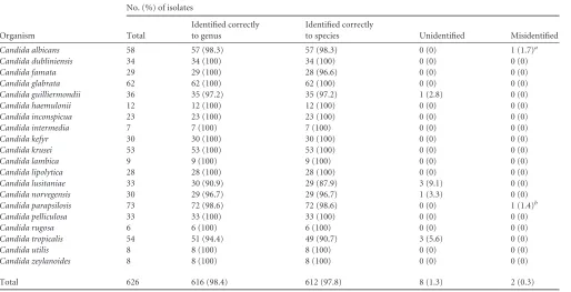
TABLE 1Performance characteristics of the Vitek MS system in identifying clinically relevantCandidaspecies
Organism
No. (%) of isolates
Total
Identified correctly to genus
Identified correctly
to species Unidentified Misidentified
Candida albicans 58 57 (98.3) 57 (98.3) 0 (0) 1 (1.7)a
Candida dubliniensis 34 34 (100) 34 (100) 0 (0) 0 (0)
Candida famata 29 29 (100) 28 (96.6) 0 (0) 0 (0)
Candida glabrata 62 62 (100) 62 (100) 0 (0) 0 (0)
Candida guilliermondii 36 35 (97.2) 35 (97.2) 1 (2.8) 0 (0)
Candida haemulonii 12 12 (100) 12 (100) 0 (0) 0 (0)
Candida inconspicua 23 23 (100) 23 (100) 0 (0) 0 (0)
Candida intermedia 7 7 (100) 7 (100) 0 (0) 0 (0)
Candida kefyr 30 30 (100) 30 (100) 0 (0) 0 (0)
Candida krusei 53 53 (100) 53 (100) 0 (0) 0 (0)
Candida lambica 9 9 (100) 9 (100) 0 (0) 0 (0)
Candida lipolytica 28 28 (100) 28 (100) 0 (0) 0 (0)
Candida lusitaniae 33 30 (90.9) 29 (87.9) 3 (9.1) 0 (0)
Candida norvegensis 30 29 (96.7) 29 (96.7) 1 (3.3) 0 (0)
Candida parapsilosis 73 72 (98.6) 72 (98.6) 0 (0) 1 (1.4)b
Candida pelliculosa 33 33 (100) 33 (100) 0 (0) 0 (0)
Candida rugosa 6 6 (100) 6 (100) 0 (0) 0 (0)
Candida tropicalis 54 51 (94.4) 49 (90.7) 3 (5.6) 0 (0)
Candida utilis 8 8 (100) 8 (100) 0 (0) 0 (0)
Candida zeylanoides 8 8 (100) 8 (100) 0 (0) 0 (0)
Total 626 616 (98.4) 612 (97.8) 8 (1.3) 2 (0.3)
a
Isolate misidentified asC. dubliniensis.
bIsolate misidentified asC. pelliculosa.
TABLE 2Performance characteristics of the Vitek MS system in identifying clinically relevant non-Candidayeast species
Organism
No. (%) of isolates
Total
Identified correctly to genus
Identified correctly
to species Unidentified Misidentified
Cryptococcus neoformans 35 35 (100) 35 (100) 0 (0) 0 (0)
Geotrichum capitatum 32 30 (93.8) 30 (93.8) 2 (6.3) 0 (0)
Geotrichum klebahnii 6 0 (0) 0 (0) 3 (50) 3 (50)a
Kodamaea ohmeri 11 10 (90.9) 10 (90.9) 1 (9.1) 0 (0)
Malassezia furfur 7 6 (85.7) 6 (85.7) 1 (14.3) 0 (0)
Malassezia pachydermatis 8 3 (37.5) 3 (37.5) 5 (62.5) 0 (0)
Rhodotorula mucilaginosa 35 35 (100) 35 (100) 0 (0) 0 (0)
Saccharomyces cerevisiae 42 41 (97.6) 41 (97.6) 1 (2.4) 0 (0)
Trichosporon asahii 32 30 (93.8) 30 (93.8) 2 (6.3) 0 (0)
Trichosporon inkin 9 9 (100) 9 (100) 0 (0) 0 (0)
Trichosporon mucoides 9 8 (88.9) 8 (88.9) 1 (11.1) 0 (0)
Total 226 207 (91.6) 207 (91.6) 16 (7.1) 3 (1.3)
aIsolates were misidentified asG. candidum.
on May 16, 2020 by guest
http://jcm.asm.org/
[image:2.585.45.545.543.715.2]Briefly, a portion of a single colony was applied directly to a disposable target slide (product no. 410893; bioMérieux, Marcy l’Etoile, France) composed of a polypropylene carrier with a stainless steel layer, using a
1-l loop (product no. 861567010; Sarstedt, Newton, NC), and was lysed
by direct application of 0.5l formic acid (25% [vol/vol], product no.
411072; bioMérieux) to the isolate immediately after application on the target plate. Immediately after the formic acid overlay was allowed to dry
at room temperature, 1l of matrix solution (3.1% [wt/vol]␣
-cyano-4-hydroxycinnamic acid, product no. 411071; bioMérieux) was applied and allowed to dry at room temperature prior to mass spectrometric analysis. Isolates were prepared for mass spectrometric analysis at the Vitek MS preparation station, and the isolate information was transferred to the Vitek MS acquisition station using Myla v2.4 middleware. The total sam-ple preparation time was approximately 1 min per isolate.
MALDI-TOF MS.Following sample preparation, samples were ana-lyzed with the Vitek MS MALDI-TOF mass spectrometer in linear posi-tive-ion mode, across the mass-to-charge ratio range of 2,000 to 20,000 Da. Each spot was irradiated with 500 laser shots at 50 Hz. Target plates were calibrated and quality controlled both before and after data
acquisi-tion by usingEscherichia coliATCC 8739. Additionally, aCandida glabrata
isolate (C. glabrataATCC MYA-2950) and a sample containing matrix
only (negative control) were assayed for quality control purposes. After the acquisition of spectra, data were transferred from the Vitek MS acqui-sition station to the Vitek MS analysis server, and identification results were displayed using Myla v2.4 middleware. The total processing and data analysis time was approximately 20 min for a single isolate; this time increased by approximately 1 min for each subsequent sample. Each op-erator participating in the study was required to analyze a proficiency panel successfully prior to beginning to test isolates for this investigation.
Data analysis.The Vitek MS identification system is based on com-parison of the characteristics of the spectra obtained with the Vitek MS v2.0 database. This database was built using spectra for known strains for each claimed species. Based on this representative data collection, a weight is assigned to each peak for each species according to its specificity. As part of the identification process, the software compares the spectrum ob-tained with peak weights defined for each claimed species. The resulting quantitative value, the confidence value, is calculated and expresses the similarity between the unknown organism and every organism or organ-ism group in the database. A single identification is displayed, with a confidence value from 60.0 to 99.9, when one significant organism or organism group is retained. “Low-discrimination” identifications are dis-played when more than one but not more than four significant organisms or organism groups are retained. In this case, the sum of confidence values is equal to 100. When more than four organisms or organism groups are found, or when no match is found, the organism is considered unidenti-fied.
Molecular identification of yeast isolates.The molecular identifica-tion of all isolates in the test collecidentifica-tion was performed by MIDI Labs (Newark, DE). The isolates were identified by sequencing the D2 region of
the 26S rRNA gene (12) using the MicroSeq D2 LSU rDNA fungal
iden-tification kit (Applied Biosystems, Foster City, CA) (20). Briefly, yeast
genomic DNA was extracted and the D2 region was amplified by PCR; the resultant PCR product was sequenced and compared with fungal se-quences in the MicroSeq D2 fungal library and other public databases,
including GenBank (http://www.ncbi.nlm.nih.gov/GenBank).
RESULTS
Overall performance of the Vitek MS system.
A collection of 852
yeast isolates, comprising 31 different species obtained primarily
from clinical microbiology laboratories located in five different
geographical regions in North America, was used to challenge the
Vitek MS v2.0 database (bioMérieux). Of the 852 isolates included
in the collection, 823 (96.6%) were identified to the genus level,
while 819 (96.1%) were identified to the species level. In total, 24
isolates (2.8%) were not identified and five isolates (0.6%) were
misidentified.
Performance of the Vitek MS system in identifying
Candida
species.
A total of 626
Candida
isolates representing 20 different
species, including 58
Candida albicans
, 62
C. glabrata
, and 53
Can-dida krusei
isolates, were analyzed (
Table 1
). Of the 626 isolates,
616 (98.4%) were identified to the genus level and 612 (97.8%)
were identified to the species level. Only eight isolates (1.3%) were
unidentified and two isolates (0.3%) were misidentified. The
iso-lates that were misidentified included one isolate of
C. albicans
that was misidentified as
Candida dubliniensis
and one isolate of
Candida parapsilosis
that was misidentified as
Candida pelliculosa
.
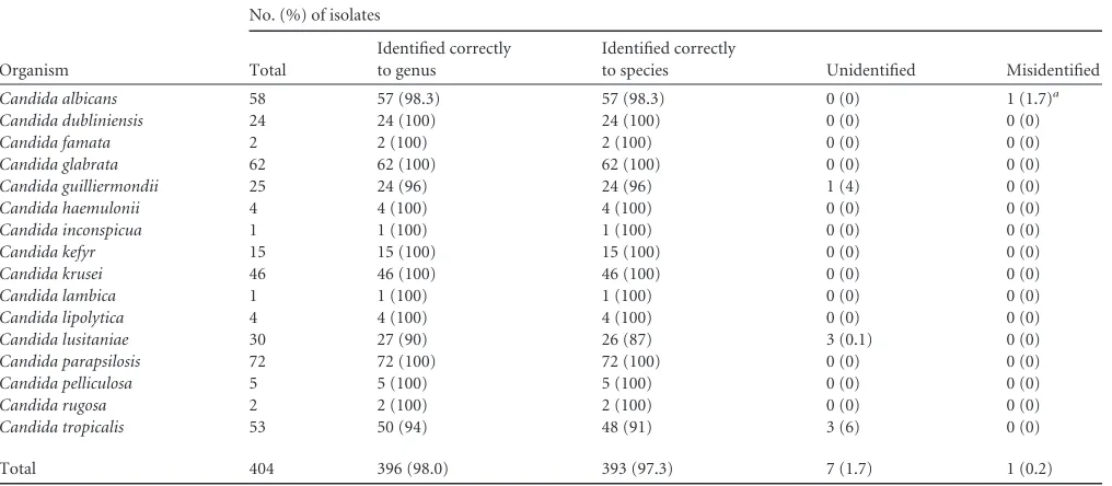
When the isolates from the bioMérieux stock collection were
excluded, 16 species of
Candida
were represented. Of these 404
isolates, 396 (98.0%) were identified correctly to the genus level
and 393 (97.3%) to the species level (
Table 3
).
Performance of the Vitek MS system in identifying non-
Can-dida
yeast isolates.
A total of 226 isolates representing 11 different
species, including 35
C. neoformans
isolates, 50
Trichosporon
iso-lates, and 35
Rhodotorula mucilaginosa
isolates, were analyzed
(
Table 2
). The number of isolates identified to both the genus and
species levels was 207 (91.6%), with all 35 (100%)
C. neoformans
isolates correctly identified to the species level. The number of
isolates that were misidentified (three isolates [1.3%]) was low.
The three misidentified isolates were
Geotrichum klebahnii
isolates
that were identified as
Geotrichum candidum
. The proportion of
isolates that were not identified in this group (16 isolates [7.1%])
was greater than the proportion of isolates that were not identified
in the
Candida
species group.
When the isolates from the bioMérieux stock collection were
excluded from this group of organisms, nine species of non-
Can-dida
yeast isolates remained. Of the 104 isolates, 99 (95.2%) were
correctly identified to both the genus and species levels (
Table 4
).
Quality control.
The
C. glabrata
quality control organism and
the negative control sample (matrix only) were tested by the Vitek
MS every day that yeast isolates were assayed and with every new
lot of target slides, formic acid, and matrix. During the trial, the
quality control organism was tested 141 times and acceptable
re-sults were obtained 139 times (98.6%). Two quality control tests
yielded no identification upon initial testing. In both instances,
however, the correct identification was obtained upon repeat
test-ing on the same day. In all instances, the negative control yielded
no identification.
DISCUSSION
Although the identification of yeast isolates has greatly improved
over the past several decades, the manual and automated
bio-chemical methods commonly used to identify contemporary yeast
isolates are time-consuming and may result in low-discrimination
identifications that require additional testing (
12
,
21
). Nucleic
ac-id-based identification techniques, such as DNA sequencing of
yeast, have high accuracy but are expensive, might have prolonged
turnaround times, and require technical expertise and equipment
that may not be available to all laboratories. MALDI-TOF MS
offers a balance between speed and highly accurate yeast
identifi-cations.
While fewer studies evaluating MALDI-TOF MS identification
of yeasts than bacteria have been published to date, the theme of
the existing literature is that the performance of MALDI-TOF MS
in identifying fungi, both yeasts and molds, is comparable or
on May 16, 2020 by guest
http://jcm.asm.org/
perior to that of conventional and nucleic acid-based
identifica-tion methods (
11
,
12
,
19
,
22
–
29
). The major advantages of
MALDI-TOF MS identification of yeasts, compared with
conven-tional methods, are the marked decreases in cost and time to
iden-tification (
30
). Antifungal susceptibility profiles generally are
pre-dictable from the species identification (
8
) and, of note, the four
species of yeast that account for the vast majority of infections, i.e.,
C. albicans
,
C. glabrata
,
C. krusei
, and
C. parapsilosis
, have distinct
susceptibility profiles (
8
). Therefore, rapid, highly accurate
idtification of yeast isolates using MALDI-TOF MS is poised to
en-hance patient care drastically and to reduce hospital-associated
costs due to fungal infections.
In this study, we evaluated the performance characteristics of
the Vitek MS with the v2.0 database for identification of medically
important yeast species. This study has a number of strengths. The
first is that this was a multicenter evaluation; therefore, a large
number of independent operators were able to demonstrate the
interlaboratory accuracy of this method. Isolates were recovered
from geographically distinct areas across North America,
enrich-ing the collection for strain heterogeneity. In addition, this study
included a large number of isolates, and the identification of all
isolates was verified using sequence analysis as a gold standard.
Finally, this is the first study to date to evaluate the performance
characteristics of the Vitek MS v2.0 database for identification of
clinically relevant yeast species.
The results of the multicenter study indicate that, independent
of the laboratory and the geographical origin of the isolates, the
Vitek MS demonstrated an overall species identification rate
com-parable or superior to those for both traditional biochemical and
nucleic acid-based yeast identification systems (
11
,
12
) but with a
significant reduction in the time to identification. This method is
technically facile and, once the laboratory has recovered the
cap-ital investment for the instrument purchase, the ongoing cost of
consumables is low.
In our study, 24 (2.8%) and 5 (0.6%) isolates were not
identi-fied and were misidentiidenti-fied, respectively. Overall, we identiidenti-fied
⬎
96% of the 852 isolates in this study to the species level. This is
comparable to the findings of other studies evaluating
MALDI-TABLE 3Performance characteristics of the Vitek MS system in identifyingCandidaspecies recovered from clinical specimens
Organism
No. (%) of isolates
Total
Identified correctly to genus
Identified correctly
to species Unidentified Misidentified
Candida albicans 58 57 (98.3) 57 (98.3) 0 (0) 1 (1.7)a
Candida dubliniensis 24 24 (100) 24 (100) 0 (0) 0 (0)
Candida famata 2 2 (100) 2 (100) 0 (0) 0 (0)
Candida glabrata 62 62 (100) 62 (100) 0 (0) 0 (0)
Candida guilliermondii 25 24 (96) 24 (96) 1 (4) 0 (0)
Candida haemulonii 4 4 (100) 4 (100) 0 (0) 0 (0)
Candida inconspicua 1 1 (100) 1 (100) 0 (0) 0 (0)
Candida kefyr 15 15 (100) 15 (100) 0 (0) 0 (0)
Candida krusei 46 46 (100) 46 (100) 0 (0) 0 (0)
Candida lambica 1 1 (100) 1 (100) 0 (0) 0 (0)
Candida lipolytica 4 4 (100) 4 (100) 0 (0) 0 (0)
Candida lusitaniae 30 27 (90) 26 (87) 3 (0.1) 0 (0)
Candida parapsilosis 72 72 (100) 72 (100) 0 (0) 0 (0)
Candida pelliculosa 5 5 (100) 5 (100) 0 (0) 0 (0)
Candida rugosa 2 2 (100) 2 (100) 0 (0) 0 (0)
Candida tropicalis 53 50 (94) 48 (91) 3 (6) 0 (0)
Total 404 396 (98.0) 393 (97.3) 7 (1.7) 1 (0.2)
a
[image:4.585.41.549.78.300.2]Isolate misidentified asC. dubliniensis.
TABLE 4Performance characteristics of the Vitek MS system in identifying non-Candidayeast isolates recovered from clinical specimens
Organism
No. (%) of isolates
Total
Identified correctly to genus
Identified correctly
to species Unidentified Misidentified
Cryptococcus neoformans 29 29 (100) 29 (100) 0 (0) 0 (0)
Geotrichum capitatum 3 3 (100) 3 (100) 0 (0) 0 (0)
Kodamaea ohmeri 1 1 (100) 1 (100) 0 (0) 0 (0)
Malassezia furfur 1 0 (0) 0 (0) 1 (100) 0 (0)
Malassezia pachydermatis 1 0 (0) 0 (0) 1 (100) 0 (0)
Rhodotorula mucilaginosa 26 26 (100) 26 (100) 0 (0) 0 (0)
Saccharomyces cerevisiae 28 27 (96) 27 (96) 1 (4) 0 (0)
Trichosporon asahii 11 9 (82) 9 (82) 2 (18) 0 (0)
Trichosporon mucoides 4 4 (100) 4 (100) 0 (0) 0 (0)
Total 104 99 (95.2) 99 (95.2) 5 (4.8) 0 (0)
on May 16, 2020 by guest
http://jcm.asm.org/
[image:4.585.44.545.570.722.2]TOF MS identification of yeasts using other instrumentation
plat-forms or spectral databases; Yaman and coworkers identified 94%
of 265 yeast isolates correctly using the Bruker Biotyper (
29
),
Bader and colleagues identified
⬎
95% of 1,192 isolates correctly
using both the Bruker Biotyper and the Saramis instruments (
26
),
Dhiman and colleagues identified
⬎
96% of 138 “common” yeasts
and 84.5% of 103 “uncommon” yeasts to the species level using
the Bruker Biotyper (
27
), and Iriart et al. identified 184 of 188
yeast isolates (97.9%) tested using the Vitek MS (
19
). In contrast
to the current study, the study by Iriart et al. (
19
) evaluated the
Vitek MS v1.0 database and included primarily
Candida
isolates
from a medical center in France, and sequencing was not the
ref-erence method for the study.
For the isolates that were misidentified in the current study, the
incorrect identifications would be unlikely to lead to adverse
clin-ical outcomes. Two of the five incorrectly identified isolates were
Candida
species, including an isolate of
C. albicans
misidentified
as
C. dubliniensis
and an isolate of
Candida parapsilosis
misiden-tified as
C. pelliculosa
. The clinical impact of misidentifying
C.
albicans
as
C. dubliniensis
is likely to be minimal, although it has
been suggested that the development of fluconazole resistance is
more likely for
C. dubliniensis
than for
C. albicans
(
31
).
C.
parap-silosis
exhibits higher MICs for the echinocandins than do most
other
Candida
species (
8
,
32
); therefore, misidentification might
be clinically significant. However, data on the susceptibility profile
of
C. pelliculosa
are sparse, and it is not obvious what empirical
therapy might be initiated based on this identification. Although
few isolates were not identified in this study, three (9.1%) of the
Candida lusitaniae
isolates tested were not identified. This is of
minor importance, compared with the overall performance
char-acteristics of this method, but this finding is of note in light of the
fact that this species can be resistant to amphotericin B, a trait
unusual for
Candida
species (
8
).
The three other misidentified isolates were
Geotrichum
klebah-nii
identified as
G. candidum
.
G. klebahnii
is in the current
data-base. While this error is unlikely to be clinically significant,
bio-Mérieux indicated that future database and software updates will
result in reporting of these two species as
G. candidum/klebahnii
rather than specific species-level identification, to circumvent this
misidentification event (bioMérieux, personal communication).
In contrast to the “direct colony” methods typically used for
MALDI-TOF MS identification of bacterial isolates, the majority
of studies to date evaluating MALDI-TOF MS methods for
iden-tification of yeasts have suggested the use of a more
labor-inten-sive formic acid/organic solvent extraction method. This method
involves a series of centrifugation steps and is thought to be
nec-essary for reliable identification of these organisms, because of the
thick, chitin-containing cell walls of yeasts (
26
,
27
,
29
,
33
,
34
).
These additional steps significantly increase the hands-on time
required for analysis and negatively affect turnaround times. For
example, using the full extraction method for sample preparation,
one study reported an average of 5.1 min of hands-on time and a
total turnaround time of 38.4 min per isolate (
27
). A recent study
conducted by Theel and coworkers evaluated a direct on-plate
extraction preparation method using 70% formic acid, and 73 of
90 isolates (81.1%) were identified to the species level using this
method (
35
). The performance of the on-plate direct extraction
method demonstrated in this study and by Theel et al. (
35
)
repre-sents improvements in both turnaround times and workflow for
MALDI-TOF MS identification of yeasts. However, one point of
caution when using a direct plate extraction preparation method
is that the early growth of some thermally dimorphic fungi, such
as
Histoplasma capsulatum
and
Coccidioides immitis
/
posadasii
,
might resemble yeast-like colonies. Therefore, clinical
laborato-ries should be mindful of growth rates and colony morphology
when using this method for yeast identification.
Despite the promising results reported in this study, there are
some limitations to our data. All except four of the isolates were
grown on SDA for MALDI-TOF MS analysis; therefore, the
per-formance characteristics of this methodology for yeast grown on
other types of media are unknown. For the 852 yeast isolates tested
in this study, all of the species identified are included in the Vitek
MS v2.0 database. It is not known if unusual taxa not represented
in the database would be misidentified or simply not identified if
tested with this system. Finally, no isolates of
Cryptococcus gattii
,
an emerging fungal pathogen (
36
), were included in the study.
Thus, the ability of the Vitek MS to differentiate
C. neoformans
from
C. gattii
, which might be of epidemiological and clinical
importance, is not known. Previous studies using other platforms
suggest that MALDI-TOF MS methods do have the potential for
species resolution of
Cryptococcus
species by permitting the
addi-tion of mass spectra to the reference database (
28
). The Vitek MS
IVD system evaluated in this study does not permit user
modifi-cations, such as the addition of spectra to the database.
In conclusion, we present the results of a multicenter study
evaluating the Vitek MS system for identification of clinically
rel-evant yeasts. Identification of yeasts using the Vitek MS is faster
and more accurate than phenotypic identification systems
cur-rently employed in clinical microbiology laboratories and affords
accuracy comparable to that of more laborious and costly
molec-ular methods. Implementation of this methodology should
streamline yeast identification in the laboratory, positively affect
patient care, and reduce health care-associated costs.
ACKNOWLEDGMENTS
This study was funded by bioMérieux.
We thank Connie Bradford for her assistance with this study. We also thank W. Michael Dunne, Jr., and Dave Pincus for their thoughtful re-views of the manuscript.
J. A. Branda, J. A. Rychert, and M. J. Ferraro have received research funding from bioMérieux and Becton, Dickinson and Co. C. C. Ginoc-chio has received research funding and consulting fees from bioMérieux and Becton, Dickinson. G. W. Procop has received research funding from bioMérieux, Bruker, the CDC, and Luminex. S. S. Richter has received research funding from bioMérieux, Nanosphere, and Forest Laboratories. C.-A. D. Burnham has received research funding from bioMérieux, Ac-clerate, Cepheid, and T2 Biosystems. The other authors have no conflicts to disclose.
REFERENCES
1.Arendrup MC, Fisher BT, Zaoutis TE.2009. Invasive fungal infections in the paediatric and neonatal population: diagnostics and management
is-sues. Clin. Microbiol. Infect.15:613– 624.
2.Muskett H, Shahin J, Eyres G, Harvey S, Rowan K, Harrison D.2011. Risk factors for invasive fungal disease in critically ill adult patients: a
systematic review. Crit. Care15:R287.
3.Parize P, Rammaert B, Lortholary O.2012. Emerging invasive fungal
diseases in transplantation. Curr. Infect. Dis. Rep.14:668 – 675.
4.Shoham S, Marr KA.2012. Invasive fungal infections in solid organ
transplant recipients. Future Microbiol.7:639 – 655.
5.Gratwohl A, Baldomero H, Aljurf M, Pasquini MC, Bouzas LF, Yoshimi A, Szer J, Lipton J, Schwendener A, Gratwohl M, Frauendorfer K, Niederwieser D, Horowitz M, Kodera Y.2010. Hematopoietic stem cell
transplantation: a global perspective. JAMA303:1617–1624.
on May 16, 2020 by guest
http://jcm.asm.org/
6.US Department of Health and Human Services.2013. Statistics and facts
for people over 50.http://www.organdonor.gov/about/statistics.html.
Ac-cessed 12 February 2013.
7.Garey KW, Rege M, Pai MP, Mingo DE, Suda KJ, Turpin RS, Bearden
DT.2006. Time to initiation of fluconazole therapy impacts mortality in
patients with candidemia: a multi-institutional study. Clin. Infect. Dis.
43:25–31.
8.Pappas PG, Kauffman CA, Andes D, Benjamin DK, Jr, Calandra TF, Edwards JE, Jr, Filler SG, Fisher JF, Kullberg BJ, Ostrosky-Zeichner L, Reboli AC, Rex JH, Walsh TJ, Sobel JD.2009. Clinical practice guide-lines for the management of candidiasis: 2009 update by the Infectious
Diseases Society of America. Clin. Infect. Dis.48:503–535.
9.Echeverria PM, Kett DH, Azoulay E.2011.Candidaprophylaxis and
therapy in the ICU. Semin. Respir. Crit. Care Med.32:159 –173.
10. Andes DR, Safdar N, Baddley JW, Playford G, Reboli AC, Rex JH, Sobel JD, Pappas PG, Kullberg BJ. 2012. Impact of treatment strategy on outcomes in patients with candidemia and other forms of invasive candi-diasis: a patient-level quantitative review of randomized trials. Clin. Infect.
Dis.54:1110 –1122.
11. Marcos JY, Pincus DH.2013. Fungal diagnostics: review of commercially
available methods. Methods Mol. Biol.968:25–54.
12. Pincus DH, Orenga S, Chatellier S. 2007. Yeast identification: past,
present, and future methods. Med. Mycol.45:97–121.
13. Freydiere AM, Odds FC.2001. Commercial kits for yeast identification:
concerns for standardisation. Eur. J. Clin. Microbiol. Infect. Dis.20:366 –
367.
14. Freydiere AM, Guinet R, Boiron P.2001. Yeast identification in the
clinical microbiology laboratory: phenotypical methods. Med. Mycol.39:
9 –33.
15. Verweij PE, Breuker IM, Rijs AJ, Meis JF.1999. Comparative study of
seven commercial yeast identification systems. J. Clin. Pathol.52:271–273.
16. Walsh TJ, Groll A, Hiemenz J, Fleming R, Roilides E, Anaissie E.2004. Infections due to emerging and uncommon medically important fungal
pathogens. Clin. Microbiol. Infect.10(Suppl 1):48 – 66.
17. Sanguinetti M, Porta R, Sali M, La Sorda M, Pecorini G, Fadda G, Posteraro B.2007. Evaluation of VITEK 2 and RapID yeast plus systems for yeast species identification: experience at a large clinical microbiology
laboratory. J. Clin. Microbiol.45:1343–1346.
18. Fenselau C, Demirev PA.2001. Characterization of intact
microorgan-isms by MALDI mass spectrometry. Mass Spectrom. Rev.20:157–171.
19. Iriart X, Lavergne RA, Fillaux J, Valentin A, Magnaval JF, Berry A, Cassaing S.2012. Routine identification of medical fungi by the new Vitek MS matrix-assisted laser desorption ionization-time of flight system with
a new time-effective strategy. J. Clin. Microbiol.50:2107–2110.
20. Hall L, Wohlfiel S, Roberts GD.2003. Experience with the MicroSeq D2 large-subunit ribosomal DNA sequencing kit for identification of com-monly encountered, clinically important yeast species. J. Clin. Microbiol.
41:5099 –5102.
21. Hata DJ, Hall L, Fothergill AW, Larone DH, Wengenack NL.2007. Multicenter evaluation of the new VITEK 2 advanced colorimetric yeast
identification card. J. Clin. Microbiol.45:1087–1092.
22. Erhard M, Hipler UC, Burmester A, Brakhage AA, Wostemeyer J.2008. Identification of dermatophyte species causing onychomycosis and tinea
pedis by MALDI-TOF mass spectrometry. Exp. Dermatol.17:356 –361.
23. Marinach-Patrice C, Lethuillier A, Marly A, Brossas JY, Gene J, Sy-moens F, Datry A, Guarro J, Mazier D, Hennequin C.2009. Use of mass
spectrometry to identify clinicalFusariumisolates. Clin. Microbiol. Infect.
15:634 – 642.
24. Marklein G, Josten M, Klanke U, Muller E, Horre R, Maier T, Wenzel T, Kostrzewa M, Bierbaum G, Hoerauf A, Sahl HG. 2009. Matrix-assisted laser desorption ionization-time of flight mass spectrometry for fast and reliable identification of clinical yeast isolates. J. Clin. Microbiol.
47:2912–2917.
25. Stevenson LG, Drake SK, Shea YR, Zelazny AM, Murray PR. 2010. Evaluation of matrix-assisted laser desorption ionization-time of flight mass spectrometry for identification of clinically important yeast species.
J. Clin. Microbiol.48:3482–3486.
26. Bader O, Weig M, Taverne-Ghadwal L, Lugert R, Gross U, Kuhns M.
2011. Improved clinical laboratory identification of human pathogenic yeasts by matrix-assisted laser desorption ionization time-of-flight mass
spectrometry. Clin. Microbiol. Infect.17:1359 –1365.
27. Dhiman N, Hall L, Wohlfiel SL, Buckwalter SP, Wengenack NL.2011. Performance and cost analysis of matrix-assisted laser desorption ioniza-tion-time of flight mass spectrometry for routine identification of yeast. J.
Clin. Microbiol.49:1614 –1616.
28. Posteraro B, Vella A, Cogliati M, De Carolis E, Florio AR, Posteraro P, Sanguinetti M, Tortorano AM.2012. Matrix-assisted laser desorption ionization-time of flight mass spectrometry-based method for
discrimi-nation between molecular types ofCryptococcus neoformansand
Crypto-coccus gattii. J. Clin. Microbiol.50:2472–2476.
29. Yaman G, Akyar I, Can S.2012. Evaluation of the MALDI TOF-MS
method for identification ofCandidastrains isolated from blood cultures.
Diagn. Microbiol. Infect. Dis.73:65– 67.
30. Tan KE, Ellis BC, Lee R, Stamper PD, Zhang SX, Carroll KC.2012. Prospective evaluation of a matrix-assisted laser desorption ionization-time of flight mass spectrometry system in a hospital clinical microbiology laboratory for identification of bacteria and yeasts: a bench-by-bench study for assessing the impact on time to identification and
cost-effectiveness. J. Clin. Microbiol.50:3301–3308.
31. Moran GP, Sullivan DJ, Henman MC, McCreary CE, Harrington BJ, Shanley DB, Coleman DC.1997. Antifungal drug susceptibilities of oral
Candida dubliniensisisolates from human immunodeficiency virus (HIV)-infected and non-HIV-infected subjects and generation of stable fluconazole-resistant derivatives in vitro. Antimicrob. Agents Chemother.
41:617– 623.
32. Beyda ND, Lewis RE, Garey KW. 2012. Echinocandin resistance in
Candidaspecies: mechanisms of reduced susceptibility and therapeutic
approaches. Ann. Pharmacother.46:1086 –1096.
33. Goyer M, Lucchi G, Ducoroy P, Vagner O, Bonnin A, Dalle F.2012. Optimization of the preanalytical steps of matrix-assisted laser desorption ionization-time of flight mass spectrometry identification provides a flex-ible and efficient tool for identification of clinical yeast isolates in medical
laboratories. J. Clin. Microbiol.50:3066 –3068.
34. Cassagne C, Cella AL, Suchon P, Normand AC, Ranque S, Piarroux R.
2013. Evaluation of four pretreatment procedures for MALDI-TOF MS
yeast identification in the routine clinical laboratory. Med. Mycol.51:371–
377.
35. Theel ES, Schmitt BH, Hall L, Cunningham SA, Walchak RC, Patel R, Wengenack NL.2012. Formic acid-based direct, on-plate testing of yeast andCorynebacteriumspecies by Bruker Biotyper matrix-assisted laser de-sorption ionization-time of flight mass spectrometry. J. Clin. Microbiol.
50:3093–3095.
36. Byrnes EJ, III, Bartlett KH, Perfect JR, Heitman J.2011.Cryptococcus gattii: an emerging fungal pathogen infecting humans and animals.
Mi-crobes Infect.13:895–907.