0095-1137/11/$12.00 doi:10.1128/JCM.01492-10
Copyright © 2011, American Society for Microbiology. All Rights Reserved.
Highly Specific and Quick Detection of
Mycobacterium avium
subsp.
paratuberculosis
in Feces and Gut Tissue of Cattle and Humans
by Multiple Real-Time PCR Assays
䌤
Can Imirzalioglu,
1Heinrich Dahmen,
2Torsten Hain,
1Andre Billion,
1Carsten Kuenne,
1Trinad Chakraborty,
1and Eugen Domann
1*
Institute of Medical Microbiology, Frankfurter Strasse 107, D-35392 Giessen, Germany,1and
Veterinary Practice, Burgring 9, D-54595 Pruem, Germany2
Received 23 July 2010/Returned for modification 23 December 2010/Accepted 7 March 2011
Mycobacterium aviumsubsp.paratuberculosisis the causative agent of Johne’s disease (JD) in cattle and may be associated with Crohn’s disease (CD) in humans. It is the slowest growing of the cultivable mycobacteria, and culture from clinical, veterinary, food, or environmental specimens can take 4 months or even longer. Currently, the insertion element IS900is used to detectM. aviumsubsp.paratuberculosisDNA. However, closely related IS900elements are also present in other mycobacteria, thus limiting its specificity as a target. Here we describe the use of novel primer sets derived from the sequences of two highly specific single copy genes, MAP2765c and MAP0865, for the quantitative detection ofM. aviumsubsp.paratuberculosiswithin 6 h by using real-time PCR. Specificity of the target was established using 40M. aviumsubsp.paratuberculosisisolates, 67 different bacterial species, and two intestinal parasites. Using the probes and methods described, we detected 27 (2.09%)M. aviumsubsp.paratuberculosis-positive stool specimens from 1,293 individual stool samples by the use of either IS900or probes deriving from the MAP2765c and MAP0865 genes described here. In general, bacterial load due toM. aviumsubsp.paratuberculosiswas uniformly low in these samples and we estimated 500 to 5,000 M. avium subsp. paratuberculosis bacteria per gram of stool in assay-positive samples. Thus, the methods described here are useful for rapid and specific detection of M. avium subsp. paratuberculosis in clinical samples.
Mycobacterium aviumsubsp.paratuberculosisis the causative agent of Johne’s disease (JD) in cattle, and it has been sug-gested that this microorganism may be associated with Crohn’s
disease (CD) in humans (14, 17).M. aviumsubsp.
paratuber-culosisbelongs to the mycobacterial speciesM. avium, which is
currently subdivided into three subspecies (33): M. avium
subsp.avium(synonym,M. avium),M. aviumsubsp.
paratuber-culosis (synonym, M. paratuberculosis), and M. avium subsp.
silvaticum(synonym,M. silvaticum). At the subspecies level,M. aviumsubsp.paratuberculosiscan be differentiated
phenotyp-ically fromM. aviumsubsp.aviumandM. aviumsubsp.
silvati-cumby its dependence on mycobactin (51) and genotypically
by the presence of multiple copies of the insertion element
IS900(2, 5, 13, 22, 45).
JD (or paratuberculosis) is a chronic, granulomatous severe form of gastroenteritis with progressive weight loss and ema-ciation affecting domestic and wild ruminants, e.g., cattle, sheep, goats, red deer, and rabbits worldwide (12, 30, 31).
Infected livestock periodically shedM. aviumsubsp.
paratuber-culosisvia feces and milk, which results in environmental
dis-tribution, whereM. aviumsubsp.paratuberculosiscan survive
for extended periods (55). Milk pasteurization trials showed that high-temperature and short-duration standard
pasteuriza-tion procedures do not effectively killM. aviumsubsp.
paratu-berculosisin milk, as clinical strains ofM. aviumsubsp. para-tuberculosishave been shown to be more thermally tolerant
than eitherM. bovisorCoxiella burnetii, the current milk
pas-teurization standard microorganisms (10, 44, 47, 48). There-fore, human contact can result from the consumption of inad-equately pasteurized milk or raw milk or of certain other dairy products, fecally contaminated vegetables, contaminated beef, or even water (12, 14, 34, 41, 47, 48).
CD is a chronic inflammatory disease of the gastrointestinal tract in humans, affecting in particular the terminal ileum, with symptoms of general malaise, weight loss, abdominal pain, and diarrhea (4, 12, 16, 40). A hallmark of CD is the histological proof of a granulomatous inflammation, which is also charac-teristic of JD and other mycobacterial diseases, leading to
suggestions thatM. aviumsubsp.paratuberculosismay be
as-sociated with CD in humans (see references 12 [and references therein], 8, 15, 16, 27, and 28).
Currently, there is controversy as to whether M. avium
subsp.paratuberculosis (i) is an innocent bystander that has
merely colonized the intestine of Crohn’s patients, (ii) could be a secondary infection but not a cause of the disease, (iii) could be the primary infectious agent and the cause of CD, (iv) acts as a superantigen, or (v) modifies the immune response in CD (6, 12, 15, 25, 32, 37, 42, 43, 50). One of the major obstacles to
resolution of the debate on the role ofM. aviumsubsp.
para-tuberculosisin CD and the controversial studies published is the requirement of reliable and contemporary detection and
identification ofM. avium subsp.paratuberculosisin complex
* Corresponding author. Mailing address: Institute of Medical Mi-crobiology, Frankfurter Strasse 107, D-35392 Giessen, Germany. Phone: 49 641-9941287. Fax: 49 641-9941259. E-mail: eugen.domann @mikrobio.med.uni-giessen.de.
䌤Published ahead of print on 23 March 2011.
1843
on May 16, 2020 by guest
http://jcm.asm.org/
specimens such as blood, biopsy samples, breast milk, and
feces.M. aviumsubsp.paratuberculosisis the slowest growing
of the cultivable mycobacteria, and primary culture from clin-ical, veterinary, food, or environmental specimens can take 4 months or even longer (12, 24, 44, 55). Moreover, the
charac-teristics ofM. aviumsubsp.paratuberculosisin JD and in CD
seem to be totally different: in CD,M. aviumsubsp.
paratuber-culosis appears as non-acid-fast coccobacilli with the ultra-structure of spheroplasts (cell-wall-deficient forms) that do not
transform into characteristicM. aviumsubsp.paratuberculosis
organisms until after several months of incubation (4). In this study, we aimed at establishing highly specific multi-ple quantitative real-time PCR (qrt-PCR) assays based on the
published genome sequence ofM. avium subsp.
paratubercu-losisstrain K-10 to enable rapid and unequivocal detection and
identification ofM. aviumsubsp.paratuberculosisdirectly from
clinical, veterinary, food, or environmental specimens as well as from pure cultures. The method developed allows the quick
and reliable detection and quantification of M. aviumsubsp.
paratuberculosisdirectly from stool samples within 6 h at rea-sonable costs.
MATERIALS AND METHODS
Human clinical samples and data.Patient fecal and tissue specimens repre-sented clinical routine diagnostic samples for the detection of pathogenic bac-teria and parasites. Testing for the presence ofM. aviumsubsp.paratuberculosis
DNA was performed in addition to DNA detection of conventional enteropatho-genic bacteria and parasites. Positive results were reported. Clinical data from patients with positive results were obtained after informed consent by record review. The study was approved by the Ethics Board of the Justus-Liebig-Uni-versity of Giessen, Faculty of Medicine.
Cattle samples.Feces and gut biopsy specimens were obtained from healthy cows and from cows with suspected paratuberculosis. The symptoms of clinical
paratuberculosis are chronic diarrhea and progressive weight loss, whereas sub-clinically infected animals mainly exhibit decreased milk production. The guts were dissected into small pieces and subdivided into inflamed and noninflamed tissue samples based on microscopic examination.
Microorganisms and standard cultivation.The mycobacterial and nonmyco-bacterial species used in the study are listed here (see Table 2). The strains were obtained from the German Resource Center for Biological Material (DSMZ) and from the strain collection of the Institute of Medical Microbiology, Justus-Liebig University of Giessen.
M. aviumsubsp.paratuberculosiswas cultivated on BD BBL Herrold’s egg yolk agar with Mycobactin J and ANV (Becton Dickinson, Heidelberg, Germany) and in modified Middlebrook 7H9 medium for up to 4 months as previously de-scribed (12, 39). All remaining bacteria were grown under optimal conditions on appropriate media as previously recommended (3).
Acid-fast staining.The acid-fast staining of specimens was done by the Ziehl-Neelsen procedure, and stained specimens examined under conditions of oil immersion. Dissected biopsy samples from gut tissue of cows were stained as previously described for tissue sections of bisons (20).
DNA extraction.DNA was extracted from pure bacterial cultures with a RTP Spin Bacteria DNA kit (Invitek, Berlin, Germany). The total DNA from human and animal fecal specimens was extracted with a PSP Spin Stool DNA kit and from tissue with an Invisorb Spin Tissue minikit (Invitek) as recommended by the vendor.
Parasitic DNA from Entamoeba histolyticaand from Giardia lamblia was obtained from the Bernhard-Nocht Institute for Tropical Medicine (Hamburg, Germany).
[image:2.585.42.539.78.357.2]Quantitative real-time PCR (qrt-PCR) and conventional PCR.The new prim-ers and probes used were designed with “Primer Express” vprim-ersion 1.0 software (Applied Biosystems, Foster City, CA). The internal probes were labeled with the fluorescent reporter dye 5-carboxyfluoroscein (FAM) on the 5⬘end and the quencher dye N⬘,N⬘,N⬘,N⬘-tetramethyl-6-carboxyrhodamine (TAMRA) on the 3⬘ end. The qrt-PCR was accomplished as described previously (21, 38). Protozoal universal 18S small-subunit rRNA (SSU-rRNA) detection was performed by conventional PCR using eukaryote-specific primers (Table 1) as described pre-viously (26). The amounts of specific target sequences present in unknown samples were calculated by measuring the threshold cycle (CT) values and using standard curves generated with a series of known quantities of target sequences. TheCTvalue represented the cycle at which the copy of the amplified target
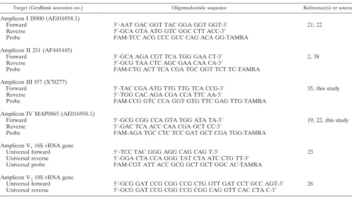
TABLE 1. Specific primers and TaqMan probes used in this study to detectM. aviumsubsp.paratuberculosis
Target (GenBank accession no.) Oligonucleotide sequence Reference(s) or source
Amplicon I IS900 (AE016958.1)
Forward 5⬘-AAT GAC GGT TAC GGA GGT GGT-3⬘ 21, 22
Reverse 5⬘-GCA GTA ATG GTC GGC CTT ACC-3⬘
Probe FAM-TCC ACG CCC GCC CAG ACA GG-TAMRA
Amplicon II 251 (AF445445)
Forward 5⬘-GCA AGA CGT TCA TGG GAA CT-3⬘ 2, 38
Reverse 5⬘-GCG TAA CTC AGC GAA CAA CA-3⬘
Probe FAM-CTG ACT TCA CGA TGC GGT TCT TC-TAMRA
Amplicon III f57 (X70277)
Forward 5⬘-TAC CGA ATG TTG TTG TCA CCG-3⬘ 35, this study
Reverse 5⬘-TGG CAC AGA CGA CCA TTC AA-3⬘
Probe FAM-CCG GTC CCA GGT GTG TTC GAG TTG-TAMRA
Amplicon IV MAP0865 (AE016958.1)
Forward 5⬘-GCG CGG CCA GTA TGG ATA TA-3⬘ 19, 22, this study
Reverse 5⬘-GAC TCA ACC CAA CGA GCT CC-3⬘
Probe FAM-AGA TGC CTC TCC GAT GCT CGA TGG-TAMRA
Amplicon V116S rRNA gene
Universal forward 5⬘-TCC TAC GGG AGG CAG CAG T-3⬘ 23
Universal reverse 5⬘-GGA CTA CCA GGG TAT CTA ATC CTG TT-3⬘
Universal probe FAM-CGT ATT ACC GCG GCT GCT GGC AC-TAMRA
Amplicon V218S rRNA gene
Universal forward 5⬘-GCG GAT CCG CGG CCG CTG GTT GAT CCT GCC AGT-3⬘ 26 Universal reverse 5⬘-GCG GAT CCG CGG CCG CGG CAG GTT CAC CTA C-3⬘
on May 16, 2020 by guest
http://jcm.asm.org/
sequence intersected the threshold or baseline. Briefly, an inoculation loop of the
M. aviumsubsp.paratuberculosisDSM 44133 type strain grown on Herrold’s egg yolk was carefully resuspended in phosphate-buffered saline (PBS) and serially diluted. The total amounts of cells were counted using a Neubauer counting chamber. We determined the total cell numbers, since complex biological ma-terial may contain both viable and dead bacteria.
Detection ofSalmonella entericaserovarenteritidis,Campylobacter jejuni, Yer-sinia enterocolitica,Clostridium difficile,Entamoeba histolytica, andGiardia lam-blia.The total DNA of human fecal specimens was extracted with a PSP Spin Stool kit (Invitek) as described above.S. enteritidis,C. jejuni,Y. enterocolitica,C. difficile,E. histolytica, andG. lambliawere detected by specific real-time PCRs as previously described (18, 29, 52, 53, 54).
Multiple displacement amplification (MDA) of IS900-positive fecal speci-mens.MDA was performed with the extracted DNA of amplicon I (IS900 )-positive but amplicon II (MAP2765c [251])-, III (MAP0865 [f57])-, and IV (MAP0865)-negative fecal samples. A commercially available GenomiPhi DNA amplification kit (Amersham Biosciences, Uppsala, Sweden), which utilizes MDA to exponentially amplify genomic DNA, was used, following the manufac-turer’s instructions. Briefly, 1l of DNA extract was added to 9l of sample buffer containing random hexamer primers and heated to 95°C. The chilled sample was mixed with 9l of reaction buffer and 1l of enzyme mix. The mixture was incubated for 14 h at 30°C and afterwards subjected to heat inacti-vation for 10 min at 65°C.
Bioinformatics.DNA sequences were aligned with MegAlign version 5.0 soft-ware (DNASTAR Inc., Madison, WI) and compared with sequences deposited in the GenBank, EMBL, DDBJ, and PDB databases using the BLASTn basic local alignment search tool (1). The published genome ofM. aviumsubsp. paratuber-culosisstrain K-10 (22; GenBank accession number AE016958.1) was analyzed and depicted with the GenomeViz bioinformatics tool (11). The standard curves to calculate the amounts ofM. aviumsubsp.paratuberculosisin complex stool and biopsy specimens were generated with SigmaPlot 2000 version 6.00 software (SPSS GmbH Software, Munich, Germany).
RESULTS
Development of MAP0865-specific real-time PCRs. Frag-ment f57 (GenBank accession no. X70277) has been previously described and used as a highly specific 620-bp-long probe for
detection ofM. aviumsubsp.paratuberculosisand diagnosis of
Johne’s disease (35, 49). The nucleotide-nucleotide BLAST (BLASTn) analysis we performed revealed that it was located in gene MAP0865, one of the 4,350 predicted open reading
frames (ORFs) ofM. aviumsubsp.paratuberculosisstrain K-10
(Fig. 1) (22). Fragment f57 covered 48.7% of this 1,272-bp-long ORF with an identity of 100% (Fig. 1). Another BLASTn analysis using the nucleotide sequence of ORF MAP0865 with at least 3.7 million sequences deposited in databases GenBank, EMBL, DDBJ, and PDB revealed it to be unique. Consensus was detected only with deposited MAP0865 and fragment f57 sequences.
By the use of Primer Express software, two new TaqMan
amplicons with primers and probes were designed for thisM.
avium subsp. paratuberculosis-specific MAP0865 ORF. The first one, designated amplicon III, covered both fragment f57 and ORF MAP0865, while the second, designated amplicon IV, covered only ORF MAP0865 (Fig. 1, Table 1).
Specificities of the MAP0865-specific real-time PCRs. The specificities of the new real-time PCRs using amplicons III and IV were demonstrated by the analysis of DNA from panels of 40M. aviumsubsp.paratuberculosis isolates, 17 other myco-bacterial species, 13 positive myco-bacterial species, 20 Gram-negative bacterial species (including 8 different species causing gastroenteritis), 7 obligate anaerobic bacterial species, and 2 intestinal parasite species (Table 2). The results showed that the real-time PCRs with amplicons III and IV, as well as those
performed with previously developed amplicons I (IS900) and
II (MAP2765c [251]), specifically amplified only theM. avium
subsp.paratuberculosisDNA but not the DNA of the
remain-ing bacteria (Table 2), indicatremain-ing that the entire ORF
MAP0865 is unique forM. aviumsubsp.paratuberculosis.
All DNA extracts gave positive results using 16S rRNA gene
universal amplicon V1for bacteria or 18S rRNA gene universal
amplicon V2for parasites (Table 2). The nucleic acid
concen-trations of the DNA extracts were calculated to be at ⬃50
ng/l.
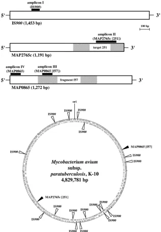
Positions of element IS900, ORF MAP2765c (251), and ORF MAP0865 (f57) on the chromosome ofM. aviumsubsp. para-tuberculosisstrain K-10.A previously published genome-scale
comparison ofM. aviumsubsp.paratuberculosiswith its closely
related subspecies M. aviumsubsp. aviumrevealed potential
new diagnostic sequences (2). Among these, target 251 with a length of 540 bp has been identified as a valuable new
se-quence for specific amplification ofM. aviumsubsp.
paratuber-culosisDNA (38; GenBank accession no. AF445445). Nucle-otide sequence alignments indicated that target 251 is located
in gene MAP2765c ofM. aviumsubsp.paratuberculosisstrain
K-10 (Fig. 1) (22).
Genome visualization using the GenomeViz bioinformatics
tool (11) revealed a random distribution of 17 copies of IS900
on theM. aviumsubsp.paratuberculosischromosome and that
both ORF MAP0865 (f57) and ORF MAP2765c (251) exist as singular sequences at diametrically opposite positions (Fig. 1).
Eleven copies of IS900and MAP0865 (f57) were located on
the positive strand, whereas the remaining six copies of IS900
and MAP2765c (251) were located on the negative strand (Fig.
1). This underlines the utility of IS900 as a highly sensitive
PCR target forM. aviumsubsp.paratuberculosisdue to its high
copy number on theM. aviumsubsp.paratuberculosis
chromo-some. Nevertheless, ORFs MAP0865 and MAP2765c are
highly specific PCR targets that can be used to identify M.
avium subsp. paratuberculosis and to confirm IS900-specific PCRs.
Optimization of PCR conditions and annealing temperature of IS900-, MAP2765c (251)-, MAP0865 (f57)-, and MAP0865-specific real-time PCRs. In order to determine the optimal PCR buffer conditions and the optimal primer annealing tem-perature for all four amplicons, we analyzed the efficiencies of
the real-time PCRs with MgCl2concentrations in the range of
1.0 to 5.0 mM and temperatures in the range of 55 to 65°C
(data not shown). The DNA used was extracted from ⬃108
cells ofM. aviumsubsp.paratuberculosis, and the experiments
were done in triplicate. For all amplicons (I through IV), the
optimal MgCl2concentration was 3.5 mM and the annealing
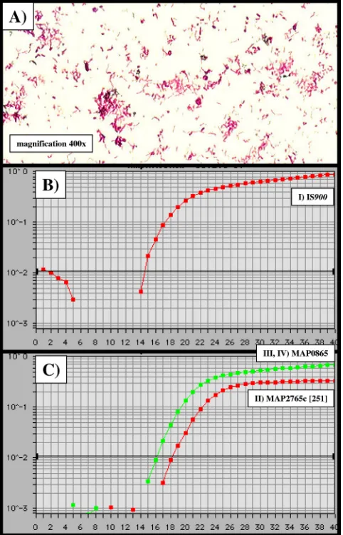
temperature 57.8°C. The ideal results obtained consisted of a
CTvalue of 14.4 for amplicon I (IS900), aCTvalue of 18.2 for
amplicon II (MAP2765c [251]), and a CT value of 16.2 for
amplicons III (MAP0865 [f57]) and IV (MAP0865) (Fig. 2). The order with respect to sensitivity and efficiency of the TaqMan-PCR was amplicon I first, amplicons III and IV sec-ond, and amplicon II third. In cases of amplicon I-positive but amplicon II- to IV-negative specimens, multiple displacement amplification (MDA) was used to exponentially amplify genomic DNA in the DNA extracts of the tested samples. The results suggest the use of MDA as a routine tool for
amplifi-cation of specimens when low concentrations of M. avium
subsp.paratuberculosis-specific DNA are suspected.
on May 16, 2020 by guest
http://jcm.asm.org/
The sensitivity of real-time PCR depends on the DNA ex-traction method used.Since the real-time PCR with amplicon I
(IS900) showed the highest efficiency, serial dilutions of the
genomic DNA of M. avium subsp. paratuberculosistype strain
DSM 44133 were used as templates to assess its sensitivity under the conditions mentioned above. The detection limit of the
real-time PCR with amplicon I (IS900) was (theoretically) determined
to be 1 to 10 CFU/reaction and therefore was identical to the
results obtained previously (21). To identify M. avium subsp.
paratuberculosisand to calculate directly the amount of the bac-teria in complex biological mabac-terial such as feces and tissue
with-out cultivation, we assessed the efficiency of the DNA extraction methods with two commercially available kits: a stock solution of 7.3⫻107M. aviumsubsp.paratuberculosisbacteria was serially
diluted to 7.3⫻106, 7.3⫻105, 7.3⫻104, 7.3⫻103, 7.3⫻102, and
7.3 ⫻ 101 bacteria, and the DNA of the bacteria from each
dilution step was extracted using commercially available kits for stool and tissue specimens (Invitek, Berlin, Germany). The stool
kit had a detection limit ofⱖ70 cells/reaction, whereas the
detec-tion limit of the tissue kit wasⱖ10 cells/reaction. Both analyses
confirmed very high correlation, as demonstrated by
[image:4.585.133.449.69.523.2]determina-tions of linear regression (r2) (Fig. 3).
FIG. 1. Graphic presentation of theM. avium subsp. paratuberculosis-specific chromosomal regions IS900, MAP2765c (target 251), and MAP0865 (fragment f57) and their positions in theM. aviumsubsp.paratuberculosisK-10 genome (GenBank accession no. AE016958.1). Outer circle, plus strand; inner circle, minus strand. The homology analyses and the graphic presentations were done using the methods BLAST, Clustal W (1), and GenomeViz (11). The black boxes represent specific TaqMan PCR amplicons I through IV, including forward and reverse primers and the internal probe labeled with FAM and TAMRA (Table 1). The amplicons were designed using Primer Express software (Applied Biosystems, Foster City, CA).
on May 16, 2020 by guest
http://jcm.asm.org/
Microscopic detection and cultivation of M. avium subsp. paratuberculosisin feces and gut tissue from cattle with Johne’s disease.We examined the feces and dissected gut tissue from 12 butchered cows by Ziehl-Neelsen staining and culture. Three cows were healthy, and the remaining nine were sus-pected to be afflicted with Johne’s disease. Diseased cows 1 to 4, 6 to 9, and 12 showed acid-fast bacteria in both feces and
inflamed gut tissue, and pure cultures of M. avium subsp.
paratuberculosiscould be obtained after 3 to 6 months of in-cubation (Fig. 2A, Table 3). The noninflamed gut tissue
sam-ples were uniformly negative forM. aviumsubsp.
paratubercu-losis. Healthy cows 5, 10, and 11 were negative forM. avium
subsp.paratuberculosisin all samples examined (Table 3). All
M. avium subsp. paratuberculosis isolates were identified by
the specific real-time PCRs with amplicons I (IS900), II
(MAP2765c [251]), III (MAP0865 [f57]), and IV (MAP0865).
The growth of allM. aviumsubsp.paratuberculosisisolates was
mycobactin dependent. Furthermore, the isolates were
as-signed to theM. aviumcomplex by partial sequencing of the
16S rRNA genes and by performing a Genotype
Mycobacte-rium CM/AS test (Hain Lifescience GmbH, Nehren, Ger-many).
Evaluation of theM. aviumsubsp.paratuberculosis-specific real-time PCRs using original bovine gut tissue and feces.The total DNA from inflamed and noninflamed dissected gut tissue and from feces of healthy and diseased cows was extracted with commercially available kits for tissue and stool specimens (Invitek, Berlin, Germany). All of the diseased cows with
pos-itiveM. aviumsubsp.paratuberculosisculture results were also
[image:5.585.45.539.80.504.2]positive for specific real-time PCR amplicons I to IV (Table 3).
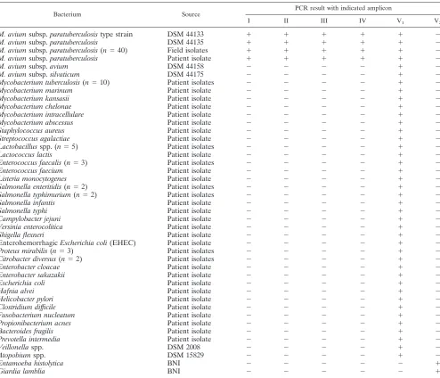
TABLE 2. Bacterial isolates (and their sources) used in this studya
Bacterium Source PCR result with indicated amplicon
I II III IV V1 V2
M. aviumsubsp.paratuberculosistype strain DSM 44133 ⫹ ⫹ ⫹ ⫹ ⫹ ⫺
M. aviumsubsp.paratuberculosis DSM 44135 ⫹ ⫹ ⫹ ⫹ ⫹ ⫺
M. aviumsubsp.paratuberculosis(n⫽40) Field isolates ⫹ ⫹ ⫹ ⫹ ⫹ ⫺ M. aviumsubsp.paratuberculosis Patient isolate ⫹ ⫹ ⫹ ⫹ ⫹ ⫺
M. aviumsubsp.avium DSM 44158 ⫺ ⫺ ⫺ ⫺ ⫹ ⫺
M. aviumsubsp.silvaticum DSM 44175 ⫺ ⫺ ⫺ ⫺ ⫹ ⫺
Mycobacterium tuberculosis(n⫽10) Patient isolates ⫺ ⫺ ⫺ ⫺ ⫹ ⫺
Mycobacterium marinum Patient isolate ⫺ ⫺ ⫺ ⫺ ⫹ ⫺
Mycobacterium kansasii Patient isolate ⫺ ⫺ ⫺ ⫺ ⫹ ⫺
Mycobacterium chelonae Patient isolate ⫺ ⫺ ⫺ ⫺ ⫹ ⫺
Mycobacterium intracellulare Patient isolate ⫺ ⫺ ⫺ ⫺ ⫹ ⫺
Mycobacterium abscessus Patient isolate ⫺ ⫺ ⫺ ⫺ ⫹ ⫺
Staphylococcus aureus Patient isolate ⫺ ⫺ ⫺ ⫺ ⫹ ⫺
Streptococcus agalactiae Patient isolate ⫺ ⫺ ⫺ ⫺ ⫹ ⫺
Lactobacillusspp. (n⫽5) Patient isolates ⫺ ⫺ ⫺ ⫺ ⫹ ⫺
Lactococcus lactis Patient isolate ⫺ ⫺ ⫺ ⫺ ⫹ ⫺
Enterococcus faecalis(n⫽3) Patient isolates ⫺ ⫺ ⫺ ⫺ ⫹ ⫺
Enterococcus faecium Patient isolate ⫺ ⫺ ⫺ ⫺ ⫹ ⫺
Listeria monocytogenes Patient isolate ⫺ ⫺ ⫺ ⫺ ⫹ ⫺
Salmonella enteritidis(n⫽2) Patient isolates ⫺ ⫺ ⫺ ⫺ ⫹ ⫺ Salmonella typhimurium(n⫽2) Patient isolates ⫺ ⫺ ⫺ ⫺ ⫹ ⫺
Salmonella infantis Patient isolate ⫺ ⫺ ⫺ ⫺ ⫹ ⫺
Salmonella typhi Patient isolate ⫺ ⫺ ⫺ ⫺ ⫹ ⫺
Campylobacter jejuni Patient isolate ⫺ ⫺ ⫺ ⫺ ⫹ ⫺
Yersinia enterocolitica Patient isolate ⫺ ⫺ ⫺ ⫺ ⫹ ⫺
Shigella flexneri Patient isolate ⫺ ⫺ ⫺ ⫺ ⫹ ⫺
EnterohemorrhagicEscherichia coli(EHEC) Patient isolate ⫺ ⫺ ⫺ ⫺ ⫹ ⫺
Proteus mirabilis(n⫽3) Patient isolates ⫺ ⫺ ⫺ ⫺ ⫹ ⫺
Citrobacter diversus(n⫽2) Patient isolates ⫺ ⫺ ⫺ ⫺ ⫹ ⫺
Enterobacter cloacae Patient isolate ⫺ ⫺ ⫺ ⫺ ⫹ ⫺
Enterobacter sakazakii Patient isolate ⫺ ⫺ ⫺ ⫺ ⫹ ⫺
Escherichia coli Patient isolate ⫺ ⫺ ⫺ ⫺ ⫹ ⫺
Hafnia alvei Patient isolate ⫺ ⫺ ⫺ ⫺ ⫹ ⫺
Helicobacter pylori Patient isolate ⫺ ⫺ ⫺ ⫺ ⫹ ⫺
Clostridium difficile Patient isolate ⫺ ⫺ ⫺ ⫺ ⫹ ⫺
Fusobacterium nucleatum Patient isolate ⫺ ⫺ ⫺ ⫺ ⫹ ⫺
Propionibacterium acnes Patient isolate ⫺ ⫺ ⫺ ⫺ ⫹ ⫺
Bacteroides fragilis Patient isolate ⫺ ⫺ ⫺ ⫺ ⫹ ⫺
Prevotella intermedia Patient isolate ⫺ ⫺ ⫺ ⫺ ⫹ ⫺
Veillonellaspp. DSM 2008 ⫺ ⫺ ⫺ ⫺ ⫹ ⫺
Atopobiumspp. DSM 15829 ⫺ ⫺ ⫺ ⫺ ⫹ ⫺
Entamoeba histolytica BNI ⫺ ⫺ ⫺ ⫺ ⫺ ⫹
Giardia lamblia BNI ⫺ ⫺ ⫺ ⫺ ⫺ ⫹
a
Positive results for amplicons I through V are indicated by a plus sign, and negative results are indicated by a minus sign.n, number of isolates; DSM, strain derived from the German Resource Center for Biological Material (DSMZ); BNI, parasite DNA obtained from the Bernhard-Nocht Institute for Tropical Medicine; amplicon I, IS900; amplicon II, MAP2765c (251); amplicon III, MAP0865 (f57); amplicon IV, MAP0865; amplicon V1, universal 16S rRNA gene; amplicon V2, universal 18S rRNA gene.
on May 16, 2020 by guest
http://jcm.asm.org/
Using the standard curves (Fig. 3), the amount ofM. avium
subsp. paratuberculosiswas calculated to be in the range of
⬃2⫻104to 6⫻107bacteria per gram of inflamed gut tissue
and⬃1⫻107to 2⫻109bacteria per gram of feces. Thus, the
concentration ofM. aviumsubsp.paratuberculosisin feces was
continuously⬃30- to 500-fold higher than in inflamed parts of
the intestinal tissue. All of the tissue samples from healthyM.
aviumsubsp.paratuberculosisculture-negative cows as well as the noninflamed tissue samples of diseased cows were also
negative in theM. aviumsubsp.paratuberculosis-specific
real-time PCRs (Table 3).
Detection ofM. aviumsubsp.paratuberculosisin stool spec-imens and gut tissue of patients with diarrhea.We examined consecutive stool specimens of 1,293 hospitalized patients with
mild or severe symptoms of diarrhea for the presence of S.
enteritidis, C. jejuni, Y. enterocolitica, C. difficile,E. histolytica,
G. lamblia, and M. avium subsp. paratuberculosis. The total
DNA was extracted with an Invitek stool kit, and theM. avium
subsp.paratuberculosisanalysis was done with highly sensitive
amplicon I (IS900). Twenty-seven patients (2.09%) gave
posi-tive results, and the concentrations ofM. aviumsubsp.
[image:6.585.300.542.70.459.2]paratu-berculosiswere calculated in the range of⬃5⫻102to 5⫻103 bacteria per gram of stool by the use of the corresponding standard curve (Fig. 3A). The results were confirmed by using amplicons II through IV. For 6 patients, the results for ampli-cons II to IV remained negative following initial amplification despite a positive amplicon I result, suggesting the presence of non-M. avium subsp. paratuberculosis mycobacterial DNA.
FIG. 2. Temperature optimization and sensitivity of IS900-, MAP2765c-, and MAP0865-specific TaqMan PCRs. Using a DNA extract of⬃108CFU of theM. aviumsubsp.paratuberculosisDSM
[image:6.585.43.284.72.447.2]44133 type strain, the optimal annealing temperature for all amplicons I through IV was determined to be uniformly 57.8°C. (A) Ziehl-Neelsen staining of the M. avium subsp.paratuberculosis wild-type isolate obtained from feces of cow 7 (Table 3). (B and C)xaxis,CT values;yaxis,⌬Rn(FAM reporter signal divided by the ROX [carboxy-X-rhodamine] passive reference signal). (B) amplicon I (IS900),CT value⫽14.4; (C) amplicon II (MAP2765c [251]),CTvalue⫽18.2; amplicons III (MAP0865 [f57]) and IV (MAP0865),CTvalue⫽16.2.
FIG. 3. Calculation of the detection limits forM. avium subsp. paratuberculosisby using commercially available DNA extraction kits and IS900TaqMan PCR (amplicon I). The DNA of 1 ml of serial dilutions of the M. avium subsp.paratuberculosis DSM 44133 type strain was extracted to generate standard curves and to determine the linear regression (r2) and the detection limit (indicated by arrows) for
each method. (A) Invitek stool kit (r2⫽0.9913); detection limit,ⱖ70
cells. (B) Invitek tissue kit (r2⫽0.9858); detection limit,ⱖ10 cells. The
calculations were done with Sigma Plot software (SPSS Inc. Software, Munich, Germany).
on May 16, 2020 by guest
http://jcm.asm.org/
However, following MDA, the presence ofM. avium subsp.
paratuberculosiswas then confirmed by positive PCR results for
amplicons II to IV for all 6 specimens. All of the 26M. avium
subsp.paratuberculosis-positive patients gave negative test
re-sults forS. enteritidis,C. jejuni,Y. enterocolitica,E. histolytica,
andG. lamblia. Only one of these patients, who suffered from
pseudomembranous colitis, subsequently tested positive forC.
difficile.
Among the 1,293 patients, we identified 11 patients with chronic inflammatory bowel disease. Of these, 6 patients had clinically confirmed CD and an additional 5 were classified
with ulcerative colitis (UC). Only two of the 27 M. avium
subsp.paratuberculosis-positive individuals were CD patients.
Additionally, a small piece of gut tissue from one CD patient
also gaveM. aviumsubsp.paratuberculosis-positive test results.
The identification was confirmed by reamplification with am-plicons II through IV, acid-fast staining, and culture. The re-maining four CD and five UC patients gave negative results for M. aviumsubsp.paratuberculosis.
DISCUSSION
The most prominent target used in several studies to detect
DNA ofM. aviumsubsp.paratuberculosisby PCR is the
inser-tion element IS900(19, 21). The multicopy (17 copies) nature
of the sequence on theM. aviumsubsp.paratuberculosis
chro-mosome makes it ideal as a target sequence for the detection ofM. aviumsubsp.paratuberculosis, since it exhibits a higher level of sensitivity compared to the use of single-copy genes as targets (22, 45). We analyzed consecutive stool specimens of
1,293 hospitalized patients by the use of target IS900.
Twenty-seven (2.09%) of the cohort gave positive test results for IS900.
Only two of these patients suffered from CD. The bacterial load was persistently low and was calculated in the range of 500
to 5,000M. aviumsubsp.paratuberculosisbacteria per gram of
stool. In addition, an analyzed section of gut tissue from one
CD patient was also positive for IS900and the bacterium could
be isolated by culture. It was not possible to isolateM. avium
subsp.paratuberculosis from the 26 other patients, since the
analyses were performed retrospectively with stored DNA ex-tracts.
Unfortunately, the specificity of target IS900 is not 100%,
since IS900insertion elements with close sequence homology
are also present on the chromosomes ofM. cookii, M.
mari-num, M. paraffinicum, andM. scrofulaceumisolates (7, 9, 21,
37, 40). Furthermore, polymorphisms detected in IS900 as
variants ofM. aviumsubsp.paratuberculosishave been
previ-ously described; such variants should be interpreted as
sugges-tive of the presence of aMycobacteriumorganism other than
M. aviumsubsp.paratuberculosisuntil the detection has been confirmed by independent methods (38). Therefore, to
en-hance the specificity of M. aviumsubsp.paratuberculosis
de-tection, it is indispensable to use multiple M. avium subsp.
paratuberculosis-specific targets. So far, several specific targets
have been used by employing different techniques: IS900and
target 251 by real-time PCR (21, 38, 44, 49), ISMap02by a
nested PCR method (46), and f57 sequences by hybridization and by PCR (35, 44, 49). Also, the completed genome
se-quence of M. avium subsp. paratuberculosis strain K-10 and
comparative genome analysis with the closely related species
M. aviumsubsp.avium, including experimental studies to iden-tifyM. aviumsubsp.paratuberculosis-specific genomic regions, revealed miscellaneous potential new diagnostic targets (2, 19,
22, 35, 38). In order to improve detection, we used severalM.
aviumsubsp.paratuberculosis-specific targets in one assay for detection and also used quantitative TaqMan real-time PCR, since the technology is highly sensitive and specific.
For a multiple real-time PCR assay, we chose IS900, target
251, and the f57 sequence, which are randomly distributed on theM. aviumsubsp.paratuberculosischromosome (Fig. 1, bot-tom). When BLASTn analysis and bioinformatic GenomeViz software were used, target 251 and sequence f57 were found in
genes MAP2765c and MAP0865 of the published M. avium
subsp.paratuberculosisK-10 strain, respectively (Fig. 1, top).
Computer-aided analysis of the entire MAP0865 ORF
re-vealed that it was unique forM. aviumsubsp.paratuberculosis,
since no corresponding sequences were detected in publicly available databases. To establish real-time PCR assays for this sequence, two new TaqMan targets were generated: amplicon III, located in the f57 sequence, and amplicon IV, located at
the 5⬘end of ORF MAP0865 (Fig. 1, top). Each PCR was run
separately and independently in corresponding unique micro-titer wells but under the same conditions with respect to PCR buffer and temperature profiles. Even though the conditions were identical, the results showed that the sensitivities of the individual PCRs were different (Fig. 2B and C). The highest
sensitivity was obtained with the IS900 PCR and the lowest
with the MAP2765c (251) PCR. Amplicons III (MAP0865 [f57]) and IV (MAP0865) showed identical levels of efficacy
and intermediate levels of sensitivity between those of IS900
PCR and MAP2765c (251) PCR. Because of the identical efficacy results determined under the applied conditions, only one curve, representing both amplicons, is depicted (Fig. 2C).
The high sensitivity of the IS900 PCR is attributed to the
multicopy nature of this element, which is present as 17 copies
on the M. avium subsp. paratuberculosis K-10 chromosome
[image:7.585.42.282.118.242.2](Fig. 1, bottom). The sensitivities determined for amplicons II through IV were lower, since ORFs MAP2765c and MAP0865 are singular targets (Fig. 1, bottom). We also attribute the
TABLE 3. Detection ofM. aviumsubsp.paratuberculosisin bovine gut tissue and feces by amplicon I (IS900)-, amplicon II
(MAP2765c关251兴)-, amplicon III (MAP0865关f57兴)-, and amplicon IV (MAP0865)-specific TaqMan PCR,
by acid-fast staining, and by culture (n⫽12)a
Cattle and specimen category
Acid-fast staining
result
Culture result
TaqMan PCR result for indicated
amplicon
I II III IV
Diseased (n⫽9)
Noninflamed gut tissue ⫺ ⫺ ⫺ ⫺ ⫺ ⫺
Inflamed gut tissue ⫹ ⫹ ⫹ ⫹ ⫹ ⫹
Feces ⫹ ⫹ ⫹ ⫹ ⫹ ⫹
Healthy (n⫽3)
Noninflamed gut tissue ⫺ ⫺ ⫺ ⫺ ⫺ ⫺
Feces ⫺ ⫺ ⫺ ⫺ ⫺ ⫺
aDiseased cattle (n⫽9) were suspected to be infected withM. avium subsp.paratuberculosisbecause of typical symptoms of Johne’s disease (para-tuberculosis).
on May 16, 2020 by guest
http://jcm.asm.org/
lower sensitivity of the MAP2765c PCR to its size (203 bp), which affected the efficacy of the PCR methods used. The recommended size for TaqMan-based amplicons is 70 to 150 bp (36), and both amplicons III and IV of MAP0865 represent an optimal size of 101 bp, which resulted in identical efficacy results for these independent PCRs.
The specificities of newly designed quantitative real-time PCR amplicons III and IV, along with those of the previously described specific amplicons I and II, were demonstrated using 2M. aviumsubsp.paratuberculosistype strains, 40 field isolates ofM. aviumsubsp.paratuberculosis(human, animal, and envi-ronmental origin), and 13 species of Gram-positive bacteria, 20 species of Gram-negative bacteria, 7 species of anaerobic bac-teria, and 2 species of intestinal parasites (Table 2). Positive
signals were obtained only for theM. aviumsubsp.
paratuber-culosis strains; all of the other microorganisms tested gave uniformly negative results. The results for the pure microor-ganisms were achieved with DNA extracted by an RTP Spin Bacteria kit. Since stool and tissue specimens are complex and represent difficult samples for DNA extraction and PCR with respect to potential DNA-degrading enzymes and PCR inhib-itors and to standardize methodology, we used commercially available stool and tissue kits from Invitek (see Materials and Methods). DNA extraction is generally achieved within 1 h.
The detection limits forM. aviumsubsp.paratuberculosis
de-termined using the tissue and the stool kits for DNA extraction of tissue and stool specimens were marginally above the the-oretical detection limit of 1 to 10 CFU and determined to be
⬃10 CFU and⬃70 CFU, respectively (Fig. 3).
The applicability of the DNA extraction methods and the specific quantitative real-time PCR assays used was assessed by acid-fast staining and by culture using tissue and stool speci-mens of three healthy cattle and of nine diseased cattle with Johne’s disease. The real-time PCR results were 100% identi-cal to the results of microscopic analysis and the culture (Table 3) but could be accomplished in 6 h. The quantification ob-tained by qrt-PCR showed that the examined cattle shed very
large amounts ofM. aviumsubsp.paratuberculosiscalculated
to be in the range of approximately 1⫻107to 2⫻109bacteria
per gram of feces, which was⬃20,000- through⬃400,000-fold
higher than in human feces. Fecal samples from cattle are easily obtained and are therefore superior to other specimens such as biopsy specimens or blood samples for analysis. In particular, the results we obtained from gut tissue of infected cattle strongly depended on the biopsy specimens used. Non-inflamed gut tissue from diseased cattle invariably gave nega-tive results, whereas inflamed gut tissue gave posinega-tive results (Table 3).
The presence ofM. aviumsubsp.paratuberculosisin human
stool specimens and one biopsy specimen as detected with
target IS900was confirmed by reamplifying the samples with
amplicons II through IV. This means that all samples that gave
IS900-positive test results truly representedM. aviumsubsp.
paratuberculosis. We are well aware that our human samples were single snapshots randomly collected and want to empha-size that our results for CD and UC patients are not meant to be representative. However, the results clearly demonstrate that the technology of DNA extraction and qrt-PCR utilized is appropriate for analysis of stool specimens and gut tissue for
the presence of M. avium subsp. paratuberculosisand could
therefore substantially contribute to the current debate about
the role of M. aviumsubsp. paratuberculosisin CD. Further
studies are necessary, and we suggest using several consecutive stool specimens, which are noninvasive compared to biopsy specimens and with which we were able to demonstrate high
sensitivity and specificity for the detection ofM. aviumsubsp.
paratuberculosis.
In conclusion, we demonstrate the development of qrt-PCR amplicons III and IV, which, in combination with amplicons I and II, enable unequivocal detection, identification, and
quan-tification of M. avium subsp. paratuberculosis directly from
clinical, veterinary, food, and environmental specimens as well
as from pure cultures. For IS900-positive but amplicon II- to
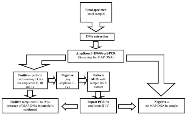
IV-negative specimens, we suggest the use of MDA and the consecutive repetition of the confirmatory PCRs. A suggested workflow is shown in Fig. 4. Fecal specimens are true
alterna-FIG. 4. Suggested workflow for the detection ofM. avium subsp.paratuberculosis DNA in stool samples. MDA, multiple displacement amplification. Amplicon I⫽IS900; amplicon II⫽MAP2765c (251); amplicon III⫽MAP0865 (f57); amplicon IV⫽MAP0865.
on May 16, 2020 by guest
http://jcm.asm.org/
[image:8.585.132.457.67.262.2]tives for the analysis of patients and cattle because of their noninvasiveness and simplicity in collection. Our data pre-sented here provide a basis for further structured studies of the
potential role ofM. aviumsubsp.paratuberculosisin CD and
other human diseases.
ACKNOWLEDGMENTS
We thank Martina Klo¨s-Langsdorf and Kirsten-Susann Bommer-sheim for excellent technical assistance, Isabell Krabs for the prepa-ration of the gut tissue of slaughtered cows, Simone Ries for running qrt-PCRs during a practical course, and Egbert Tannich from the Bernhard-Nocht Institute for Tropical Medicine (Hamburg, Germany) for providing parasite DNA obtained fromEntamoeba histolyticaand fromGiardia lamblia.
This work was supported by grants from the Bundesministerium fuer Bildung und Forschung, Germany, within the framework of the Na-tional Genome Research Network (NGFN) (contract no. 01GS0401).
REFERENCES
1.Altschul, S. F., W. Gish, W. Miller, E. W. Myers, and D. J. Lipman.1990. Basic local alignment search tool. J. Mol. Biol.215:403–410.
2.Bannantine, J., P. E. Baechler, Q. Zhang, L. Li, and V. Kapur.2002. Ge-nome scale comparison ofMycobacterium aviumsubsp.paratuberculosiswith
Mycobacterium aviumsubsp.aviumreveals potential diagnostic sequences. J. Clin. Microbiol.40:1303–1310.
3.Chapin, K. C., and T.-L. Lauderdale.2007. Reagents, stains, and media: bacteriology, p. 334–364.InP. R. Murray, E. J. Baron, J. H. Jorgensen, M. L. Landry, and M. A. Pfaller (ed.), Manual of clinical microbiology, 9th ed. ASM Press, Washington, DC.
4.Chiodini, R. J.1989. Crohn’s disease and the mycobacterioses: a review and comparison of two disease entities. Clin. Microbiol. Rev.2:90–117. 5.Collins, D. M., and G. W. de Lisle.1986. Restriction endonuclease analysis
of various strains ofMycobacterium paratuberculosis isolated from cattle. Am. J. Vet. Res.47:2226–2229.
6.Committee on Diagnosis and Control of Johne’s Disease.2003. Johne’s disease and Crohn’s disease, p. 104–120.InDiagnosis and control of Johne’s disease: report for the National Research Council of the National Academy of Science. The National Academic Press, Washington, DC.
7.Cousins, D. V., et al.1999. Mycobacteria distinct fromMycobacterium avium
subsp.paratuberculosisisolated from the faeces of ruminants possess IS900 -like sequences detectable IS900polymerase chain reaction: implications for diagnosis. Mol. Cell Probes13:431–442.
8.Dalziel, T. K.1913. Chronic interstitial enteritis. Br. Med. J.2:1068–1070. 9.Englund, S., G. Bolske, and K. E. Johansson.2002. An IS900-like sequence
found in aMycobacteriumsp. other thanMycobacterium aviumsubsp. para-tuberculosis.FEMS Microbiol. Lett.209:267–271.
10.Gao, A., L. Mutharia, S. Chen, K. Rahn, and J. Odumeru.2002. Effect of pasteurisation on survival ofMycobacterium paratuberculosisin milk. J. Dairy Sci.85:3198–3205.
11.Ghai, R., T. Hain, and T. Chakraborty.2004. GenomeViz: visualizing mi-crobial genomes. BMC Bioinformatics5:198.
12.Grant, I. R.2005. Zoonotic potential ofMycobacterium aviumsubsp. para-tuberculosis: the current position. J. Appl. Microbiol.98:1282–1293. 13.Green, E. P., et al.1989. Sequence and characteristics of IS900, an insertion
element identified in a human Crohn’s disease isolate ofMycobacterium paratuberculosis. Nucleic Acids Res.17:9063–9073.
14.Harris, N. B., and R. G. Barletta.2001.Mycobacterium aviumsubsp. para-tuberculosisin veterinary medicine. Clin. Microbiol. Rev.14:489–512. 15.Hermon-Taylor, J.2001. Protagonist:Mycobacterium aviumsubspecies
para-tuberculosisis a cause of Crohn’s disease. Gut49:755–756.
16.Hermon-Taylor, J., and T. J. Bull.2002. Crohn’s disease caused by Myco-bacterium aviumsubsp.paratuberculosis: a public health tragedy whose res-olution is long overdue. J. Med. Microbiol.51:3–6.
17.Hermon-Taylor, J., et al.2000. Causation of Crohn’s disease by Mycobacte-rium aviumsubspeciesparatuberculosis. Can. J. Gastroenterol.14:521–539. 18.Hoorfar, J., P. Ahrens, and P. Radstro¨m.2000. Automated 5⬘nuclease PCR
assay for identification ofSalmonella enterica. J. Clin. Microbiol.38:3429– 3435.
19.Hruska, K., M. Bartos, P. Kralik, and I. Pavlik.2005.Mycobacterium avium
subsp.paratuberculosisin powdered infant milk: paratuberculosis in cattle— the public health problem to be solved. Vet. Med.-Czech50:327–335. 20.Huntley, J. F. J., R. H. Whitlock, J. P. Bannantine, and J. R. Stabel.2005.
Comparison of diagnostic detection methods forM. aviumsubsp. paratuber-culosisin North American bison. Vet. Pathol.42:42–51.
21.Kim, S. G., et al.2002. Development and application of quantitative poly-merase chain reaction assay based on the ABI 7700 system (TaqMan) for detection and quantification ofMycobacterium aviumsubsp.paratuberculosis.
J. Vet. Diagn. Invest.14:126–131.
22.Li, L., et al.2005. The complete genome sequence ofMycobacterium avium
subspeciesparatuberculosis. Proc. Natl. Acad. Sci. U. S. A.102:12344–12349. 23.Martin, F. E., M. A. Nadkami, N. A. Jacques, and N. Hunter.2002. Quan-titative microbiological study of human carious dentine by culture and real-time PCR: association of anaerobes with histopathological changes in chronic pulpitis. J. Clin. Microbiol.40:1698–1704.
24.McFadden, J. J., P. D. Butcher, R. J. Chiodini, and J. Hermon-Taylor.1987. Crohn’s disease-isolated mycobacteria are identical toMycobacterium para-tuberculosis, as determined by DNA probes that distinguish between myco-bacterial species. J. Clin. Microbiol.25:796–801.
25.Mendoza, J. L., R. Lana, and M. Díaz-Rubio.2009.Mycobacterium avium
subspeciesparatuberculosisand its relationship with Crohn’s disease. World J. Gastroenterol.15:417–422.
26.Mu¨ller, A., et al. 2000. Detection ofIsospora belli by polymerase chain reaction using primers based on small-subunit ribosomal RNA sequences. Eur. J. Clin. Microbiol. Infect. Dis.19:631–634.
27.Naser, S. A., D. Schwartz, and I. Shafran.2000. Isolation ofMycobacterium aviumsubsp.paratuberculosisfrom breast milk of Crohn’s disease patients. Am. J. Gastroenterol.95:1094–1095.
28.Naser, S. A., G. Ghobrial, C. Romero, and J. F. Valentine.2004. Culture of
Mycobacterium aviumsubspeciesparatuberculosisfrom the blood of patients with Crohn’s disease. Lancet364:1039–1044.
29.Nogva, H. K., A. Bergh, A. Holck, and K. Rudi.2000. Application of the 5⬘-nuclease PCR assay in evaluation and development of methods for quan-titative detection of Campylobacter jejuni. Appl. Environ. Microbiol. 66: 4029–4036.
30.Olsen, I., G. Sigurgardottir, and B. Djonne.2002. Paratuberculosis with special reference to cattle. Vet. Q.24:12–28.
31.Ott, S. L., S. J. Wells, and B. A. Wagner.1999. Herd-level economic losses associated with Johne’s disease on US dairy operations. Prev. Vet. Med. 40:179–192.
32.Parrish, N. M., et al.2009. Absence ofMycobacterium aviumsubsp. paratu-berculosisin Crohn’s patients. Inflamm. Bowel Dis.15:558–565.
33.Pfyffer, G. E.2007.Mycobacterium: general characteristics, laboratory detec-tion, and staining procedures, p. 543–572.InP. R. Murray, E. J. Baron, J. H. Jorgensen, M. L. Landry, and M. A. Pfaller (ed.), Manual of clinical micro-biology, 9th ed. American Society for Microbiology Press, Washington, DC. 34.Pierce, E. S.2009. Possible transmission ofMycobacterium aviumsubspecies
paratuberculosisthrough potable water: lessons from an urban cluster of Crohn’s disease. Gut Pathog.23:17.
35.Poupart, P., M. Coene, H. Vanheuverswyn, and C. Cocito.1993. Preparation of a specific RNA probe for detection ofMycobacterium paratuberculosisand diagnosis of Johne’s disease. J. Clin. Microbiol.31:1601–1605.
36.Proudnikov, D., et al.2003. Optimizing primer-probe design for fluorescent PCR. J. Neurosci. Methods123:31–45.
37.Quirke, P.2001. Antagonist:Mycobacterium aviumsubspecies paratubercu-losisis a cause of Crohn’s disease. Gut49:757–760.
38.Rajeev, S., Y. Zhang, S. Sreevatsan, A. S. Motiwala, and B. Byrum.2005. Evaluation of multiple genomic targets for identification and confirmation of
Mycobacterium aviumsubsp.paratuberculosisisolates using real-time PCR. Vet. Microbiol.105:215–221.
39.Schwartz, D., et al.2000. Use of short-term culture for identification ofM. aviumsubsp.paratuberculosisin tissue from Crohn’s disease patients. Clin. Microbiol. Infect.6:303–307.
40.Semret, M., C. Y. Turenne, and M. A. Behr.2006. Insertion sequence IS900
revisited. J. Clin. Microbiol.44:1081–1083.
41.Shankar, H., et al.2010. Presence, characterization, and genotype profiles of
Mycobacterium aviumsubspeciesparatuberculosisfrom unpasteurized indi-vidual and pooled milk, commercial pasteurized milk, and milk products in India by culture, PCR, and PCR-REA methods. Int. J. Infect. Dis.14:121– 126.
42.Shivananda, S., et al.1996. Incidence of inflammatory bowel disease across Europe: is there a difference between north and south? Results of the European Collaborative Study in Inflammatory Bowel Disease (EC-IBD). Gut39:690–697.
43.Sibartie, S., et al.2010.Mycobacterium aviumsubsp.paratuberculosis(MAP) as a modifying factor in Crohn’s disease. Inflamm. Bowel Dis.16:296–304. 44.Slana, I., M. Liapi, M. Moravkova, A. Kralova, and I. Pavlik.2009.
Myco-bacterium aviumsubsp.paratuberculosisin cow bulk tank milk in Cyprus detected by culture and quantitative IS900and F57 real-time PCR. Prev. Vet. Med.89:223–226.
45.Soumya, M. P., R. M. Pillai, P. X. Antony, H. K. Mukhopadhyay, and V. N. Rao.2009. Comparison of faecal culture and IS900PCR assay for the detection ofMycobacterium aviumsubsp.paratuberculosisin bovine faecal samples. Vet. Res. Commun.33:781–791.
46.Stabel, J. R., and J. P. Bannantine.2005. Development of a nested PCR method targeting a unique multicopy element, ISMap02, for detection of
Mycobacterium aviumsubsp.paratuberculosisin fecal samples. J. Clin. Mi-crobiol.43:4744–4750.
47.Sung, N., and M. T. Collins.1998. Thermal tolerance ofMycobacterium paratuberculosis. Appl. Environ. Microbiol.64:999–1005.
48.Sung, N., and M. T. Collins.2000. Effect of three factors in cheese
on May 16, 2020 by guest
http://jcm.asm.org/
tion (pH, salt and heat) onMycobacterium aviumsubsp.paratuberculosis
viability. Appl. Environ. Microbiol.66:1334–1339.
49.Tasara, T., and R. Stephan.2005. Development of an F57 sequence-based real-time PCR assay for detection ofM. aviumsubsp.paratuberculosisin milk. Appl. Environ. Microbiol.71:5957–5968.
50.Thomas Dow, C.2008. Cows, Crohn’s and more: isMycobacterium paratu-berculosisa superantigen? Med. Hypotheses71:858–861.
51.Thorel, M. F., M. Krichevsky, and V. V. Levy-Frebault.1990. Numerical taxon-omy of mycobactin-dependent mycobacteria, emended description of Mycobac-terium avium, and description ofMycobacterium aviumsubsp.aviumsubsp. nov.,
Mycobacterium aviumsubsp.paratuberculosissubsp. nov., andMycobacterium aviumsubsp.silvaticumsubsp. nov. Int. J. Syst. Bacteriol.40:254–260. 52.van den Berg, R. J., et al.2005. Prospective multicenter evaluation of a new
immunoassay and real-time PCR for rapid diagnosis ofClostridium difficile -associated diarrhea in hospitalized patients. J. Clin. Microbiol.43:5338– 5340.
53.Verweij, J. J., et al.2004. Simultaneous detection ofEntamoeba histolytica,
Giardia lamblia, andCryptosporidium parvumin fecal samples by using mul-tiplex real-time PCR. J. Clin. Microbiol.42:1220–1223.
54.Vishnubhatla, A., et al.2000. Rapid 5⬘nuclease (TaqMan) assay for detec-tion of virulent strains ofYersinia enterocolitica. Appl. Environ. Microbiol. 66:4131–4135.
55.Whittington, R. J., D. J. Marshall, P. J. Nicholls, I. B. Marsh, and L. A. Reddacliff. 2004. Survival and dormancy ofMycobacterium aviumsubsp.
paratuberculosisin the environment. Appl. Environ. Microbiol.70:2989– 3004.