N A N O E X P R E S S
Open Access
Magnetic-Field-Assisted Assembly of
Anisotropic Superstructures by Iron Oxide
Nanoparticles and Their Enhanced
Magnetism
Chengpeng Jiang
1, Chi Wah Leung
2and Philip W. T. Pong
1*Abstract
Magnetic nanoparticle superstructures with controlled magnetic alignment and desired structural anisotropy hold promise for applications in data storage and energy storage. Assembly of monodisperse magnetic nanoparticles under a magnetic field could lead to highly ordered superstructures, providing distinctive magnetic properties. In this work, a low-cost fabrication technique was demonstrated to assemble sub-20-nm iron oxide nanoparticles into crystalline superstructures under an in-plane magnetic field. The gradient of the applied magnetic field contributes to the anisotropic formation of micron-sized superstructures. The magnitude of the applied magnetic field
promotes the alignment of magnetic moments of the nanoparticles. The strong dipole-dipole interactions between the neighboring nanoparticles lead to a close-packed pattern as an energetically favorable configuration. Rod-shaped and spindle-Rod-shaped superstructures with uniform size and controlled spacing were obtained using spherical and polyhedral nanoparticles, respectively. The arrangement and alignment of the superstructures can be tuned by changing the experimental conditions. The two types of superstructures both show enhancement of coercivity and saturation magnetization along the applied field direction, which is presumably associated with the magnetic anisotropy and magnetic dipole interactions of the constituent nanoparticles and the increased shape anisotropy of the superstructures. Our results show that the magnetic-field-assisted assembly technique could be used for fabricating nanomaterial-based structures with controlled geometric dimensions and enhanced magnetic properties for magnetic and energy storage applications.
Keywords:Iron oxide nanoparticles, Nanoparticle superstructures, Nanoparticle assembly, Magnetic properties
Background
Enormous progress has been made in chemical synthesis of magnetic nanoparticles, enabling us to precisely con-trol their morphology and composition [1–4]. To find their applications in data storage and energy storage, it is of fundamental and practical interest to organize these magnetic nanoparticles into structures with controlled shape, spacing, and alignment [4–7]. Self-assembly strat-egy by applying a magnetic field offers a simple and effi-cient route to assemble magnetic nanoparticles as the building blocks into well-ordered structures, providing
distinctive magnetic properties related to coercivity and anisotropy energy [4, 7–9].
Generally, the magnetic-field-guided assembly of mag-netic nanoparticles can be categorized into two types: dynamic self-assembly, being a nanoparticle organization process which occurs in liquid interfaces or solutions, with continuous energy input to grant the interactions responsible for the growing and sustaining of ordered structures; and static self-assembly, in which the nano-particles assemble on solid surfaces or substrates, lead-ing to stable structures or patterns without dissipatlead-ing energy after the completion of assembly [10, 11]. A rep-resentative example of the liquid-phase assembly of magnetic nanoparticles is colloidal assembly of photonic crystals, and a number of research works have reported * Correspondence:[email protected]
1Department of Electrical and Electronic Engineering, The University of Hong
Kong, Hong Kong, Hong Kong
Full list of author information is available at the end of the article
the dynamic assembly of magnetic colloidal crystals under applied field. Wang’s group demonstrated a mag-netically actuated liquid crystal by controlling the assem-bly orientation and hence the transmittance intensity of magnetic nanorods using a weak magnetic field [12]. They further developed the magnetically responsive pho-tonic structures built from magnetic nanoellipsoids, whose diffraction properties can be tuned reversibly [13]. Xia’s group introduced the colloidal clusters of magnetite nanocrystals into polymer matrix via photo-polymerization, and photonic bandgap of the resulting photonic crystals can be modulated by tuning the cluster size and field strength during self-assembly [14]. Timonen and his coworkers presented a switchable dynamic self-assembly technique by driving the ferrofluid droplets of iron oxide nanoparticles on super-hydrophobic surface under a time-vary magnetic field [15]. For the dynamic assembly of photonic crystals, manipulating the orientation or controlling the spacing of assem-bled structures is usually implemented by changing the applied magnetic field for the purpose of modu-lating optical properties [16, 17]. However, if magnetic storage application is envisioned, the assembled struc-tures should be physically robust and mechanically stable without the presence of an external magnetic field. In this regard, magnetic-field-guided static as-sembly strategy is much preferred. Recent works on this topic mostly involve fabricating magnetic nano-particle superstructures with alignment and shape an-isotropy through evaporative or convective deposition process, during which geometric constraints or exter-nal stimuli could be applied to pattern or direct the
assembled superstructures [18–21]. Park et al.
achieved the production of crystalline superstructures in rod and wire shapes composed of 10-nm cobalt nanoparticles, which show enhanced coercivity associ-ated with easy-axis anisotropy [22]. Li et al. demon-strated a solvent-evaporation assembly process to obtain micro-rod superstructures made of 21-nm magnetite nanoparticles, exhibiting specific crystallo-graphic orientations [23]. Singh et al. developed an interfacial deposition method to assemble 13-nm magnetite nanocubes into aligned superstructures with stranded helical shapes [24]. Although periodic chain or rod structures in micron size with a high aspect ratio could be prepared in these reports and also in the recently developed strategies for static assembly of magnetic nanoparticles under a uniform magnetic field [22–26], fabrication of sub-10-μm nanoparticle superstructures with homogeneous morphology and controlled spacing still remains challenging. Besides, it will be of great value to investigate how the shape of the constituent nanoparticles and the gradient of the magnetic field influence the assembly process and
measure the anisotropy-associated magnetic properties of the resulting superstructures. From the application viewpoint, the assembled superstructures with both shape anisotropy and magnetic anisotropy could be exploited as unique magnetic systems for studying nanomagnetism, and they could also be used for fab-ricating functional materials and solid-state magnetic devices with controlled magnetization direction and desired anisotropy energy.
In this work, sub-20-nm iron oxide nanoparticles (IONPs) with spherical and polyhedral shapes were syn-thesized by thermal pyrolysis in organic phase. An evap-orative deposition method was applied to assemble the IONPs into superstructures under the applied magnetic field. The direction and rate of the assembly process and the magnitude and gradient of the magnetic field were controlled. Morphologies of the IONPs and their super-structures were characterized, and the assembly
mech-anism was analyzed. Effects of the experimental
conditions and nanoparticle shape on the morphology and alignment of the superstructures were investigated. Magnetic properties of the superstructures were mea-sured, and the enhancement of their coercivity and satu-rated magnetization along the applied field direction was investigated by calculating the magnetic anisotropy en-ergy and dipole interaction enen-ergy of the IONPs.
Methods
Iron (III) oxyhydroxide (99 %), iron (III) acetylacetonate (99.9 %), 1,2-tetradecanediol (90 %), oleic acid (90 %), lauric acid (98 %), and decanoic acid (98 %) were pur-chase from Aldrich. Octadecene (90 %) and benzyl ether (98 %) were obtained from Acros. All chemicals were used as received.
Spherical IONPs capped by lauric acid were synthe-sized using iron (III) oxyhydroxide (3 mmol), lauric acid (9 mmol), oleic acid (1 mmol), and octadecene (15 ml). The reaction mixture was vigorously stirred under N2, and it was first heated to 200 °C for 30 min and then heated to reflux for 1 h. By removing the heating source, the resulting black mixture was cooled to 50 °C and kept in a water bath. Excess acetone was added to the mix-ture, and a black-brown material was precipitated by centrifugation and then dissolved in hexane. This purifi-cation step was repeated two more times, and the
ob-tained nanoparticles were dispersed in 6 ml of
chloroform and toluene (v:v= 9:1).
Polyhedral IONPs capped by decanoic acid were synthe-sized using iron (III) acetylacetonate (3 mmol), decanoic acid (18 mmol), benzyl ether (60 ml), and 1,2-tetradecane-diol (15 mmol). The heating process and the purification process were the same as those for spherical IONPs.
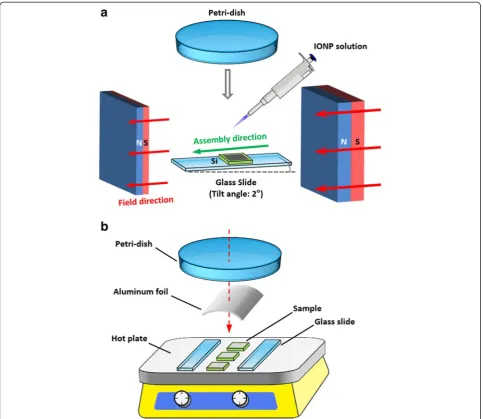
concentration 160 mM based on Fe) onto a flat Si sub-strate (8 mm × 8 mm, thermally oxidized) subjected to an in-plane magnetic field generated by NdFeB magnets (Fig. 1a). The evaporation rate of the solvent was con-trolled by covering the sample with a petri dish, while the translation direction of the air-solution-substrate contact line was controlled by tilting the substrate at 1.5° with respect to the ground. The magnetic field direction was adjusted to be parallel to the translation direction of the three-phase contact line. The magnitude and gradi-ent of the magnetic field (typical values at the initial as-sembly stage 200 Oe and 60 Oe/mm, measured at the sample center) were controlled by changing the distance and location of the magnets. After the solvent evapo-rated completely, the sample was transferred to a hot
plate for annealing. Here, a hot plate was used instead of a convection oven due to that it heats the sample from the bottom by thermal conduction, which can prevent skin effect and shorten the heating time for thin film an-nealing. As shown in Fig. 1b, two glass slides were placed parallelly to fix the sample, and a curved aluminum foil and a glass petri dish were covered on top but without contacting the sample to prevent air flow and reduce heat dissipation. The sample was then heated at 200 °C for 15 min in air to remove the organic substances. After the heat treatment, a solid coating was obtained on the substrate surface.
The IONPs were characterized by transmission elec-tron microscope (TEM). The nanoparticle superstruc-tures were characterized using optical microscope and
[image:3.595.57.541.284.703.2]scanning electron microscope (SEM), and their magnetic
properties were measured using vibrating sample
magnetometer (VSM) and magnetic force microscope (MFM).
Results and Discussion
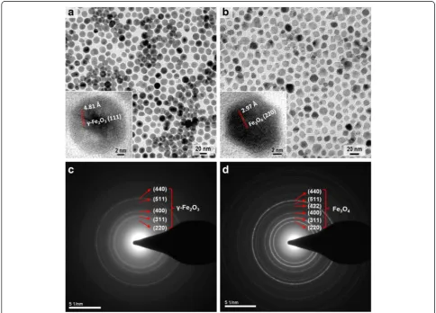
Morphology and composition of the synthesized nano-particles were investigated by bright-field TEM, high-resolution TEM (HRTEM), and selected-area electron diffraction (SAED). Figure 2a shows that the lauric acid-capped nanoparticles are in spherical shape with an average size of 19 nm and their measured lattice spacing based on the diffraction rings in Fig. 2c matches well with thed-spacing of hklplanes inγ-Fe2O3(JCPDF #04-0755). Figure 2b shows that the decanoic acid-capped nanoparticles are in polyhedral shape with an average size of 16 nm and their diffraction rings in Fig. 2d can be well indexed to the lattice spacing of Fe3O4 (JCPDF #19-0629). The TEM and HRTEM images demonstrate that the spherical nanoparticles are polycrystalline with a narrow size distribution of 8 %, while the
polyhedral nanoparticles are single crystalline with a size distribution of 15 %. The ring features in both SAED patterns indicate the random orientations of the nanoparticles.
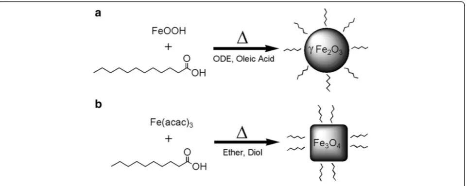
Synthesis protocols of the IONPs with different sizes and shapes are inspired by thermal pyrolysis procedures developed by William [27] and Sun [28] It was found that the chemical property of the precursor material and the coordinating ability of the organic solvent have great influence on the morphology of the IONPs. As illus-trated in Fig. 3, the use of inorganic precursor and non-coordinating solvent led to isotropic growth of the IONPs with spherical shape, while using organometallic precursor and weakly coordinating solvent resulted in anisotropic growth of the IONPs with polyhedral shape. Surfactants with carboxylic groups were chosen as the capping agents for the IONPs due to their strong bind-ing ability with the surface atoms of the IONPs, which contributes to the formation of nearly monodisperse
nanoparticles with high yield and high stability.
Decanoic acid and lauric acid were used instead of oleic
[image:4.595.55.540.356.704.2]acid since they have lower boiling point and shorter chain length, which could facilitate the removal of or-ganic surfactant during heat treatment and also decrease the spacing between the neighboring IONPs for enhan-cing magnetic coupling. A temporal separation of the nucleation stage and the growth stage by kinetic and thermodynamic parameters was achieved using a two-step heating strategy, which helps to narrow the size dis-tribution of the IONPs.
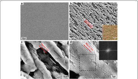
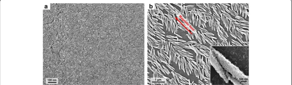
Morphology of the assembled structures formed by spherical IONPs is investigated by SEM. As shown in Fig. 4a, without the applied magnetic field, a homoge-neous film composed of well-ordered spherical IONPs could be obtained. The nanoparticle film is compact and continuous without void or defect at micron scale, illus-trating that the assembly technique by controlling the evaporation rate and assembly direction could lead to self-organized structures formed by monodisperse nano-particles. As shown in Fig. 4b, under the in-plane ap-plied magnetic field, rod-shaped superstructures with a length of 2.8μm, width of 0.5μm, and aspect ratio of ~6 were obtained, and they are homogeneously distributed
with controlled spacing of 0.7 μm. By measuring the
intersecting angle (β) between the applied magnetic field and the longitudinal axis of each rod-shaped superstruc-ture, it is found that the superstructures show good alignment with the magnetic field (β= 4.0° ± 2.7°). The orientation of the superstructure may not accurately re-flect the direction of the magnetic moments of its con-stituent IONPs, but it is obvious that the assembly directions of the superstructures correlate with the mag-netic field direction. Besides, some dot-shaped super-structures were formed probably due to the incomplete growth of the assembled structures. Park et al. also re-ported the shape evolution of supercrystal dots during
the magnetic assembly of supercrystal rods [22]. En-larged SEM image in Fig. 4c reveals that the rod-shaped superstructures are composed of closely packed spher-ical IONPs without coalescence or crack and the dark regions between the adjacent superstructures are capped by a monolayer of IONPs. The ordered stacking struc-tures in the close-up SEM image (Fig. 4d) and the six symmetric bright spots in the fast Fourier transform (FFT) pattern of the superstructure (inset of Fig. 4d) demonstrate that highly ordered hexagonal packing patterns were formed by uniform nanoparticles in the rod-shaped superstructures as an energetically favor-able configuration [29].
[image:5.595.60.541.87.279.2]all the experimental conditions should be adjusted and tuned within proper ranges; otherwise, anisotropic super-structures would not form during the magnetic-field-assisted assembly process. This is because nanoparticle self-assembly has nonlinear nature and it is highly sensi-tive to the experimental conditions, which will affect the growth and arrangement of the assembled structures [30]. Other experimental conditions such as the boiling point
and evaporation rate of the dissolving solvent and also the temperature of the substrate will be investigated in our fu-ture research.
Effects of the nanoparticle shape on the morphology and alignment of the superstructures were further inves-tigated. Here, polyhedral IONPs were used instead of spherical IONPs for preparing the sample, and inher-ently, they could assemble into a closely packed and
Fig. 5SEM images of the rod-shaped superstructures prepared under different conditions.aSEM image of the superstructures prepared by increasing the tilt angle of the substrate.bSEM image of the superstructures prepared by decreasing the concentration of the nanoparticle solution.cSEM image of the superstructures prepared by reducing the magnitude and gradient of the applied magnetic field
[image:6.595.56.540.87.366.2] [image:6.595.64.538.536.694.2]defect-free film at micron scale as shown in Fig. 6a. Under the in-plane applied magnetic field, spindle-shaped superstructures with a length of 0.8μm, width of 0.15 μm, and aspect ratio of ~5 were formed as pre-sented in Fig. 6b. The spindle-shaped superstructures have uniform size, but their alignment with the applied field (intersecting angle β= 12.9° ± 9.7°) are not as good as the rod-shaped superstructures, and they do not have uniform spacing. The lack of spatial order in these su-perstructures could be due to the fact that the spindle-shaped superstructures tend to aggregate together, and the shape anisotropy of these superstructures and their constituent polyhedral IONPs should also be taken into consideration. These spindle-shaped superstructures ob-served here are similar to the loosely aligned micro-rod superstructures reported by Li et al. [23]. The structural difference between these spindle-shaped superstructures and the previously mentioned rod-shaped superstruc-tures could be mainly attributed to the different shapes of their constituent IONPs, which influence the packing and ordering of nanoparticles during self-assembly. Moreover, nanoparticle compositions (related to mag-netic properties) and nanoparticle surfactants (affecting diffusion and interaction of nanoparticles in organic solvent) also affect the assembly process and hence the superstructure shape, although their differences are triv-ial between the two superstructures.
The magnetic-field-assisted assembly of IONPs is a particle deposition process, which depends on the bal-ance between evaporation-driven assembly and field-driven assembly [31, 32]. During the assembly process, convection of the organic solvent due to evaporation drives the IONPs to transport and deposit at the three-phase contact line. The evaporation rate mainly deter-mines the assembly rate, which is quite fast due to that chloroform used for dispersing the IONPs is a highly volatile solvent. The evaporation direction relates to the translation direction of the contact line [33], which
determines the assembly direction, and the applied field direction was experimentally adjusted to be parallel to this assembly direction. At the first stage of the assembly process, a gradient magnetic field (typical values magni-tude 200 Oe, gradient 60 Oe/cm) was applied in plane, and the dominant forces exerted on IONPs are capillary force induced by solvent evaporation and magnetic force caused by external field and dipolar interactions, while effects of the thermal fluctuations and the local interac-tions (such as van der Waals attraction and steric repul-sion) are negligible [15, 21]. Under the magnetic field, each IONP behaves as a magnetic dipole with magnetic momentm=μ0MV, whereμ0is the permeability of free
space and M and Vare the domain magnetization and
volume of the IONP, respectively [32]. Magnetic dipole-dipole interaction between two magnetically aligned IONPs is given byFdip= 3m2(1−3 cos2α)/d4, whereαis the angle between their magnetic moment and the line connecting their centers and d is the inter-particle dis-tance [25, 34]. This interaction is attractive along the field direction but repulsive perpendicular to the field direction [34], which results in the anisotropic growth of spaced superstructures with preferred alignment along the field direction. The gradient of the magnetic field also in-duces a force along the field direction Fmag=∇(m⋅B), with B being the magnetic field, and this force together with the capillary force expedites the packing of the IONPs and causes the formation of micron-sized struc-tures. After the evaporation of chloroform (~80 s), assem-bled structures with uniform size started to develop. At the second stage of the assembly process, a strong and uniform magnetic field with a magnitude of 2 kOe was ap-plied in plane. The IONPs closely packed into condensed superstructures, and their magnetic moments may also get aligned with this strong field. After complete dewet-ting of the nanoparticle solution (~120 s), the assembled structures became solid coatings on Si. The two stages of the assembly process both affect the morphology and
[image:7.595.57.539.563.703.2]property of the superstructures. Specifically, applying the first stage alone led to micron-sized superstructures with-out significantly enhanced magnetism, while solely imple-menting the second stage produced parallel strip patterns
with a long length (up to 100 μm) and large spacing
(~2μm).
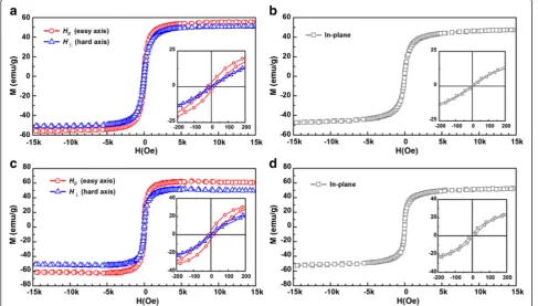
To probe the magnetic properties of the tures, hysteresis loops for the rod-shaped superstruc-tures (Fig. 7a) and spindle-shaped superstrucsuperstruc-tures (Fig. 7c) were measured at 300 K along two in-plane di-rections, namely, the direction along the applied field (H//) and the direction perpendicular to the applied field (H⊥). As a comparison, in-plane hysteresis loops for the spherical IONPs (Fig. 7b) and polyhedral IONPs (Fig. 7d) assembled as uniform films in the absence of a magnetic field were measured, respectively. For the rod-shaped
superstructures, coercivity Hc of 22 Oe and
satur-ation magnetizsatur-ation Ms of 56 emu/g were observed
along H//, which are larger than the results obtained along H⊥ (Hc= 8 Oe, Ms= 51 emu/g). For the
spindle-shaped superstructures, increased Hc of 18 Oe and
increased Ms of 62 emu/g were also obtained along
H// compared to the results measured along H⊥ (Hc= 6 Oe, Ms= 52 emu/g). The results indicate that the magnetic easy axes of both superstructures are along the applied field direction and the superstructures
show larger coercivity and higher saturation magnetization at their easy axes compared to the uniform films assem-bled from the same IONPs.
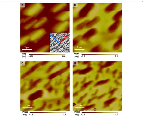
To gain insights into the magnetic interactions of the superstructures, MFM imaging was performed for the sample of the rod-shaped superstructures in different magnetization states. The measurements were per-formed in tapping mode using a magnetized tip (Mag-netic Multi75-G, BudgetSensors) through a two-pass process. In the first pass, AFM image of surface topog-raphy (Fig. 8a) was recorded, and further analysis of the topographical data indicates a thickness of 0.68 μm for the rod-shaped superstructures, close to the measured
width of 0.5 μm from the corresponding SEM image
(inset of Fig. 8a). In the second pass, the tip is lifted above the sample surface and scanned at a constant height of 400 nm following the acquired AFM topog-raphy. Magnetic contrast of the superstructures was ob-served on the demagnetized sample by comparing the MFM image (Fig. 8b) with the AFM topography (Fig. 8a) at the same region. This magnetic contrast is probably caused by the MFM tip magnetizing the originally demagnetized sample. It is well known that a MFM tip, being a source of stray field itself, could cause magnetization to the sample it is scanning. Particularly, the building material of our sample is magnetically soft
Fig. 7M-H curves of the assembled structures at 300 K.aRod-shaped superstructures of spherical IONPs assembled under a magnetic field.b
[image:8.595.56.544.416.693.2]iron oxide nanoparticles with a small coercivity (<25 Oe) and small size (<20 nm), which show nearly superpara-magnetic behavior at room temperature; thus, the nano-particles could be easily magnetized by the stray field of the MFM tip, and the nanoparticle dipole coupling within each superstructure led to the phase difference of the MFM cantilever oscillation [35]. The sample-tip interaction was also reported by Confalonieri et al. in their magnetic system containing a monolayer of 20-nm Fe2O3nanoparticles [36]. MFM images of the sample in a remanent state after saturation along H// (easy axis) and after saturation along H⊥ (hard axis) are shown in Fig. 8c, d, respectively. In all the three MFM images (Fig. 8b–d), the superstructures exhibit the same mag-netic contrast, indicating that the neighboring super-structures are magnetically coupled together [35]. However, it is not easy to identify the difference between
Fig. 8c and d because their magnetic contrasts are simi-lar and also, these two images cannot be directly com-pared since they are not from exactly the same region. Therefore, the VSM results are more useful and convin-cing to reflect the magnetism of the superstructures at the easy axis and at the hard axis.
To quantitatively interpret the origin of the enhanced magnetism observed in the VSM results, magnetic anisot-ropy energy and dipole interaction energy are calculated for the two types of IONPs, respectively. For single-domain non-interacting magnetic nanoparticles, the ef-fective magnetic anisotropy energy constant Keff can be calculated as Keff= 25kBTB/Vp, where kB is Boltzmann constant,TBis the blocking temperature of nanoparticles, Vpis the volume of a nanoparticle (Vp≈1:33πd3p=8,dpis nanoparticle diameter), and Keff is the sum of several terms including magnetocrystalline anisotropy, shape Fig. 8MFM characterization of the rod-shaped superstructures.aAFM image of the superstructures, with aninsetof SEM image.bMFM image of the superstructures in a demagnetized state.cMFM image of the superstructures in a remanent state after saturation alongH//.dMFM image
[image:9.595.60.538.85.472.2]anisotropy, surface anisotropy, and stress anisotropy [37]. The calculatedKeff (2.16 × 10⁵erg/cm3 for 19-nm spher-ical maghemite IONP; 3.70 × 10⁵erg/cm3for 16-nm poly-hedral magnetite IONP) using TB of the nanocrystals in powder form with similar size and shape as our IONPs (225 K; 230 K) [37–39] is much larger than the reported values for bulk maghemite (0.46 × 10⁵erg/cm3) and mag-netite (1.35 × 10⁵erg/cm3). This enhancement of magnetic anisotropy is associated with dipole interactions between IONPs. According to the Stoner-Wohlfarth theory, mag-netocrystalline anisotropy energy Ea of a single-domain magnetic nanoparticle is approximated byEa=K1Vpsin2θ, where K1 is the magnetocrystalline anisotropy constant (K1≈Keff/12) andθ is the angle between the easy axis of the nanoparticle and the direction of the field-induced magnetization [38]. The estimated Ea is 1.62 × 10⁻¹⁶ erg for a spherical IONP using K1= 1.8 × 10⁴ emu/cm3 and sin θ= 0.05, while Ea is 7.64 × 10⁻¹⁵erg for a polyhedral IONP using K1= 3.1 × 10⁴ emu/cm3 and sin θ= 0.34. Similarly, shape anisotropy energyEshcan be approximated as Esh=KshVpsin2θ, where Ksh is 0:5M2s easyð ÞðNb−NaÞ, with Na and Nbbeing the demagnetization factors of the nanoparticle in the short-axis and long-axis directions and Ms(easy)being the saturation magnetization at the easy axis [40, 41]. The estimatedEshis 1.71 × 10⁻¹⁷erg for a spherical IONP using Nb−Na= 0.05, Ms(easy)= 56 emu/g = 274.4 emu/cm3, andKsh= 1.9 × 10³emu/cm3, while Esh is 3.72 × 10⁻¹⁵erg for a polyhedral IONP usingNb−Na= 0.3, Ms(easy)= 62 emu/g = 316.2 emu/cm3, and Ksh= 1.5 × 10⁴ emu/cm3. Besides, surface anisotropy also has signifi-cant contribution to the effective magnetic anisotropy due to surface spin disorder, while stress anisotropy is much less pronounced considering that the lattice constants of the IONPs are nearly equal to their bulk counterparts [40, 41]. For the assembled superstructure composed of magnetic-ally aligned nanoparticles under the applied magnetic field, the dipole interaction energy of one nanoparticle in the superstructure can be approximated asEdip¼2μ0μ2p=4πr3 using the macro-spin approximation, whereμpis the dipole moment of the nanoparticle and r is the mean center-to-center distance between the neighboring nanoparti-cles [42]. By measuring the average inter-particle distance from the SEM images (22 nm for spherical IONP; 18 nm for polyhedral IONP) and assuming μp= Ms(easy)Vpin a remanent state along the easy axis, the esti-mated Edip is 1.45 × 10⁻¹⁴ erg for a spherical IONP, and Edip is 1.25 × 10⁻¹⁴erg for a polyhedral IONP. Note that accurate calculation of total dipole interactions in a super-structure is not trivial, due to their long-range effects, highly anisotropic nature, and their dependence on the ar-rangement of the constituent nanoparticles and the direc-tion of nanoparticle dipole moments [43]. Here, the estimated dipole interaction energy is comparable to the estimated magnetic anisotropy energy for both types of
IONPs, indicating that it is also of great importance in de-termining the magnetic properties of the interacting nano-particles. Furthermore, by modeling the superstructures as aligned arrays of nanowires, the corresponding coercivity along the easy axis can be evaluated as Hc easyð Þ¼4L2exq2 Ms easyð Þ=D2wþPcK21d
2
p=Ms easyð ÞAex, where the first term is related to the shape anisotropy energy of nanowires, the second term is related to the magnetocrystalline anisot-ropy energy of nanoparticles [35, 42, 44], Lex is the ex-change length, q is the smallest solution of the Bessel function relating to the aspect ratio (1.84),Dwis the aver-age diameter of nanowires, Pc is a coefficient (0.5), and Aex is the exchange stiffness constant (1 × 10⁻⁶erg/cm). The estimatedHc(easy)is 6.6 Oe for the rod-shaped super-structures using Lex= 5 nm and Dw= 500 nm, while Hc(easy) is 11 Oe for the spindle-shaped superstructures using Lex= 2.4 nm and Dw= 208 nm. Both of the calcu-lated Hc(easy) values are smaller than the experimentally obtainedHcvalues (22 Oe; 18 Oe), due to that the estima-tion of coercivity using the nanowire model does not consider the magnetic interactions between different su-perstructures and the dipole interactions between different nanoparticles [35]. Above all, the enhanced magnetism in the superstructures is mainly attributed to the magnetic anisotropy of the constituent nanoparticles, the strong di-pole interactions between the neighboring nanoparticles within each superstructure, and the increased shape an-isotropy of the superstructures. Additionally, the building blocks of the superstructures are magnetically soft IONPs with sub-20-nm sizes, which show nearly superparamag-netic behavior at room temperature. If other magsuperparamag-netic nanoparticles with strong ferromagnetism (for example, CoFe2O4) [45] are used for the magnetically directed as-sembly, much more significant enhancement of coercivity and saturation magnetization in the resulting superstruc-tures could be anticipated. Future works will be carried out to fabricate anisotropic superstructures using mag-netically hard nanoparticles (sub-20-nm cobalt ferrite nanoparticles) and also develop uniform superstructures with controllable size (from 2μm down to 200 nm) and tunable magnetic properties (including strength of mag-netic coercivity and orientation of magmag-netic easy axis) for the application of high-density magnetic storage de-vices and magnetic sensors.
Conclusions
In summary, sub-20-nm IONPs with spherical shape and polyhedral shape were chemically synthesized, and self-assembly of these IONPs into anisotropic super-structures under the in-plane magnetic field was
demon-strated. The solvent-evaporation assembly process
experimental conditions exhibit influence on the size and spacing of the superstructures, while the shape of the building nanoparticles affects the morphology and alignment of the superstructures. Well-aligned rod-shaped superstructures were obtained using spherical IONPs, and loosely aligned spindle-shaped superstruc-tures were prepared with polyhedral IONPs. The two types of superstructures both show magnetic easy axes along the applied field direction, and they also show en-hancement of coercivity and saturation magnetization along their easy axes. The enhanced magnetism in the superstructures is associated with magnetic anisotropy and dipole interactions of the nanoparticles and shape anisotropy of the superstructures. This low-cost tech-nique of magnetically directed nanoparticle assembly could be used for fabricating nanomaterial-based struc-tures with controlled geometric dimensions and en-hanced magnetic properties for magnetic and energy storage applications. The anisotropic superstructures prepared here are promising for studying fundamental physics related to magnetic interactions and collective behaviors of magnetic nanomaterials.
Competing Interests
The authors declare that they have no competing interests.
Authors’Contributions
CJ and PWTP designed the experiments and measurements. CJ performed the synthesis, fabrication, and characterization of the samples. CWL executed the magnetic property measurement of the samples. CJ drafted the manuscript. CJ, PWTP, and CWL discussed the results and examined the manuscript. All authors read and approved the final manuscript.
Acknowledgements
This work was supported by the Seed Funding Program for Basic Research and Small Project Funding Program from The University of Hong Kong, ITF Tier 3 funding (ITS/104/13, ITS/214/14), RGC-GRF grant (HKU 704911P), and University Grants Committee of Hong Kong (AoE/P-04/08). CWL acknowledges support by RGC, HKSAR (PolyU 153015/14P), and PolyU (1-ZE25).
Author details
1Department of Electrical and Electronic Engineering, The University of Hong
Kong, Hong Kong, Hong Kong.2Department of Applied Physics, The Hong
Kong Polytechnic University, Kowloon, Hong Kong.
Received: 7 February 2016 Accepted: 4 April 2016
References
1. Vreeland EC, Watt J, Schober GB, Hance BG, Austin MJ, Price AD et al (2015) Enhanced nanoparticle size control by extending LaMer’s mechanism. Chem Mater 27:6059–6066
2. Ling D, Lee N, Hyeon T (2015) Chemical synthesis and assembly of uniformly sized iron oxide nanoparticles for medical applications. Accounts Chem Res 48:1276–1285
3. Zeng H, Sun S, Li J, Liu JP, Wang ZL (2004) Tailoring magnetic properties of core/shell nanoparticles. Appl Phys Lett 85:792–794
4. Frey NA, Peng S, Cheng K, Sun S (2009) Magnetic nanoparticles: synthesis, functionalization, and applications in bioimaging and magnetic energy storage. Chem Soc Rev 38:2532–2542
5. Bharti B, Fameau A-L, Rubinstein M, Velev OD (2015) Nanocapillarity-mediated magnetic assembly of nanoparticles into ultraflexible filaments and reconfigurable networks. Nat Mater 14:1104–1109
6. Kralj S, Makovec D (2015) Magnetic assembly of superparamagnetic iron oxide nanoparticle clusters into nanochains and nanobundles. ACS Nano 9:9700–9707
7. Yu Y, Mendoza‐Garcia A, Ning B, Sun S (2013) Cobalt‐substituted magnetite nanoparticles and their assembly into ferrimagnetic nanoparticle arrays. Adv Mater 25:3090–3094
8. Giersig M, Hilgendorff M (2005) Magnetic nanoparticle superstructures. Eur J Inorg Chem 2005:3571–3583
9. Singamaneni S, Bliznyuk VN, Binek C, Tsymbal EY (2011) Magnetic nanoparticles: recent advances in synthesis, self-assembly and applications. J Mater Chem 21:16819–16845
10. Whitesides GM, Grzybowski B (2002) Self-assembly at all scales. Science 295:2418–2421
11. Bharti B, Velev OD (2015) Assembly of reconfigurable colloidal structures by multidirectional field-induced interactions. Langmuir 31:7897–7908 12. Wang M, He L, Zorba S, Yin Y (2014) Magnetically actuated liquid crystals.
Nano Lett 14:3966–3971
13. Wang M, He L, Xu W, Wang X, Yin Y (2015) Magnetic assembly and field‐tuning of ellipsoidal‐nanoparticle‐based colloidal photonic crystals. Angew Chem Int Ed 54:7077–7081
14. Xia H, Zhang L, Chen Q-D, Guo L, Fang H-H, Li X-B et al (2009) Band-gap-controllable photonic crystals consisting of magnetic nanocrystal clusters in a solidified polymer matrix. J Phys Chem C 113:18542–18545
15. Timonen JV, Latikka M, Leibler L, Ras RH, Ikkala O (2013) Switchable static and dynamic self-assembly of magnetic droplets on superhydrophobic surfaces. Science 341:253–257
16. Vogel N, Retsch M, Fustin C-A, del Campo A, Jonas U (2015) Advances in colloidal assembly: the design of structure and hierarchy in two and three dimensions. Chem Rev 115:6265–6311
17. Lu Z, Yin Y (2012) Colloidal nanoparticle clusters: functional materials by design. Chem Soc Rev 41:6874–6887
18. He L, Wang M, Ge J, Yin Y (2012) Magnetic assembly route to colloidal responsive photonic nanostructures. Acc Chem Res 45:1431–1440 19. Swan JW, Bauer JL, Liu Y, Furst EM (2014) Directed colloidal self-assembly in
toggled magnetic fields. Soft Matter 10:1102–1109
20. Nie Z, Petukhova A, Kumacheva E (2010) Properties and emerging applications of self-assembled structures made from inorganic nanoparticles. Nat Nanotechnol 5:15–25
21. Grzelczak M, Vermant J, Furst EM, Liz-Marzán LM (2010) Directed self-assembly of nanoparticles. ACS Nano 4:3591–3605
22. Park J-I, Jun Y-w, Choi J-s, Cheon J (2007) Highly crystalline anisotropic superstructures via magnetic field induced nanoparticle assembly. Chem Commun 43:5001–5003.
23. Li L, Yang Y, Ding J, Xue J (2010) Synthesis of magnetite nanooctahedra and their magnetic field-induced two-/three-dimensional superstructure. Chem Mater 22:3183–3191
24. Singh G, Chan H, Baskin A, Gelman E, Repnin N, Král P et al (2014) Self-assembly of magnetite nanocubes into helical superstructures. Science 345:1149–1153
25. Bizdoaca E, Spasova M, Farle M, Hilgendorff M, Liz-Marzan L, Caruso F (2003) Self-assembly and magnetism in core-shell microspheres. J Vac Sci Technol A 21:1515–1518
26. Hu L, Zhang R, Chen Q (2014) Synthesis and assembly of nanomaterials under magnetic fields. Nanoscale 6:14064–14105
27. William W (2004) Synthesis of monodisperse iron oxide nanocrystals by thermal decomposition of iron carboxylate salts. Chem Commun 40:2306–2307 28. Sun S, Zeng H (2002) Size-controlled synthesis of magnetite nanoparticles.
J Am Chem Soc 124:8204–8205
29. Talapin DV, Shevchenko EV, Murray CB, Titov AV, Král P (2007) Dipole-dipole interactions in nanoparticle superlattices. Nano Lett 7:1213–1219 30. Jiang C, Li L, Pong PW (2015) Controlled convective self-assembly of silver
nanoparticles in volatile organic solvent and its application in electronics. RSC Adv 5:98747–98756
31. Gao Y, Bao Y, Beerman M, Yasuhara A, Shindo D, Krishnan KM (2004) Superstructures of self-assembled cobalt nanocrystals. Appl Phys Lett 84:3361–3363
32. Mousavi NS, Khapli SD, Kumar S (2015) Direct observations of field-induced assemblies in magnetite ferrofluids. J Appl Phys 117:103907
34. He L, Hu Y, Kim H, Ge J, Kwon S, Yin Y (2010) Magnetic assembly of nonmagnetic particles into photonic crystal structures. Nano Lett 10:4708–4714
35. Duong B, Khurshid H, Gangopadhyay P, Devkota J, Stojak K, Srikanth H et al (2014) Enhanced magnetism in highly ordered magnetite nanoparticle‐filled nanohole arrays. Small 10:2840–2848
36. Confalonieri GAB, Szary P, Mishra D, Benitez MJ, Feyen M, Lu AH et al (2010) Magnetic coupling mechanisms in particle/thin film composite systems. Beilstein J Nanotechnol 1:101–107
37. Lin C-R, Chiang R-K, Wang J-S, Sung T-W (2006) Magnetic properties of monodisperse iron oxide nanoparticles. J Appl Phys 99:08N710
38. Yang H, Ogawa T, Hasegawa D, Takahashi M (2008) Synthesis and magnetic properties of monodisperse magnetite nanocubes. J Appl Phys 103:07D526 39. Shavel A, Rodríguez‐González B, Spasova M, Farle M, Liz‐Marzán LM (2007)
Synthesis and characterization of iron/iron oxide core/shell nanocubes. Adv Funct Mater 17:3870–3876
40. Cullity BD, Graham CD (2011) Introduction to magnetic materials. New Jersey: John Wiley & Sons; p. 234–237.
41. Lima E Jr, Brandl A, Arelaro A, Goya G (2006) Spin disorder and magnetic anisotropy in Fe3O4nanoparticles. J Appl Phys 99:083908
42. Majetich S, Sachan M (2006) Magnetostatic interactions in magnetic nanoparticle assemblies: energy, time and length scales. J Phys D Appl Phys 39:R407 43. Fleutot S, Nealon GL, Pauly M, Pichon BP, Leuvrey C, Drillon M et al (2013)
Spacing-dependent dipolar interactions in dendronized magnetic iron oxide nanoparticle 2D arrays and powders. Nanoscale 5:1507–1516
44. Mørup S, Hansen MF, Frandsen C (2010) Magnetic interactions between nanoparticles. Beilstein J Nanotechnol 1:182–190
45. Peddis D, Orrù F, Ardu A, Cannas C, Musinu A, Piccaluga G (2012) Interparticle interactions and magnetic anisotropy in cobalt ferrite nanoparticles: influence of molecular coating. Chem Mater 24:1062–1071
Submit your manuscript to a
journal and benefi t from:
7Convenient online submission 7Rigorous peer review
7Immediate publication on acceptance 7Open access: articles freely available online 7High visibility within the fi eld
7Retaining the copyright to your article