Elevated interleukin-12 in progressive multiple
sclerosis correlates with disease activity and is
normalized by pulse cyclophosphamide
therapy.
M Comabella, … , H L Weiner, S J Khoury
J Clin Invest.
1998;
102(4)
:671-678.
https://doi.org/10.1172/JCI3125
.
Multiple sclerosis is postulated to be a Th1-type cell-mediated autoimmune disease. We
investigated cytokine profiles in patients with progressive multiple sclerosis by using
intracytoplasmic staining. We found increased IL-12 production by monocytes and
increased IFN-gamma production by T cells in untreated patients as compared with
controls. In patients treated with methotrexate, methylprednisolone, or
cyclophosphamide/methylprednisolone (CY/MP), only CY/MP treatment normalized the
elevated IL-12 production. Furthermore, CY/MP-treated patients had decreased IFN-gamma
and increased IL-4, IL-5, and TGF-beta expression. Patients followed prospectively before
and after starting CY/MP treatment showed a gradual decrease in IL-12 and IFN-gamma
production and an increase in IL-4 and IL-5. In vitro, addition of
4-hydroperoxycyclophosphamide, a metabolite of cyclophosphamide decreased IL-12
production in mononuclear cell cultures. When patients were classified as having active or
stable disease, IL-12 production correlated with disease activity. In summary, our results
demonstrate a Th1-type cytokine bias in peripheral blood mononuclear cells of untreated
progressive MS patients that is reversed by CY/MP treatment and is associated with Th2
and TGF-beta (Th3) type responses. These findings provide a basis for immune monitoring
of patients with MS and suggest that treatments that downregulate IL-12 may prove to be
beneficial in progressive MS.
Research Article
Find the latest version:
http://jci.me/3125/pdf
J. Clin. Invest.
© The American Society for Clinical Investigation, Inc. 0021-9738/98/08/0671/08 $2.00
Volume 102, Number 4, August 1998, 671–678 http://www.jci.org
Elevated Interleukin-12 in Progressive Multiple Sclerosis Correlates with Disease
Activity and Is Normalized by Pulse Cyclophosphamide Therapy
Manuel Comabella, Konstantine Balashov, Shohreh Issazadeh, Derek Smith, Howard L. Weiner, and Samia J. Khoury Center for Neurologic Diseases, Brigham and Women’s Hospital, Boston, Massachusetts 02115
Abstract
Multiple sclerosis is postulated to be a Th1-type cell-medi-ated autoimmune disease. We investigcell-medi-ated cytokine profiles in patients with progressive multiple sclerosis by using in-tracytoplasmic staining. We found increased IL-12 produc-tion by monocytes and increased IFN-g production by T cells in untreated patients as compared with controls. In pa-tients treated with methotrexate, methylprednisolone, or cy-clophosphamide/methylprednisolone (CY/MP), only CY/MP treatment normalized the elevated IL-12 production. Fur-thermore, CY/MP-treated patients had decreased IFN-g and increased IL-4, IL-5, and TGF-b expression. Patients followed prospectively before and after starting CY/MP treatment showed a gradual decrease in IL-12 and IFN-g production and an increase in IL-4 and IL-5. In vitro, addi-tion of 4-hydroperoxycyclophosphamide, a metabolite of cy-clophosphamide decreased IL-12 production in mononu-clear cell cultures. When patients were classified as having active or stable disease, IL-12 production correlated with disease activity. In summary, our results demonstrate a Th1-type cytokine bias in peripheral blood mononuclear cells of untreated progressive MS patients that is reversed by CY/MP treatment and is associated with Th2 and TGF-b (Th3) type responses. These findings provide a basis for im-mune monitoring of patients with MS and suggest that treatments that downregulate IL-12 may prove to be benefi-cial in progressive MS. (J. Clin Invest. 1998. 102:671–678.) Key words: intracellular immunofluorescence • cyclophos-phamide • interleukin-12 • Th2 • Th3 • multiple sclerosis
Introduction
Multiple sclerosis (MS)1 is a chronic inflammatory disease of
the central nervous system characterized by perivascular T cell and macrophage infiltrates leading to demyelination.
Al-though the etiology of MS is unknown, it is considered to be a T cell–mediated autoimmune disease (1). In the mouse system, two distinct T cell subsets have been defined. Th1 cells secrete IL-2 and IFN-g and mediate delayed-type hypersensitivity, whereas Th2 cells secrete predominantly IL-4, IL-5, and IL-10 and mediate humoral immunity (2). Cells that secrete predom-inantly TGF-b have been termed Th3 cells (3, 4). In the exper-imental autoimmune encephalomyelitis (EAE) mouse model, T cells producing Th1 cytokines can transfer disease (5, 6), while spontaneous recovery from EAE correlates with a switch to TGF-b and Th2 cytokines (7–10). IL-12 is a het-erodimeric cytokine produced mostly by phagocytic cells and induces cytokine production, primarily of IFN-g, from T cells. Several studies in humans (11, 12) and in the mouse (13, 14) have assigned a role to IL-12 as the promoter of Th1 cell gen-eration, acting in antagonism with IL-4, the major promoter of Th2 responses. Administration of IL-12 to mice after transfer of encephalitogenic cells resulted in increased severity and du-ration of EAE; treatment with anti–IL-12 antibodies substan-tially reduced the incidence and severity of adoptively trans-ferred EAE (15). Elevated serum levels of IL-12 as well as increase in T cell receptor–mediated IL-12 secretion have been reported in the chronic progressive form of MS (16, 17). The results of these studies suggest that IL-12 plays an important role in the pathogenesis of EAE and MS and that immune strategies aimed at downregulating IL-12 may be efficacious therapies.
Cyclophosphamide is a cytotoxic drug that interferes with DNA synthesis and has its major pharmacological action on dividing cells. Cyclophosphamide was one of the first drugs shown to reverse the course of EAE even when given after symptoms appeared (18, 19) and provided a rationale for treat-ing patients with MS. Early studies on the immunologic effects of cyclophosphamide demonstrated a decrease in the absolute number of T cells (20), a reduction in circulating B cells (21), a decrease in the synthesis of IgG within the blood–brain barrier (22), and decreased spontaneous proliferation of lymphocytes (23). We have investigated the use of pulse cyclophosphamide to treat progressive MS and found a positive clinical effect in younger patients with actively progressive disease of short du-ration (24, 25), although not all studies of cyclophosphamide therapy found a positive effect (26). Moreover, we reported an increase in the anti-CD3–induced IL-4 secretion by T cells in MS patients treated with pulse cyclophosphamide methylpred-nisolone (CY/MP; 27) and have recently found that CY/MP induces myelin antigen specific IL-4–secreting T cells in MS patients (28). Other immunosuppressive drugs such as meth-otrexate, cladribine, and monthly intravenous methylpredniso-lone have also been used to treat progressive MS patients, and the mechanism by which these agents may affect disease pro-cess in MS is also unknown.
Several techniques have been used to analyze cytokine syn-thesis ex vivo at the single-cell level: ELISPOT, in situ hybrid-ization, limiting dilution, and single-cell PCR (29). However,
Address correspondence to Samia J. Khoury, M.D., Center for Neu-rologic Diseases, Brigham and Women’s Hospital, Harvard Medical School, 77 Louis Pasteur Ave., Boston, MA 02115. Phone: 617-525-5370; FAX: 617-525-5252; E-mail: [email protected]
Received for publication 16 February 1998 and accepted in revised form 17 June 1998.
all of these techniques have significant drawbacks, requiring either high technical proficiency or laborious manual scoring of data. Cytokine production measured by intracytoplasmic staining and flow cytometry has the advantage of rapidly de-termining the cytokine production of a large number of indi-vidual cells. In addition, flow cytometry allows the identifica-tion of the phenotype and quantificaidentifica-tion of the frequency of cytokine producing cells as defined by membrane antigens and intracellular cytokines. We used this new approach to analyze a panel of cytokines in the peripheral blood of progressive MS patients with or without immunosuppressive therapies. We found that IL-12 production was linked to disease activity in patients with progressive disease, and that a decrease in IL-12 production with an increase in Th2 cytokines and TGF-b was associated with CY/MP treatment.
Methods
Patients and treatment regimens.MS patients were followed at the outpatient MS clinic at the Brigham and Women’s Hospital. Neuro-logic examination including physician assessment of disease activity and scoring on the Expanded Disability Status Scale (EDSS) was per-formed at every visit. There were 18 untreated progressive MS pa-tients who had never received immunosuppressive therapy and who had not received steroid treatment in the 6 mo before blood drawing (female/male 5 12:6; average age 5 50.262.5; average EDSS 5 5.660.3). Patients on pulse cyclophosphamide received 600–1,200 mg/m2 intravenous cyclophosphamide every 4–8 wk together with 1
gm of intravenous methylprednisolone (CY/MP). Cyclophosphamide dose was adjusted to produce a white blood cell nadir of 2,000 cells/ cm3 10–14 d after therapy. 21 patients treated with pulse
cyclophos-phamide were studied (F/M 5 18:3; average age 5 42.762.0; average EDSS 5 5.160.3). 11 progressive MS patients were receiving monthly infusions of intravenous methylprednisolone only (F/M 5 5:6; average age 5 49.963.8; average EDSS 5 5.760.3). 11 progres-sive MS patients were receiving methotrexate (F/M 5 8:3; average age 5 50.763.8; average EDSS 5 5.760.3). The control group con-sisted of 14 healthy individuals (F/M 5 6:8, average age 5 44.663.6). Disease activity was assessed by the examining physician at the time of blood drawing, and each patient rated as stable or active based on clin-ical history and neurologic examination. A longitudinal study was per-formed in three progressive MS patients with active disease before starting treatment with pulse cyclophosphamide and monthly for 3 mo after starting treatment of monthly CY/MP pulses. Three healthy do-nors were also followed as controls for the same time. Peripheral blood was collected immediately before cyclophosphamide infusion in pa-tients followed prospectively.
Antibodies and reagents. FITC-conjugated mouse anti–human IgG2a CD14 and Cy-Chrome mouse anti–human IgG2a CD3 were obtained from PharMingen (San Diego, CA). FITC and Cy-Chrome mouse IgG2a (PharMingen) were used as isotype controls. The following mAb against intracytoplasmic cytokines (all phycoerythrin [PE]-con-jugated) were obtained from PharMingen: mouse anti–human IgG1 p40/p70 IL-12, mouse anti–human IgG1 IFN-g, mouse anti–human IgG1 TNF-a, mouse anti–human IgG1 IL-4, rat anti–human IgG2a IL-10, and rat anti–human IgG2a IL-5. PE-conjugated mouse IgG1 and rat IgG2a were used as isotype controls. Unlabeled mouse anti–human IL-12 was used as specificity control. The following FITC-conjugated antibodies were also obtained from PharMingen: mouse anti–human IgG1 CD40, mouse anti–human IgG1 CD86, and mouse anti–human IgG2a class II MHC. Mouse IgG1 and mouse IgG2a, both FITC-con-jugated, were used as isotype controls. Mouse anti–human IgG1 CD80 was from Immunotech (Westbrook, ME). Purified mouse anti– human IgG1 TGF-b1 mAb was obtained from R&D Systems (Gai-thersburg, MD). Purified mouse anti–human IgG1 (PharMingen) was
used as isotype control. PE-conjugated goat anti–mouse IgG1 (Caltag Labs, South San Francisco, CA) was used as a secondary reagent.
Phorbol 12-myristate 13-acetate (PMA), ionomycin, LPS, sapo-nin, and monensin were obtained from Sigma Chemical Co. (St. Louis, MO); PHA was from Murex Diagnostics Limited (Dartford, England); recombinant human IFN-g (rhIFN-g) was from PharMin-gen. 4-Hydroperoxycyclophosphamide (4HC) was kindly provided by Michael Colvin (Johns Hopkins Oncology Center, Baltimore, MD).
Cell separation and culture conditions. PBMC were isolated by Ficoll-Hypaque density gradient centrifugation and resuspended (2 3 106/ml) in complete culture media consisting of RPMI 1,640 medium
supplemented with 10% fetal bovine serum, 4 mM L-glutamine, 25 mM Hepes buffer, 50 units/ml penicillin, and 50 mg/ml streptomycin (all from BioWhittaker, Walkersville, MD). Optimal conditions for mononuclear cell stimulation were determined by kinetic studies us-ing sus-ingle stimulatus-ing agents or combination of agents. The time of maximal cytokine production was used for the studies reported be-low. Maximal stimulation of monocytes for IL-12 production was ob-tained with sequential stimulation with IFN-g and LPS as follows: PBMC were placed in polypropylene culture tubes (Fisher Scientific Co., Pittsburgh, PA) in a volume of 1 ml and stimulated with rhIFN-g (10 ng/ml) for 2 h and LPS (100 ng/ml) for 22 h. To determine IL-10, TNF-a, and TGF-b secretion, cells were cultured either in medium alone or with LPS (100 ng/ml) for 6 h. Maximal T cell cytokine pro-duction was achieved with a combination of PHA, PMA, and iono-mycin as follows: to determine IL-4, IL-5, IL-10, IFN-g, TNF-a, and TGF-b by T cells, PBMC were placed in 6-well plates (Costar Corp., Cambridge, MA) in a volume of 6 ml and cultured in medium alone or with a combination of PMA (100 ng/ml), ionomycin (250 ng/ml), and PHA (1 mg/ml) for 6 h. In all cases, monensin (3 mM) was added to the PBMC cultures from the beginning to inhibit cytokine secretion.
Intracytoplasmic staining. Cultured cells were washed in staining buffer (DPBS without Mg21 or Ca21, 0.1% sodium azide, 1%
heat-inactivated FCS) and resuspended in staining buffer containing 20% of human antibody serum (Sigma) to block Fc receptors. Antibodies to cell surface markers were added for 30 min at the end of the stimu-lation culture, i.e., at 24 h for IL-12 cultures and at 6 h for other cy-tokine cultures. The cells were washed twice in staining buffer then fixed with 4% paraformaldehyde and left for 20 min or overnight in the dark. Cells were washed twice, resuspended in permeabilization buffer (DPBS without Mg21 or Ca21, 0.1% sodium azide, 1%
heat-inactivated FCS, 0.1% saponin) and incubated with antibodies against cytokines or isotype controls in a concentration ranging from 0.25 to 1
mg per 106 cells for 30 min. The cells were washed in permeabilization
buffer then twice in staining buffer. For TGF-b staining, incubation with anti–TGF-b was followed by incubation with goat anti–mouse antibody (1/25 dilution) for 10 min. Purified mouse IgG1 plus PE-labeled goat anti–mouse served as negative control. All staining steps were carried out at 48C in the dark. Cytokine blocking experiments were performed by the addition of an excess of unlabeled anti-cyto-kine mAb for 20 min before the addition of the labeled anti-cytoanti-cyto-kine mAb (Fig. 1).
In vitro experiments using 4HC. PBMC (2 3 106/ml) were
cul-tured in polypropylene tubes for 1 h with or without 4HC at 1029,
1027, and 1025 M. Cells were washed three times in complete media
and stimulated with either LPS for 6 h or rhIFN-g plus LPS for 24 h. Intracytoplasmic staining of TNF-a and IL-10 or IL-12 was per-formed as previously described. In additional experiments, mono-cytes were purified based on CD14 expression and pretreated with 4HC for 1 h then stimulated with rhIFN-g plus LPS for 24 h and stained for IL-12. Isolation of CD141 monocytes was performed by
incubation of cells with mouse anti–human CD14 microbeads (Mil-tenyi Biotec Inc., Bergisch Gladbach, Germany). Positive selection of CD141 cells was performed by magnetic cell separation using the
MiniMACSTM system (Miltenyi Biotec Inc.). The purity of CD141
above. In brief, after 18 h of stimulation (time for maximal expression of costimulatory molecules), the cells were washed in staining buffer, incubated with antibodies for 30 min, and then washed twice in stain-ing buffer and fixed with 1% paraformaldehyde.
Flow cytometry. Cells were analyzed using a FACSort flow cy-tometer (Becton Dickinson Immunocytometry Systems, San Jose, CA) equipped with CellQuest software. Cytokine expression by T cells and monocytes was determined based on CD3 and CD14 stain-ing. 20,000 events (for T cells) and 40,000 events (for monocytes) were acquired. The percentage of cytokine positive cells was calcu-lated by subtracting the isotype control antibody signal from the spe-cific anti-cytokine antibody signal.
Statistical analysis. The Kruskal-Wallis test was used to analyze differences in cytokine production among groups. If significant differ-ences (P, 0.05) were found, a two-tailed unpaired Mann-Whitney’s test was then used to test for significant differences between two groups. Results are presented as mean6SEM values.
Results
Increased IL-12 expression in patients with progressive MS.
We investigated the production of IL-12 by monocytes of un-treated progressive MS patients. Under unstimulated condi-tions very few cells (, 1%) produce IL-12 in MS patients or normal individuals. Thus, intracellular staining for IL-12 was performed in 61 patients and 14 controls after rhIFN-g plus LPS stimulation. As seen in Fig. 2, patients with progressive MS on no treatment (n5 18) had a significantly (P5 0.005) higher percentage of IL-12 secreting monocytes (mean 26.362.7%) than healthy individuals (n5 14, mean 14.962.1%). Patients with progressive MS treated with MP or methotrexate had lev-els similar to the untreated patients, whereas patients treated with CY/MP had decreased percentage of monocytes express-ing IL-12 to the level seen in healthy individuals (mean 13.061.8%). We investigated whether other PBMCs may pro-duce IL-12; natural killer (NK) cells propro-duced no detectable IL-12 before or after stimulation, and less than 1% of B cells produced IL-12 after LPS stimulation in normal individuals and MS patients.
Measurement of TNF-a and IL-10 production by mono-cytes under stimulated and unstimulated conditions (Table I) showed no significant differences among patients (treated or untreated and controls).
Increased IFN-g production by T cells from untreated pro-gressive MS patients and normalization of Th1 and Th2 cytokine balance in treated patients. PBMCs from patients and healthy controls were stimulated in vitro for 6 h with a combination of
PMA (100 ng/ml), ionomycin (250 ng/ml), and PHA (1 mg/ml). Intracellular staining for IL-4, IL-5, IL-10, IFN-g, and TNF-a was measured in T cells (Table II).
[image:4.612.65.553.57.162.2]Under unstimulated conditions, T cells expressed IL-4 and IL-5 while IFN-g and TNF-a were not detectable. In patients treated with MP, the percentage of IL-4–expressing cells was significantly higher than in untreated progressive MS patients (P5 0.01). In patients treated with CY/MP, there were signifi-cantly higher percentages of IL-4– and IL-5–producing cells Figure 1. Intracellular staining of IL-12 in monocytes. (A) Monocyte gate on CD141 cells. (B) Negative control using mouse IgG1 antibodies. (C) Specific IL-12 staining showing the percentage of CD141 cells that express IL-12. (D) Specificity control: IL-12 staining was blocked by pre-incubation of fixed and permeabilized cells with excess unlabeled anti–IL-12 antibody for 20 min before the addition of labeled anti–IL-12 anti-body. SSC, side scatter.
Figure 2. LPS-stimulated IL-12 expression in progressive MS
pa-tients and healthy controls. 2 3 106 PBMC were primed with rhIFN-g
for 2 h and then activated with LPS for 22 h. Monensin was added in the last 22 h to suppress the secretion of cytokines. Cells were incu-bated with anti-CD14 antibody and fixed and permeabilized before staining with anti–IL-12 antibody. Data represent the percentage of IL-121 CD14 cells for each patient. CY,
cyclophosphamide/methyl-prednisolone; MTX, methotrexate; MP, methylprednisolone. IL-12 expression was significantly decreased in the cyclophosphamide group (*P 5 0.0005 vs. untreated progressive MS; P 5 0.007 vs. MTX;
[image:4.612.317.556.363.581.2]compared with the other groups (Table II). IL-10 production by T cells did not differentiate among groups.
After mitogenic stimulation, IFN-g production by T cells was increased in untreated progressive MS patients (P 5 0.03 compared with normal controls). In contrast, both CY/MP and methylprednisolone treatments were associated with a signifi-cant reduction (P 5 0.004 and 0.04 compared with untreated patients, respectively) in IFN-g expression (Table II). No dif-ferences in IL-10, IL-4, IL-5, and TNF-a expression was ob-served among groups after mitogenic stimulation.
TGF-b expression by T cells and monocytes is increased in treated patients. TGF-b expression by T cells and monocytes was determined in PBMC after 6 h incubation with and with-out the appropriate stimulation, as described above. The per-centage of T cells expressing TGF-b in controls and untreated progressive MS was , 1%. Immunosuppressive treatment was associated with an increase in TGF-b expression, reaching sta-tistical significance in methotrexate (1.260.3, P 5 0.001 vs. controls) and CY/MP-treated patients (2.061.2, P 5 0.0003 vs. controls; P 5 0.02 vs. untreated progressive MS; Fig. 3). Base-line TGF-b expression by monocytes was , 2% in all groups except the CY/MP treatment group where the percentage of monocytes secreting TGF-b was significantly higher (4.561.5) compared with controls (P 5 0.02) and methotrexate groups (P 5 0.02; Fig. 3). No differences in TGF-b expression were observed after mitogenic stimulation.
Prospective study of patients before and after cyclophospha-mide treatment. To establish that the observed changes in
cy-tokine expression were linked to treatment we followed three active progressive MS patients before starting treatment with
[image:5.612.316.555.55.272.2]CY/MP and for 3 mo after treatment. Three healthy controls were also followed for the same time. The time course of cy-tokine changes before and after treatment is shown in Fig. 4. IL-12 production by monocytes and IFN-g expression by T cells in MS patients were elevated compared with control be-fore CY/MP treatment and decreased after therapy started. No significant changes in IL-12 or IFN-g production were seen in the healthy controls over the same period. On the other hand, IL-4 and IL-5 expression in MS patients was comparable with control before CY/MP treatment and increased during the follow-up period. These cytokine changes were noted after the first month of treatment. IL-4 and IL-5 expression re-mained stable in the controls (Fig. 4).
Table I. Percentage of TNF-a and IL-10 Positive Monocytes
Unstimulated LPS-stimulated
TNF-a IL-10 TNF-a IL-10
Control (n 5 8) 7.765.1 1.761.4 78.963.1 17.862.9 Untreated (n 5 8) 17.369.7 3.061.6 74.365.5 18.963.9 MTX (n 5 7) 8.363.9 2.061.2 80.863.8 20.462.4 MP (n 5 8) 14.064.6 3.061.2 78.162.0 15.764.0 CY (n 5 8) 9.364.6 4.962.0 81.063.3 18.362.7
Results are expressed as mean6SEM. MTX, methotrexate; MP, methyl-prednisolone; CY, cyclophosphamide.
Table II. Percentage of Cytokine Positive CD3 Cells
Unstimulated Stimulated
IL-4 IL-5 IL-10 IL-4 IL-5 IL-10 IFN-g TNF-a
Control (n 5 8) 0.660.1 1.460.3 1.360.5 2.760.4 2.760.7 4.261.9 19.063.0* 46.364.7 Untreated (n 5 8) 0.460.1 0.960.2 2.361.0 4.260.9 4.660.8 4.561.5 29.663.4 55.465.4 MTX (n 5 7) 0.660.1 0.860.2 1.460.6 6.161.5 4.160.9 3.861.6 25.364.2 60.468.4 MP (n 5 8) 1.660.4‡ 1.560.4 1.560.5 6.161.4 3.460.7 2.260.8 19.761.9§ 54.365.7
CY (n 5 8) 2.460.4i 4.060.7¶ 3.761.5 6.161.2 6.161.2 2.761.0 15.262.4** 49.963.2
*P 5 0.03 vs. untreated MS. ‡P 5 0.01 vs. untreated MS. §P 5 0.04 vs. untreated MS. iP 5 0.0002 vs. untreated MS, P 5 0.0003 vs. MTX, P 5 0.001 vs.
[image:5.612.57.299.72.176.2]controls. ¶P 5 0.006 vs. untreated MS, P 5 0.004 vs. MTX, P 5 0.009 vs. controls or MP. **P 5 0.004 vs. untreated MS, P 5 0.04 vs. MTX. Results are expressed as mean6SEM. MTX, methotrexate; MP, methylprednisolone; CY, cyclophosphamide.
[image:5.612.58.557.598.704.2]In vitro pretreatment of PBMC with 4HC decreased IL-12 expression. To investigate the effect of cyclophosphamide on
IL-12 production, we studied the effects of 4HC—one of the known cyclophosphamide metabolites—on monocytes in vitro. PBMCs were cultured for 1 h with different concentrations of the metabolite before stimulation with rhIFN-g and LPS for 24 h. As shown in Fig. 5, IL-12 expression decreased by z 50% with the addition of 4HC. The inhibition was dose dependent and was not due to decreased viability of monocytes, since the number of cells recovered from cultures were similar in the cultures with and without 4HC. No effect of 4HC on IL-10 or
TNF-a expression by monocytes was seen after LPS stimula-tion for 6 h.
[image:6.612.56.442.57.353.2]To determine whether 4HC acts directly on monocytes, we performed experiments in which monocytes were purified based on CD14 expression, pretreated with 4HC for 1 h, and then stimulated for 24 h. As shown in Fig. 5 A, IL-12 expres-sion by purified monocytes after rhIFN-g and LPS stimulation was lower compared with IL-12 production following stimula-tion of PBMC. Nonetheless, the addistimula-tion of 4HC to purified monocytes was associated with a small dose-dependent de-crease in IL-12 expression.
Figure 4. Prospective study of
cyto-kine production during cyclophos-phamide treatment. Cytokine pro-duction by monocytes and T cells were determined before starting cyclophosphamide (PRE) and monthly for the first 3 mo of treat-ment (x-axis). Filled symbols repre-sent data from MS patients, and open circles are from healthy indi-viduals studied at the same time. IL-12 production was determined in CD141 cells after stimulation with IFN-g and LPS. IFN-g produc-tion by CD31 cells was determined after PMA, PHA, and ionomycin stimulation. IL-4 and IL-5 produc-tion are from unstimulated, cultured CD31 cells. The results are the mean6SEM of three MS patients and three healthy controls.
Figure 5. Effect of 4HC on IL-12
and costimulatory molecules expres-sion. (A) PBMCs (filled squares; 2 3 106/ml) or purified CD141 (filled cir-cles; 2 3 106/ml) were cultured for 1 h
[image:6.612.56.488.535.728.2]The effect of 4HC on decreased IL-12 production may be secondary to decreased expression of costimulatory molecules. Thus, we examined the expression of CD40, CD80, CD86, and Class II MHC on monocytes after incubation with 4HC in vitro. As seen in Fig. 5 B, incubation with 4HC induced a sig-nificant dose-dependent downregulation of CD40 expression on monocytes. A slight decrease in CD80 expression was seen at high doses. No change in CD86 or Class II MHC (not shown) expression was noted.
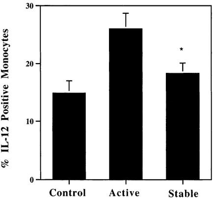
IL-12 expression in MS monocytes correlates with disease activity. We were interested in determining if there was a
rela-tionship between IL-12 production and disease activity. Pa-tients were classified into two groups: active or stable by their treating physician at the time of blood drawing, and IL-12 ex-pression was determined without knowledge of the clinical classification. IL-12 expression was compared among patients (treated or untreated) with stable disease (n 5 36) and patients with active disease (n 5 25). We found a statistically significant increase in IL-12 production in the group with active disease compared with patients with stable disease (26.062.7 vs. 18.361.8, P 5 0.02; Fig. 6). There was no correlation between IL-12 production and EDSS score.
Discussion
In this study, we investigated cytokine production in PBMC from MS patients by using intracellular detection of cytokines by flow cytometry (30, 31). This method enables detection of cytokine production by individual cell populations and simul-taneously determines their phenotype based upon surface an-tigen expression. Unstimulated monocytes produce little or no IL-12 spontaneously; thus we used in vitro stimulation to de-tect IL-12–secreting cells. NK cells did not produce IL-12 be-fore or after stimulation, and a very small percentage of B cells (, 1%) had detectable IL-12 production.
We found that the frequency of IL-12–producing mono-cytes in untreated MS patients was higher than in controls.
These results are in agreement with previous reports in which an elevated production of IL-12 by PBMC was found in pro-gressive MS (16, 17). Furthermore, IL-12 expression segre-gated with the disease activity as assessed by the treating phy-sician suggesting a role for IL-12 in disease progression. The frequency of TNF-a–producing monocytes was not statistically different among the patient subgroups but showed a trend to-wards increased frequency in untreated progressive patients. This may be due to a selective effect of CY/MP on IL-12 pro-duction, to an effect of CY/MP on subpopulations of mono-cytes that produce mainly IL-12, or due to the smaller sample size analyzed for TNF-a. We also found increased IFN-g– expressing T cells in patients with progressive MS on no treat-ment in agreetreat-ment with a previous report of increased IFN-g mRNA-expressing PBMC in MS patients (32) and with the known deleterious effect of IFN-g in MS (33–35).
The second major finding in our study is the change in cy-tokine balance induced by immunosuppressive treatments. Treatment with methotrexate increased TGF-b production by T cells. Methylprednisolone (MP) monthly treatment was as-sociated with a decrease in IFN-g–expressing cells and an in-crease in IL-4–expressing cells. Although in vitro dein-crease in IL-12 production by dexamethasone in monocytes has recently been reported (36), we did not observe any effect on IL-12 production in the group of patients treated with monthly MP. That may be explained by either a difference in the biological effects of MP and dexamethasone or a predominant effect of dexamethasone in the non-CD14 population of adherent cells. CY/MP treatment was associated with multiple cytokine ef-fects. There was a significant decrease in IL-12 and IFN-g and an increase in IL-4, IL-5, and TGF-b. The latter was increased both in T cells and monocytes. Thus, CY/MP treatment was as-sociated with a shift in cytokines from Th1 to Th2/Th3 pattern. These results agree with our recent report of increased anti-CD3–induced IL-4 secretion by T cells in patients treated with cyclophosphamide (27). The increase in percentage of IL-4, IL-5, and TGF-b–producing cells in CY/MP treated patients was much less than the changes we observed for IL-12 and IFN-g. Given the low numbers of IL-4–, IL-5–, and TGF-b– secreting cells in untreated MS patients, the increases we ob-served are likely to be biologically significant, although the re-sults should be interpreted with caution. The differences noted between untreated and CY/MP treated patients in percentage of cells producing IL-4– and IL-5–producing cells were seen under unstimulated conditions, no differences in stimulated IL-4– or IL-5–producing cells were noted. A similar phenome-non was reported in patients treated with rIFN-b1b, where the
percentage of monocytes spontaneously producing IL-10 was increased compared with untreated patients, but this differ-ence disappeared after stimulation (37).
[image:7.612.57.274.491.692.2]The finding of decreased IL-12 in CY/MP-treated patients also was observed in patients followed-up prospectively before and after treatment. In these patients the cytokine shifts occur subsequent to the start of treatment. Corticosteroids are known to inhibit the production of most cytokines in activated T cells (38, 39), and may increase IL-4 synthesis in T cells (40, 41). Because pulse cyclophosphamide treatment includes methylprednisolone infusions, we evaluated the effects of monthly MP treatment. We found that these patients had a modest but significant increase in IL-4 expression and a de-crease in IFN-g expression. These results suggest that cortico-steroids promote a Th2 cytokine response in T cells. Thus, Figure 6. IL-12 expression correlates with disease activity. IL-12
methylprednisolone may have a synergistic effect with cyclo-phosphamide in inducing immune deviation.
TGF-b is a regulator of immune responses and plays an im-portant role in recovery from EAE (7, 10, 42, 43), and investi-gators have reported that there is less severe disease in MS pa-tients with increased levels of TGF-b secreting cells (44). It was recently shown that IL-12 and IFN-g are important regu-lators of TGF-b production both in vivo and in vitro (45). Thus, our finding of an increase in TGF-b expression by T cells and monocytes in patients treated with CY/MP is consistent with the decreased IL-12 and IFN-g expression in these pa-tients. Whether this increased production of TGF-b is a direct effect of treatment or is secondary to the change in Th1/Th2 balance remains to be determined.
In vitro studies suggest that the decreased IL-12 production is a direct effect of cyclophosphamide metabolite on monocytes. Thus addition of 4HC in vitro resulted in decreased IL-12 ex-pression. 4HC is a synthetic compound that spontaneously hy-drolyzes in aqueous solution to 4-hydroxycyclophosphamide, the initial metabolite formed by liver microsomal activation of cyclophosphamide. 4HC mimics the immunosuppressive effects of cyclophosphamide observed in the intact organism (46, 47). Surprisingly, the inhibition of IL-12 expression by 4HC in vitro was selective, because expression of other cytokines produced by monocytes, such as TNF-a and IL-10, was not affected. How-ever, an indirect effect of cyclophosphamide treatment on other cytokine production cannot be ruled out. Purified stimulated monocytes had lower IL-12 expression compared with stimula-tion of PBMCs, probably reflecting the absence of signaling from CD40 ligand usually provided by T cells in the PBMC cultures. CD40–CD40L interaction is critical for IL-12 produc-tion by monocytes under antigen driven condiproduc-tions (16, 48), but monocytes can produce IL-12 via a CD40/CD40L independent pathway in response to LPS or heat-killed Listeria monocy-togenes, and this pathway does not require interaction of APC with T cells (49). Nevertheless, IL-12 expression by purified monocytes was slightly decreased by the addition of the metab-olite, suggesting some direct effect of cyclophosphamide on monocytes. Although the mechanism by which cyclophospha-mide affects IL-12 secretion remains to be defined, it is unlikely to be due to the alkylating properties of cyclophosphamide, as proliferating cells are more vulnerable to the cytotoxic effects of cyclophosphamide, and monocytes are not considered to be actively proliferating cells. Furthermore, in vitro addition of 4HC to the cell cultures was not associated with decreased cell viability. Thus, cyclophosphamide may have a direct effect on monocyte IL-12 production and an indirect effect (T cell de-pendent) through decreased CD40 expression on monocytes.
In summary, our results demonstrate a Th1-type cytokine bias in peripheral blood mononuclear cells of untreated pro-gressive MS patients that is reversed by CY/MP treatment and is associated with Th2 and TGF-b (Th3) type responses. These findings provide a basis for immune monitoring of patients with MS. If the linkage between IL-12 production and re-sponse to treatment is validated in longitudinal studies, this would suggest that treatments that downregulate IL-12 are likely to be beneficial in progressive MS.
Acknowledgments
Supported by National Institutes of Health (NS-23132-11A), the Na-tional Multiple Sclerosis Society, and the Nancy Davis Center
With-out Walls. M.C. was supported by a grant from the Fondo de Investi-gacion Sanitaria (ref. 97/5462).
References
1. Khoury, S.J., H.L. Weiner, and D.A. Hafler. 1996. Immunological mech-anisms in multiple sclerosis. In Handbook of Multiple Sclerosis. S.D. Cook, edi-tor. Marcell Dekker Inc., New York. 145–155.
2. Mosmann, T.R., H. Cherwinski, M.W. Bond, M.A. Giedlin, and R.L. Coffman. 1986. Two types of murine helper T cell clone. I. Definition according to profiles of lymphokine activities and secreted proteins. J. Immunol. 136: 2348–2357.
3. Chen, Y., V.K. Kuchroo, J.-I. Inobe, D.A. Hafler, and H.L. Weiner. 1994. Regulatory T cell clones induced by oral tolerance: suppression of autoimmune encephalomyelitis. Science. 265:1237–1240.
4. Weiner, H.L. 1997. Oral tolerance: immune mechanisms and treatment of autoimmune diseases. Immunol. Today. 18:335–343.
5. Ando, D.G., J. Clayton, D. Kono, J.L. Urban, and E.E. Sercarz. 1989. En-cephalitogenic T cells in the B10.PL model of experimental allergic encephalo-myelitis (EAE) are of the Th-1 lymphokine subtype. Cell. Immunol. 124:132– 143.
6. Racke, M.K., A. Bonomo, D.E. Scott, B. Cannella, A. Levine, C.S. Raine, E.M. Shevach, and M. Rocken. 1994. Cytokine-induced immune devia-tion as a therapy for inflammatory autoimmune disease. J. Exp. Med. 180:1961– 1966.
7. Khoury, S.J., W.W. Hancock, and H.L. Weiner. 1992. Oral tolerance to myelin basic protein and natural recovery from experimental autoimmune en-cephalomyelitis are associated with downregulation of inflammatory cytokines and differential upregulation of transforming growth factor beta, interleukin 4, and prostaglandin E expression in the brain. J. Exp. Med. 176:1355–1364.
8. Kennedy, M.K., D.S. Torrance, K.S. Picha, and K.M. Mohler. 1992. Anal-ysis of cytokine mRNA expression in the central nervous system of mice with experimental autoimmune encephalomyelitis reveals that IL-10 mRNA expres-sion correlates with recovery. J. Immunol. 149:2496–2505.
9. Issazadeh, S., A. Ljungdahl, B. Hojeberg, M. Mustafa, and T. Olsson. 1995. Cytokine production in the central nervous system of Lewis rats with ex-perimental autoimmune encephalomyelitis: dynamics of mRNA expression for interleukin 10, interleukin 12, cytolysin, tumor necrosis factor-a and tumor ne-crosis factor-b. J. Neuroimmunol. 61:205–212.
10. Chen, Y., W.W. Hancock, R. Marks, P.A. Gonnella, and H.L. Weiner. 1998. Mechanisms of recovery from experimental allergic encephalomyelitis: T cell deletion and immune deviation in myelin basic protein receptor transgenic mice. J. Neuroimmunol. 82:149–159.
11. Manetti, R., P. Parronchi, M.G. Giudizi, M.P. Piccinni, E. Maggi, G. Trinchieri, and S. Romagnani. 1993. Natural killer cell stimulatory factor (inter-leukin 12 [IL-12]) induces T helper type 1 (Th1)-specific immune responses and inhibits the development of IL-4-producing Th cells. J. Exp. Med. 177:1199– 1204.
12. Manetti, R., F. Gerosa, M.G. Giudizi, R. Biagiotti, P. Parronchi, M.P. Piccinni, S. Sampognaro, E. Maggi, S. Romagnani, G. Trinchieri, et al. 1994. In-terleukin 12 induces stable priming for interferon gamma (IFN-gamma) pro-duction during differentiation of human T helper (Th) cells and transient IFN-gamma production in established Th2 cell clones. J. Exp. Med. 179:1273–1283.
13. Seder, R.A., R. Gazzinelli, A. Sher, and W.E. Paul. 1993. Interleukin 12 acts directly on CD41 T cells to enhance priming for interferon gamma produc-tion and diminishes interleukin 4 inhibiproduc-tion of such priming. Proc. Natl. Acad. Sci. USA. 90:10188–10192.
14. Hsieh, C.S., S.E. Macatonia, C.S. Tripp, S.F. Wolf, A. O’Garra, and K.M. Murphy. 1993. Development of TH1 CD41 T cells through IL-12 pro-duced by Listeria-inpro-duced macrophages. Science. 260:547–549.
15. Leonard, J.P., K.E. Waldburger, and S.J. Goldman. 1995. Prevention of experimental autoimmune encephalomyelitis by antibodies against interleukin 12. J. Exp. Med. 181:381–386.
16. Balashov, K.E., D.R. Smith, S.J. Khoury, D.A. Hafler, and H.L. Weiner. 1997. Increased interleukin 12 production in progressive multiple sclerosis: in-duction by activated CD41 T cells via CD40 ligand. Proc. Natl. Acad. Sci. USA. 94:599–603.
17. Nicoletti, F., F. Patti, C. Cocuzza, P. Zaccone, A. Nicoletti, R. DiMarco, and A. Reggio. 1996. Elevated serum levels of interleukin-12 in chronic pro-gressive multiple sclerosis. J. Neuroimmunol. 70:87–90.
18. Paterson, P.Y., and M.A. Hanson. 1969. Cyclophosphamide inhibition of experimental allergic encephalomyelitis and cellular transfer of the disease in Lewis rats. J. Immunol. 103:1311–1316.
19. Paterson, P.Y., and D.G. Drobish. 1969. Cyclophosphamide: effect on experimental allergic encephalomyelitis in Lewis rats. Science. 165:191–192.
20. Brinkman, C.J., W.M. Nillesen, and O.R. Hommes. 1984. The effect of cyclophosphamide on T lymphocytes and T lymphocyte subsets in patients with chronic progressive multiple sclerosis. Acta Neurol. Scand. 69:90–96.
dis-continuation of long-term cyclophosphamide treatment. J. Neuroimmunol. 14: 175–182.
22. Lamers, K.J., B.M. Uitdehaag, O.R. Hommes, W. Doesburg, R.A. Wevers, and W.J. von Geel. 1988. The short-term effect of an immunosuppres-sive treatment on CSF myelin basic protein in chronic progresimmunosuppres-sive multiple scle-rosis. J. Neurol. Neurosurg. Psychiatry. 51:1334–1337.
23. Hafler, D.A., J. Orav, R. Gertz, L. Stazzone, and H.L. Weiner. 1991. Im-munologic effects of cyclophosphamide/ACTH in patients with chronic pro-gressive multiple sclerosis. J. Neuroimmunol. 32:149–158.
24. Hauser, S.L., D.M. Dawson, J.R. Lehrich, M.F. Beal, S.V. Kevy, R.D. Propper, J.A. Mills, and H.L. Weiner. 1983. Intensive immunosuppression in progressive multiple sclerosis. A randomized, three-arm study of high-dose in-travenous cyclophosphamide, plasma exchange, and ACTH. N. Engl. J. Med. 308:173–180.
25. Weiner, H.L., G.A. Mackin, E.J. Orav, D.A. Hafler, D.M. Dawson, Y. LaPierre, R. Herndon, J.R. Lehrich, S.L. Hauser, A. Turel, et al. 1993. Intermit-tent cyclophosphamide pulse therapy in progressive multiple sclerosis: final re-port of the Northeast Cooperative Multiple Sclerosis Treatment Group. Neu-rology. 43:910–918.
26. Group., T.C.C.M.S.S. 1991. The Canadian Cooperative trial of cyclo-phosphamide and plasma exchange in progressive multiple sclerosis. Lancet. 337:442–446.
27. Smith, D.R., K.E. Balashov, D.A. Hafler, S.J. Khoury, and H.L. Weiner. 1997. Immune deviation following pulse cyclophosphamide/methylpredniso-lone treatment of multiple sclerosis: increased interleukin-4 production and as-sociated eosinophilia. Ann. Neurol. 42:313–318.
28. Takashima, H., D. Smith, H. Fukaura, S. Khoury, D. Hafler, and H. Weiner. 1998. Pulse cyclophosphamide plus methylprednisolone induces mye-lin antigen specific IL-4 secreting T cells in multiple sclerosis patients. Cmye-lin. Im-munol. Immunopathol. 88:28–34.
29. Lewis, C.E. 1991. Detecting cytokine production at the single-cell level. Cytokine. 3:184–188.
30. Prussin, C. 1997. Cytokine flow cytometry: understanding cytokine biol-ogy at the single-cell level. J. Clin. Immunol. 17:195–204.
31. Jung, T., U. Schauer, C. Heusser, C. Neumann, and C. Rieger. 1993. De-tection of intracellular cytokines by flow cytometry. J. Immunol. Meth. 159:197– 207.
32. Link, J., M. Soderstrom, T. Olsson, B. Hojeberg, A. Ljungdahl, and H. Link. 1994. Increased transforming growth factor-beta, interleukin-4, and inter-feron-gamma in multiple sclerosis. Ann. Neurol. 36:379–386.
33. Panitch, H.S., R.L. Hirsch, A.S. Haley, and K.P. Johnson. 1987. Exacer-bations of multiple sclerosis in patients treated with gamma interferon. Lancet. 1:893–895.
34. Beck, J., P. Rondot, L. Catinot, E. Falcoff, H. Kirchner, and J. Wietzerbin. 1988. Increased production of interferon gamma and tumor necro-sis factor precedes clinical manifestations in multiple scleronecro-sis: do cytokines trigger off exacerbations? Acta Neurol. Scand. 78:318–323.
35. Olsson, T., W.-Z. Wang, B. Höjeberg, V. Kostulas, Y.-P. Jiang, G. Anderson, H.-P. Ekre, and H. Link. 1990. Autoreactive T-lymphocytes in mul-tiple sclerosis determined by antigen-induced secretion of interferon-gamma. J. Clin. Invest. 86:981–985.
36. Blotta, M.H., R.H. DeKruyff, and D.T. Umetsu. 1997. Corticosteroids inhibit IL-12 production in human monocytes and enhance their capacity to in-duce IL-4 synthesis in CD41 lymphocytes. J. Immunol. 158:5589–5595.
37. Crucian, B., P. Dunne, H. Friedman, R. Ragsdale, S. Pross, and R. Widen. 1996. Detection of altered T helper 1 and T helper 2 cytokine produc-tion by peripheral blood mononuclear cells in patients with multiple sclerosis utilizing intracellular cytokine detection by flow cytometry and surface marker analysis. Clin. Diagn. Lab. Immunol. 3:411–416.
38. Scheinman, R.I., P.C. Cogswell, A.K. Lofquist, and A.J. Baldwin. 1995. Role of transcriptional activation of I kappa B alpha in mediation of immuno-suppression by glucocorticoids. Science. 270:283–286.
39. Auphan, N., J.A. DiDonato, C. Rosette, A. Helmberg, and M. Karin. 1995. Immunosuppression by glucocorticoids: inhibition of NF-kappa B activity through induction of I kappa B synthesis. Science. 270:286–290.
40. Ramirez, F., D.J. Fowell, M. Puklavec, S. Simmonds, and D. Mason. 1996. Glucocorticoids promote a TH2 cytokine response by CD41 T cells in vitro. J. Immunol. 156:2406–2412.
41. Zieg, G., G. Lack, R.J. Harbeck, E.W. Gelfand, and D.Y. Leung. 1994. In vivo effects of glucocorticoids on IgE production. J. Allergy Clin. Immunol. 94:222–230.
42. Racke, M.K., S. Dhib-Jalbut, P.S. Cannella, P.S. Albert, and D.E. Mc-Farlin. 1991. Prevention and treatment of chronic relapsing experimental aller-gic encephalomyelitis by transforming growth factor-b1. J. Immunol. 146:3012– 3017.
43. Johns, L., K. Flanders, G. Ranges, and S. Sriram. 1991. Successful treat-ment of experitreat-mental allergic encephalomyelitis with transforming growth fac-tor-b1. J. Immunol. 147:1792–1796.
44. Olsson, T. 1995. Critical influences of the cytokine orchestration on the outcome of myelin antigen-specific T-cell autoimmunity in experimental au-toimmune encephalomyelitis and multiple sclerosis. Immunol. Rev. 144:245– 268.
45. Marth, T., W. Strober, R.A. Seder, and B.L. Kelsall. 1997. Regulation of transforming growth factor-beta production by interleukin-12. Eur. J. Immunol. 27:1213–1220.
46. Diamantstein, T., M. Klos, H. Hahn, and S.H. Kaufmann. 1981. Direct in vitro evidence for different susceptibilities to 4-hydroperoxycyclophospha-mide of antigen-primed T cells regulating humoral and cell-mediated immune responses to sheep erythrocytes: a possible explanation for the inverse action of cyclophosphamide on humoral and cell-mediated immune responses. J. Immu-nol. 126:1717–1719.
47. Kaufmann, S.H., H. Hahn, and T. Diamantstein. 1980. Relative suscep-tibilities of T cell subsets involved in delayed-type hypersensitivity to sheep red blood cells to the in vitro action of 4-hydroperoxycyclophosphamide. J. Immu-nol. 125:1104–1108.
48. Cella, M., D. Scheidegger, L.K. Palmer, P. Lane, A. Lanzavecchia, and G. Alber. 1996. Ligation of CD40 on dendritic cells triggers production of high levels of interleukin-12 and enhances T cell stimulatory capacity: T-T help via APC activation. J. Exp. Med. 184:747–752.