Induction of the chemokine stromal-derived
factor-1 following DNA damage improves
human stem cell function
Tanya Ponomaryov, … , Dov Zipori, Tsvee Lapidot
J Clin Invest. 2000;
106(11)
:1331-1339.
https://doi.org/10.1172/JCI10329
.
The chemokine stromal-derived factor-1 (SDF-1) controls many aspects of stem cell
function. Details of its regulation and sites of production are currently unknown. We report
that in the bone marrow, SDF-1 is produced mainly by immature osteoblasts and
endothelial cells. Conditioning with DNA-damaging agents (ionizing irradiation,
cyclophosphamide, and 5-fluorouracil) caused an increase in SDF-1 expression and in
CXCR4-dependent homing and repopulation by human stem cells transplanted into
NOD/SCID mice. Our findings suggest that immature osteoblasts and endothelial cells
control stem cell homing, retention, and repopulation by secreting SDF-1, which also
participates in host defense responses to DNA damage.
Article
Introduction
Hematopoietic stem cells reside within the bone marrow (BM) microenvironment in direct contact with stromal cells. The latter regulate the continuous production of maturing blood cells and their release into the blood cir-culation while maintaining a small pool of primitive, undifferentiated stem cells (1). During development, blood-forming stem cells migrate from the fetal liver into the circulation and home to the BM microenvironment (2). Similarly, in clinical and experimental stem cell trans-plantation, intravenously injected stem cells migrate through the blood and home to the BM, repopulating it with immature and maturing myeloid and lymphoid blood cells (3). Functional in vivo assays for human stem cells based on transplantation into immune-deficient SCID, and more recently into NOD/SCID or B2m-null NOD/SCID mice, have been developed by us, as well as in other laboratories (4–7). These assays identified a small population of primitive human severe combined immun-odeficiency (SCID) repopulating cells (SRC), by virtue of their functional ability to repopulate transplanted recip-ients with both myeloid and lymphoid cells (8). Deter-mining the role of stromal cells in stem cell homing, retention, and repopulation will help to identify the mechanisms that govern stem cell development and may advance clinical applications to stem cell transplantation.
Stromal-derived factor-1 (SDF-1), also known as pre–B-cell growth-stimulating factor, is produced in many organs, including the BM (2, 9). However, details of its expression pattern and mode of regulation are currently unknown. In contrast to proinflammatory chemokines, SDF-1 is produced by BM stromal cells also in the absence of stimuli generated by viral or bac-terial infections, suggesting a major role for SDF-1 in steady-state homeostatic processes such as leukocyte trafficking (2, 9). In accordance with this notion has been the identification of SDF-1 as a powerful chemoattractant for immature and mature hematopoi-etic cells of several lineages (2, 10–12). Mice that lack either SDF-1 or its receptor CXCR4 exhibit many defects, including impaired hematopoiesis in the fetal BM (9, 13). Recently, we discovered that human stem cell engraftment of NOD/SCID mice is dependent on the expression of SDF-1 and CXCR4 (14). Interesting-ly, the BM of mice transplanted with fetal liver cells from CXCR4-deficient mice was repopulated by donor cells (15, 16). However, release of differentiating cells from the marrow into the circulation and the levels of donor-derived hematopoiesis in the BM of these mice was significantly reduced. These results demonstrate that long-term repopulation by pluripotent stem cells is impaired in CXCR4-null fetal liver cells, since these
Induction of the chemokine stromal-derived factor-1
following DNA damage improves human stem cell function
Tanya Ponomaryov,
1Amnon Peled,
1Isabelle Petit,
1Russell S. Taichman,
2Liliana Habler,
3Judith Sandbank,
3Fernando Arenzana-Seisdedos,
4Aude Magerus,
4Antonio Caruz,
5Nobutaka Fujii,
6Arnon Nagler,
7Meir Lahav,
8Martin Szyper-Kravitz,
8Dov Zipori,
9and Tsvee Lapidot
11Department of Immunology, The Weizmann Institute of Science, Rehovot, Israel
2University of Michigan Dental School, Department of Periodontics, Ann Arbor, Michigan, USA 3Assaf Harofeh Tsrifin Medical Center, Tsrifin, Israel
4Institute Pasteur, Paris, France 5University of Jaen, Jaen, Spain 6University of Kyoto, Kyoto, Japan
7Hadassah University Hospital, Jerusalem, Israel 8Meir Medical Center, Kfar-Saba, Israel
9Department of Molecular Cell Biology, The Weizmann Institute of Science, Rehovot, Israel
Address correspondence to: Tsvee Lapidot, Department of Immunology, The Weizmann Institute of Science, PO Box 26, Rehovot 76100, Israel. Phone: 972-8-9342481; Fax: 972-8-9344141; E-mail: [email protected].
Received for publication May 15, 2000, and accepted in revised form October 11, 2000.
The chemokine stromal-derived factor-1 (SDF-1) controls many aspects of stem cell function. Details of its regulation and sites of production are currently unknown. We report that in the bone marrow, SDF-1 is produced mainly by immature osteoblasts and endothelial cells. Conditioning with DNA-damaging agents (ionizing irradiation, cyclophosphamide, and 5-fluorouracil) caused an increase in SDF-1 expression and in CXCR4-dependent homing and repopulation by human stem cells trans-planted into NOD/SCID mice. Our findings suggest that immature osteoblasts and endothelial cells control stem cell homing, retention, and repopulation by secreting SDF-1, which also participates in host defense responses to DNA damage.
cells fail to durably proliferate and differentiate in the absence of CXCR4. In support of this notion, recent studies demonstrated that SDF-1 is a survival factor for both murine and human immature progenitor cells (17–19). Other studies indicate that SDF-1 is a pre–B-cell growth factor, that it is also involved in the devel-opment of megakaryocytes, that it is an inhibitor of myelopoiesis, and that it prevents primitive human cells from entering the cell cycle (20–23). We have demonstrated that SDF-1 plays a pivotal role in the conversion of CD34+cell-rolling adhesions on
endothe-lial cells into firm adhesions by activating VLA-4 and LFA-1 (24). Furthermore, it has been shown that SDF-1 is expressed by human and murine BM endothe-lium in vivo, while murine endothelial cells from the lung or human umbilical cord did not express the chemokine (24, 25). Activation of LFA-1, VLA-4, and VLA-5 on human CD34+cells by SDF-1 is crucial for
their engraftment in transplanted NOD/SCID (26). Lastly, preliminary results indicate that CXCR4-null fetal liver cells can also interact with SDF-1 independ-ently of CXCR4, which partially compensates for the absence of signaling via CXCR4 (27). In support of our work, adult mice reconstituted with genetically modi-fied BM cells with the SDF-1 intrakine gene, had sig-nificantly reduced levels of both lymphoid pre–B cells and myeloid cells in their BM (28).
SDF-1 is thus important in stem cell trafficking and development. It is not known, however, which BM cells are primarily responsible for the produc-tion of SDF-1, nor is it known how SDF-1 expression is regulated. The hematopoietic system is the most sensitive to DNA-damaging agents, such as irradia-tion or cytotoxic drugs. This vulnerability of blood-forming stem cells reduces regeneration after treat-ment or accidental exposure to these agents and thus impinges on long-term survival (29). In the present study, we identified BM cell types that express SDF-1 in vivo and in vitro. We further tested the effect of DNA-damaging agents such as irradia-tion, cyclophosphamide (Cy), or 5-fluorouracil (5-FU) on the expression of SDF-1 in the BM of mice or in cultured stromal cells. In parallel, we deter-mined the time-dependent effect of irradiation or other DNA-damaging agents on the CXCR4-depend-ent repopulation potCXCR4-depend-ential of human stem cells in transplanted NOD/SCID mice.
Methods
Human and mouse cells. Human cord blood (CB) cells were obtained from full-term deliveries after informed consent and were used in accordance with approved procedures by the human experimentation and ethics committees of the Weizmann Institute. CB samples were diluted in PBS, supplemented with 1% FCS (Bio-logical Industries, Beit Haemek, Israel). Low-density mononuclear cells were collected after standard sepa-ration on Ficoll-Paque (Pharmacia Biotech AB, Upp-sala, Sweden), and washed with PBS.
Enrichment of human CD34+cells. Enrichment of human CD34+cells was performed with a magnetic bead
sepa-ration kit (mini MACS; Miltenyi Biotec, Bergisch Glad-bach, Germany) according to the manufacturer’s instructions. The purity of the enriched CD34+cells was
60–85% when cells were passed over one column, and greater than 98% when passed over two columns.
Human umbilical vein endothelial cells (HUVECs) were isolated from umbilical cord veins, according to the method of Jaffe et al. (30). Human adherent stromal cells were prepared and grown as described previously from leftover BM cells for allogeneic transplantation (31). Mouse BM stromal-cell lines (MBA-2.1, 14F1.1, MBA-15) were grown as described previously (32, 33). U373-MG, a human glioblastoma cell line (ATTC num-ber HTB17; American Type Culture Collection, Rockville, Maryland, USA), was cultured in DMEM. MS-5, a murine stromal cell line, is a kind gift from J.C. Gutierrez-Ramos (Millennium Pharmaceuticals, Cam-bridge, Massachusetts, USA). Cells were grown in RPMI-1640 complemented with 50 nM 2-mercaptoethanol. Both culture media were enriched with 10% FCS. Con-ditioned media (CM) from confluent layers of murine and human stroma cells was collected after 3 days in culture. Mouse CM were prepared from supernatants containing single-cell suspensions of flushed murine BM and spleen cells in RPMI with 10% FCS after cen-trifugation and storage at –20°C.
Osteosarcoma cell lines and primary human osteoblast-like cells. Human osteosarcoma cell lines were purchased from the American Tissue Type Collection, including MG-63 (ATCC CRL1424) and SaOS-2 (ATCC 85-HTB). These cell lines were maintained in either DMEM with Earle’s salts (HOS and MG-63) or McCoy’s 5A medium (G-292, SaOS-2, U2-OS) with 10% heat-inactivated FBS and antibiotics. Enriched human osteoblast-like cell (HOB) cultures were established using modifications of methods described by Robey and Termine (34). Nor-mal human trabecular bone was obtained from patients undergoing orthopedic surgery in accordance with the University of Michigan’s Investigational Review Board. Bone cleaned of loosely adherent tissue was ground to produce a uniform particle size (size ≤1 mm2) (BioComp Minimill; Walter Lorenz Surgical Inc.,
Jacksonville, Florida, USA) and incubated in 1 mg/ml bacterial collagenase (Type P; Boehringer Mannheim Biochemicals Inc., Indianapolis, Indiana, USA). The explants were placed into culture until confluent monolayers were produced in a 1:1 (vol/vol) mixture of Ham’s F12/DMEM (Biofluids, Rockville, Maryland, USA) with low Ca2+ and 10% heat-inactivated FBS.
Thereafter, the cultures were maintained in calcium-replete Ham’s F12/DMEM (1:1 vol/vol) medium con-taining 10% heat-inactivated FCS, antibiotics 10 mM
Mice. NOD-SCID mice (NOD/LtSzPrKdc scid/PrKdc scid) were bred and maintained under defined flora condi-tions in individually ventilated (HEPA-filtered air) ster-ile micro-isolator cages (Techniplast, Gazzada, Italy) at the Weizmann Institute. All the experiments were approved by the Animal Care Committee of the Weiz-mann Institute. Mice, 8 weeks old, were irradiated with a sublethal dose of 375 cGy (67 cGy/min.) from a cobalt source before transplantation or treatment with Cy (200 mg/kg) or 5-FU (200–250 mg/kg). Human CD34+
-enriched cells (60–85% purity, 2 × 10⁵/mouse) or
mononuclear cells (1–2 × 10⁷/mouse) were injected into
the tail veins of mice irradiated immediately, or 48 hours earlier, in 0.5 ml of RPMI with 10% FCS. Trans-planted mice were sacrificed after 4–5 weeks or the fol-lowing day in homing experiments, and the cells from the spleen, femur, tibia, and pelvis bones were flushed with a syringe. BALB/c and C57BL mice were purchased from Harlan Laboratories (Rehovot, Israel). Mice were injected intraperitoneally with Cy (200 mg/kg) or 5-FU (150 mg/kg) and sacrificed at the indicated time points.
Semiquantitative RT-PCR. Total RNA was isolated from MBA-2.1, 14F1.1, and MBA-15 cells (with or without treatment with 150 µg/ml 5-FU or 200 µg/ml Cy), human stromal cells, and mice tissues (BM and spleen) using TRI-Reagent (Molecular Research Center Inc., Cincinnati, Ohio, USA) according to the manufactur-er’s protocol. Each RNA sample (1 µg) was subjected to cDNA synthesis in 30 µl of reaction mixture containing 1 µl Oligo dT 15 primer (500 µg/ml; Promega Corp., Madison, Wisconsin, USA), 2 µl dNTP’s mixture (10 mM, PCR grade; Boehringer Mannheim Biochemicals Inc.), 3 µl DTT (0.1 M; Promega Corp.), 1 µl RNasin (40 U/µl; Promega Corp.) and 1 µl MMLV-RT (200 U/µl; Promega Corp.) in the supplied reaction buffer (5×, 250 mM Tris-HCl, pH 8.3, 375 mM KCl, 15 mM MgCl2, 50 mM DTT; Promega Corp.) for 1 hour at 42°C. The PCR was performed in a 50-µl reaction mixture using 5 µl of cDNA, Taq DNA Polymerase (Promega Corp.), 1 µl of dNTP’s mixture (10 mM; Boehringer Mannheim Bio-chemicals Inc.), and specific primers for 35 cycles at appropriate annealing temperatures. As a control for primer contamination or dimerization, the same reac-tion mixture without cDNA was prepared. Semiquanti-tative analysis for SDF-1 expression in murine tissues and cell lines was performed for 25 cycles: Tann = 65°C, 45 seconds. The resulting PCR products were separated on 1.6% agarose gel (SeaKem LE agarose; FMC Corp., Rockland, Maine, USA).
Semiquantitative RT-PCR for HOBs and osteosarcoma cell lines. Cells were plated at an initial density of 2 ×10⁵
cells/cm2with complete medium changes in medium
containing freshly prepared β-glycerol phosphate (10 mM) and L-ascorbate (50 µg/ml) on days 5, 7, 10, and 14. One hundred fifty micrograms per milliliter 5-FU (Sigma Chemical Co., St. Louis, Missouri, USA) was added to the cultures on day 10. On day 14, total cellu-lar RNA was recovered, reverse transcribed, and PCR was performed at 94°C for 1 minute, 60°C for 1 minute, and
72°C for 1 minute for 27 or 35 cycles (semiquantitative and nonquantitative, respectively, followed by a 10-minute extension at 72°C (Cetus DNA thermal cycler; Perkin Elmer Applied Biosystems, Foster City, Califor-nia, USA) as described previously with sense and anti-sense primers prepared by the oligonucleotide synthesis core at the University of Michigan (36).
Primer sequences and lengths of products. Primer sequences used were: murine SDF-1 (5′GGA CGC CAA GGT CGT CGC CGT G, 3′TTG CAT CTC CCA CGG ATG TCA G, 335 bp); murine CXCR4 (5′GAC GGA CAA GTA CCG GCT GC, 3′GAC AGC TTA GAG ATG ATG AT, 482 bp); human SDF-1 (5′CGT CAG CCG CAT TGC CCG CT, 3′GGT CTA GCG GAA AGT CCT, 380 bp); human CXCR4 (5′CTG AGA AGC ATG ACG GAC AA, 3′TGG AGT GTG ACA GCT TGG AG, 484 bp); GADPH (5′GAC AAC AGC CTCAAG ATC ATC AGC, 3′AAG TCA GAG GAG ACC ACC TGG TGC); β-actin (5′TCC TGT GGC ATC CAT GAA ACT ACA TTC AAT TCC, 3′GTG AAA ACG CAG CTC AGT AAC AGT CCG CCT AG, 347 bp).
Flow-cytometry analysis of human cells. In mouse BM or spleen: for immunostaining, approximately 105cells
were resuspended in staining buffer (PBS, 0.1% BSA, 0.02% sodium azide), incubated with 10 µg/ml (1:50) of purified anti-mouse CD16/CD32 (FcR) (PharMin-gen, San Diego, California, USA) and 1% human plas-ma for 20 minutes at 4°C. Cells were then stained with human-specific, direct-labeled Ab’s and incubated for 30 minutes on ice. Dead cells were gated out by stain-ing with propidium iodide (Sigma Chemical Co.). Human cells from engrafted mice were analyzed by double-staining with anti-CD45 FITC (Immuno Qual-ity Products, Gromingen, The Netherlands) and anti-CD19 phycoerythrin (PE) (lymphoid) or anti-CD33 (myeloid) (Coulter, Miami, Florida, USA). The levels of immature cells in the Transwell migration assay were analyzed by double-staining with anti-CD34 FITC (Becton Dickinson Immunocytometry Systems, San Jose, California, USA) and anti-CD38PE (Coulter). After staining, cells were washed twice in the same buffer and analyzed on a FACSort (Becton Dickinson Immunocytometry Systems), using CellQuest software (Becton Dickinson Immunocytometry Systems).
Cell sorting. Cell sorting was performed on a FACStar plus (Becton Dickinson Immunocytometry Systems) as described previously (8, 14). In brief, single-cell suspen-sions of human CD34+-enriched cells (MiniMacs) were
labeled with anti-human CD34 FITC (Becton Dickinson Immunocytometry Systems) and anti-human CD38 PE (Coulter) mAb’s. The purity of sorted CD34+/CD38–/low
and CD34+/CD38+cells was greater than 99%.
Chemotaxis assay. Chemotaxis experiments with human CD34+cells were assayed by using Costar
Tran-swells (6.5 mm/diameter, 5 mm/pore; Corning Costar, Cambridge, Massachusetts, USA). One hundred micro-liters of chemotaxis buffer (RPMI-1640, 1% FCS) con-taining 2 ×10⁵cells were added in the upper chamber,
added to the bottom chamber. Cells migrating within 4 hours to the bottom chamber of the Transwell were counted for 30 seconds using a FACSort (Becton Dick-inson Immunocytometry Systems). SDF-1 was pur-chased from R&D Systems Inc. (Minneapolis, Min-nesota, USA). For the inhibition of chemotaxis the preincubation of CD34+cells with anti-CXCR4 mAb
12g5 (PharMingen) or T22 CXCR4–binding peptide was performed for 30 minutes before the migration.
Colony assay. Semisolid progenitor cultures were per-formed as described previously (5). In brief, the cells were plated in 0.9% methylcellulose (Sigma Chemical Co.), 30% FCS, 5 ×10–52 ME (Sigma Chemical Co.), 50 ng/ml stem
cell factor (SCF), 5 ng/ml IL-3, 5 ng/ml GM-CSF (R&D Systems Inc.), and 2 U/ml erythropoietin (EPO; Orto Bio Tech, Don Mills, Ontario, Canada). BM cells from trans-planted mice were cultured under conditions selected for growth of human colonies only by replacing 15% FCS with 15% human plasma. Plating concentrations for enriched CD34+cells were 3 ×10³cells/ml, and for BM
cells from transplanted mice, 200 ×103cells/ml. The
cul-tures were incubated at 37°C in a humidified atmosphere containing 5% CO2and were scored 14 days later.
[image:5.612.69.528.52.194.2]Immunohistology of BM sections. Immunohistochemical staining was performed on formalin-fixed, paraffin-embedded human bone sections, using the avidin-biotin-peroxidase–complex method and diaminobenzidine tetrahydrochloride (DAB) chromogen kit (DAKO A/S, Glostrup, Denmark). Human bone sections were stained with the anti–SDF-1 mAb K15C (1:50 dilution; refs. 24, 37). The immunohistochemical staining was performed using the LSAB+ peroxidase kit (DAKO A/S). To enhance immunostaining, the antigen was preretrieved in citrate buffer using pressure cooking within a calibrated microwave, heated for 15 minutes, and then reheated for additional 2.5 minutes. Slides were counterstained with Mayer’s hematoxylin. The specificity of the anti–SDF-1 mAb was confirmed by the complete absence of peroxi-dase staining on identical tissue sections using an iso-type-matched control mAb (data not shown).
Figure 1
Anti–SDF-1 reactive immature BM osteoblasts lining the endosteum region (OB) and stromal cells (ST; arrows, ×200) (a), venules (arrow) (b), and arteriole (arrow) (c) in human BM sections (×1000). No peroxidase staining of control mAb was observed on identical tissue sec-tions (data not shown).
Figure 2
[image:5.612.65.539.514.653.2]ELISA. The amount of SDF-1 released in the cell-cul-ture supernatants was estimated by ELISA. SDF-1 was captured by an anti-human/mouse monoclonal SDF-1 Ab (Research Diagnostics Inc., Flanders, New Jersey, USA). Immobilized SDF-1 was detected using a biotiny-lated polyclonal SDF-1 Ab (R&D Systems Inc.) coupled to horseradish peroxidase–labeled streptavidin.
Results
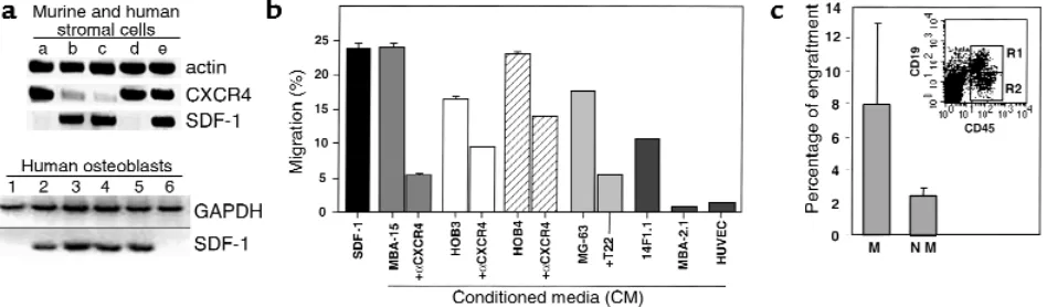
SDF-1 is highly expressed by human BM osteoblasts and endothelial cells. The potential role of SDF-1 in human stem and progenitor cell migration and localization in the BM has been studied by staining human bone tis-sue sections with mAb specific for SDF-1 (24, 37). We found that SDF-1 is produced mainly by immature osteoblasts lining the bone endosteum region (Figure 1a) and by endothelial cells lining the small and large vessels, including periarterial regions (Figure 1, b and c) as well as the blood capillaries of the bone (24). Stro-mal cells in the mesenchyme were also stained by the Ab (Figure 1a). We next tested the expression and pro-duction of SDF-1 in different cell lines representing various murine BM stroma cell types. By RT-PCR analy-sis we found that murine cell lines with osteoblastic characteristics such as MBA-15 (Figure 2a, lane c), and most HOBs (Figure 2a, lanes 1–4) and/or human osteosarcomas (Figure 2a, lanes 5 and 6), express high levels of SDF-1 mRNA. SDF-1 is also expressed by murine mesenchymal adipocyte 14F1.1 cells (lane b) and by primary human stromal cells (lane e) (Figure 2a). Interestingly, BM-derived murine endothelial cells (MBA-2.1, lane a), as well as the HUVECs (lane d), did not express SDF-1 (Figure 2a). CXCR4, the receptor for SDF-1, has been shown to be functionally expressed by endothelial cells such as HUVECs (38). We found that CXCR4 was not expressed by the mesenchymal-derived
BM stromal cells MBA-15 and 14F1.1, but was expressed by murine MBA-2.1 endothelial cells (Figure 2a). We further found that in primary human stroma cultures, both SDF-1 and CXCR4 are expressed (Figure 2a). We then tested the ability of human CD34+cells to
migrate, in a Transwell assay, in response to CM col-lected from different BM stromal cell lines. In correla-tion with the expression data, we found that CM col-lected from osteoblasts, expressing high levels of SDF-1 (murine MBA-15, human MG-63, and HOB3-4), induced the migration of CD34+cells to percentages
comparable to purified recombinant SDF-1 protein at 125 ng/ml (Figure 2b). The chemoattractant activity produced by the stromal cells was significantly inhib-ited by pretreatment of CD34+cells with neutralizing
anti-CXCR4 Ab’s (α-CXCR4, Figure 2b) or by neutral-izing CXCR4 with the T22 peptide (39) (Figure 2b), indicating that SDF-1 is responsible for the activity found in the CM. Moreover, CM from endothelial cells (MBA-2.1, HUVECs) that do not express SDF-1 mRNA, poorly attract human CD34+cells (Figure 2b). We have
shown previously that SDF-1 preferentially induces the migration of SCID repopulating cells (SRC) (14). Sim-ilarly, human CD34+cells, which migrated in response
to SDF-1 present in the CM of osteoblasts (MBA-15), were capable of repopulating the BM of NOD/SCID mice with multilineage hematopoiesis, while nonmi-grating cells gave rise to only low levels of engraftment (Figure 2c). Similar results were obtained with CM from human osteoblasts (data not shown).
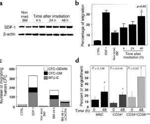
[image:6.612.233.535.487.737.2]DNA-damaging agents increase the production of SDF-1 and the function of human SRC/stem cells. Clinical BM trans-plantation requires conditioning of the recipient with radiation or chemotherapy before stem cell transplan-tation. Similarly, most experimental transplantation protocols also involve total body irradiation (TBI) of the
Figure 3
recipient mice to achieve engraftment. We therefore tested the effects of irradiation on the expression of SDF-1 mRNA, using RT-PCR, and on the secretion of the chemokine, by analyzing its activity in CM prepared from the BM of BALB/c, C57b/6, and NOD/SCID mice before or 4, 24, and 48 hours after sublethal TBI. We found that the expression levels of SDF-1 mRNA increased significantly 24 and 48 hours after irradiation (Figure 3a). In parallel, CM showed a time-dependent increase in the potential to attract human CD34+cells
(Figure 3b). SDF-1 derived from the BM of irradiated mice induced the migration of primitive multilineage human progenitor cells in a CXCR4-dependent manner (Figure 3c). We also tested the engraftment potential of human cord blood cells immediately or 48 hours after TBI of NOD/SCID mice. One month after transplanta-tion, mice were sacrificed, and the percentage of human cells in the mouse BM was assayed. When CD34+cells
were transplanted 48 hours after TBI, we found a sig-nificant (P< 0.016) threefold increase in the levels of engrafted cells compared with those mice transplanted immediately after irradiation (Figure 3d). Moreover, when a population enriched with more primitive human CD34+CD38–/lowwas used as donor cells, we
found the highest levels of engraftment in mice trans-planted 48 hours after irradiation (Figure 3d). Human engraftment in the murine BM included both lymphoid and myeloid cells (data not shown).
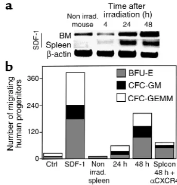
Increase in the expression of SDF-1 was also observed in the spleen of irradiated mice (Figure 4a). The increase in the expression of SDF-1 correlated well with an increase in the chemoattractant activity of the CM collected from spleen cells. Spleen cell–derived SDF-1 induced the migration of multilineage progen-itor cells, and this migratory effect was blocked by Ab’s to CXCR4 (Figure 4b).
Our data suggest that within the BM, stromal cells respond directly to irradiation with increased produc-tion of SDF-1. To test this possibility, RNA was pre-pared from BM stromal cell lines collected before, or 48 hours after, in vitro irradiation. By using RT-PCR analysis, we found that both mesenchymal cell lines, i.e., osteoblast (MBA-15) and in the adipocyte (14F1.1) cells, the expression levels of SDF-1 increased 48 hours after irradiation (Figure 5a). In contrast, no increase in SDF-1 transcription after irradiation was observed when endothelial (MBA-2.1) cells that do not produce SDF-1 were examined (Figure 5a). Moreover, while the levels of SDF-1 in the CM from the stromal mes-enchymal cell line MS5 increased in a dose-dependent manner in response to irradiation, irradiation had no effect on SDF-1 production by the astrocyte cell line U373, which is not from the BM stroma (Figure 5b). Interestingly, stimulation with IL-1β increased SDF-1 production by this cell line (Figure 5b).
[image:7.612.73.245.52.232.2]DNA-damaging agents, such as Cy and 5-FU are used in a broad spectrum of clinical applications, including chemotherapy and mobilization of stem and progenitor cells from the BM into the circulation (40, 41). 5-FU is a DNA-damaging agent that is also used for enrichment of quiescent stem cell populations in experimental trans-plantation protocols (42). We tested the effect of Cy and 5-FU on the expression of SDF-1 by the BM of treated BALB/c mice and by the murine osteoblast cell line
Figure 5
Influence of DNA-damaging treatment on SDF-1 expression. (a) Semiquantitative RT-PCR for murine cell lines MBA-2.1, 14F1.1, and MBA-15, before and 48 hours after 300-cGy radiation. (b) ELISA for CM of astrocytes U373 and BM mesenchymal stromal cells MS5 48 hours after stimulation with IL-1β(Promega Corp.) or irradia-tion. (c) Semiquantitative RT-PCR for SDF-1 mRNA from BM of Cy-or 5-FU–treated BALB/c mice, murine MBA-15 osteoblasts (two mice or two tissue-culture plates per time point), human primary HOB3, and control levels of β-actin expression.
Figure 4
[image:7.612.304.540.398.627.2]MBA-15, or by human osteoblasts (HOB3). Mice treated with either Cy or 5-FU had increased levels of SDF-1 expression in the BM (Figure 5c). Similarly, murine and human osteoblasts also produced higher levels of SDF-1 in response to treatment with DNA-damaging agents (Figure 5c). Preconditioning of NOD/SCID mice 48 hours before transplantation, with 5-FU, Cy, or irradia-tion, increased the homing to the BM and spleen by human mononuclear cells (Figure 6, a and b). Total num-bers of human CD45+cells homing to the murine BM
and spleen, were also dependent on the transplantation time after irradiation (immediately or 48 hours after TBI) (Figure 6, a and b). This effect was dependent on SDF-1, since Ab’s to CXCR4 inhibited both homing and engraft-ment by human stem cells (ref. 14, and data not shown).
Discussion
Homing of human hematopoietic stem cells to the BM, their retention and repopulation, is essential for the establishment of intact hematopoiesis (43, 44). Recent reports demonstrated that the chemokine SDF-1 is essential for the repopulation of the BM by hematopoi-etic stem cells during murine embryonic development and in transplantation of human SRC (9, 13, 14, 44, 45). In the present study we demonstrate that immature human osteoblasts, as well as stromal and endothelial cells within the human BM, express the chemokine SDF-1. We found that osteoblast cell lines and mesenchymal stromal cells that are capable of supporting hematopoiesis in vitro express high levels of SDF-1. This pattern of SDF-1 expression by bone-forming cells is of great interest, since the most primitive, undifferentiated hematopoietic progenitors have been shown to be local-ized mostly in areas along the bone endosteum and also in periarterial regions (46, 47). Interestingly, we found increased expression of SDF-1 in these areas by immature osteoblasts and BM endothelial cells. It is therefore pos-sible that early stem cells, which express CXCR4 and are known to have increased migration potential toward SDF-1, home to areas near the bone endosteum where SDF-1 is highly produced by immature osteoblasts and also to periarterial regions in which SDF-1 is produced by endothelial cells. During development, SDF-1 and its receptor CXCR4 are expressed by a wide variety of adja-cent tissue pairs, including ectoderm/mesoderm and mesoderm/endothelium (2). Although CXCR4-depend-ent migration toward SDF-1 is increased in undifferCXCR4-depend-enti- undifferenti-ated cell types, the expression and function of CXCR4/SDF-1 is also associated with differentiation of both pre–B cells and the megakaryocytic progenitors (21, 48). In addition, SDF-1 is widely needed when there is requirement for cell movement (2). Mice lacking CXCR4 have defective formation of the large vessels supplying the gastrointestinal tract and also defects in the development of the neuronal system (13, 45). Moreover, some endothe-lial and neuronal cells were shown to express CXCR4 and to migrate to SDF-1 (13, 38, 49, 50). SDF-1 was found to synergize with VEGF in the formation of blood vessels (49). We have shown that SDF-1 can induce the arrest of
rolling CD34+cells on human endothelium under shear
flow in vitro, and that in vivo, human BM endothelial cells express SDF-1 (Figure 1; ref. 24). In agreement with our results, murine BM, but not lung endothelium, also express SDF-1 (25, 51). In addition, primitive progenitors also localize in periarterial regions of the BM (46, 47).
[image:8.612.304.540.318.574.2]SDF-1 is constitutively expressed by a variety of tissues, and its expression does not require proinflammatory stimulants (2, 52). The factors that regulate SDF-1 expression during development and in steady-state adult homeostasis are not known. Conditioning by radiation or other DNA-damaging agents is commonly used to ensure successful stem cell transplantation in the clinic. This pretreatment creates space within the BM microen-vironment by eliminating host hemopoietic cells and also reduces the load of malignant cells in disease states in which the BM is afflicted. In allogeneic transplantation, irradiation also prevents the host immune response toward the graft (1, 3). A significant increase in mRNA levels of IL-1α, IL-6, and TNF-αin murine hematopoiet-ic tissues such as the BM and spleen have been reported
Figure 6
to occur following sublethal irradiation (53). We found that sublethal irradiation or treatment with 5-FU increas-es both the mRNA as well as the protein levels of SDF-1 in the BM and spleen of BALB/c, C57BL, or NOD/SCID mice. Irradiation or cytotoxic drugs also promote SDF-1 expression in human and murine bone forming osteoblasts. In parallel, the potential of human stem and progenitor cells to engraft the BM of transplanted NOD/SCID mice increases dramatically when the cells are transplanted 24–48 hours after treatment, when the expression of SDF-1 peaks. Since SDF-1 is also a survival factor for stem cells and is involved in the development of different hematopoietic lineages, SDF-1 can also increase repopulation by promoting the survival and pro-liferation of transplanted stem cells. In addition, SDF-1 can also promote homing of macrophages that partici-pate in clearing apoptotic cells and/or induce apoptosis of damaged host hematopoietic cells in the irradiated BM or spleen. Indeed, Visser et al. reported increased levels of transplanted stem cells in the liver which also produces SDF-1 and reduced levels of stem cell homing to the BM but not to the spleen in mice transplanted 4 hours after TBI in a murine stem cell–homing model compared with untreated recipients (54). In support of our work, Weiss et al. have demonstrated in a syngeneic mouse model that whole BM transplantation at limiting dilution cell doses immediately after TBI or 24 and 48 hours after TBI clear-ly demonstrated significantclear-ly increased levels of survival in mice transplanted 24 or 48 hours after TBI compared with mice transplanted immediately after TBI (55). Our data suggest that the increased levels of SDF-1 secreted 24–48 hours after TBI increased donor stem cell function, i.e., homing and repopulation that in turn increased the survival of the recipient mice.
Overexpression of human CXCR4 and CD4 receptors on murine T cells led to enhanced levels of these cells in the murine BM and to a dramatic decrease in their lev-els in the circulation (56). Furthermore, injection of SDF-1 into the murine spleen was shown to increase the homing of the murine progenitor cell line FDCP-mix cells (10). Human SDF-1 injected into the BM or spleen of nonirradiated NOD/SCID mice also attracted human SRC/stem cells in a CXCR4-dependent manner (57). Considered together with our findings, these results suggest that increase in the concentration of SDF-1 within the BM stromal microenvironment may increase the homing, retention, and repopulation of hematopoietic stem and progenitor cells. The DNA-damaging agent, 5-FU is widely used to enrich for qui-escent hematopoietic stem cells (42). We found that 5-FU, like ionizing irradiation, induces the expression of SDF-1 by murine and human BM-derived osteoblasts and significantly increases homing and engraftment of SRC. It is suggested that increase in production of SDF-1 during alarm situations is part of host defense mech-anism that counteracts the effects of DNA-damaging agents leading to cell death and anemia (17, 18). The expression of SDF-1 demonstrated here, in conjunction with irradiation and chemotherapy treatment that are
commonly used in clinical protocols, may also increase repopulation by malignant cells, since many cancer cells express CXCR4 and respond to stimulation with SDF-1. These include various types of leukemia cells, breast cancer cells, and prostate cancer cells (58–62). Interest-ingly, many malignant cells, such as multiple myeloma, and some breast cancer and prostate cancer cells, can infiltrate into the BM and in some cases also create bone lesions that could be in response to stimulation with SDF-1 secreted by the patient’s BM stromal cells in response to chemotherapy (63, 64).
In summary, we found that immature bone-forming cells produce high levels of SDF-1, which may direct the migration and repopulation of stem cells to specific “stem cell niches” localized mostly near the endosteum and also in periarterial sites. Furthermore, we show that the expression levels of SDF-1 increase after treatment with irradiation or other DNA-damaging agents such as Cy or 5-FU and that the increase in SDF-1 production correlates well with an increase in the levels of BM hom-ing and/or repopulation by primitive human SRC/stem cells. We therefore suggest that molecules found to increase SDF-1 production may be considered for clini-cal protocols with the aim of improving the outcome of stem cell transplantation provided that there are no CXCR4+malignant cells that can also respond to
stimu-lation with SDF-1. Our findings delineate key steps in the process of SDF-1–dependent stem cell homing/repopu-lation and suggest a major role for SDF-1 in alarm situa-tions, as part of host defense processes that protect stem cells from DNA-damaging agents.
Acknowledgments
This work was supported in part by grants from Pas-teur/Weizmann (to F. Arenzana-Seisdedos and T. Lapi-dot), The Israel Science Foundation, Concern Founda-tion, Israel Cancer Research Foundation (to T. Lapidot), and NIH DE-11283 (to R. Taichman). T. Lapi-dot is incumbent of the Pauline Recanati Career Devel-opment Chair of Immunology.
1. Weissman, I.L. 2000. Stem cells: units of development, units of regeneration, and units in evolution. Cell. 100:157–168.
2. McGrath, K.E., Koniski, A.D., Maltby, K.M., McGann, J.K., and Palis, J. 1999. Embryonic expression and function of the chemokine SDF-1 and its recep-tor, CXCR4. Dev. Biol. 213:442–456.
3. Moore, M.A. 1999. “Turning brain into blood”: clinical applications of stem-cell research in neurobiology and hematology. N. Engl. J. Med. 341:605–607. 4. McCune, J.M., et al. 1988. The SCID-hu mouse: murine model for the analy-sis of human hematolymphoid differentiation and function. Science. 241:1632–1639.
5. Lapidot, T., et al. 1992. Cytokine stimulation of multilineage hematopoiesis from immature human cells engrafted in SCID mice. Science. 255:1137–1141. 6. Cashman, J.D., et al. 1997. Kinetic evidence of the regeneration of multilin-eage hematopoiesis from primitive cells in normal human bone marrow transplanted into immunodeficient mice. Blood. 89:4307–4316. 7. Kollet, O., et al. 2000. β2 microglobulin-deficient (β2mnull) NOD/SCID mice
are excellent recipients for studying human stem cell function. Blood. 95:3102–3105.
8. Larochelle, A., et al. 1996. Identification of primitive human hematopoietic cells capable of repopulating NOD/SCID mice using retroviral gene mark-ing and cell purification: implications for gene therapy. Nat. Med. 2:1329–1337.
10. Aiuti, A., Webb, I.J., Bleul, C., Springer, T., and Gutierrez-Ramos, J.C. 1997. The chemokine SDF-1 is a chemoattractant for human CD34+ hematopoi-etic progenitor cells and provides a new mechanism to explain the mobi-lization of CD34+ progenitors to peripheral blood. J. Exp. Med. 185:111–120. 11. Bleul, C.C., Fuhlbrigge, R.C., Casasnovas, J.M., Aiuti, A., and Springer, T.A. 1996. A highly efficacious lymphocyte chemoattractant, stromal cell-derived factor 1 (SDF-1). J. Exp. Med. 184:1101–1109.
12. Jo, D.Y., Rafii, S., Hamada, T., and Moore, M.A. 2000. Chemotaxis of primi-tive hematopoietic cells in response to stromal cell-derived factor-1. J. Clin. Invest. 105:101–111.
13. Zou, Y.R., Kottmann, A.H., Kuroda, M., Taniuchi, I., and Littman, D.R. 1998. Function of the chemokine receptor CXCR4 in haematopoiesis and in cere-bellar development. Nature. 393:595–599.
14. Peled, A., et al. 1999. Dependence of human stem cell engraftment and repopulation of NOD/SCID mice on CXCR4. Science. 283:845–848. 15. Ma, Q., Jones, D., and Springer, T.A. 1999. The chemokine receptor CXCR4
is required for the retention of B lineage and granulocytic precursors with-in the bone marrow microenvironment. Immunity. 10:463–471. 16. Kawabata, K., et al. 1999. A cell-autonomous requirement for CXCR4 in
long-term lymphoid and myeloid reconstitution. Proc. Natl. Acad. Sci. USA. 96:5663–5667.
17. Lataillade, J.J., et al. 2000. Chemokine SDF-1 enhances circulating CD34(+) cell proliferation in synergy with cytokines: possible role in progenitor sur-vival. Blood. 95:756–768.
18. Grafte-Faure, S., et al. 2000. Recruitment of primitive peripheral blood cells: synergism of interleukin 12 with interleukin 6 and stromal cell-derived fac-tor-1. Cytokine. 12:1–7.
19. Broxmeyer, H.E., Hangoc, G., Cooper, S., and Kim, C.H. 1999. Enhanced myelopoiesis in SDF-1 transgenic mice: SDF-1 modulates myelopoiesis by regulating progenitor cell survival and inhibitory effects of myelosuppresive chemokines. Blood. 94:2886. (Abstr.)
20. Nagasawa, T., Kikutani, H., and Kishimoto, T. 1994. Molecular cloning and structure of a pre-B-cell growth-stimulating factor. Proc. Natl. Acad. Sci. USA. 91:2305–2309.
21. Hodohara, K., et al. 2000. Stromal cell-derived factor-1 (SDF-1) acts togeth-er with thrombopoietin to enhance the development of megakaryocytic pro-genitor cells (CFU-MK). Blood. 95:769–775.
22. Sanchez, X., et al. 1997. Activation of HIV-1 coreceptor (CXCR4) mediates myelosuppression. J. Biol. Chem.272:27529–27531.
23. Cashman, J., Clark-Lewis, I., and Eaves, C. 2000. SDF-1 and TGF-β enhance the detection of transplantable human stem cells regenerating in NOD/SCID mice. Exp. Hematol.28:85. (Abstr.)
24. Peled, A., et al. 1999. The chemokine SDF-1 stimulates integrin-mediated arrest of CD34(+) cells on vascular endothelium under shear flow. J. Clin. Invest. 104:1199–1211.
25. Imai, K., et al. 1999. Selective secretion of chemoattractants for haemopoi-etic progenitor cells by bone marrow endothelial cells: a possible role in hom-ing of haemopoietic progenitor cells to bone marrow. Br. J. Haematol. 106:905–911.
26. Peled, A., et al. 2000. The chemokine SDF-1 activates the integrins LFA-1, VLA-4 and VLA-5 on immature human CD34+ cells: role in transendothe-lial/stromal migration and engraftment of NOD/SCID mice. Blood. 95:3289–3296.
27. Kollet, O., et al. 2000. SDF-1 induces survival, adhesion and migration in 3D ECM like gels of murine CXCR4 null fetal liver cells via another GPCR. The American Society of Hematology.In press. (Abstr.)
28. Onai, N., et al. 2000. Impairment of lymphopoiesis and myelopoiesis in mice reconstituted with bone marrow-hematopoietic progenitor cells expressing SDF-1-intrakine. Blood. 96:2074–2080.
29. Domen, J., and Weissman, I.L. 1999. Self-renewal, differentiation or death: regulation and manipulation of hematopoietic stem cell fate. Mol. Med. Today. 5:201–208.
30. Jaffe, E.A., Nachman, R.L., Becker, C.G., and Minick, C.R. 1973. Culture of human endothelial cells derived from umbilical veins. Identification by mor-phologic and immunologic criteria. J. Clin. Invest.52:2745–2756. 31. Teixido, J., Hemler, M.E., Greenberger, J.S., and Anklesaria, P. 1992. Role of
beta 1 and beta 2 integrins in the adhesion of human CD34hi stem cells to bone marrow stroma. J. Clin. Invest.90:358–367.
32. Zipori, D., Toledo, J., and von der Mark, K. 1985. Phenotypic heterogeneity among stromal cell lines from mouse bone marrow disclosed in their extra-cellular matrix composition and interactions with normal and leukemic cells. Blood. 66:447–455.
33. Zipori, D., et al. 1985. Cultured mouse marrow stromal cell lines. II. Distinct subtypes differing in morphology, collagen types, myelopoietic factors, and leukemic cell growth modulating activities. J. Cell. Physiol. 122:81–90. 34. Robey, P.G., and Termine, J.D. 1985. Human bone cells in vitro. Calcif. Tissue
Int. 37:453–460.
35. Taichman, R.S., and Emerson, S.G. 1994. Human osteoblasts support hematopoiesis through the production of granulocyte colony-stimulating factor. J. Exp. Med. 179:1677–1682.
36. Taichman, R.S., Reilly, M.J., and Matthews, L.S. 2000. Human
osteoblast-like cells and osteosarcoma cell lines synthesize macrophage inhibitory pro-tein 1alpha in response to interleukin 1beta and tumour necrosis factor alpha stimulation in vitro. Br. J. Haematol. 108:275–283.
37. Coulomb-L’Hermin, A., et al. 1999. Stromal cell-derived factor 1 (SDF-1) and antenatal human B cell lymphopoiesis: expression of SDF-1 by mesothelial cells and biliary ductal plate epithelial cells. Proc. Natl. Acad. Sci. USA. 96:8585–8590.
38. Murdoch, C., Monk, P.N., and Finn, A. 1999. CXC chemokine receptor expression on human endothelial cells. Cytokine. 11:704–712.
39. Murakami, T., et al. 1997. A small molecule CXCR4 inhibitor that blocks T cell line-tropic HIV-1 infection. J. Exp. Med. 186:1389–1393.
40. Passos-Coelho, J.L., et al. 1995. Predictive factors for peripheral-blood pro-genitor-cell collections using a single large-volume leukapheresis after cyclophosphamide and granulocyte-macrophage colony-stimulating factor mobilization. J. Clin. Oncol.13:705–714.
41. Craddock, C.F., et al. 1992. Circulating stem cells in mice treated with cyclophosphamide. Blood. 80:264–269.
42. Berardi, A.C., Wang, A., Levine, J.D., Lopez, P., and Scadden, D.T. 1995. Func-tional isolation and characterization of human hematopoietic stem cells. Science. 267:104–108.
43. Quesenberry, P.J., and Becker, P.S. 1998. Stem cell homing: rolling, crawling, and nesting. Proc. Natl. Acad. Sci. USA. 95:15155–15157.
44. Ma, Q., et al. 1998. Impaired B-lymphopoiesis, myelopoiesis, and derailed cerebellar neuron migration in CXCR4- and SDF-1-deficient mice. Proc. Natl. Acad. Sci. USA. 95:9448–9453.
45. Tachibana, K., et al. 1998. The chemokine receptor CXCR4 is essential for vascularization of the gastrointestinal tract. Nature. 393:591–594. 46. Lambertsen, R.H., and Weiss, L. 1984. A model of intramedullary
hematopoietic microenvironments based on stereologic study of the distri-bution of endocloned marrow colonies. Blood. 63:287–297.
47. Lord, B.I. 1990. The architecture of bone marrow cell populations. Int. J. Cell Cloning. 8:317–331.
48. Honczarenko, M., et al. 1999. SDF-1 responsiveness does not correlate with CXCR4 expression levels of developing human bone marrow B cells. Blood. 94:2990–2998.
49. Salcedo, R., et al. 1999. Vascular endothelial growth factor and basic fibrob-last growth factor induce expression of CXCR4 on human endothelial cells: in vivo neovascularization induced by stromal-derived factor-1alpha. Am. J. Pathol. 154:1125–1135.
50. Bajetto, A., et al. 1999. Glial and neuronal cells express functional chemokine receptor CXCR4 and its natural ligand stromal cell-derived factor 1. J. Neu-rochem. 73:2348–2357.
51. Imai, K., et al. 1999. Selective transendothelial migration of hematopoietic progenitor cells: a role in homing of progenitor cells. Blood. 93:149–156. 52. Pablos, J.L., et al. 1999. Stromal-cell derived factor is expressed by dendritic
cells and endothelium in human skin. Am. J. Pathol. 155:1577–1586. 53. Chang, C.M., et al. 1997. Sublethal gamma irradiation increases IL-1alpha,
IL-6, and TNF-alpha mRNA levels in murine hematopoietic tissues. J. Inter-feron Cytokine Res. 17:567–572.
54. Hendrikx, P.J., Martens, C.M., Hagenbeek, A., Keij, J.F., and Visser, J.W. 1996. Homing of fluorescently labeled murine hematopoietic stem cells. Exp. Hematol. 24:129–140.
55. Weiss, L., Bullorsky, E., Ashkenazi, Y.J., and Slavin, S. 1988. Optimal time interval between myeloablative whole body irradiation and reconstitution with syngeneic bone marrow graft. Bone Marrow Transplant. 3:207–210. 56. Sawada, S., et al. 1998. Disturbed CD4+ T cell homeostasis and in vitro
HIV-1 susceptibility in transgenic mice expressing T cell line-tropic HIV-HIV-1 recep-tors. J. Exp. Med.187:1439–1449.
57. Kollet, O., Peled, A., and Lapidot, T. 1999. Exclusive homing of human CD 38-/lowCXCR4+stem cells to the spleen and bone marrow of immune defi-cient mice within 1-16 hours. Blood. 94:1731. (Abstr.)
58. Sehgal, A., Keener, C., Boynton, A.L., Warrick, J., and Murphy, G.P. 1998. CXCR-4, a chemokine receptor, is overexpressed in and required for prolif-eration of glioblastoma tumor cells. J. Surg. Oncol. 69:99–104.
59. Bradstock, K.F., et al. 2000. Effects of the chemokine stromal cell-derived fac-tor-1 on the migration and localization of precursor-B acute lymphoblastic leukemia cells within bone marrow stromal layers. Leukemia. 14:882–888. 60. Mohle, R., et al. 1998. The chemokine receptor CXCR-4 is expressed on CD34+ hematopoietic progenitors and leukemic cells and mediates transendothelial migration induced by stromal cell-derived factor-1. Blood. 91:4523–4530.
61. Koshiba, T., et al. 2000. Expression of stromal cell-derived factor 1 and CXCR4 ligand receptor system in pancreatic cancer: a possible role for tumor progression. Clin. Cancer Res. 6:3530–3535.
62. Mitra, P., et al. 1999. CXCR4 mRNA expression in colon, esophageal and gas-tric cancers and hepatitis C infected liver. Int. J. Oncol. 14:917–925. 63. Lalle, M., De Rosa, L., Marzetti, L., and Montuoro, A. 2000. Detection of
breast cancer cells in the bone marrow or peripheral blood: methods and prognostic significance. Tumori. 86:183–190.