The differing tempo of growth in bone size,
mass, and density in girls is region-specific
Shona Bass, … , Aaron Tabensky, Ego Seeman
J Clin Invest.
1999;
104(6)
:795-804.
https://doi.org/10.1172/JCI7060
.
The differing tempo and direction of growth of the periosteal and endocortical surfaces, and
the differing tempo of growth of the axial and appendicular skeleton, may predispose to
regional deficits in bone size, bone mineral content (BMC), and volumetric bone mineral
density (vBMD). These traits were measured during 2 years by dual x-ray absorptiometry in
109 girls. By 7 years of age, bone size was approximately 80% of its maturational peak, and
BMC was approximately 40% of its peak. Before puberty, the legs grew more rapidly than
the trunk. During puberty, the growth spurt was truncal. Between 7 and 17 years, femoral
and lumbar spine BMC increased by 50–150% because bone size increased. vBMD
increased by 10–30%. Thus, growth builds a bigger, but only moderately denser, skeleton.
Regions growing rapidly, or distant from their peak, may be more severely affected by
illness than those growing slowly or nearer completion of growth. Depending on the age of
exposure to disease, deficits may occur in limb dimensions (prepuberty), spine dimensions
(early puberty), or vBMD by interference with mineral accrual (late puberty). As vBMD is
independent of age before puberty, the position of an individual’s vBMD in the population
distribution is established early in life. Bone fragility in old age may have its foundations in
growth.
Article
Introduction
Fractures are a public health problem affecting up to 40% of women and 15% of men (1). The bone fragility predis-posing to fractures is conferred, in part, by a reduction in areal bone mineral density (aBMD). This deficit is often greatest at the fracture site. For example, women with spine fractures have lower spine than hip aBMD, women with femoral neck fractures have lower femoral neck than spine aBMD, and women with distal radial fractures have greater deficits at the radius than at other sites (2–5).
As all persons lose bone with advancing age, the greater deficit in aBMD at the site of fracture is com-monly attributed to excessive bone loss (3, 4). However, aBMD in the elderly is a function of the amount of bone gained during growth and the amount of bone lost dur-ing agdur-ing. The time interval between the attainment of peak aBMD in the second or third decade and the occurrence of vertebral fractures more than 3 decades later probably precludes prospective evaluation of the relative contribution of reduced peak aBMD to the deficit in aBMD in patients with fractures. Nevertheless, some insight has been obtained from studies of the familial resemblance of aBMD in patients with fractures and in their offspring (6–9).
Finding reduced aBMD in the offspring of patients with fractures is consistent with the notion that attainment of a low peak aBMD during growth is likely to account part-ly for the deficit in aBMD in these patients (6–9). As the deficit in aBMD in offspring (relative to their age-matched
peers) is about half the deficit in their parents (relative to their age-matched peers), and parents and offspring share half their genes, the low peak aBMD may be a sufficient explanation for the reduced aBMD; bone loss, in excess of that seen in the population, need not be invoked.
Furthermore, the greater deficits in aBMD at the frac-ture site in patients with osteoporosis may be due to regional deficits in peak bone size, aBMD, or both (rather than excessive bone loss) because daughters of women with spine fractures have greater deficits in aBMD at the spine than femur (6); daughters of women with femoral neck fractures have deficits in aBMD con-fined to the femoral neck and midshaft (7); women and men with spine fractures have reduced vertebral body size, but not femoral neck size (10–12); and men with hip fractures have reduced femoral neck size but normal ver-tebral body size (12). Thus, factors operating during the first 15 years of life may determine whether an individ-ual has a smaller or larger bone with lower or higher aBMD at one site but not another. Hence, studies of skeletal growth may advance our understanding of the pathogenesis of bone fragility in old age.
The purpose of this study was to describe the age- and pubertal stage–specific tempo of growth in bone size and mineral content of the axial and appendicular skeleton. On the basis of the observations made in this study, we propose that the differing tempo of growth of size and mass of a region, and the differing tempo of growth of one region relative to another, predispose to region-specific
The differing tempo of growth in bone size, mass, and
density in girls is region-specific
Shona Bass,
1Pierre D. Delmas,
2Georgina Pearce,
1Elke Hendrich,
1Aaron Tabensky,
1and Ego Seeman
11Endocrine Unit and Department of Medicine, Austin and Repatriation Medical Centre, University of Melbourne,
Melbourne 3084, Australia
2INSERM Research Unit 403, E. Herriot Hospital, cedex 0369437, Lyon, France
Address correspondence to: Ego Seeman, Endocrine Department, Austin and Repatriation Medical Centre, Heidelberg, Melbourne 3084, Australia. Phone: 61-3-9496-5489; Fax: 61-3-9496-3365; E-mail: [email protected]. Shona Bass’s present address is: the School of Health Sciences, Deakin University, Burwood, Melbourne 3125, Australia.
Received for publication April 12, 1999, and accepted in revised form August 5, 1999.
The differing tempo and direction of growth of the periosteal and endocortical surfaces, and the differ-ing tempo of growth of the axial and appendicular skeleton, may predispose to regional deficits in bone size, bone mineral content (BMC), and volumetric bone mineral density (vBMD). These traits were meas-ured during 2 years by dual x-ray absorptiometry in 109 girls. By 7 years of age, bone size was approximately 80% of its maturational peak, and BMC was approximately 40% of its peak. Before puberty, the legs grew more rapidly than the trunk. During puberty, the growth spurt was truncal. Between 7 and 17 years, femoral and lumbar spine BMC increased by 50–150% because bone size increased. vBMD increased by 10–30%. Thus, growth builds a bigger, but only moderately denser, skeleton. Regions growing rapidly, or distant from their peak, may be more severely affected by illness than those growing slowly or nearer com-pletion of growth. Depending on the age of exposure to disease, deficits may occur in limb dimensions (prepuberty), spine dimensions (early puberty), or vBMD by interference with mineral accrual (late puber-ty). As vBMD is independent of age before puberty, the position of an individual’s vBMD in the popula-tion distribupopula-tion is established early in life. Bone fragility in old age may have its foundapopula-tions in growth.
deficits in bone size, mass, and density, which in turn con-tribute to the differing types of fractures in old age.
Methods
Subjects. One hundred and nine volunteers from the Ivan-hoe Girls Grammar School were recruited after written permission was obtained from the girls, their parents, and the school principal. All the subjects were Caucasian, healthy, and with no diseases, exposure to contracep-tives, or drugs known to affect bone. Data were collect-ed at 6 monthly intervals for 2 years. The study was approved by the Austin and Repatriation Medical Cen-tre Ethics Committee.
Bone densitometry, anthropometry, and bone age. Bone mass was measured using dual x-ray absorptiometry (Lunar DPX-L, version 1.3z;Lunar Corp., Madison, Wis-consin, USA) (13). Results were expressed as bone min-eral content (BMC; g) and aBMD (BMC/projected area of the region scanned; g/cm2). Regional BMC was deter-mined by using the “region of interest” option from the total body scan. Spine BMC refers to the spine includ-ing third cervical to fifth lumbar vertebrae. The coeffi-cient of variation (CV) was 2–4%.
BMC within a 4 cm2projected area centered at the midshaft of the femur was determined using the pedi-atric antero-posterior spine program. The ruler func-tion was used to determine the periosteal and endo-cortical diameters. The CV was 1.5%. Femoral midshaft volumetric bone mineral density (vBMD) was calculat-ed as BMC dividcalculat-ed by the total shaft volume (i.e., corti-cal plus medullary volumes, assuming these to be cylin-drical). True BMD of the femoral cortex (i.e., the cortical bone “inside” periosteum and “outside” the endocortical envelope) was calculated as BMC/cortical volume (total shaft minus medullary volumes). The true BMD (mineral mass per unit volume of mineral-ized bone matrix itself) is about 2.05 g/cm3by chemi-cal methods (14). Accuracy errors may occur in the der-ivation of true BMD because intracortical canals and intracanalicular volumes are ignored (15), and the esti-mate assumes that the midshaft of the femur is a cylin-der. All the other measures of “density,” namely, BMC, aBMD, vBMD, are “apparent” density measurements of the whole bone (marrow space included), although not prefaced in this article by the word “apparent” (16). Periosteal and endocortical diameters at the midpoint of the third metacarpal were measured using radi-ogrametry. vBMD of the third lumbar vertebra (L3) was calculated as BMC/vertebral body volume, where vol-ume = area3/2(17). The CV was 0.9–2.9%. The vBMD calculation is likely to be an overestimate (18), as recti-linear postero-anterior scanning includes the mass of the posterior processes; the growth in size of these structures is not taken into account by the Carter method of deriving vBMD (17).
Standing height (cm) was measured using a Holtain stadiometer. Femur length (the distance from the infe-rior border of the lateral epicondyle to the supeinfe-rior bor-der of the greater trochanter) and tibia, humerus, and ulna lengths (mm) were measured using a Harpenden anthropometer. Bone age was determined at baseline and 18 months using the Greulich and Pyle method.
Bone age was used because any delay in puberty will result in a reduced bone mass and size relative to chronological age but not necessarily relative to bone age. Pubertal status was assessed using Tanner breast staging (1 = prepubertal, 2–4 = peripubertal, postpu-bertal = after menarche).
Biochemical measurements. A 12-hour overnight urine sample was collected before testing. Serum from a fast-ing blood sample was stored at –40°C. Osteocalcin was measured with a human specific immunoradiometric assay (OC, ng/mL; ELSA-OSTEO, Cis Biointernational, Salcay, France) (19). Bone-specific alkaline phosphatase was measured using an immunoradiometric assay (BSAP, ng/mL; Ostare; Hybritech Inc., San Diego, Cali-fornia, USA) (20). Collagen propeptide of type 1 colla-gen was measured with a 2-site ELISA (PICP, ng/mL; Procollagen-C; Metra Biosystems, Mountain View, Cal-ifornia, USA) (21). Bone resorption was assessed by measuring urinary type 1 C-telopeptide breakdown products with an ELISA (µL/mmol, CrossLaps; Osteometer A/S, Herlev, Denmark) (22). The intra- and interassay CVs were 7–10% and 7–13%, respectively.
Serum estradiol (E2, pmol/L) and testosterone (nmol/L) were assayed using an ACS:180 Automated Chemiluminescent System (Ciba-Corning Diagnostics Corp., Medfield, Massachusetts, USA). The E2 and testosterone assays had sensitivities of 55 pmol/L and 0.5 nmol/L, respectively, and interassay CV of 8% and 7%, respectively. Commercial RIA kits were used to measure growth hormone (GH, mIU/L; Spectria, Human Growth Hormone, Orion Diagnostica, Espoo, Finland), dehydro-epiandrosterone sulfate (DHEAS, ng/mL; DHEAS Direct, Biotecx, Houston, Texas, USA), and androstenedione (ng/mL; Direct Androstenedione, Diagnostics Biochem Canada Inc., Ontario, Canada). The interassay CVs were 4%, 9%, and 14%, respectively. IGF-1 (ng/mL) was measured by RIA using anti-human IGF-1 antibodies raised in rabbits (GroPep, Adelaide, Australia) and had an interassay CV of 5%.
with those found in 18 women age 24.7 ± 0.5 years, height 164.2 ± 1.5 cm, total body BMC 2,599 ± 93 g, and spine BMC 250 ± 10 g. To compare the growth-related increases in BMC and vBMD, we regressed each trait on age and compared the ratios of the slope coef-ficient/residual standard error and the increase expressed in standard deviation scores.
A mixed cross-sectional longitudinal design was used, as this gives an accurate age-specific mean for the trait being studied (24). There was continuity in the cross-sectional and longitudinal data as the absolute values and variance for anthropometric and bone mass meas-urements did not differ between the subjects tested for the first time at a given age and subjects now at the same age having their first measurement 2 years earlier.
The associations between bone size and mass with sex hormones, GH and IGF-1, were determined during the accelerating and the decelerating phases of growth. Baseline periosteal and endocortical diameters were correlated with hormone measurements. Significantly correlated results were entered into a stepwise regres-sion to identify independent predictors of each trait. All correlations were adjusted for bone age.
Results
At the onset of the study, the participants had a mean bone age of 12 years (range, 6.5–17) and mean chrono-logical age of 12 years (range, 6.9–16.6). Bone age and chronological age did not differ (mean difference = –0.03 ± 0.1 years). Thirty-six girls were prepubertal, with a bone age of 9.3 years (range, 6.3–12) and a chronological age of 9.4 years (range, 6.9–11.9); 26 girls were peripubertal, with a bone age 11.8 years (range, 9.6–15) and a chronological age of 12.0 years (range, 10.1–15.1); 36 girls were postpu-bertal, with a bone age of 14.9 years (range, 12–17) and a chronological age of 14.6 years (range, 11.4–16.6). Five girls refused assessment of pubertal status.
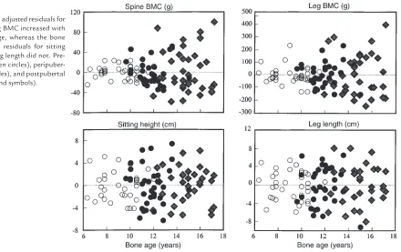
[image:4.612.185.540.334.738.2]Growth in bone length and bone mass. Table 1 shows the absolute values for anthropometric and bone mass measurements. Figure 1 shows the results expressed as a percentage of the predicted adult value versus bone age. The shaded region in Figure 1 represents pubertal growth from Tanner stage 2 to menarche. In prepuber-tal girls with a bone age of 7 years, height, sitting height, and region lengths were approximately 80% of the pre-dicted adult value; total and regional BMC were approx-imately 40% of the predicted adult value. At menarche
Figure 1
(12.7 ± 0.1 years), bone lengths were within 3% of their adult peak; total and regional BMC were 15–20% below their predicted peak.
Figure 2 shows that the variance in the age-adjusted BMC residuals increased with advancing bone age, whereas age-adjusted residuals for bone length did not. The pre-, peri-, and postpubertal BMC residuals (g2) increased, being, respectively, 174 ± 34.6, 626 ± 139, and 1,838 ± 381 (spine, P < 0.001), and 4,777 ± 1729, 8,775 ± 2,229, and 27,237 ± 5,539 (legs, P < 0.05 to 0.001). The respective pre-, peri-, and postpubertal length residuals (cm2) did not increase significantly and were 6.1 ± 1.3, 12.9 ± 2.7, 9.7 ± 1.9 (sitting height), and 9.6 ± 1.7, 17.5 ± 10.6, 17.3 ± 3.7 (leg length).
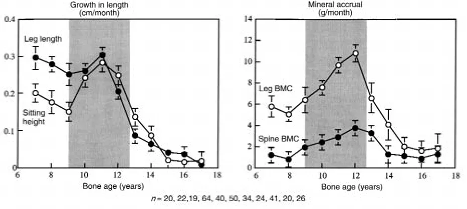
Figure 3 shows the prospectively derived rates of total and regional growth in bone size and BMC versus bone age. Growth velocity of sitting height slowed before puber-ty, accelerated, and then decelerated at approximately 12 years. Growth velocity of the legs was more rapid than that of sitting height before puberty. In contrast to the spine, the legs showed no significant acceleratory phase before decelerating rapidly at approximately 11 years. There was no detectable increase in sitting height after 14 years of age or in leg length after 16 years of age.
[image:5.612.101.541.49.326.2]At approximately 7 years, mineral accrual at the legs was higher than at the spine (5.1 ± 0.6 versus 0.8 ± 0.5 g/month). At approximately 12 years, mineral accrual was higher in the legs than spine (10.8 ± 0.8 versus 3.8 ± Figure 2
[image:5.612.65.537.561.731.2]The bone-age adjusted residuals for spine and leg BMC increased with advancing age, whereas the bone age adjusted residuals for sitting height and leg length did not. Pre-pubertal (open circles), peripuber-tal (filled circles), and postpuberperipuber-tal (filled diamond symbols).
Table 1
Height, sitting height, appendicular segment lengths, total and regional bone mineral content in 109 girls aged 7 to 17 years (mean ± SEM)
Bone age (years) 7 8 9 10 11 12 13 14 15 16 17
n (23) (23) (27) (72) (53) (65) (51) (36) (57) (34) (34)
Bone length
Height (cm) 127.9 ± 1.1 132.4 ± 0.9 136.8 ± 0.9 142.8 ± 0.7 148.0± 0.8 154.7± 0.8 160.2± 1.0 162.2 ± 1.1 162.3± 0.9 163.8± 1.2 164.6 ± 0.9 Sitting height (cm) 68.3 ± 0.6 70.0± 0.4 71.7 ± 0.5 74.7 ± 0.3 77.7 ± 0.5 80.6 ± 0.4 84.1 ± 0.5 86.0 ± 0.6 87.0 ± 0.4 88.1 ± 0.5 87.3 ± 0.5 Humerus (mm) 268 ± 2 281 ± 2 289 ± 3 303 ± 2 312 ± 2 329 ± 2 343 ± 3 347 ± 3 344 ± 2 344 ± 3 345 ± 3 Radius-ulna (mm) 199 ± 2 206 ± 2 214 ± 2 222 ± 1 229 ± 1 240 ± 1 249 ± 2 250 ± 2 251 ± 2 247 ± 3 249 ± 2 Femur (mm) 321 ± 4 339 ± 3 352 ± 4 370 ± 3 382 ± 3 403 ± 3 417 ± 3 420 ± 4 415 ± 3 416 ± 4 418 ± 3 Tibia (mm) 288 ± 3 301 ± 3 314 ± 3 327 ± 2 335 ± 3 352 ± 3 365 ± 3 362 ± 4 357 ± 3 355 ± 4 362 ± 2
Bone mineral content
0.6 g/month, respectively) despite growth in leg and spine lengths being similar (∼0.3 cm/month). Decelera-tion in mineral accrual occurred 1 year later than decel-eration in bone length at both sites. Bone mass accrual continued at the spine and legs until 16 years of age.
Between 7 and 17 years, 1,491 g of mineral was accrued: 42% (630 g) in the legs; 25% (376 g) in the ribs, sternum, pectoral, and pelvic girdles; 12% (176 g) in the arms; 11% (157 g) in the skull; and 10% (156 g) in the spine. Between the 7–11 years (prepuberty), 11–14 years (puber-ty), and 14–17 years (postpuber(puber-ty), the increases in spine BMC were 38, 72, and 46 g, respectively. The correspon-ding increases in leg BMC were 240, 241, and 149 g.
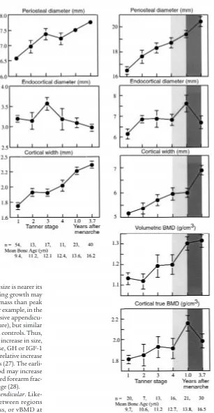
Growth in BMC and vBMD. Midshaft metacarpal and femoral cortical widths were approximately 70% of the predicted adult peak in prepubertal girls (Tanner stage 1). As shown in Figure 4, periosteal diameter increased at both sites with little change in endocortical diameter. Thus, cortical width increased, particularly when endo-cortical diameter contracted at Tanner stage 3 at the metacarpal, and after menarche at the femur. Of final cortical width, endocortical contraction contributed 13% at the metacarpal and 7% at the femur.
Bone mass measurements were available for the femur, not metacarpal. As shown in Figure 4, as periosteal and endocortical diameters of the femoral midshaft increased comparably, cortical width remained constant between Tanner stage 4 and menarche. Despite this, femoral mid-shaft vBMD increased, presumably because true BMD of the cortex increased (lighter shaded column). After menarche, periosteal diameter increased and endocorti-cal (medullary) diameter decreased resulting in an increase in cortical width (Figure 4, darkly shaded col-umn). Despite the increasing femoral midshaft cortical width, vBMD remained unchanged, perhaps because true BMD of the cortex decreased.
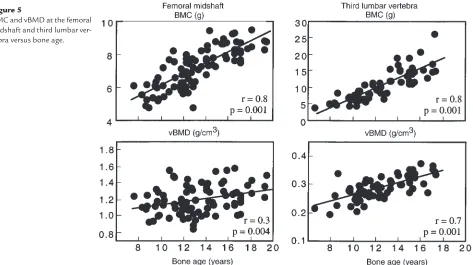
As shown in Figure 5, BMC at the femoral midshaft increased across age by 50%, or 3.4 SD. This was 4 times
the increase of 0.86 SD in femoral midshaft vBMD. L3 BMC increased across age by 150%; a 3.7 SD increase, 1.5 times the increase of 2.6 SD in vBMD.
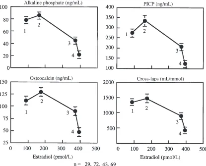
Biochemical and hormonal measurements. Biochemical measures of bone remodeling and serum E2 increased, reached a peak at bone age approximately 12 years (Tan-ner stage 4) and then decreased at menarche at a serum E2 of 200–400 pmol/mL (Table 2; Figures 6 and 7). Change in total bone mass (grams per month) correlat-ed with alkaline phosphatase (r = 0.20), osteocalcin (r = 0.38), P1CP (r = 0.32), and CrossLaps (r = 0.24) (all P < 0.02). Serum E2 accounted for 28% of the variance in leg BMC accrual in the accelerating phase of growth, 19% of the variance in the spine BMC accrual, and 49% of the variance in cortical width in the decelerating phase of growth (both, P < 0.05). IGF-1 accounted for 21% of the variance in the spine BMC in the accelerating phase of growth and for 14% of the variance in endocortical diam-eter (both P < 0.03). Bone age was the only independent predictor of leg bone mass accrual accounting for 35% of the variance (P < 0.005).
Discussion
We report that BMC and aBMD increased during growth. However, the changes were trait, surface, and region spe-cific: the age of onset, peak growth velocity, and comple-tion of growth of a region’s external dimensions preceded the mineral accrued within its periosteal envelope. Before puberty, appendicular growth was more rapid than axial growth. In early puberty, appendicular growth remained constant with no acceleration, whereas axial growth accel-erated. Growth of both regions then decelerated in late puberty. Cortical bone width increased due to periosteal expansion followed by endocortical contraction. vBMD of the midshaft of the femur and spine increased.
[image:6.612.65.530.487.695.2]Growth in size and mass within a region. The differing tempo of growth of bone size and mass within a region may con-tribute to deficits in bone size, BMC, and vBMD found in
Figure 3
patients with fractures (12). As a region’s size is nearer its peak than its mass (25), illness interrupting growth may result in a greater deficit in peak bone mass than peak bone size, leading to a reduced vBMD. For example, in the growing rat, ovariectomy results in excessive appendicu-lar growth (due to failed epiphyseal closure), but simiappendicu-lar mineral accrual relative to sham operated controls. Thus, the increase in mass is less relative to the increase in size, resulting in reduced vBMD (26). Likewise, GH or IGF-1 administration to rats results in a greater relative increase in size than mass so that vBMD decreases (27). The earli-er growth in size than mass in childhood may increase bone fragility and account for the increased forearm frac-tures observed at around 12–13 years of age (28).
Growth between regions: axial versus appendicular. Like-wise, the differing tempo of growth between regions may predispose to deficits in size, mass, or vBMD at one region but not another. Rapidly growing regions, or regions further from their peak, may be more severe-ly affected by illness than are slowsevere-ly growing regions or regions that have completed their growth. Before puberty, growth of the legs was more rapid than growth of the spine; 40% of the 630 g of mineral to be accrued in the legs was accrued by 11 years of age, whereas only 24% of spine mineral mass had been accrued. At puber-ty, growth of the spine accelerated while growth of the
legs slowed without a detectable acceleration phase. Hypogonadism or delayed puberty produces increased leg length due to failed epiphyseal closure but reduced trunk length due to failed pubertal acceleration of trunk growth (29).
[image:7.612.232.539.41.633.2]vBMD: the result of the relative growth of bone size and the amount of bone accrued within its periosteal envelope. vBMD, Figure 4
the mass per unit volume of bone, is determined by the rel-ative growth of the external dimensions of the bone and the mineral mass accrued within it (16). vBMD of the femoral midshaft remained relatively constant in early puberty (Tanner 1–4; Figure 4). Similarly, constancy of vBMD of the femoral shaft, radial shaft, and vertebral body has been has been reported by several investigators (30–34). The increase in vBMD at the spine reported here is likely to be an overestimate because of inclusion of the posterior processes in the measurement (see Methods). This constancy of vBMD during the prepubertal years is not widely recognized because most studies of growth report BMC or aBMD, size-dependent expressions of “density” that increase with age largely because bone size increases (16, 35–37). This constancy of vBMD before puberty suggests that the position of an individual’s peak vBMD relative to other individuals in the population dis-tribution is likely to be partly determined during early growth. That is, whether a person’s vBMD is at the 5th, 50th, or 95th percentile may be partly determined early in life by genetic factors coregulating the relative growth
of bone size and mass. vBMD “tracks” because the increase in size during growth is matched by a propor-tional increase in mass within it, at least before puberty. In morphological terms, the increase in femoral midshaft size was matched by a proportional increase in the corti-cal width of the enlarging bone. In the spine, vBMD remains constant because the increasing vertebral body size is matched by an increase in the thickness of the existing trabeculae within it (38, 39).
[image:8.612.66.539.50.315.2]By contrast, vBMD increased during late puberty and after menarche. For vBMD to increase — for there to be more bone in the growing bone — the increase in min-eral mass within the periosteal envelope must be rela-tively greater than the increase in the external bone size. In morphological terms, vBMD increased at the femoral midshaft because cortical thickness increased by periosteal expansion, slower endocortical expansion, followed by endocortical contraction after menarche (40). (If both surfaces expand at the same rate, the enlarging bone will not have a thicker cortex. If the endocortical surface expands more rapidly than the Figure 5
BMC and vBMD at the femoral midshaft and third lumbar ver-tebra versus bone age.
Table 2
GH, IGF-1, serum estradiol, testosterone, androstenedione and, DHEAS according to Tanner stage (mean ± SEM).
Tanner stage Years after menarche
1 2 3 4 1.0 ± 0.1 3.7 ± 0.1
(n = 45) (n = 31) (n = 42) (n = 42) (n = 75) (n = 118)
Concentration (mean ± SEM)
GH (mIU/L) 40 ± 28 18 ± 3 18 ± 2 24 ± 3 22 ± 2 17 ± 1
[image:8.612.59.527.615.724.2]expanding periosteum, cortical thickness will decrease.) vBMD of the vertebral body increases during puberty due to continued, and perhaps accelerated, thickening of trabeculae (32, 34, 39). The numbers of trabeculae, determined early in life at the growth plate, remain con-stant from early life to adulthood (39).
The increase in vBMD of the femoral midshaft may have also been partly due to an increase in the true density of the cortical shell at menarche because this increase in vBMD of the shaft occurred when cortical width did not change, whereas true BMD increased (Figure 4). After menarche, endocortical contraction occurred. Despite this, vBMD remained constant because true BMD decreased, perhaps because secondary mineralization of the newly accumulated matrix had not yet occurred. These inferences will require verification using bone histomor-phometry given that the calculation of true BMD assumes that the midshaft of the femur is a cylinder.
Although vBMD increased in late puberty, it is likely that vBMD increased more in some individuals than others. The variance in bone size did not increase, sug-gesting that bone size may “track,” i.e., individuals may gain a similar proportion of their starting value during growth (41). However, the variance in BMC increased during puberty and after menarche (Figure 2),
suggest-ing that BMC may not track; some individuals may accrue a greater “dose” of sex hormone–dependent bone mass per unit bone volume, whereas others accrue less bone mass per unit bone volume. In morphological terms, for a given bone size, some individuals may gain a thicker cortex than others by less expansion of the endocortical surface before puberty by less resorptive modeling or by more endocortical apposition during puberty than others. Persons with higher spine vBMD may have formed greater trabeculae numbers at the growth plate during early development or may have increased trabecular thickness during pre- and peripu-bertal development (more than others with the same starting bone size). The magnitude of the population variance of each of these structural components of bone need to be defined at each age and for both genders. Then, the genetic and environmental components of the variance can be studied (42, 43).
[image:9.612.72.530.52.386.2]In conclusion, the skeleton is not a single functioning entity; the effect of growth, aging, disease, and treatment vary according to whether a region is axial or appendic-ular, cortical or trabecappendic-ular, and whether the surface is periosteal or endosteal (endocortical, intracortical, tra-becular) (44). Each surface has a unique developmental pattern. The earlier growth in size than mass within a Figure 6
region, the differing tempo and direction of growth of the periosteal and endocortical surfaces, and the differ-ing tempo of growth of the axial and appendicular skele-ton suggests that each trait is regulated differently.
The heterogeneity in the growth of these traits pro-vides the setting for the development of region-specific deficits in mass, size, or density should illness occur. That is, unlike in adulthood, the effect of exposure to a risk factor during growth depends on the maturational level of the region exposed (as well as the exposure “dose”). Thus, bone fragility is likely to have its origins established during growth. A better understanding of the pathogenesis of osteoporosis can be gained by iden-tifying the genetic and environmental factors regulating the periosteal and endosteal surfaces of bone as the absolute and relative changes on these surfaces during growth and aging determine the size, mass, architecture, and strength of the skeleton (45).
Acknowledgments
The study was supported by the Dairy Research and Development Corporation of Australia. The authors thank the students and staff of the Ivanhoe Girls
Gram-mar School and Sister Jan Edmonds for making this work possible.
1. Cooper, C., and Melton, L.J., III. 1996. Magnitude and impact of osteo-porosis and fractures. In Osteoporosis. R. Marcus, D. Feldman, and J. Kelsey, editors. Academic Press Inc. San Diego, CA. 419–434. 2. Riggs, B.L., et al. 1981. Differential changes in bone mineral density of
the appendicular and axial skeleton with aging: relationship to spinal osteoporosis. J. Clin. Invest. 67:328–335.
3. Riggs, B.L., and Melton, L.J., III. 1986. Involutional osteoporosis. N. Engl. J. Med. 314:1676–1686.
4. Eastell, R., et al. 1989. Unequal decrease in bone density of lumbar spine and ultradistal radius in Colles’ and vertebral fracture syndromes. J. Clin. Invest. 83:168–174.
5. Mautalen, C., Vega, E., Ghiringhelli, G., and Fromm, G. 1990. Bone diminution of osteoporotic females at different sites. Calcif. Tissue Int. 46:217–221.
6. Seeman, E., et al. 1989. Reduced bone mass in daughters of women with osteoporosis. N. Engl. J. Med.320:554–558.
7. Seeman, E., Hopper, J.L., Tsalamandris, C., and Formica, C. 1994. Bone density in daughters of women with hip fractures. J. Bone Miner. Res. 9:739–743.
8. Evans, R.A., et al. 1998. Bone mass is low in relatives of osteoporotic patients. Ann. Intern. Med. 109:870–873.
9. Ristevski, S., Yeung, S., Poon, C., Wark, J.W., and Ebeling, P. 1997. Osteopaenia is common in young first-degree male relatives of men with osteoporosis. Australian and New Zealand Bone and Mineral Society, Annual Scientific Meeting.Abstract book.Volume 7. Canberra, Australia. pp. 65. 10. Vega, E., et al. 1998. Bone mineral density and bone size in men with
[image:10.612.93.508.53.388.2]pri-mary osteoporosis and vertebral fractures. Calcif. Tissue Int. 62:465–469. Figure 7
11. Gilsanz, V., et al. 1995. Gender differences in vertebral size in adults: bio-mechanical implications. J. Clin. Invest. 95:2332–2337.
12. Duan, Y., Parfitt, M., and Seeman, E. 1999. Vertebral bone mass, size and volumetric bone mineral density in premenopausal women, and post-menopausal women with and without spine fractures. J. Bone Miner. Res. In press.
13. Mazess, R.B. 1983. Non-invasive bone measurements in skeletal research. Vol-ume 9. Academic Press. New York, NY. 277–343.
14. Robinson, R.A. 1960. Chemical analysis and electron microscopy of bone. In Bone as a tissue. K. Rodhal, J.T. Nicholson, and E.M. Brown, edi-tors. McGraw-Hill Book Co. New York, NY. 186–250.
15. Parfitt, A.M. 1998. A structural approach to renal bone disease. J. Bone Miner. Res. 13:1213–1220.
16. Seeman, E. 1998. Growth in bone mass and size: are racial and gender differences in bone density more apparent than real? J. Clin. Endocrinol. Metab. 83:1–6.
17. Carter, D.R., Bouxsein, M.L., and Marcus, R. 1992. New approaches for interpreting projected bone densitometry data. J. Bone Miner. Res. 7:137–145.
18. Tabensky, A., Williams, J., Deluca, V., Brigante, E., and Seeman, E. 1996. Bone mass, areal and volumetric bone density are equally accurate, sen-sitive, and specific surrogates of the breaking strength of the vertebral body: an in vitro study. J. Bone Miner. Res. 11:1981–1988.
19. Garnero, P., et al. 1992. Measurement of serum osteocalcin with a human-specific two-site immunoradiometric assay. J. Bone Miner. Res. 7:1389–1397.
20. Garnero, P., and Delmas, P.D. 1993. Assessment of the serum levels of bone alkaline phosphatase with a new immunoradiometric assay in patients with metabolic bone disease. J. Clin. Endocrinol. Metab. 77:1046–1053. 21. Winterbottom, N., Vernon, S., Freeman, K., Dankoff, G., and Sevedin, S.
1993. A serum immunoassay for the C-terminal propeptide of type I col-lagen. J. Bone Miner. Res. 8(Suppl. 1):S341. (Abstr.)
22. Garnero, P., Gineyts, E., Riou, P.R., and Delmas, P.D. 1994. Assessment of bone resorption with a new marker of collagen degradation in patients with metabolic bone disease. J. Bone Miner. Res. 79:780–785. 23. Theintz, G., et al. 1992. Longitudinal monitoring of bone mass
accu-mulation in healthy adolescents: evidence for a marked reduction after 16 years of age at the levels of the lumbar spine and femoral neck in female subjects. J. Clin. Endocrinol. Metab.75:1060–1065.
24. Marubini, E. 1978. Mathematical handling of long-term longitudinal data. In Human growth: principles and prenatal growth. F. Falkner and J.M. Tanner, editors. Volume 1. 1st edition. Plenum Press. New York, NY, and London, United Kingdom. 209–225.
25. Fournier, P.-E., Rizzoli, R., Slosman, D.-O., Theintz, G., and Bonjour, J.-P. 1997. Asynchrony between the rates of standing height gain and bone mass accumulation during puberty. Osteoporos. Int. 7:525–532. 26. Zhang, X.Z., Kalu, D.N., Erbas, B., Hopper, J.L., and Seeman, E. 1999. The
effect of gonadectomy on bone size, mass and volumetric density in growing rats may be gender-, site-, and growth hormone-dependent. J.
Bone Miner. Res. 14:802–809.
27. Rosen, H.N., et al. 1995. Treatment with growth hormone and IGF-1 in growing rats increases bone mineral content but not bone mineral den-sity. J. Bone Miner. Res. 10:1352–1358.
28. Bailey, D.A., Wedge, J.H., McCulloch, R.G., Martin, A.D., and Bernhardson, S.C. 1989. Epidemiology of fractures of the distal end of the radius in chil-dren as associated with growth. J. Bone Joint Surg. Am. 71:1225–1231. 29. Schibler, D., Brooks, C.G., Kind, H.P., Zachmann, M., and Prader, A.
1974. Growth and body proportions in 54 boys and men with Klinefel-ters syndrome. Helv. Paediatr. Acta. 29:325–333.
30. Lu, P.W., Cowell, C.T., Lloyd-Jones, S.A., Briody, J., and Howman-Giles, R. 1996. Volumetric bone mineral density in normal subjects, aged 5–27 years. J. Clin. Endocrinol. Metab. 81:1586–1590.
31. Warner, J., et al. 1998. Measured and predicted bone mineral content in healthy boys and girls aged 6–18 years: adjusted for size and puberty. Acta Paediatr. 87:244–249.
32. Gilsanz, V., et al. 1988. Vertebral bone density in children: effect of puber-ty. Radiology. 166:847–850.
33. Dunnill, M.S., Anderson, J.A., and Whitehead, R. 1967. Quantitative his-tological studies on age changes in bone. J. Pathol. Bacteriol. 94:275–291. 34. Gilsanz, V., et al. 1998. Differential effects of race on the axial and
appen-dicular skeleton of children. J. Clin. Endocrinol. Metab. 83:1420–1427. 35. Katzman, D., Bachrach, L., Carter, D., and Marcus, R. 1991. Clinical and
anthropometric correlates of bone mineral acquisition in healthy ado-lescent girls. J. Clin. Endocrinol. Metab. 73:1332–1339.
36. Glastre, C.C., et al. 1990. Measurement of bone mineral content of the lumbar spine by dual energy x-ray absorptiometry in normal children: correlations with growth parameters. J. Clin. Endocrinol. Metab. 70:1330–1333.
37. Mazess, R.B., and Cameron, J.R. 1971. Skeletal growth in school children: maturation and bone mass. Am. J. Phys. Anthropol. 35:399–408. 38. Parfitt, A.M. 1997. Genetic effects on bone mass and turnover: relevance
to black/white differences. J. Am. Coll. Nutr. 16:325–333.
39. Parfitt, A.M., Rauch, F., Travers, R., and Glorieux, F.H. 1999. A new model of cancellous bone growth. J. Bone Miner. Res. In press. 40. Garn, S. 1970. Nutritional perspectives.Charles C. Thomas. Springfield, IL.
3–120.
41. Smith, D.W., et al. 1976. Shifting linear growth during infancy: illustra-tion of genetic factors in growth from fetal life through infancy. J. Pedi-atr. 89:225–230.
42. Seeman, E., and Hopper, J.L. 1997. Problems in the study of the genetics of osteoporosis. Osteoporos. Int. 7(Suppl. 3):s10–s16.
43. Hopper, J.L., et al. 1998. Genetic, common environment, and individual specific components of variance for bone mineral density in 10- to 26-year-old females: a twin study. Am. J. Epidemiol. 147:17–29.
44. Seeman, E., et al. 1982. The differential effects of endocrine dysfunction on the axial and appendicular skeleton. J. Clin. Invest. 69:1302–1309. 45. Seeman, E. 1997. From density to structure: growing up and growing old