Copyright © 2004, American Society for Microbiology. All Rights Reserved.
DNA Microarray Format for Detection and Subtyping of
Human Papillomavirus
Corne´ H. W. Klaassen,
1,2* Clemens F. M. Prinsen,
1Hanneke A. de Valk,
2Alphons M. Horrevorts,
2Marcel A. F. Jeunink,
1and Frederik B. J. M. Thunnissen
1,3Departments of Pathology
1and Medical Microbiology and Infectious Diseases,
2Canisius Wilhelmina
Hospital, Nijmegen, and Dot Diagnostics, Beuningen,
3The Netherlands
Received 17 November 2003/Accepted 30 December 2003
A new human papillomavirus (HPV) assay using high-density DNA microarrays is described. An HPV DNA
fragment from the 3
ⴕ
end of the E1 gene was amplified and digoxigenin labeled by PCR, and the resulting
amplicons were hybridized onto type-specific oligonucleotides immobilized on high-density DNA microarrays.
For detection, a simple immunohistochemical staining procedure was used with a substrate that has both
colorimetric and fluorescent properties. This detection chemistry enables the rapid identification of reactive
spots by regular light microscopy and semiquantification by laser scanning. Both single and multiple HPV
infections are recognized by this assay, and the corresponding HPV types are easily identified. With this assay,
53 mucosal HPV types were detected and identified. A total of 45 HPV types were identified by a single
type-specific probe, whereas the remaining 8 mucosal HPV types could be identified by a specific combination
of probes. The simple assay format allows usage of this assay without expensive equipment, making it
accessible to all diagnostic laboratories with PCR facilities.
To date, more than 80 human papillomavirus (HPV) types
have been described that are involved in either cutaneous or
genitomucosal lesions of the skin. Because of the strong
asso-ciation of certain HPV types with cervical cancers, the
genital-mucosal HPV types have been divided into clusters of types
with a relative high risk or a relative low risk of an HPV
infection progressing into cervical cancer. The data from
com-bined case-control studies (19) suggest that some HPV types
should be regarded high-risk or carcinogenic (e.g., HPV16, -18,
-31, -33, -35, -39, -45, -51, -52, -56, -58, -59, -68, -73, and -82)
and that some should be considered probably carcinogenic
(HPV26, -53, and -66), whereas the relative risk associated
with certain other HPV types may be only low (e.g., HPV6, -11,
-40, -42, -43, -44, -54, -61, -70, -72, and -81).
Several DNA-based assays have already been developed for
the simultaneous broad identification and subtyping of HPV
types. The majority are PCR-based assays with a variety of
PCR primers being used, such as the MY09-MY11, GP5
⫹
-GP6
⫹
, SPF, and PGMY09-PGMY11 primer combination(s)
(5, 7, 15, 18). For typing purposes these assays have been
combined with dot blots (10, 18, 29), microtiter enzyme
immu-noassays (11, 15), reverse hybridization line probe assays (4, 8,
14, 26), cycle sequencing (28), T-ladder generation (20), and
pyrosequencing (6). Several non-PCR methods have been
de-signed as well, such as the hybrid capture assay (17) and the in
situ hybridization approach (25).
The large variety of techniques available for detection and
subtyping of the multitude of HPV types illustrates the fact
that no single technique provides the ultimate and complete
solution to this effect. This is especially indicated by the
ob-served differences between existing techniques with respect to
analytical sensitivity, ability to discriminate between different
HPV types, and ability to recognize multiple infections (4, 7,
12, 14, 15, 22, 26–28). Moreover, existing assays still fail to
identify all of the possible HPV types involved since they were
designed to detect only a subset of the mucosal HPV types.
This circumstance justifies the development of new and
im-proved HPV assays.
Here, we introduce a new broad HPV assay format based on
high-density DNA microarrays. Digoxigenin-labeled
HPV-de-rived PCR amplicons are hybridized onto type-specific
biotin-ylated HPV probes immobilized on streptavidin-coated glass
slides. Hybridized amplicons are visualized by using a simple
immunohistochemical staining procedure with a substrate for
alkaline phosphatase that has both colorimetric and
fluores-cent properties. This detection chemistry enables rapid
iden-tification of reactive spots with regular light microscopy, as well
as semiquantification by laser scanning. Single and multiple
HPV infections are easily recognized, and the corresponding
HPV types can be identified. The simple assay format allows
usage of this assay without expensive equipment, making it
accessible to all diagnostic laboratories with PCR facilities.
With this assay format it is possible to detect and identify 53
genital HPV types, the largest HPV panel thus far.
MATERIALS AND METHODS
HPV sequences.The following sequences from officially recognized HPV types were included in the present study (with the GenBank accession number in parentheses [latest update for HPV search, 12 February 2003]): HPV1 (U06714 and V01116), HPV2 (X55964), HPV3 (X74462), HPV4 (X70827), HPV5 (M22961, M17463, and D90252), HPV6 (AF092932, L41216, and X00203), HPV7 (X74463), HPV HPV8 (M12737), HPV9 (X74464), HPV10 (X74465), HPV11 (M14119), HPV12 (X74466), HPV13 (X62843 and S43933), HPV14 (X74467), HPV15 (X74468), HPV16 (K02718, U89348, and AF125673), HPV17
* Corresponding author. Mailing address: Department of Pathology
C66, Canisius Wilhelmina Hospital, Weg door Jonkerbos 100, 6532 SZ
Nijmegen, The Netherlands. Phone: 7514. Fax:
31-24-365-7516. E-mail: [email protected].
2152
on May 15, 2020 by guest
http://jcm.asm.org/
(X74469), HPV18 (X05015), HPV19 (X74470), HPV20 (U31778), HPV21 (U31779), HPV22 (U31780), HPV23 (U31781), HPV24 (U31782), HPV25 (X74471), HPV26 (X74472), HPV27 (X74473), HPV28 (U31783), HPV29 (U31784), HPV30 (X74474), HPV31 (J04353), HPV32 (X74475), HPV33 (A07020, A12360, and M12732), HPV34 (X74476), HPV35 (M74117 and X74477), HPV36 (U31785), HPV37 (U31786), HPV38 (U31787), HPV39 (M62849), HPV40 (X74478), HPV41 (X56147), HPV42 (M73236), HPV44 (U31788), HPV45 (X74479), HPV47 (M32305), HPV48 (U31789), HPV49 (X74480), HPV50 (U31790), HPV51 (M62877), HPV52 (X74481), HPV53 (X74482), HPV54 (U37488), HPV55 (U31791), HPV56 (X74483), HPV57 (U37537 and X55965), HPV58 (D90400), HPV59 (X77858), HPV60 (U31792), HPV61 (U31793), HPV63 (X70828), HPV65 (X70829), HPV66 (U31794), HPV67 (D21208), HPV69 (AB027020), HPV70 (U21941), HPV71 (AB040456), HPV72 (X94164), HPV73 (X94165), HPV74 (AF436130), HPV75 (Y15173), HPV76 (Y15174), HPV77 (Y15175), HPV80 (Y15176), HPV82 (AB027021), HPV83 (AF151983), HPV84 (AF293960), HPV85 (AF131950), HPV86 (AF349909), HPV87(AJ400628), HPV89 (AF436128), HPV90 (AY057438), HPV91 (AF419318), and HPV92 (AF531420).
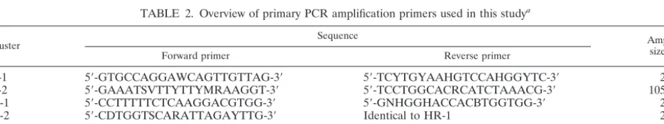
Selection of a target region and design of PCR primers.Available HPV DNA sequences (whole genome, individual genes as well as gene specific fragments) were separately and systematically analyzed for sequence homology by creating multiple DNA sequence alignments by using the CLUSTAL X program (24). A target region was selected based on the following considerations: (i) DNA se-quence alignment of the target region should yield a clear separation into a minimal number of clusters containing primarily low-risk, high-risk, or cutaneous HPV types. This condition would facilitate and simplify the design of cluster- and thus risk-specific PCR amplification primers; (ii) the target region should contain sufficient DNA heterogeneity to allow design of type-specific oligonucleotide probes in order to discriminate between as many HPV types as possible; and yet (iii) should be flanked by regions with sufficient DNA sequence homology to allow formulation of (degenerate) PCR primers and (iv) the target region should not be too long, which might complicate efficient DNA amplification by PCR. According to these considerations, a suitable target region was selected at the C terminus of the HPV E1 gene. Based on the alignments, HPV sequences were subdivided into six clusters containing either low-risk mucosal HPV types (clus-ters LR-1 and LR-2), high-risk mucosal types (clus(clus-ters HR-1, HR-2, and HR-3), or cutaneous HPV types (cluster C) (Table 1). Degenerate PCR primers were formulated for each of the genital HPV clusters to allow amplification of HPV types in a cluster-specific manner (Table 2).
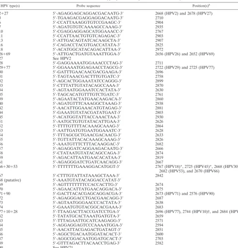
Design of type-specific HPV probes.HPV type-specific oligonucleotide probes (20-mers) were formulated and checked for specificity by BLAST analysis (1)
(Table 3). Each probe contained a minimum of two mismatches (around the center of the probe) to all other HPV types wherever possible. In a few cases, no discrimination between certain HPV types could be made on theoretical grounds (for instance, for the closely related HPV types 2 and 27, with which there is only one nucleotide difference between both amplicons). For these cases, either a single probe was used to detect both types or a unique combination of probes was used to establish the differentiation (Table 3). All probes were synthesized with
a biotinylated tag at the 5⬘end. The biotin label was introduced for
immobili-zation of the probes onto streptavidin-coated glass slides, and the additional tag was used for quality control procedures (see Results) and for reduction of possible steric hindrance by the solid support.
Samples.To obtain as many different HPV types as possible to validate the specificity of this novel assay, two different clinical sample collections were screened. The first collection consisted of 30 archival formaldehyde-fixed, par-affin-embedded biopsies with proven HPV presence, as determined by using the HPV PCR-enzyme immunoassay approach (11). These samples contained at least one high-risk and/or low-risk HPV type. The second sample collection contained 100 liquid-based cytology samples. These samples were routinely col-lected as part of cervical screening procedures.
DNA isolation.DNA was isolated from 10-m-thick paraffin sections by using a Puregene DNA isolation kit from Gentra Systems (Biozym B.V., Landgraaf, The Netherlands) according to the instructions supplied by the manufacturer. After centrifugation of the liquid-based cytology samples, the cell pellet was
resuspended in 180 l of phosphate-buffered saline (PBS). Next, DNA was
isolated by using a QIAamp blood kit (Qiagen, Hilden, Germany) according to the recommended protocol. Isolated DNA was quantitated by using UV absor-bance measurements, taking into account an optical density conversion factor at
260 nm of 1 at⬃50g of DNA/ml (23). The isolated DNA was of excellent
purity, as judged by theA260/A280value of⬃1.8.
Primary PCR amplification.Target amplification was performed in a T1 or T
gradient thermocycler (Biometra, Go¨ttingen, Germany) in five separate 50-l
reactions (one for each cluster) containing 100 ng of template DNA, 1M
concentrations of both forward and reverse amplification primers, 0.2 mM de-oxynucleoside triphosphates, 1 U of HotGoldStar DNA polymerase
(Eurogen-tec, Seraing, Belgium), 1⫻reaction buffer, and 2 mM MgCl2. Cycling conditions
were as follows: 10 min of denaturation at 95°C, followed by 40 cycles of 30 s of denaturation at 95°C, 30 s of annealing at 52°C, and 1 min of extension at 72°C. Before the reactions were cooled to room temperature, an additional incubation for 10 min at 72°C was performed. All temperature transitions were performed with maximal heating and cooling settings (5°C/s). In order to check for the presence of PCR inhibitors in the DNA samples, an additional PCR was
per-TABLE 1. Subdivision of 84 HPV types (53 mucosal and 31 cutaneous) into six clusters
aCluster HPV types
LR-1 ... 6, 7, 11, 13, 32, 40, 42, 44, 55, 74, 91
LR-2 ... 2, 3, 10, 27, 28, 29, 54, 57, 61, 71, 72, 77, 83, 84, 86, 87, 89
HR-1 ... 16, 31, 33, 35, 52, 58, 67
HR-2 ... 18, 39, 45, 59, 68, 70, 85
HR-3 ... 26, 30, 34, 51, 53, 56, 66, 69, 73, 82
C ... 1, 4, 5, 8, 9, 12, 14, 15, 17, 19, 20, 21, 22, 23, 24, 25, 36, 37, 38, 41, 47, 48, 49, 50, 60, 63, 65, 75, 76, 80, 92
aLR, low-risk HPV types; HR, high-risk or probably high risk HPV types; C, cutaneous HPV types. The basis for this division was a phylogenetic analysis of available
[image:2.603.43.542.78.154.2]HPV E1 gene sequences. No E1 sequence was found for HPV43, -46, -62, -64, -68, -78, -79, -81, and -88. HPV68 was grouped into cluster HR-2 due to the high level of similarity in the L1 region of this HPV type to HPV39 and HPV70.
TABLE 2. Overview of primary PCR amplification primers used in this study
aCluster Sequence Ampliconsize (bp)
Forward primer Reverse primer
LR-1
5
⬘
-GTGCCAGGAWCAGTTGTTAG-3
⬘
5
⬘
-TCYTGYAAHGTCCAHGGYTC-3
⬘
258
LR-2
5
⬘
-GAAATSVTTYTTYMRAAGGT-3
⬘
5
⬘
-TCCTGGCACRCATCTAAACG-3
⬘
105–117
HR-1
5
⬘
-CCTTTTTCTCAAGGACGTGG-3
⬘
5
⬘
-GNHGGHACCACBTGGTGG-3
⬘
245
HR-2
5
⬘
-CDTGGTSCARATTAGAYTTG-3
⬘
Identical to HR-1
232
HR-3
5
⬘
-TTTBHAAATVCATTTCCAWTWGA-3
⬘
5
⬘
-TAAACGHTKRSAHAGNKTCTCCAT-3
⬘
151–154

-Globin
5
⬘
-CAACTTCATCCACGTTCACC-3
⬘
5
⬘
-GAAGAGCCAAGGACAGGTAC-3
⬘
71
aFor labeling purposes in a second consecutive asymmetric PCR reaction, each of the HPV cluster-specific reverse primers was extended at the 5⬘end with an M13
universal primer tag (5⬘-GTTTTCCCAGTCACGAC-3⬘). Degenerate primer positions are indicated according to the single-letter convention: R⫽A and G; Y⫽C
and T; W⫽A and T; S⫽C and G; M⫽A and C; K⫽G and T; B⫽C, G, and T; D⫽A, G, and T; H⫽A, C, and T; V⫽A, C, and G; N⫽A, C, G, and T.
on May 15, 2020 by guest
http://jcm.asm.org/
[image:2.603.58.540.610.700.2]formed under the same experimental conditions with primers targeting the
human-globin gene (Table 1). In order to check for positive PCR results, 5l
of the PCR was analyzed on an agarose gel according to standard procedures (23).
Digoxigenin labeling and generation of single-stranded amplification prod-ucts.Labeling of the obtained HPV PCR amplicons was established with an asymmetrical PCR procedure similar to the conditions described above. The cluster-specific forward primer was used, and the sequence of the reverse primer
was identical to the sequence of the tag added to all HPV reverse amplification
primers, which was labeled at the 5⬘end with digoxigenin. The concentration of
the primers was 0.05M for the forward primer and 1.0M for the reverse
primer, respectively. Each labeling reaction contained 1l of the PCR original
[image:3.603.47.539.79.590.2]amplification reaction that tested positive. By applying the asymmetrical PCR, all reaction products were simultaneously labeled, as well as amplified into single-stranded products that could be used in the microarray hybridization procedure without further purification or treatment. In case of multiple positive primary
TABLE 3. Overview of type-specific HPV probes used in this study
aHPV type(s) Probe sequence Position(s)b
2
⫹
27
5
⬘
-AGAGGAGCAGGACGACAATG-3
⬘
2668 (HPV2) and 2678 (HPV27)
3
5
⬘
-TGAAGACGAGGAGGACAATG-3
⬘
2710
6
5
⬘
-CCATTAAAGGTGTCCGAAGC-3
⬘
2904
7
5
⬘
-AGATGTGTCAAAAGCCAAAG-3
⬘
2935
10
5
⬘
-CGAGGAGGAGCATGGAAACC-3
⬘
2767
11
5
⬘
-CCATTAACTGTGTCAGAGAC-3
⬘
2903
13
5
⬘
-ATTGACAGTATCACAAGCTA-3
⬘
2907
16
5
⬘
-CAGACCTACGTGACCATATA-3
⬘
2825
18
5
⬘
-ACATGGCATACAGACATTAA-3
⬘
2972
26
⫹
69
5
⬘
-ATTGACTGATGTAAATTGGA-3
⬘
2656 (HPV26) and 2652 (HPV69)
27
See HPV2
28
5
⬘
-GAGGAAAATGGAAACCCTAG-3
⬘
2711
29
⫹
77
5
⬘
-GGAAAATGGAGAACCTAGCG-3
⬘
2722 (HPV29) and 2725 (HPV77)
30
5
⬘
-GATTTGAACAACGACGAAGA-3
⬘
2696
31
5
⬘
-TAGTAAACGACTTTGTGATC-3
⬘
2758
32
5
⬘
-AGCACTGGAAATATCCAGGG-3
⬘
2899
33
5
⬘
-CTTTATTGTATACAGCCAAA-3
⬘
2870
34
5
⬘
-AGTAATGGAAATCCACTATA-3
⬘
2630
35
5
⬘
-TAGCACATGTTTGTCTGATC-3
⬘
2761
39
5
⬘
-AGAATACTATGAACAAGACA-3
⬘
2860
40
5
⬘
-AGATGTTTCAAAGGCTAAAG-3
⬘
2938
42
5
⬘
-AACATTGGAAACATGTAGAG-3
⬘
2881
44
5
⬘
-GAAATGTATACGATATGAAT-3
⬘
2803
45
5
⬘
-ACATGGTATTACCAAACTAA-3
⬘
2930
51
5
⬘
-AATGCTGTGTATACATTGAA-3
⬘
2626
52
5
⬘
-TTTTGTTTTACAAAGCAAAG-3
⬘
2864
53
5
⬘
-AATTGATGTGAATGGAAATC-3
⬘
2628
54
5
⬘
-TTTAGCGCTGAACGACAACG-3
⬘
2633
55
5
⬘
-TGTTATTACACAAAGCAAAG-3
⬘
2826
56
5
⬘
-AAATGTTTCTTTACAAGGAC-3
⬘
2682
57
5
⬘
-AGAGGATCAGGAAGACAATG-3
⬘
2666
58
5
⬘
-CTATAATGTATACAGCCAGA-3
⬘
2874
59
5
⬘
-AGACATTAATGAACACATAA-3
⬘
2819
61
5
⬘
-AGAGGGATCTGATCAACAGG-3
⬘
2667
66
⫹
30
⫹
53
5
⬘
-TTTTTTTGAAAGGACATGGT-3
⬘
2767 (HPV18)*, 2725 (HPV45)*, 2668 (HPV30),
2682 (HPV53), and 2670 (HPV66)
67
5
⬘
-CTTTGTATTATAAAGCTAAA-3
⬘
2842
68 (putative)
5
⬘
-AAATGTATACAGGACCATAT-3
⬘
69
5
⬘
-AGTTTTTTTTCCACCACTTG-3
⬘
2674
70
5
⬘
-AGAACATTATGAACAGGACA-3
⬘
2875
71
⫹
90
5
⬘
-GACTTACACGAGCAGGACGA-3
⬘
2673 (HPV71) and 2576 (HPV90)
72
5
⬘
-AGAGGGACCTGACGAACAGG-3
⬘
2687
73
5
⬘
-AGTAATGGGAACCCACTATA-3
⬘
2638
74
5
⬘
-GAAATGTGTACGGCACGAAA-3
⬘
2683
77
⫹
10
⫹
28
5
⬘
-TTAAGACTTACCGATCCTGA-3
⬘
2696 (HPV77), 2744 (HPV10)†, and 2684 (HPV28)†
82
5
⬘
-TATATGCACTAAATGATGTA-3
⬘
2659
83
5
⬘
-TTTAGAATTGCATCAAGAGG-3
⬘
2571
84
5
⬘
-AGGAGGAGTCCCAAAATGGA-3
⬘
2594
85
5
⬘
-AACATTACGAGACTGATAGT-3
⬘
2851
86
5
⬘
-AGGCTGACAATGGATACACT-3
⬘
2600
87
5
⬘
-AGGCGGACAATGGATGCACT-3
⬘
2703
89
5
⬘
-GTTTAGACTTACAACCTGAG-3
⬘
2582
90
See HPV71
91
5
⬘
-AAAGCTAGAAGTATCACGAG-3
⬘
2990
aFor immobilization and quality control purposes, all probes are extended at the 5⬘end with a biotinylated tag (see the text for details). For each probe, the position
of its 5⬘end in the corresponding HPV genome(s) is given.
bAlthough the probe sequence marked with an asterisk is also present in the HPV18 and HPV45 genome, it is not present in the HPV18 and HPV45 amplicons.
Therefore, this probe will not give rise to a hybridization signal with these two HPV types. The probe sequence marked with a dagger (†) contains one mismatch with respect to this HPV type. Type-specific probes for HPV10 and HPV28 are also present on the array.
on May 15, 2020 by guest
http://jcm.asm.org/
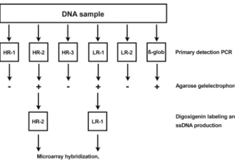
amplification reactions, they were further processed separately. A flowchart describing the amplification and labeling procedure is given in Fig. 1.
Preparation of the microarrays.HPV type-specific probes were spotted onto streptavidin-coated glass slides (Dot Diagnostics, Beuningen, The Netherlands)
at a concentration of 12M in spotting buffer (3⫻SSC [1⫻SSC is 0.15 M NaCl
plus 0.015 M sodium citrate], 1.5 M betaine [pH 7.0]) by using a SDDC-2 array spotting robot (ESI, Toronto, Canada) equipped with Stealth Microspotting Pins 3 (Telechem International, Inc., Sunnyvale, Calif.). These pins deliver ca. 0.6 nl
at each spotting site, resulting in spots of ca. 100m in diameter. Spots were
printed in triplicate, with a dot spacing inxandyaxes of 400m. The relative
humidity during spotting varied between 45 and 60%, whereas the temperature was kept constant at 22°C. After visual inspection of the slides after printing (see Results), the slides were stored. This could be done for a minimum of 3 months without negative effects on the results (data not shown).
For orientation purposes after completion of the entire hybridization, staining, and detection procedure, a randomized double-stranded oligomer (20 bp) was
used in the spotting process. This oligomer contained on one strand a 5⬘biotin
label for immobilization onto the glass slides and on the other strand a 5⬘
digoxigenin label for visualization. A number of negative control spots were also generated by applying only spotting buffer to the slides.
Microarray hybridization.All hybridization incubations and washing steps were performed at room temperature in disposable coverplates (Thermo-Shan-don, Amsterdam, The Netherlands). Standard microscope slides fit in these coverplates and are held in a vertical position during the entire procedure. A
distance of 80m is maintained between the surface of the slides and the
disposable coverplate. In this setup, the incubation mixture is being held in
position by capillary forces (effective working volume of⬃80l). Washing the
slides is performed simply by adding wash buffer to the upper reservoir (ca. 3 ml) and allowing it to pass the array by gravity. Printed arrays were first washed in
phosphate-buffered saline (PBS)–Tween for 10 min (PBS-Tween; 1 mM KH2
PO4, 0.15 M NaCl, 3 mM Na2HPO4 䡠7H2O [pH 7.4], 0.05% [wt/vol] Tween
20). Next, the microarrays were prehybridized in 40% formamide–3.3⫻SSC–1.7
mM EDTA–17 mM HEPES–0.12% Tween 20 (pH 7.3) for 5 min and
subse-quently hybridized in the same solution containing 20 l of the unpurified
single-stranded digoxigenin-labeled PCR product for 1 h. Five subsequent
washes of 5 min each were performed with PBS-Tween, 2⫻SSC–0.1% sodium
dodecyl sulfate, 1⫻SSC–0.1% sodium dodecyl sulfate, 0.1⫻SSC, and 0.05⫻
SSC, respectively.
Immunodetection and visualization of the microarrays. Detection of the digoxigenin-labeled and hybridized PCR products was performed as described before (21). In short, a 1:100 dilution of a mouse monoclonal antibody to digoxigenin was incubated (Roche Diagnostics, Almere, The Netherlands). Next, a 45-min incubation with a 1:20 dilution of alkaline phosphatase-conjugated rabbit anti-mouse immunoglobulins (Dako, Amsterdam, The Netherlands) was performed. Alkaline phosphatase activity was detected by using Vector Blue as a substrate (Vector Laboratories, Burlingame, Calif.) according to the instruc-tions of the manufacturer. The reaction product of Vector Blue can be seen by using both bright-field and fluorescence microscopy. Slides analyzed by laser scanning were washed twice for 5 min in PBS-Tween, rinsed in water, washed for 3 min in ethanol, and subsequently air dried in the dark. The Vector Blue reaction product yields blue spots visible by light microscopy and red spots upon excitation at 635 nm by laser scanning.
Imaging.Slides were scanned with a GenePix 4000A microarray laser scanner and data were analyzed with GenePix Pro 4.1 software (both from Axon Instru-ments, Foster City, Calif.). This scanner is equipped with a 532-nm laser and a
635-nm laser. Sixteen-bit TIFF images at 10-m resolution were subtracted for
local background intensity. Alternatively, when a low-magnification lens is used
(⫻2.5), the whole array can be viewed in one image without the need for special
filters or adaptors (total magnification including ocular,⫻25). For
documenta-tion, the result was recorded with a charge-coupled device camera (Sony DSC-S75) attached to the microscope.
DNA sequence analysis.PCR amplicons were purified by using a High-Pure PCR product purification kit (Roche Diagnostics) according to the instructions of the manufacturer. Purified amplicons were sequenced with the respective amplification primers on an ABI 3700 automated DNA analysis platform (Ap-plied Biosystems, Nieuwerkerk aan den Ijssel, The Netherlands) by using con-ditions recommended by the manufacturer. The identity of the obtained se-quences was verified by using basic local alignment search tool (BLAST) analysis (1).
RESULTS
Quality control procedures.
In the design of this novel assay,
a number of internal control procedures were included to
check the various steps of the entire assay procedure
(ampli-fication, spotting, hybridization, visualization, and
interpreta-tion).
Amplification control.
Apart from the HPV cluster-specific
PCRs, a

-globin PCR was included to verify the quality of the
obtained DNA. This is particularly relevant when no HPV
amplicon is obtained. All DNA samples used contained PCR
quality DNA (not shown).
Spotting control.
Since extremely tiny volumes are spotted
onto the glass slides (on average,
⬃
0.6 nl), it is virtually
im-possible to check the spotting process at this stage. Upon
removal of the slides from the spotting cabinet, these tiny
amounts of buffer are vaporized in seconds, leaving nothing to
see with the naked eye. However, upon addition of 1.5 M
betaine to the spotting buffer, spots remain visible until the
betaine is washed away (not shown). Furthermore, each probe
is spotted in triplicate to compensate for incidental misprints
(see examples in Fig. 2).
Visualization control.
Separate marker spots are created
containing a digoxigenin label. These are printed on the slides
in serial dilutions. By including these marker spots, not only
the immunohistochemical staining procedure itself but also the
relative sensitivity can be checked. Likewise, this controls
serves to monitor daily variations in the staining procedure
(Fig. 2).
[image:4.603.46.280.69.229.2]Interpretation control.
All marker spots are printed in an
easily recognized asymmetric pattern to aid in orientation of
the results upon completion of the entire assay procedure
compared to a reference chart (Fig. 2a). The diagonally placed
FIG. 1. Experimental flow chart showing the primary amplification
and labeling procedure. Sample DNA (100 ng) is amplified in six
separate primary PCRs: five for each HPV cluster and one for

-globin. The

-globin test is to ensure the presence of PCR quality
DNA in the sample. To check for positive PCRs, an aliquot (5
l) of
the primary PCR is run on a standard agarose gel. One microliter of
each positive primary reaction was reamplified in a second asymmetric
PCR with a low concentration (0.05
M) of the original forward PCR
primer and a high concentration (1
M) of a digoxigenin-labeled
universal primer (see the text for more details). The resulting
digoxi-genin-labeled and single-stranded PCR products are used without
fur-ther processing in microarray hybridization, washing, and detection
experiments. In this example the process is shown for a sample
con-taining both a high-risk and low-risk HPV type.
on May 15, 2020 by guest
http://jcm.asm.org/
on May 15, 2020 by guest
http://jcm.asm.org/
marker spots serve to indicate the columns as well as the rows
of the microarray.
Demonstration of the HPV microarray.
In Fig. 2, a number
of typical results obtained by using this novel HPV detection
and identification method are presented. The images shown
for regular light microscopy and laser scanning were obtained
from the same slide (Fig. 2b and c). This clearly shows that
these visualization methods share the same level of sensitivity
but also that a regular light microscope suffices for qualitative
HPV typing. The ability to detect and identify multiple HPV
infections is also demonstrated in Fig. 2. Examples are given
for mixed infections containing HPV6 and -16 or HPV33 and
-45. No discrepancies were found between light-microscopic
examination and laser scanning of the results.
All microarray results from samples in which a positive PCR
yielded a single HPV type were confirmed by DNA sequence
analysis of the obtained amplicons. No discrepancies were
found. Only one of the obtained amplicons did not hybridize to
any of the spots in a preliminary version of this array setup.
DNA sequence analysis of this amplicon yielded a sequence
closely related to both HPV39 and HPV70. We assumed that
this amplicon was derived from HPV68, a high-risk HPV type
reported to be most closely related to exactly these two other
HPV types (16). For HPV68, no E1 gene sequence has been
reported yet. A probe was formulated and included on the final
microarray for this HPV type as well (Table 2). At present, of
the 51 spotted mucosal HPV types on the array, the following
30 HPV types were already encountered in our clinical sample
collection: HPV2, -6, -11, -16, -18, -27, -31, -32, -33, -35, -39,
-42, -44, -45, -52, -53, -55, -56, -57, -58, -59, -66, -67, -68, -69,
-70, -74, -82, -85, and -89. Among these HPV types are the
most frequently occurring HPV types, as determined by other
existing assays: HPV16, -18, and -45 (19). All of these HPV
types have properly been identified by using this novel HPV
microarray assay procedure. These results nicely illustrate the
proof of principle for the design and applicability of this novel
HPV microarray assay.
DISCUSSION
We have developed a new and simple assay format for the
detection and subtyping of HPV based on DNA microarrays.
The assay is easy to perform and able to recognize and
dis-criminate between the largest number of different genital HPV
types thus far. In the design of the assay, special considerations
were made to keep the need for expensive specialty laboratory
equipment and reagents as low as possible, making it
accessi-ble, affordaaccessi-ble, and easily implemented in any diagnostic
lab-oratory. Basically, only a regular thermocycler and a light
mi-croscope are needed. The easy implementation is further
established by the experimental design, including the
dispos-able coverplates, and by performing hybridization and
visual-ization steps at room temperature, eliminating the need for
water baths or incubation ovens. Moreover, the detection
chemistry used allows interpretation of the results by eye
through a regular light microscope, without compromising
ei-ther the sensitivity or the final result of the assay, ei-thereby
eliminating the need for the expensive laser-scanning devices
often used in microarray experiments.
Previously reported HPV assays (14, 26) were designed to
amplify multiple HPV types, including both low-risk and
high-risk HPV types. With these assays, no discrimination between
the two groups can be made at the amplification level.
How-ever, from a clinical perspective, there would be no need to
screen for low-risk HPV types, and analyses could be restricted
to high-risk types only. In the design of this novel assay, we
chose to perform separate PCRs for high-risk and low-risk
HPV types. This condition allows analysis of a user-defined
selection for testing only high-risk HPV types, low-risk HPV
types or combinations thereof based on the users’ interests. Of
course, this is only possible if there is no or only incidental
cross-reactivity between the different groups at the DNA
am-plification level. Due to DNA sequence homology in the PCR
primer region between high-risk HPV clusters, some level of
cross-reactivity between these high-risk clusters can
theoreti-cally occur and has been observed at the DNA amplification
level (data not shown). However, despite this cross-reactivity
between high-risk HPV types, this has not interfered with
proper identification of the HPV type(s) involved at the
mi-croarray hybridization level. A minor disadvantage of this
ap-proach is that multiple primary amplification reactions have to
be performed. However, in the majority of the cases only one
of these reactions will be positive, so only one labeling reaction
and microarray hybridization experiment will have to be
per-formed per sample. Only in the case of two (occasional event)
or three (rare event) positive primary amplification reactions,
do multiple labeling reactions and microarray hybridization
experiments have to be performed. At least theoretically, these
can be combined into one labeling reaction and microarray
hybridization experiment. This, however, has not been
exam-ined in the present study.
The concept of HPV typing by microarray has been reported
before. An et al. reported a HPV microarray assay based on a
degenerate GP5
⫹
-GP6
⫹
primer combination (2). However,
the reported assay format still required a DNA chip scanner
and was designed to detect a subset of only 22 HPV types. The
same assay was reportedly used in other studies as well (3, 9,
13). With our novel HPV microarray assay, 53 genital HPV
types could be detected and subtyped. This includes 27 HPV
types, including all high-risk types, for which the classification
into high-risk or low-risk types is supported by epidemiological
data (19). Only two HPV types with a supported classification
FIG. 2. (a) Reference chart showing an overview of the microarray layout. Diagonally placed marker spots (M) were used to indicate the rows
and the columns of the array and to indicate the corners. On the left, a column with serial dilution of the marker spots was applied to monitor
the sensitivity of the staining procedure and the daily variations in the staining process. Blank spots (“-”) were included to serve as negative
controls. All items were spotted in triplicate. (b and c) Example of HPV16 detection by fluorescence (b) and in an absorption image (c) of the same
slide. (d to m) Examples of a single HPV32 (d), HPV45 (e), HPV56 (f), HPV58 (g), HPV85 (h), HPV89 (i), HPV18 (j), and HPV31 (k). (l and
m) Examples of multiple HPV infections with HPV33 and HPV45 (l) and HPV6 and HPV16 (m).
on May 15, 2020 by guest
http://jcm.asm.org/
(HPV43 and -81 [both low risk]) could not be included due to
the unavailability, in the public domain, of a DNA sequence
for the corresponding region of the HPV E1 gene for these
HPV types. For six more HPV types (HPV46, -62, -64, -78, -79,
[image:7.603.44.537.78.629.2]and -88 [all with an undetermined risk]), no corresponding
genomic DNA sequences were found in the public databases.
However, these HPV types were also not included in
previ-ously reported HPV assays. Our grouping in high-risk and
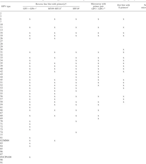
TABLE 4. Overview of genital HPV types detected in several published broad HPV detection and typing assays
HPV type Reverse line blot with primer(s)
a: Microarray with
primer pair
GP5⫹–GP6⫹b
Dot blot with
E-primersc microarrayNovel d
GP5⫹–GP6⫹e MY09–MY11f SPF10g
2
x
3
x
6
x
x
x
x
x
x
7
x
10
x
11
x
x
x
x
x
x
13
x
16
x
x
x
x
x
x
18
x
x
x
x
x
x
26
x
x
x
27
x
28
x
29
x
30
x
x
31
x
x
x
x
x
x
32
x
33
x
x
x
x
x
x
34
x
x
x
x
x
35
x
x
x
x
x
x
39
x
x
x
x
x
x
40
x
x
x
x
x
x
42
x
x
x
x
x
x
43
x
x
x
44
x
x
x
x
45
x
x
x
x
x
x
51
x
x
x
x
x
x
52
x
x
x
x
x
x
53
x
x
x
x
x
54
x
x
x
x
55
x
x
x
56
x
x
x
x
x
x
57
x
x
x
x
58
x
x
x
x
x
x
59
x
x
x
x
x
61
x
x
66
x
x
x
x
x
67
x
68
x
x
x
x
x
69
x
x
70
x
x
x
71
x
x
72
x
x
73
x
x
74
x
x
77
x
81
x
82/MM4
x
x
x
83
x
x
84
x
x
85
x
86
x
87
x
89/CP6108
x
x
90
x
91
x
aTarget gene: L1.
bTarget gene: L1 (An et al. [2]).
cTarget gene: E1 5⬘end (Ylitalo et al. [29]).
dTarget gene: E1 3⬘end (the present study).
evan den Brule et al. (26)
fGravitt et al. (8).
gKleter et al. (14).
on May 15, 2020 by guest
http://jcm.asm.org/
low-risk clusters, based on DNA sequence homology, is in
excellent agreement with epidemiological data (with the sole
exception of HPV70). Based on DNA sequence homology, 25
HPV types with unknown risk may putatively be grouped
ei-ther into the low-risk HPV clusters (HPV2, -3, -7, -10, -13, -27,
-28, -29, -32, -55, -57, -74, -77, -83, -84, -86, -87, -89, -90, and
HPV91) or high-risk clusters (HPV30, -34, -67, -69, and -85)
(Table 1). Although this homology-based classification seems
logical, it remains speculative until verified by epidemiological
data. Inclusion of these HPV types in novel broad HPV assays
in which all of these HPV types can be detected, like the novel
microarray assay described here, may provide such
epidemio-logical data.
In a small number of cases, no discrimination could be made
between closely related HPV types with a single probe. In our
experience, a minimum of a two-nucleotide mismatch in
20-mer hybridization probes was necessary for efficient
discrimi-nation between highly homologous HPV types. However, in
most of these cases, discrimination between highly homologous
HPV types can easily be established by using multiple probes in
such a way that a specific combination of responding probes
can be used to identify a specific HPV type. Although this may
obscure certain specific combinations of multiple HPV
infec-tions, it seems a plausible way to deal with this. With an
ever-expanding number of HPV types being reported, it seems
almost inevitable, with any assay that has been developed so
far, to encounter such a limitation. This may be particularly
relevant for assays in which only a small amplicon is produced,
as with the SPF10 assay (14). Although targeting only a small
PCR amplicon may have certain advantages at the DNA
am-plification level, in the SPF10 assay an interprimer region of
only 22 bp is available for making the discrimination between
different HPV types. With this assay, multiple pairs of different
HPV types differ by only 1 bp making it a challenge to reliably
discriminate between these HPV types at the DNA
hybridiza-tion level. In our novel assay, where the amplicon size varies
between 112 and 258 bp, there is enough sequence variation to
be able to discriminate between almost all HPV types.
We have limited ourselves here to the development of a
novel assay for the detection and subtyping of mucosal genital
HPV types. A total of 53 mucosal HPV types could be detected
and identified, including 16 HPV types that are not included in
many other previously reported assays (Table 4). Several of
these additional HPV types were already detected in our
clin-ical sample collection (e.g., HPV2, -27, -32, -67, and -85),
giving a clear demonstration of the expanded applicability of
this assay.
In conclusion, the microarray assay described here takes
advantage of a straightforward approach for selection and
de-sign of PCR primers and probes to be used for detection and
subtyping of HPV. The experimental setup, with a minimal
request for expensive specialty equipment and the simple and
affordable detection chemistry used, makes this approach
ac-cessible to all diagnostic laboratories with PCR facilities.
REFERENCES
1. Altschul, S. F., T. L. Madden, A. A. Schaffer, J. Zhang, Z. Zhang, W. Miller, and D. J. Lipman.1997. Gapped BLAST and PSI-BLAST: a new generation
of protein database search programs. Nucleic Acids Res.25:3389–3402.
2. An, H. J., N. H. Cho, S. Y. Lee, I. H. Kim, C. Lee, S. J. Kim, M. S. Mun, S. H. Kim, and J. K. Jeong.2003. Correlation of cervical carcinoma and
precan-cerous lesions with human papillomavirus (HPV) genotypes detected with
the HPV DNA chip microarray method. Cancer97:1672–1680.
3. Cho, N. H., H. J. An, J. K. Jeong, S. Kang, J. W. Kim, Y. T. Kim, and T. K. Park.2003. Genotyping of 22 human papillomavirus types by DNA chip in Korean women: comparison with cytologic diagnosis. Am. J. Obstet.
Gy-necol.188:56–62.
4. Coutlee, F., P. Gravitt, J. Kornegay, C. Hankins, H. Richardson, N. Lapointe, H. Voyer, and E. Franco.2002. Use of PGMY primers in L1 consensus PCR improves detection of human papillomavirus DNA in genital
samples. J. Clin. Microbiol.40:902–907.
5. de Roda Husman, A. M., J. M. Walboomers, A. J. van den Brule, C. J. Meijer, and P. J. Snijders.1995. The use of general primers GP5 and GP6
elongated at their 3⬘ends with adjacent highly conserved sequences
im-proves human papillomavirus detection by PCR. J. Gen. Virol.76(Pt. 4):
1057–1062.
6. Gharizadeh, B., M. Kalantari, C. A. Garcia, B. Johansson, and P. Nyren.
2001. Typing of human papillomavirus by pyrosequencing. Lab. Investig.
81:673–679.
7. Gravitt, P. E., C. L. Peyton, T. Q. Alessi, C. M. Wheeler, F. Coutlee, A. Hildesheim, M. H. Schiffman, D. R. Scott, and R. J. Apple.2000. Improved
amplification of genital human papillomaviruses. J. Clin. Microbiol.38:357–
361.
8. Gravitt, P. E., C. L. Peyton, R. J. Apple, and C. M. Wheeler.1998. Geno-typing of 27 human papillomavirus types by using L1 consensus PCR prod-ucts by a single-hybridization, reverse line blot detection method. J. Clin.
Microbiol.36:3020–3027.
9. Hwang, T. S., J. K. Jeong, M. Park, H. S. Han, H. K. Choi, and T. S. Park.
2003. Detection and typing of HPV genotypes in various cervical lesions by
HPV oligonucleotide microarray. Gynecol. Oncol.90:51–56.
10. Jacobs, M. V., A. M. de Roda Husman, A. J. van den Brule, P. J. Snijders, C. J. Meijer, and J. M. Walboomers.1995. Group-specific differentiation between high- and low-risk human papillomavirus genotypes by general primer-mediated PCR and two cocktails of oligonucleotide probes. J. Clin.
Microbiol.33:901–905.
11. Jacobs, M. V., P. J. Snijders, A. J. van den Brule, T. J. Helmerhorst, C. J. Meijer, and J. M. Walboomers.1997. A general primer GP5⫹/GP6⫹ -me-diated PCR-enzyme immunoassay method for rapid detection of 14 high-risk and 6 low-risk human papillomavirus genotypes in cervical scrapings. J. Clin.
Microbiol.35:791–795.
12. Karlsen, F., M. Kalantari, A. Jenkins, E. Pettersen, G. Kristensen, R. Holm, B. Johansson, and B. Hagmar.1996. Use of multiple PCR primer sets for
optimal detection of human papillomavirus. J. Clin. Microbiol.34:2095–
2100.
13. Kim, C. J., J. K. Jeong, M. Park, T. S. Park, T. C. Park, S. E. Namkoong, and J. S. Park.2003. HPV oligonucleotide microarray-based detection of HPV
genotypes in cervical neoplastic lesions. Gynecol. Oncol.89:210–217.
14. Kleter, B., L.-J. van Doorn, L. Schrauwen, A. Molijn, S. Sastrowijoto, J. ter Schegget, J. Lindeman, B. ter Harmsel, M. Burger, and W. Quint.1999. Development and clinical evaluation of a highly sensitive PCR-reverse hy-bridization line probe assay for detection and identification of anogenital
human papillomavirus. J. Clin. Microbiol.37:2508–2517.
15. Kleter, B., L.-J. van Doorn, J. ter Schegget, L. Schrauwen, K. van Krimpen, M. Burger, B. ter Harmsel, and W. Quint.1998. Novel short-fragment PCR assay for highly sensitive broad-spectrum detection of anogenital human
papillomaviruses. Am. J. Pathol.153:1731–1739.
16. Longuet, M., S. Beaudenon, and G. Orth.1996. Two novel genital human papillomavirus (HPV) types, HPV68 and HPV70, related to the potentially
oncogenic HPV39. J. Clin. Microbiol.34:738–744.
17. Lorincz, A. T.1992. Diagnosis of human papillomavirus infection by the new
generation of molecular DNA assays. Clin. Immunol. Newsl.12:123–128.
18. Manos, M. M., T. Ting, D. K. Wright, A. J. Lewis, T. R. Broker, and S. M. Wolinsky.1989. Use of polymerase chain reaction amplification for the
detection of genital human papillomaviruses. Cancer Cells7:209–214.
19. Munoz, N., F. X. Bosch, S. de Sanjose, R. Herrero, X. Castellsague, K. V. Shah, P. J. F. Snijders, C. J. L. M. Meijer, et al.2003. Epidemiologic classification of human papillomavirus types associated with cervical cancer.
N. Engl. J. Med.348:518–527.
20. Nelson, J. H., G. A. Hawkins, K. Edlund, M. Evander, L. Kjellberg, G. Wadell, J. Dillner, T. Gerasimova, A. L. Coker, L. Pirisi, D. Petereit, and P. F. Lambert.2000. A novel and rapid PCR-based method for genotyping
human papillomaviruses in clinical samples. J. Clin. Microbiol.38:688–695.
21. Prinsen, C. F. M., C. H. W. Klaassen, and F. B. J. M. Thunnissen.2003. Microarray as a model for quantitative visualization chemistry. Appl.
Immu-nohistochem. Mol. Morphol.11:168–173.
22. Qu, W., G. Jiang, Y. Cruz, C. J. Chang, G. Y. Ho, R. S. Klein, and R. D. Burk.
1997. PCR detection of human papillomavirus: comparison between MY09/
MY11 and GP5⫹/GP6⫹primer systems. J. Clin. Microbiol.35:1304–1310.
23. Sambrook, J., E. F. Fritsch, and T. Maniatis.1989. Molecular cloning: a laboratory manual, 2nd ed. Cold Spring Harbor Laboratory, Cold Spring Harbor, N.Y.
24. Thompson, J. D., T. J. Gibson, F. Plewniak, F. Jeanmougin, and D. G. Higgins.1997. The CLUSTAL X windows interface: flexible strategies for
on May 15, 2020 by guest
http://jcm.asm.org/
multiple sequence alignment aided by quality analysis tools. Nucleic Acids
Res.25:4876–4882.
25. Unger, E. R.2000. In situ diagnosis of human papillomaviruses. Clin. Lab.
Med.20:289–301.
26. van den Brule, A. J. C., R. Pol, N. Fransen-Daalmeijer, L. M. Schouls, C. J. L. M. Meijer, and P. J. F. Snijders.2002. GP5⫹/6⫹PCR followed by reverse line blot analysis enables rapid and high-throughput
identifi-cation of human papillomavirus genotypes. J. Clin. Microbiol.40:779–
787.
27. van Doorn, L.-J., W. Quint, B. Kleter, A. Molijn, B. Colau, M.-T. Martin,
Kravang-In, N. Torrez-Martinez, C. L. Peyton, and C. M. Wheeler.2002. Genotyping of human papillomavirus in liquid cytology cervical specimens by the PGMY line blot assay and the SPF10 line probe assay. J. Clin. Microbiol.
40:979–983.
28. Vernon, S. D., E. R. Unger, and D. Williams.2000. Comparison of human papillomavirus detection and typing by cycle sequencing, line blotting, and
hybrid capture. J. Clin. Microbiol.38:651–655.
29. Ylitalo, N., T. Bergstrom, and U. Gyllensten.1995. Detection of genital human papillomavirus by single-tube nested PCR and type-specific
oligonu-cleotide hybridization. J. Clin. Microbiol.33:1822–1828.