Georgia State University
ScholarWorks @ Georgia State University
Public Health Theses School of Public Health
Fall 1-5-2018
Relationship Between F2 – Isoprostanes and Adult
Metabolically Healthy Obesity
Tatiana Piccoli
Follow this and additional works at:https://scholarworks.gsu.edu/iph_theses
This Thesis is brought to you for free and open access by the School of Public Health at ScholarWorks @ Georgia State University. It has been accepted for inclusion in Public Health Theses by an authorized administrator of ScholarWorks @ Georgia State University. For more information, please contact [email protected].
Recommended Citation
Piccoli, Tatiana, "Relationship Between F2 – Isoprostanes and Adult Metabolically Healthy Obesity." Thesis, Georgia State University, 2018.
RELASHIONSHIPBETWEEN F2 – ISOPROSTANES AND ADULT METABOLICALLY HEALTHY OBESITY
By
Tatiana Victoria Piccoli 2017
Introduction: Obesity-related morbidity continues to increase worldwide. Obesity is one of the primary conditions for the development of metabolic syndrome which is characterized by a
combination of different components, including metabolic, physiological, and biochemical
factors that influence the development of cardiovascular disease, diabetes mellitus type two and
contribute to all-cause mortality. The development of metabolic abnormalities is related to
oxidative stress which increases among obese individuals. The obese population is not
homogenous and represented by individuals with altered and with a normal metabolic profile,
those with the normal metabolic profile are recognized as Metabolically Healthy Obese (MHO).
The lipid peroxidation is the main hallmark of oxidative stress and this process can be evaluated
via the measurement of the end products such as F2 -Isoprostanes.
Aim: The goal of this study was to investigate the relationship between metabolically healthy
and unhealthy obesity with markers of oxidative stress (urinary F2 -Isoprostanes) and assess
whether free radical-induced oxidative stress influences transition from metabolically healthy to
the metabolically unhealthy group. Metabolic health was defined as blood pressure below 130/85
mmHg, fasting blood glucose level as below 126 mg/dl and HDL-cholesterol above 40 mg/dl for
Method: The cohort of 857 participants included Hispanic, Hispanic black, and
non-Hispanic white from 40 to 69 years of age and had an obesity prevalence of 29% (n=244) at
baseline. Among this group, 11.07% (n=27) were metabolically healthy and 88.93% (n=217)
were metabolically unhealthy based on criteria for MHO. Among the MHO group, after 5-year
follow-up, 37% remained metabolically stable and 63% developed metabolic abnormalities and
among the MUO group, 5.6% became metabolically healthy and 94.4% remained metabolically
unhealthy. The association between different types of metabolic health and F2 -Isoprostanes
species was measured using Wilcoxon rank-sum test.
Result: The MUO status had a direct association with greater weight, Hispanic ethnicity,
impaired glucose tolerance, decreased insulin sensitivity, and decreased fasting insulin level. No
association was found between metabolic health status and levels of F2 -Isoprostanes at baseline
and follow-up.
Conclusion: The result suggests that elevated levels of F2 -Isoprostanes do not promote the
transformation of metabolically healthy obesity into metabolically unhealthy obesity.
Keywords: Obesity, metabolic health, metabolically healthy obesity, oxidative stress, F2
RELATIONSHIP BETWEEN F2 – ISOPROSTANES AND ADULT METABOLICALLY HEALTHY OBESITY
by
TATIANA VICTORIA PICCOLI
MD, GEORGIA STATE UNIVERSITY, GA 30303
A Thesis Submitted to the Graduate Faculty of Georgia State University in Partial Fulfillment of the Requirements for the Degree
MASTER OF PUBLIC HEALTH
ATLANTA, GA 30303
ii
APPROVAL PAGE
RELATIONSHIP BETWEEN F2 -ISOPROSTANES LEVEL AND ADULT METABOLICALLY HEALTHY OBESITY
by
TATIANA VICTORIA PICCOLI
Approved:
___Dr. Dora Il`yasova______ Committee Chair
__Dr. Emily Graybill_____ Committee Member
__12/01/2017_____________ Date
Acknowledgements
I wish to express my gratitude to my thesis chair, Dr. Il`yasova, who guided me through the research with patience and wise advice. My special thanks go to my committee member Dr. Graybill, I am very grateful for your time and recommendations.
iv Author`s Statement
In presenting this thesis as a partial fulfillment of the requirements for an advanced degree from Georgia State University, I agree that the Library of the University shall make it available for inspection and circulation in accordance with its regulations governing materials of this type. I agree that permission to quote from, to copy from, or to publish this thesis may be granted by the author or, in his/her absence, by the professor under whose direction it was written, or in his/her absence, by the Associate Dean, School of Public Health. Such quoting, copying, or publishing must be solely for scholarly purposes and will not involve any potential financial gain. It is understood that any copying from or publication of this dissertation which involves potential financial gain will not be allowed without written permission of the author.
Notice to Borrowers
All these deposited in the Georgia State University Library must be used in accordance with the stipulations described by the author in the preceding statement.
The author of the thesis is:
Tatiana Victoria Piccoli
3555 Monticello Commons Peachtree Corners, GA 30092
The chair of the committee for this thesis is:
Dora Il`yasova, PhD
Georgia State School of Public Health Diversion of Epidemiology & Biostatistics Georgia State University
P.O. Box 3995
Atlanta, GA 30302-3995
Users of this thesis who not regularly enrolled as a student at Georgia State University are required to attest acceptance of the preceding stipulation by signing below. Libraries borrowing this thesis for the use of their patrons are required to see that each user records here the
information requested.
NAME OF USER ADDRESS DATE TYPE OF USE
vi
TABLE OF CONTENTS TITLE PAGE………. i
APPROVAL PAGE………ii
ACKNOWLEDGEMENTS………. iii
LIST OF TABLES………...viii
ABBREVIATIONS………...……….……ix
INTRODUCTION………...1
1.1Background………...1
1.2 Purpose of study………...3
1.3 Research question and hypothesis………...4
1.4 Thesis organization……….……...……… ….5
REVIEW OF THE LITERATURE………....….6
2.1 The relationship between metabolic syndrome and oxidative stress……….….6
2.2 F2 -Isoprostanes as markers of oxidative stress ……….….7
2.3 MHO definition and prevalence……….….9
2.4 Relationship between inflammatory markers and markers of oxidative stress and MHO status……….……….…10
METHODS………...13
3.1 Data source……….…13
3.3 Variable Measurement………...…………14
3.4 The MHO phenotype definition……….15
3.5 Statistical analysis………...16
RESULTS……….18
4.1 Description of the study sample………...18
4.2 Prevalence of metabolically healthy obesity among study population………20
4.3 Association between metabolically healthy obesity and F2 – Isoprostanes………22
DISCUSSION AND CONCLUSION………...……25
5.1 Discussion of Research Question...………...25
5.2 Study strengths and limitations………...27
5.3 Conclusion……….28
viii
LIST OF TABLES Table 1: Baseline characteristics of the entire IRAS nondiabetic cohort………...19
Table 2 Metabolically Healthy and Unhealthy Obesity status at baseline and follow-up……….….21
Table 3 Oxidative status of MHO and MUO……….……….…23
Table 4 Oxidative status at follow-up among MHO stable and MHO declined……….…24
LIST OF FIGURES
Figure 1. Changes in metabolic status among MHO and MUO population.
ABBREVIATIONS
BMI Body Mass Index
CVD Cardiovascular Disease
DM Diabetes Mellitus
F2 -IsoPs F2 - Isoprostanes
HDL High Density Lipoprotein
HOMA Homeostasis Model Assessment
HOMA-IR Homeostasis Model Assessment for Insulin Resistance
IGT Impaired Glucose Tolerance
IL Interleukin
IRAS Insulin Resistance and Atherosclerosis Study
LDL Low-Density Lipoprotein
MetS Metabolic Syndrome
MHO Metabolically Healthy Obesity
MUO Metabolically Unhealthy Obesity
NCEP/ATPΙΙΙ National Cholesterol Education Program Adult Treatment Panel ΙΙΙ
NHANS National Health and Nutrition Examination Survey
ox-LDL Oxidized Low-Density Lipoprotein
P-value Probability value (observed significant level)
ROS Reactive Oxygen Species
1 CHAPTER Ι INTRODUCTION
1.1 Background
Increase in obesity prevalence worldwide contributes to the growth of obesity-related
morbidity (Landsberg et al., 2013). The study conducted by Finucane et al. (2011) investigated
the obesity increase in 199 countries from 1980 to 2008. The result of the study demonstrated
that 502 million adults had BMI above or equal to 30 kg/m 2 that allowed qualifying them as
obese. Obesity is one of the primary conditions for the development of metabolic syndrome
(Montague and O'rahilly, 2000, Furukawa et al., 2017), which is characterized by a combination
of different components, including metabolic, physiological, and biochemical factors that
influence the development of cardiovascular disease, diabetes mellitus type two and contribute to
all-cause mortality (Kaur, 2014). The mechanism of development of metabolic syndrome is
closely related to systemic oxidative stress (Keaney et al., 2003), which is more prominent
among the obese population due to the tendency of adipose tissue to produce reactive oxygen
species (ROS) in higher amount compared with other tissues (Furukawa et al., 2017). On one
hand, the obesity has an association with diabetes, hypertension, cancer and other numerous
health conditions, hence contributing to increase of the healthcare expenditure (Allen, Thorpe,
and Joski,2015). On another hand, the health programs targeting this global health problem
showed very little success on a population level (Ng et.al., 2014). Additionally, the expenditure
of community-based programs targeting obesity can vary due to the kind of approach and variety
medical spending on obese individual $1,429 higher per year compared with a person of normal
weight
Nevertheless, the obese population is heterogeneous and consists of individuals with a
variety of degree of obesity and metabolic abnormalities (Blüher,2014). Considering variability
of individuals representing the obese population, it would be beneficial to allocate this
population into groups according to their degree of metabolic abnormalities for being able to
address their needs more specifically and cost-efficiently. Among the obese population, we can
identify a group of individuals that do not have obesity-related comorbidities and do not
demonstrate any evidence of metabolic abnormalities, including insulin sensitivity and lipid
profile in the normal range and normal blood pressure, based on these criteria this subdivision is
identified as metabolically healthy obese (MHO) (Primeau et al., 2011). However, it is not clear
how healthy this group is and whether it progresses to metabolically unhealthy obesity (MUO).
The large body of literature supports the evidence about beneficial inflammation profile of MHO
individuals compared with metabolically unhealthy obese (MUO) population. Moreover,
comparing MHO population with the non-obese group, no association was found between the
MHO status and cardiovascular disease (CVD) prevalence and all-cause mortality (Calori et
al.,2011, Hamer and Stamatakis, 2012, Iacobellis et al.,2007, Karelis et al., 2005). However,
some researchers provide the evidence about the temporary condition of metabolic health among
obese individuals who with time will develop metabolic abnormalities (Appleton et al., 2013).
Additionally, some researchers found that although MHO population has not demonstrated any
signs of metabolic abnormalities, their metabolic profile was inferior to the profile of non-obese
individuals, including a lower level of HDL-cholesterol and higher non-HDL cholesterol (Manu
3
changes, such as early atherosclerosis (Oflaz et al.,2003). Based on contradicting results of
several studies Denis and Obin (2013) suggested that MHO group should be viewed as a “cluster
of traits” instead of as a category that can have a prognostic importance. Similarly, Plourde and
Korelis (2014) questioned whether MHO is a permanent status or just a stage in the development
of metabolic abnormalities accompanying obesity. Reviewing several longitudinal studies with
controversial results, the authors concluded that future metabolic health of individuals belonging
to MHO population cannot be guaranteed.
A level of influence of systemic oxidative stress in developing metabolic abnormalities
among metabolically healthy obese can be assessed by measuring the association between the
products of oxidative stress and MHO status. The lipid peroxidation is the main hallmark of
oxidative stress, where free radicals activate the process. The degree of lipid peroxidation can be
evaluated via the measurement of the end products of this process (Milne et al., 2005). The group
of the secondary end products of lipid peroxidation includes prostaglandin-like products called
F2 – Isoprostanes (F2 -IsoPs) that represent the result of peroxidation of arachidonic acid induced
by free radicals. The F2 -Isoprostanes species demonstrated a higher accuracy of the reflection of
oxidative stress compared to other markers (Fam and Morrow, 2003).
1.2 Purpose of the study
The rich body of literature investigated the role of different lifestyle factors, such as
physical activity and diet on metabolic health (Phillips et al., 2013), the association between a
proinflammatory profile and metabolic status (Plourde and Korelis,2014), and F2 -Isoprostanes as
the markers of oxidative status (Morrow et al., 1995, Il`yasova et al., 2015). To the best of our
association between different types of metabolic status of the overweight/obese population and
the 8-epi-prostaglandins F2α, that represent the most common form of F2 -Isoprostanes (Kim et
al., 2013). But no study has been conducted to evaluate the association between four types of F2
– Isoprostanes and metabolically healthy obesity (MHO).
The purpose of this study was to explore the relationship between the different metabolic
profiles and level of four different forms of F2 - Isoprostanes among the obese population, in a
prospective cohort, and to determine whether the level of F2 -Isoprostanes can predict the
transformation from MHO into MUO. Additionally, to determine what health characteristics
determining metabolic health have a correlation with the level of F2 -Isoprostanes.
1.3 Research question and hypothesis
1). Is there a cross-sectional association between concentration of F2-Isoprostanes and
Metabolically Unhealthy Obesity (MUO) status?
Compared to Metabolically Unhealthy Obese (MUO) population the MHO population
shows a lower level of F2 -Isoprostanes
2). Is there an association between level of F2 -Isoprostanes and transition from MHO to
MUO?
Compared to MHO population that continue to stay MHO after 5 years of follow-up the
5 1.4 Thesis Organization
This thesis is presented in five chapters. The first chapter contains an introduction with the
description of the background information, purpose of the study, research questions and
hypothesis. The literature review is presented in chapter two. The methods, sample, and
measures described in chapter three. Next chapter presents the result of the study. The last
chapter contents discussion, recommendations, strength and limitation of the study.
CHAPTER ΙΙ
LITERATURE REVIEW
2.1 The relationship between metabolic syndrome and oxidative stress
The increasing prevalence of metabolic syndrome (MetS) worldwide and the related
increase in CVD and diabetes type two prevalence became one of the main concerns of public
health (Ceriello and Motz, 2004, Sjorgen, 2005). The changes in lifestyle, such as overnutrition
and lack of physical activity, contribute to the development of the bundle of pathophysiological
divergences that incorporate dyslipidemia, insulin resistance, high blood pressure, and impaired
glucose tolerance. Additionally, all these changes are frequently associated with obesity (Eckel et
al., 2005, Kahn et al.,2006, Xu et al.,2012). The mechanism of the development of metabolic
syndrome is not completely understood (Bonomini et al., 2015). Even though obesity is one of
the main drivers of MetS, there is a population of normal weight individuals with MetS
(Ruderman et al., 1998, St-Onge et al., 2004). The study conducted by Park et al. (2003)
evaluated the prevalence of metabolic syndrome among a multiethnic sample from the third
National Health and Nutrition Examination Survey (NHANS) demonstrating that 4.6% of normal
weight participants had metabolic syndrome with obese individuals showing a higher prevalence
of metabolic syndrome compared to non-obese (59.6% vs 4.6%.) Throughout the last years, “metabolically triggered inflammation” is recognized as a characteristic of obesity and a
contributor to the development of metabolic syndrome (Hotamisligil, 2006). Similarly, the
development of metabolic abnormalities that lead to dyslipidemia (Zelzer et al.,2011),
hypertension (Russo et al., 1998) and diabetes ((Keaney et al.,2003) is influenced by oxidative
7
species (ROS) and capacity of antioxidants (Roberts and Sindhu, 2009) that results in increased
lipid peroxidation and damage to cellular structures (Yakes and Van Houten, 1997). The
evidence about the relationship of metabolic syndrome and oxidative stress is supported by the
decreased level of antioxidants among patients with MetS (Armutcu et al., 2008). Palmieri and
Sblendorio 2006, showed the increased frequency of metabolic syndrome among
postmenopausal women and thought to be due to the elevated free fatty acids (FFA) level and
subsequent oxidative stress. Numerous studies demonstrated a positive association between
metabolic status and markers of oxidative stress (Guilder et al., 2006, Hansel et al., 2004,
Holvoet et al.,2004), confirming the hypothesis about a vital role of oxidative stress in the
development of metabolic syndrome.
2.2 F2 -Isoprostanes as markers of oxidative stress
Throughoutthe last decades, the group of prostaglandin-like structures called F2 –
Isoprostanes became a well-recognized tool for assessing oxidative status (Basu, 2008,Il'yasova
et al.,2012). Roberts and Milne (2009) stated that due to the stability of molecules of F2 -
Isoprostanes, their level the most accurately reflects the grade of oxidative injury in vivo. The
circulation of free F2 – Isoprostanes in plasma and excretion with the urine gives an opportunity
to evaluate oxidative status in humans and animals (Li et al.,1999, Roberts and Morrow,2000).
Moreover, the quantification of the level of F2 – Isoprostanes in body fluids that reflect normal
oxidative status allowed identifying the condition when lipid peroxidation exceeds the capacity
of antioxidant defense and is recognized as oxidative stress (Milne et al.,2005). Analyzing the
result of the study investigating the relationship between the level of F2 - Isoprostanes and
hypercholesterolemia, Milne et al. (2005) concluded that this positive association is likely due to
found among patients with diabetes. Comparing groups of patients with and without diabetes,
Gopaul et al. (1995) showed that the group with diabetes had 3.3- fold elevation in F2 –
Isoprostanes level compared to the nondiabetic group. Another confirmation about the elevation
of F2 – Isoprostanes during oxidative stress was provided by Morrow et al. (1995) showing the
increased level of F2 – Isoprostanes among smokers compared to nonsmokers. The authors
concluded that the negative effect of smoking can be attributed to oxidative injury caused by
toxic products inhaled during the smoking process. An article by Milne et al. (2015) provided an
overview in the field of F2 – Isoprostanes research for 25 years since the discovery of F2 –
Isoprostanes by Morrow and Roberts. Since that time numerous studies have been conducted
with the purpose of exploring the possibilities of using these compounds as biomarkers for
illnesses. Several studies investigated the association between toxic agents causing oxidative
damage and level of F2 – Isoprostanes as markers of this injury and reported a positive
relationship between these variables (Il'yasova et al.,2010, Kadiiska et al., 2005). Another study
examining the levels of F2 – Isoprostanes among the population of Inuit, showed an elevated
level of these markers among the participants with metabolic syndrome compared to individuals
with normal metabolic health (Alkazemi et al.,2012). A similar result was presented by the study
conducted by Black et al. 2016, showing a positive association between F2 – Isoprostanes and all
constituents of metabolic syndrome including systolic blood pressure, triglycerides level, waist
circumference, and LDL- level, but also surprisingly showing a positive correlation with HDL.
Although the research conducted by Melton et al., (2017), did not show any association between
F2 – Isoprostanes index and hypertension. The authors concluded that free radical-induced
oxidative stress did not play an important role in the development of hypertension.
9 2.3 MHO definition and prevalence
Despite the longtime existing knowledge of such phenomenon as metabolically healthy obesity (MHO), there is still no common adopted definition of MHO. Similar to variability in the
definition of metabolic syndrome (Borch-Johnsen, 2007) variety of sets of criteria are used to
define MHO. Presently, about 15 different sets of criteria have been used to describe MHO status
(Plourde and Karelis, 2013). Many researchers agree to use lipid profile, glycemic status, and
blood pressure as criteria to define metabolic health among the obese population, others claim
that it is important to use the additional components (Velho et al., 2010). Based on different
criteria, the prevalence of MHO among obese individuals varied from 6.8% to 36.6% (Phillips et
al., 2013). Analyzing the results of several studies, Phillips (2013) stressed attention to MHO
prevalence inconsistency across the studies that partially can be attributed to geographic, ethnic,
age, and sample size differences, but mainly due to differences in criteria used for the definition
of MHO. Furthermore, an even more significant variability of MHO prevalence from 6% to 75%
in different studies was presented by Rey-Lopez et al. (2014). Although most studies
incorporated the basic criteria of metabolic health such as the level of HDL-cholesterol, results
of blood pressure measurements, blood glucose level, and triglycerides, also many of them used
additional criteria, such as waist circumference, level of LDL-cholesterol, and others. The usage
of these extra criteria can explain a variability in MHO prevalence (Rey-Lopez et al., 2014).
Similarly, the result of the study conducted by Velho et al. (2010) showed that MHO prevalence
varied from 3.3% to 32.1% among men and from 11.4% to 43.3% among women, depends on
criteria used. This study investigated the prevalence of MHO among the same group of
MHO prevalence was based on differences in used criteria. However, despite the utilization of a
variety of criteria for MHO definition, many studies found a positive association between sex
and MHO status, with women showing a higher prevalence of MHO, and age, with younger age
being healthier (Rey-Lopez et al., 2014).
2.4Relationship between inflammatory markers and markers of oxidative stress and MHO
status.
Reviewing several studies Vincent and Taylor (2006) admitted the positive relationship
between obesity and oxidative stress. Although the association of obesity and F2 - Isoprostanes as
markers of oxidative stress has been demonstrated in several studies (Il`yasova et al.,2005,
Keaney et al., 2003, Stojiljkovic et al.,2002, Wu et al. 2009), literature on evaluation of oxidative
stress and specifically level of F2 - Isoprostanes among MHO population is limited.The results
of prospective cohort study examining the relationship between MHO, MUO, markers of
oxidative stress and development of cognitive impairment was presented by Farah et al. (2016).
Researchers found the positive association between oxidative stress, mild cognitive deterioration
and MUO status. This study did not use F2 – Isoprostanes as markers of oxidative stress. Another
study conducted by Bañuls et al. (2017) investigated the relationship between markers of
oxidative stress, endoplasmic reticulum stress and status of metabolic health among the obese
population. The result of the study showed a higher level of proinflammatory cytokines, such as
IL-6 among MUO compared to MHO. Moreover, the production of ROS was lower among
MHO compared to MUO, demonstrating a more significant level of oxidative stress among
MUO population. Similarly, this study did not utilize F2 – Isoprostanes as markers of oxidative
11
and status of metabolic health among postmenopausal women was presented by Kim et al.
(2013). The group of 1846 individuals was divided into four groups according to their body mass index (BMI), and metabolic health status. Participants with BMI≥25 kg/m2 considered to be
overweight/obese and were divided into two groups based on the criteria for metabolic health
defined by the modified National Cholesterol Education Program Adult Treatment Panel ΙΙΙ (NCEP/ATPΙΙΙ). All participants with BMI<25 kg/m2 were also divided into two groups with
MetS and without MetS. For evaluating oxidative stress, researchers measured the level of
inflammatory markers such as circulating oxidized LDL (ox-LDL) and 8-epi-prostaglandins F2α
(8-epi-PGF2α). The design of the study was cross-sectional, allowing to see the association
between the variables but not the causation. The result of the study demonstrated that
participants from overweight/obese group had less favorable metabolic profile compared to the
normal weight group regardless of their metabolic health. Additionally, comparing subgroups
with MetS and without of MetS, investigators saw a better metabolic profile among individuals
without MetS regardless of their BMI. Overweight/obese individuals without MetS demonstrated
a higher level of ox-LDL compared to the normal weight group without MetS, but lower than
among the group with MetS. The level of 8-epi-PRG2α was higher among participants with MetS
compared to women without MetS. Researchers concluded that postmenopausal women with
MetS with normal weight demonstrated a higher level of oxidative stress compared to
overweight/obese with normal metabolic health. Additionally, between the markers of oxidative
stress, no association was found that draw a conclusion about representation by these markers
different stages of oxidative changes. The result of this study contradicts the result of the
group was presented by individuals without any risk factors for MetS, another with 1-2 risk
factors, and the third group by participants with MetS. Examining the relationship between
markers of oxidative stress, such as ox-LDL and 8-iso-PGF2α with the metabolic status of
13
CHAPTER ΙΙΙ
METHODOLOGY
3.1 Data source
The collected data from Insulin Resistance Atherosclerosis Study (IRAS) became the source of our secondary data analysis. This study was the first epidemiological study intended to
evaluate the association between constituents of insulin resistance syndrome, CVD, and other
risk factors (Wagenknecht et al., 1995). The study began in 1992 with a follow-up period of
approximately five years and was approved by the Institutional Review Board of Wake Forest
University School of Medicine (Melton at al., 2017)
3.2 Sample selection and participant observation
The study aimed to observe multiethnic cohort that was achieved with the recruitment of 1626 participants representing the Hispanic population (n=548), Non-Hispanic black (n=464),
and Non-Hispanic white (n=614). Female population represented 56% of the sample. The
participants were from 40 to 69 years of age. Aiming to have equal representation of individuals
with a different level of glucose tolerance, the study oversampled nondiabetic individuals with an
elevated fasting plasma glucose level and those who were previously identified as having
impaired glucose tolerance (IGT). The participants were recruited at four clinical centers located
in Los Angeles, California; Oakland, California; San Luis Valley, Colorado; and San Antonio,
Texas (Wagenknecht et al., 1995). Each participant gave written informed consent. The complete
examination of each participant at the baseline was achieved during two visits, each lasted
approximately four hours, with the variation of the interval between the appointments from one
to thirty days. During these appointments, the participants were evaluated for glucose tolerance
using a 75g glucose load and insulin resistance using an insulin injection. CVD and peripheral
vascular disease were assessed using an interview, performed in their preferred language, and
noninvasive testing, such as blood pressure measurement, electrocardiography, and
ultrasonography. Additionally, the participants completed a questionnaire self-reporting their
race/ethnicity, smoking, alcohol intake, physical activity, and nutrient intake. Anthropometric
measurements were performed by trained medical personnel. Moreover, for examining F2 –
Isoprostanes level 901 enrollees provided a urine sample at the baseline. The examination of the
participants began in October 1992 and was completed in April 1994. After approximately five
years of the follow-up period, in 1997-1998, the study`s participants were examined again
according to the protocol of the study. The participants free of diabetes mellitus (DM) type two
at the baseline were included in the study analysis, that reduced the sample to 1125 participants,
among those, 20 % were lost to follow-up.
3.3 Variable Measurement
World Health Organization criteria (WHO,1999) provided guidance for assessing glucose
tolerance. A blood sample was taken before administering 75g of glucose and two hours after.
Insulin resistance was measured after an injection of 50% glucose solution (0.3 g/kg) in 20 minutes followed by an injection of insulin (0.03 U/kg). During a three hours’ period, blood was
collected 12 times through another intravenous line and was evaluated for the concentration of
15
lipids, fasting blood glucose, and components of the biochemical profile. At the baseline
examination, morning urine samples were collected and kept at – 70o C until the analysis. A total
number of 901 samples of urine were collected, among those 857 samples were satisfyingly
measured for F2 – Isoprostanes concentration. The measurement of F2 – Isoprostanes was
performed utilizing liquid chromatography/tandem mass spectrometry, the result was calibrated
according to the urinary concentration of creatinine. Four different F2 – Isoprostanes isomers
were measured including iPF(2a)-ΙΙΙ, 2,3-dinor-iPF(2a)-ΙΙΙ, iPF(2a)-lV, 8,12-iso-iPF(2a)-lV. The
F2 -Isopostanes index was created based on the result for four F2 – Isoprostanes isomers and
allowed to rank participants based on this calculation [(X1i – M1)/SD1 +(X2i -M2)/SD2 +(X3i
-M3)/SD3 +(X4i -M4)/SD4)/4. In this formula, “i” is a code for a participant, X1-4 represent values of
four F2 – Isoprostanes isomers, M1-4 mean of these four isomers, and SD1-4 standard deviation
values. The body mass index, as a measurement of general adiposity, was calculated as a
relationship between body mass in kilograms divided by height in square meters.
3.4 The MHO phenotype definition
We identified two different groups of MHO based on two different definitions of this
phenomenon. One of the most commonly used definitions of MHO status included the absence
of diabetes, hypertension, and dyslipidemia (Muñoz-Garach et al.,2016). We used 126 mg/dl of
fasting blood glucose level as a cut-off for diabetes. Enrollees with fasting plasma glucose level
<126 mg/dl were considered nondiabetic. Individuals with systolic blood pressure equal or below
130 mmHg and diastolic blood pressure equal or below 85 mmHg and not taking blood pressure
lowering medication were considered normotensive. Lipid profile was represented by the level of
HDL-cholesterol, which was considered of normal value if its level was above or equal to
criteria for the first definition were united in the group named MHO1. Another definition of
MHO status was based on modified criteria suggested by Wildman et al. (2008). This group included individuals with normotensive status (BP ≤130/85 mmHg) who did not undergo
antihypertensive treatment. The absence of diabetes with fasting blood glucose level ≤ 100mg/dl
was another criterion. Moreover, for being included in this MHO group additional criteria had to
be met, such as homeostasis model assessment of insulin resistance (HOMA-IR) ≤ 5.1 and
triglycerides HDL-cholesterol ratio ≤ 1.65 for male and ≤1.32 for female. This group was named
MHO2.
3.5 Statistical analysis
Statistical analysis was performed using SAS statistical software (version 9.4: SAS Institute, Cary, NC). A total number of 857 nondiabetic participants were included in this
analysis at the baseline. The descriptive statistical analysis was applied to the baseline sample. The individuals with BMI ≥ 30kg/m2 were selected and the final sample was represented by 244
participants. At the follow-up visit, this sample reduced to 241 participants. The association
between categorical variables, including a crude association between age category, sex, ethnicity
and MHO1 and MHO2 groups, was measured using chi-square test. Univariate analysis was
perfumed to identify a median and interquartile range for continuous variables. We used
Wilcoxon rank-sum test to evaluate how different were continuous variables, including F2 –
Isoprostanes levels, between groups with MHO and MUO. We analyzed the differences between
MHO and MUO groups at the baseline and at the follow-up appointment. Additionally, at the
follow-up, we analyzed metabolic changes including how many MHO participants remained
17
health and evaluated their association with oxidative stress markers among the participants. A
p-value was considered statistically significant if it was less than 0.05
CHAPTER ΙV
RESULTS
4.1 Description of the study sample
The study population at the baseline included a slightly higher number of females (58%) compared to males (42%). Racial/ethnic diversity was represented by Hispanic (32%),
Non-Hispanic black (28%) and Non-Non-Hispanic white (40%). Among the age categories the highest
representation was in the group from 50 to 59 years of age (36%), groups from 40 to 49 years of
age and 60 to 69 years of age were represented equally 32% each. Almost half of the baseline
population never smoked (46%), former smokers represented 40% of the population, and current
smokers 14 %. Among the baseline population, 40% had hypertension and 60 % did not have
increased blood pressure and did not take hypertensive medication. Approximately two-thirds of
the baseline population (67.5%) belonged to the group with normal glucose tolerance and
one-third (32.5%) had impaired glucose tolerance. The baseline sample did not include participants with diabetes. Prevalence of obesity (BMI≥30kg/m2) was 29%, (n=244) of the baseline
19
[image:31.612.68.533.136.628.2]
Table 1. Baseline characteristics of the entire IRAS nondiabetic cohort.
Continuous variables N (missing values) Means (SD)
Age (years) 857 (0) 54.6 (8.315)
BMI (kg/m2 ) 855 (2) 28.5 (5.655)
Fasting Glucose (mg/dl) 857 (0) 98.6 (11.3)
Insulin sensitivity (SI, x10-4minutes-1/µUml)
795 (62) 2.20 (2.05)
Acute Insulin Response (microU/ml) 834 (23) 486.8 (494.3)
iPF(2a)-ΙΙΙ (ng/mg CN) 853 (4) 0.249 (0.194)
2,3-dinor-iPF(2a)-ΙΙΙ (ng/mg CN) 853 (4) 4.35 (2.10)
iPF(2a)-lV (ng/mg CN) 853 (4) 6.49 (4.16)
8,12-iso-iPF(2a)-lV (ng/mg CN) 853 (4) 4.15 (2.87)
F2 -IsoP Index* 853 (4) -0.001 (0.821)
2-hour Glucose (mg/dl) 857 (0) 124.3 (33.5)
HDL (mg/dl) 854 (3) 47.11 (15.17)
Triglycerides (mg/dl) 853 (4) 130.7 (81.37)
Fasting Insulin (uU/ml) 856 (1) 15.69 (15.13)
Categorical variables N (missing values) Percent
Gender 855 (2) 100
Males 363 42.46 Females 492 57.54
Race/ethnicity 855 (2) 100
Hispanic 276 32.28 Non-Hispanic black 237 27.72 Non-Hispanic white 342 40.0
Smoking status 855 (2) 100
Never smoked 397 46.43 Former smoker 338 39.53 Current smoker 120 14.04
Hypertension 855 (2) 100
Yes 339 39.65 No 516 60.35
Obesity 244 (0) 100
MUO 217 88.93
MHO 27 11.07
Glucose tolerance status 855 (2) 100
NGT 577 67.49 IGT 278 32.51
4.2 Prevalence of metabolically healthy obesity among study population
Among the obese population, 27 individuals (11.07%) met criteria for MHO1 and 3 participants
(1.23%) were recognized as metabolically healthy based on the criteria for MHO2 definition. All
participants who met criteria for MHO2 definition also met the criteria for MHO1 definition. At
follow-up, the sample from the obese population decreased (n=241) and included 22 participants
(9.1%) belonging to the metabolically healthy (MH) group and 219 participants (90.9%) were
metabolically unhealthy (MU). These 22 MHO individuals met the criteria for MHO1 definition.
Additionally, 10 participants (4.1%) met criteria for MHO2 definition at follow-up. All
participants who can be considered MHO2 also met the criteria for MHO1. Age category and
sex did not show a statistically significant association with MHO status at baseline and
follow-up. Smoking status did not demonstrate an association with MHO status at baseline, showing an
equal prevalence of MHO among former smokers and participant who never smoked (44.4%
each). At follow-up, this association increased with 45.4% of individuals who never smoked
representing the MHO group of which 36.4% were former smokers, current smokers represented
less than 20% of MHO population on both visits (P=.06). At baseline participants belonging to
the Hispanic group demonstrated the lowest prevalence of MHO (14.8%) and the highest
prevalence of MUO (37.3%) compared to two other racial/ethnic groups (P=.05). Although this
trend continued to the follow-up, it became less statistically significant (P=.08). As anticipated,
the MHO status was associated with glucose tolerance status, insulin sensitivity, and fasting
insulin. Although an association between MHO status and BMI was not statistically significant at
baseline, at follow-up there was a statistically significant association demonstrating higher
21
[image:33.612.60.582.129.720.2]
Table 2 Metabolically Healthy and Unhealthy Obesity status at baseline and follow-up.
Baseline N= 244 (percent) p-value Follow-up N=241 (percent) p-value
Categorical Variable* MHO
N=27 MUO N=217 MH N=22 MU N=219
Age 0.07 0.5
40-49 10 (37.04) 71 (32.72) 9 (40.91) 70 (31.96) 50-59 13 (48.15) 69 (31.80) 8 (36.36) 74 (33.79) 60-69 4 (14.81) 77 (35.48) 5 (22.73) 75 (34.25)
Gender 0.9 0.7
Male 9 (33.33) 70 (32.26) 8 (36.36) 71 (32.42) Female 18 (66.67) 147 (67.64) 14 (63.64) 148 (67.58)
Ethnicity 0.05 0.08
Hispanic 4 (14.81) 81 (37.33) 3 (13.64) 81 (36.99) Non-Hispanic black 12 (44.44) 60 (27.65) 8 (36.36) 64 (29.22) Non-Hispanic white 11 (40.74) 76 (35.02) 11 (50.00) 74 (33.79)
Smoking status 0.76 0.06
Never smoked 12 (44.4)
111 (51.2)
10 (45.4) 109 (50.7)
Former smoker 12 (44.4)
81 (37.3)
8 (36.4) 95 (44.2)
Current smoker 3 (11.2)
25 (11.5)
4 (18.2) 11 (5.1)
Hypertension N, %
N/A N/A
Yes 0 143 (65.90)
0 163 (74.43)
No 27 (100.00)
74 (34.10)
22 (100.00) 56 (25.57)
Glucose tolerance status
N, %
0.01 < .001
NGT 20 (74.06)
103 (47.47)
19 (86.36) 82 (37.44)
IGT 7 (25.94)
114 (52.53)
2 (9.09) 68 (31.05)
Type 2 diabetes N/A N/A 1 (4.55) 69 (31.51)
Continuous Variable Median ±
IQR
Median ± IQR
Median ± IQR Median ±
IQR BMI Kg/m2 33.08 (31.23-36.33) 34.27 (31.89- 37.705)
0.126 30.29
(29.02- 36.00) 35.45 (31.99- 38.53) 0.01 Fasting Glucose (mg/dl) 98.0 (91-104) 102.0 (95-111)
N/A 96.0
(90.0-104.5)
105.5 (95.5-121.5)
N/A
(SI, x10-4minutes-1/µUml) (1.28-2.62) (0.370-
1.54)
(0.96-2.50) (0.0-0.98)
Acute Insulin response (microU/ml) 565.0 (161.6- 710.8) 417.6 (137.4-790.4)
0.552 428.5
(354.8- 735.8) 346.2 (98.00- 847.1) 0.19 2-hour Glucose (mg/dl) 116 (95-141) 142 (120-167)
<.001 108.0 (89.0-122.5)
155.5
(124.5- 213.7)
< .001
HDL (mg/dl) 55.0
(51.0-57.0)
40.0 (33.0-46.5)
N/A 58.5 (51.0-65.0) 43.0 (35.0-51.0) N/A Triglycerides (mg/dl) 104.0 (74.0-183.0) 139.0 (90.0-182.0)
N/A 76.0
(59.0-127.0)
124.0 (86.0-179.0)
N/A
Fasting Insulin (uU/ml) 14.0
(11.0-21.0)
19.0 (14.0-26.0)
0.005 15.0 (11.0-18.0)
23.0 (16.0-33.0)
< .001
• Variables value correspond to each time point. All categorical variables reported in N, %, p-value for categorical variables assessed using Chi-square. All continuous variables reported in median ± IQR assessed using univariate test, p-value for continuous variables assessed using Wilcoxon test.
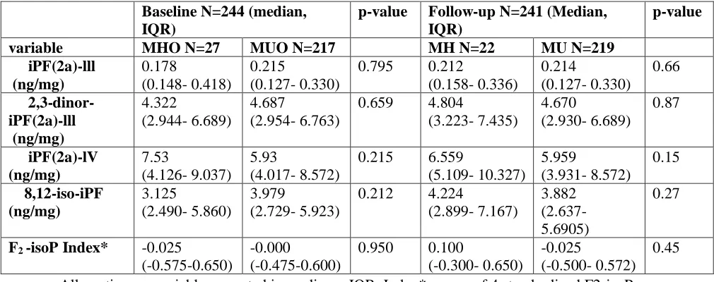
4.3 Association between metabolically healthy obesity and F2 – Isoprostanes
Each individual F2 – Isoprostanes isomer demonstrated the variability of distribution.
Examined association between MHO status and level of each isomer of F2 – Isoprostanes was not
statistically significant. Majority of F2- Isoprostanes species demonstrated an inverse association
with MHO status, such iPF (2a)-ΙΙΙ, 2,3-dinor – iPF (2a)-ΙΙΙ, 8,12-iso-iPF, and F2 -Isoprostanes
Index all were slightly higher among MUO compared with MHO group at baseline. Although
iPF(2a)-lV had a direct association with MHO status being higher among MHO (median=7.53
ng/mg) compared to MUO (median= 5.93 ng/mg). None of the associations were statistically
significant. Assessment of the association between levels of F2 – Isoprostanes and MHO status
on follow-up demonstrated an increase in the number of F2 – Isoprostanes having direct
associations with MHO status with only iPF(2a)-lll being slightly higher among MUO (0.214
23
The associations between F2 – Isoprostanes isomers and MHO status on follow-up were not
[image:35.612.66.571.156.356.2]statistically significant (Table 3).
Table 3 Oxidative status of MHO and MUO
Baseline N=244 (median, IQR)
p-value Follow-up N=241 (Median,
IQR)
p-value
variable MHO N=27 MUO N=217 MH N=22 MU N=219
iPF(2a)-lll (ng/mg) 0.178 (0.148- 0.418) 0.215 (0.127- 0.330)
0.795 0.212
(0.158- 0.336) 0.214 (0.127- 0.330) 0.66 2,3-dinor-iPF(2a)-lll (ng/mg) 4.322 (2.944- 6.689) 4.687 (2.954- 6.763)
0.659 4.804
(3.223- 7.435) 4.670 (2.930- 6.689) 0.87 iPF(2a)-lV (ng/mg) 7.53 (4.126- 9.037) 5.93 (4.017- 8.572)
0.215 6.559
(5.109- 10.327) 5.959 (3.931- 8.572) 0.15 8,12-iso-iPF (ng/mg) 3.125 (2.490- 5.860) 3.979 (2.729- 5.923)
0.212 4.224
(2.899- 7.167)
3.882 (2.637- 5.6905)
0.27
F2 -isoP Index* -0.025
(-0.575-0.650)
-0.000
(-0.475-0.600)
0.950 0.100
(-0.300- 0.650)
-0.025
(-0.500- 0.572) 0.45
• All continuous variables reported in median ± IQR. Index*- mean of 4 standardized F2-isoPs.
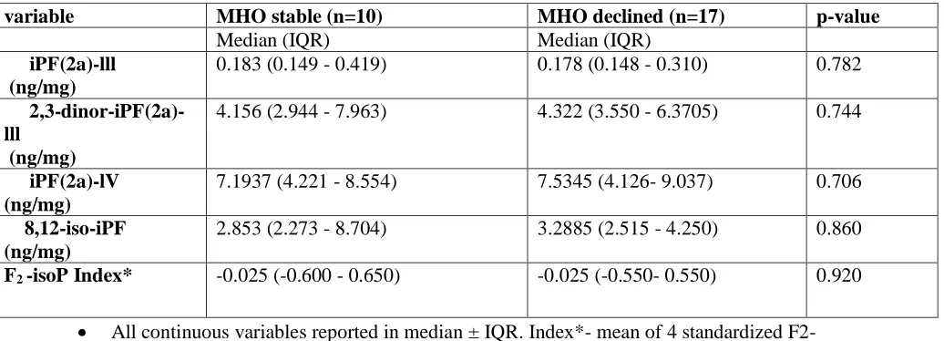
Comparison of baseline and follow-up MHO status showed that from 27 individuals
representing MHO at the baseline, 17 participants progressed into an unhealthy state and 10
participants remained stable metabolically healthy. Among those who were MUO (n=217) at
baseline, 12 individuals demonstrated metabolic improvement and moved into a group of
metabolically healthy and the rest of the group remained unhealthy. The association between
level of F2 – Isomers and metabolically stable state was not statistically significant with a median
of F2 -isoP index being of the same value (-0.025ng/mg) among MHO stable and metabolically
Table 4 Oxidative status at follow-up among MHO stable and MHO declined
variable MHO stable (n=10) MHO declined (n=17) p-value
Median (IQR) Median (IQR)
iPF(2a)-lll (ng/mg)
0.183 (0.149 - 0.419) 0.178 (0.148 - 0.310) 0.782
2,3-dinor-iPF(2a)-lll
(ng/mg)
4.156 (2.944 - 7.963) 4.322 (3.550 - 6.3705) 0.744
iPF(2a)-lV (ng/mg)
7.1937 (4.221 - 8.554) 7.5345 (4.126- 9.037) 0.706
8,12-iso-iPF (ng/mg)
2.853 (2.273 - 8.704) 3.2885 (2.515 - 4.250) 0.860
F2 -isoP Index* -0.025 (-0.600 - 0.650) -0.025 (-0.550- 0.550) 0.920
25
CHAPTER V
DISCUSSION
5.1 Discussion of Research Question
The purpose of the study was to examine whether increased oxidative status can promote a
transition from metabolically healthy obesity (MHO) into metabolically unhealthy obesity
(MUO). The increased mass of adipose tissue is characterized by the changed structure of
adipocytes, including their hypertrophy and hyperplasia that affect the property of adipocytes,
such as reaction to insulin due to decreased density of receptors for insulin (Fernández-Sánchez
et al., 2011). The possibility of adipose tissue to produce certain bioactive molecules, including
leptin and proinflammatory cytokines, such as IL-6 and tumor necrosis factor-alfa (TNF-α)
promotes higher oxidative and inflammatory status of this tissue (BouloumiÉ et al.,1999,
Perwez Hussain and Harris, 2007). This property of adipose tissue advances metabolic
misbalance, including changed lipid profile and altered glucose metabolism. What contributes
to a diversity of metabolic state among obese remained not well understood. Several studies
identified an association between age, gender and MHO status (van Vliet-Ostaptchouk et
al.,2014). Numerous conducted research provided conflicting data on the effect of lifestyle, diet, or
behavior on metabolic health (Phillips, 2013). Some researchers provided evidence about the
positive effect of physical exercise on metabolic health of the obese population (Phillips et al.,2013,
Velho et al., 2010). Additionally, it has been shown that MHO population has a more favorable
inflammatory profile compared to MUO (Phillips, 2013). We hypothesized that F2 -Isoprostanes
as markers of free radical oxidative stress can become a predictor of deteriorating metabolic
health among still metabolically healthy obese individuals. Based on the result of our study, we
did not identify any statistically significant association between level of F2 -Isoprostanes and
MUO compared to MHO. Neither did we find any statistically significant difference in F2
-Isoprostanes association between the population who were MHO stable after five years of
follow-up and those who progressed from MHO to MUO. The result of our study reflects the
result of the study conducted by Sjorgen et al. (2005), where no association was found between
markers of oxidative stress and different metabolic health status among obese men. The result of
our study can be partially attributed to the small sample of metabolically healthy individuals, 27
participants at baseline and 22 at follow-up and can be considered as one of the limitations of our
study. Another explanation for this result can be related to the criteria we used to identify MHO.
There are a variety of criteria used to define metabolic health among obese and the debates about
which combination of criteria is more correct are still ongoing (Phillips et al., 2013). The criteria
we used for MHO1 were very basic and included the absence of hypertension, diabetes and
healthy HDL-cholesterol level. It is possible to think that this MHO group already had
underlying metabolic changes that had not been manifested yet in the form of diabetes or
hypertension. Although criteria for MHO2 were stricter and incorporated HOMA,
triglycerides/HDL-cholesterol ratio and lower level of fasting glucose, such as 100mg/dl as
recommended by National Cholesterol Education Program Adult Treatment Panel ΙΙΙ
(NCEP/ATPΙΙΙ) (Lorenzo et al.,2007), the number of individuals who met these criteria was very
small for drawing any conclusion about the association between F2 -Isoprostanes level and
27
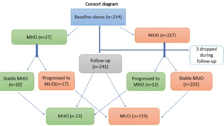
One of the main findings of the study was the identification of the changeable status of
metabolic health among the obese population. After 5years of the follow-up period, some of the
participants with initially healthy metabolic profile remained healthy and some developed
metabolic abnormalities. Similarly, the group of obese participants who were metabolically
unhealthy on the baseline demonstrated a bifurcation with some participants remained
[image:39.612.82.537.310.567.2]metabolically unhealthy and some demonstrated an improvement in metabolic health (Figure 1).
Figure 1. Changes in metabolic status among MHO and MUO populations
5.2 Study strengths and limitations
One of the strengths of this study is the utilization of prospective cohort data that
causation. Moreover, it provided an opportunity to see the trend of metabolic changes
among the obese population over a five-year period. Additionally, this study allowed to
examine the relationship between four F2 - Isoprostanes isomers and metabolic health of
the nondiabetic obese population in a multiethnic cohort.
One of the limitations of the study was the small sample of the obese population
(n=244) with a small group of MHO. Also, we did not analyze the changes in the diet,
exercises, and weight in this initially obese cohort group that could affect the transition
from one category of metabolic health into another, but it was not the purpose of this
study.
5.3 Conclusions and recommendations
Based on the result of our study we did not identify any causal relationship between
free radical oxidative stress and development of metabolic abnormalities among the
metabolically healthy obese adult population. The difference in the level of oxidative
stress between MHO and MUO was statistically insignificant and at the same time, both
groups of obese population demonstrated a higher level of oxidative stress compared to
non-obese. We can conclude that MHO population has invisible pathological processes
that in the future can manifest as metabolic abnormalities. Considering this result, we
recommend including MHO group in all programs targeting obesity that can provide
benefits to the health of obese population regardless of their metabolic profile. Future
research is needed to investigate what factors can trigger metabolic changes or prevent
29
REFERENCES
Alkazemi, D., Egeland, G. M., Roberts, L. J., & Kubow, S. (2012). Isoprostanes and isofurans as non-traditional risk factors for cardiovascular disease among Canadian Inuit. Free radical research, 46(10), 1258-1266.
Allen, L., Thorpe, K., & Joski, P. (2015). The effect of obesity and chronic conditions on medicare spending, 1987-2011. PharmacoEconomics, 33(7), 691-697
Appleton, S. L., Seaborn, C. J., Visvanathan, R., Hill, C. L., Gill, T. K., Taylor, A. W., ... & North West Adelaide Health Study Team. (2013). Diabetes and cardiovascular disease outcomes in the metabolically healthy obese phenotype. Diabetes care, 36(8), 2388-2394. Armutcu, F., Ataymen, M., Atmaca, H., & Gurel, A. (2008). Oxidative stress markers, C-reactive
protein and heat shock protein 70 levels in subjects with metabolic syndrome. Clinical chemistry and laboratory medicine, 46(6), 785-790.
Bañuls, C., Rovira-Llopis, S., Lopez-Domenech, S., Diaz-Morales, N., Blas-Garcia, A., Veses, S., ... & Hernandez-Mijares, A. (2017). Oxidative and endoplasmic reticulum stress is impaired in leukocytes from metabolically unhealthy vs healthy obese
individuals. International journal of obesity (2005).
Basu, S. (2008). F2-isoprostanes in human health and diseases: from molecular mechanisms to clinical implications. Antioxidants & redox signaling, 10(8), 1405-1434.
Black, C. N., Bot, M., Scheffer, P. G., & Penninx, B. W. (2016). Sociodemographic and lifestyle determinants of plasma oxidative stress markers 8-OHdG and F2-isoprostanes and associations with metabolic syndrome. Oxidative medicine and cellular longevity, 2016.
Blüher, M. (2014). MECHANISMS IN ENDOCRINOLOGY: Are metabolically healthy obese individuals really healthy? European journal of endocrinology, 171(6), R209-R219. Bonomini, F., Rodella, L. F., & Rezzani, R. (2015). Metabolic syndrome, aging and involvement
of oxidative stress. Aging and disease, 6(2), 109.
Borch-Johnsen, K. (2007). The metabolic syndrome in a global perspective. The public health
impact--secondary publication. Dan Med Bull, 54(2), 157-159.
BouloumiÉ, A., Marumo, T., Lafontan, M., & Busse, R. (1999). Leptin induces oxidative stress in human endothelial cells. The FASEB Journal, 13(10), 1231-1238.
Calori, G., Lattuada, G., Piemonti, L., Garancini, M. P., Ragogna, F., Villa, M., ... & Ruotolo, G. (2011). Prevalence, metabolic features, and prognosis of metabolically healthy obese Italian individuals. Diabetes care, 34(1), 210-215.
Ceriello, A., & Motz, E. (2004). Is oxidative stress the pathogenic mechanism underlying insulin resistance, diabetes, and cardiovascular disease? The common soil hypothesis
revisited. Arteriosclerosis, thrombosis, and vascular biology, 24(5), 816-823. Denis, G. V., & Obin, M. S. (2013). ‘Metabolically healthy obesity’: origins and
implications. Molecular aspects of medicine, 34(1), 59-70.
Eckel, R. H., Grundy, S. M., & Zimmet, P. Z. (2005). The metabolic syndrome. The lancet, 365(9468), 1415-1428.
Fam, S. S., & Morrow, J. D. (2003). The isoprostanes: unique products of arachidonic acid oxidation-a review. Current medicinal chemistry, 10(17), 1723-1740.
Farah, R., Gilbey, P., Grozovski, M., Asli, H., Khamisy-Farah, R., & Assy, N. (2016).
Antioxidant enzyme activity and cognition in obese individuals with or without metabolic risk factors. Experimental and Clinical Endocrinology & Diabetes, 124(09), 568-571. Fernández-Sánchez, A., Madrigal-Santillán, E., Bautista, M., Esquivel-Soto, J.,
Morales-González, Á., Esquivel-Chirino, C., ... & Morales-Morales-González, J. A. (2011). Inflammation, oxidative stress, and obesity. International journal of molecular sciences, 12(5), 3117-3132.
Finkelstein, E. A., Trogdon, J. G., Cohen, J. W., & Dietz, W. (2009). Annual medical spending attributable to obesity: payer-and service-specific estimates. Health affairs, 28(5), w822-w831
Finucane, M. M., Stevens, G. A., Cowan, M. J., Danaei, G., Lin, J. K., Paciorek, C. J., ... & Farzadfar, F. (2011). National, regional, and global trends in body-mass index since 1980: systematic analysis of health examination surveys and epidemiological studies with 960 country-years and 9· 1 million participants. The Lancet, 377(9765), 557-567.
Furukawa, S., Fujita, T., Shimabukuro, M., Iwaki, M., Yamada, Y., Nakajima, Y., ... &
Shimomura, I. (2017). Increased oxidative stress in obesity and its impact on metabolic syndrome. The Journal of clinical investigation, 114(12), 1752-1761.
Gopaul, N. K., Änggård, E. E., Mallet, A. I., Betteridge, D. J., Wolff, S. P., & Nourooz-Zadeh, J. (1995). Plasma 8-epi-PGF2α levels are elevated in individuals with non-insulin
dependent diabetes mellitus. FEBS letters, 368(2), 225-229.
Guilder, G. P., Hoetzer, G. L., Greiner, J. J., Stauffer, B. L., & DeSouza, C. A. (2006). Influence of metabolic syndrome on biomarkers of oxidative stress and inflammation in obese adults. Obesity, 14(12), 2127-2131.
Hamer, M., & Stamatakis, E. (2012). Metabolically healthy obesity and risk of all-cause and cardiovascular disease mortality. The Journal of Clinical Endocrinology & Metabolism, 97(7), 2482-2488.
Hansel, B., Giral, P., Nobecourt, E., Chantepie, S., Bruckert, E., Chapman, M. J., & Kontush, A. (2004). Metabolic syndrome is associated with elevated oxidative stress and
dysfunctional dense high-density lipoprotein particles displaying impaired antioxidative activity. The Journal of Clinical Endocrinology & Metabolism, 89(10), 4963-4971. Holvoet, P., Kritchevsky, S. B., Tracy, R. P., Mertens, A., Rubin, S. M., Butler, J., ... & Harris,
T. B. (2004). The metabolic syndrome, circulating oxidized LDL, and risk of myocardial infarction in well-functioning elderly people in the health, aging, and body composition cohort. Diabetes, 53(4), 1068-1073.
Hotamisligil, G. S. (2006). Inflammation and metabolic disorders. Nature, 444(7121), 860. Iacobellis, G., Ribaudo, M. C., Zappaterreno, A., Iannucci, C. V., & Leonetti, F. (2007). Small,
dense low-density lipoprotein and C-reactive protein in obese subjects with and without other criteria for the metabolic syndrome. Journal of clinical lipidology, 1(6), 599-604. Il'yasova, D., Morrow, J. D., & Wagenknecht, L. E. (2005). Urinary F2‐isoprostanes are not
associated with increased risk of type 2 diabetes. Obesity, 13(9), 1638-1644. Il'yasova, D., Scarbrough, P., & Spasojevic, I. (2012). Urinary biomarkers of oxidative
31
Il'yasova, D., Spasojevic, I., Wang, F., Tolun, A. A., Base, K., Young, S. P., ... & Millington, D. S. (2010). Urinary biomarkers of oxidative status in a clinical model of oxidative
assault. Cancer Epidemiology and Prevention Biomarkers, 19(6), 1506-1510. Il’yasova, D., Wagenknecht, L. E., Spasojevic, I., Watkins, S., Bowden, D., Wang, F., &
D’Agostino, R. B. (2015). Urinary F2-isoprostanes and metabolic markers of fat oxidation. Oxidative medicine and cellular longevity, 2015.
Imes, C. C., & Burke, L. E. (2014). The obesity epidemic: the USA as a cautionary tale for the rest of the world. Current epidemiology reports, 1(2), 82-88.
Kadiiska, M. B., Gladen, B. C., Baird, D. D., Germolec, D., Graham, L. B., Parker, C. E., ... & Brot, N. (2005). Biomarkers of oxidative stress study II: are oxidation products of lipids, proteins, and DNA markers of CCl 4 poisoning?. Free Radical Biology and
Medicine, 38(6), 698-710.
Kahn, S. E., Hull, R. L., & Utzschneider, K. M. (2006). Mechanisms linking obesity to insulin resistance and type 2 diabetes. Nature, 444(7121), 840.
Karelis, A. D., Faraj, M., Bastard, J. P., St-Pierre, D. H., Brochu, M., Prud’homme, D., & Rabasa-Lhoret, R. (2005). The metabolically healthy but obese individual presents a favorable inflammation profile. The Journal of Clinical Endocrinology & Metabolism,
90(7), 4145-4150.
Kaur, J. (2014). A comprehensive review on metabolic syndrome. Cardiology research and practice, 2014.
Keaney, J. F., Larson, M. G., Vasan, R. S., Wilson, P. W., Lipinska, I., Corey, D., ... & Benjamin, E. J. (2003). Obesity and systemic oxidative stress. Arteriosclerosis, thrombosis, and vascular biology, 23(3), 434-439.
Kim, M., Paik, J. K., Kang, R., Kim, S. Y., Lee, S. H., & Lee, J. H. (2013). Increased oxidative stress in normal-weight postmenopausal women with metabolic syndrome compared with metabolically healthy overweight/obese individuals. Metabolism, 62(4), 554-560.
Landsberg, L., Aronne, L. J., Beilin, L. J., Burke, V., Igel, L. I., Lloyd‐Jones, D., & Sowers, J. (2013). Obesity‐related hypertension: Pathogenesis, cardiovascular risk, and treatment— A position paper of the The Obesity Society and the American Society of Hypertension. Obesity, 21(1), 8-24.
Li, H., Lawson, J. A., Reilly, M., Adiyaman, M., Hwang, S. W., Rokach, J., & FitzGerald, G. A. (1999). Quantitative high performance liquid chromatography/tandem mass spectrometric analysis of the four classes of F2-isoprostanes in human urine. Proceedings of the
National Academy of Sciences, 96(23), 13381-13386.
Lorenzo, C., Williams, K., Hunt, K. J., & Haffner, S. M. (2007). The National Cholesterol Education Program–Adult Treatment Panel III, International Diabetes Federation, and World Health Organization definitions of the metabolic syndrome as predictors of incident cardiovascular disease and diabetes. Diabetes care, 30(1), 8-13.
Matsuda, M., & Shimomura, I. (2013). Increased oxidative stress in obesity: implications for metabolic syndrome, diabetes, hypertension, dyslipidemia, atherosclerosis, and cancer. Obesity research & clinical practice, 7(5), e330-e341.
Melton, C. D., Luo, R., Wong, B. J., Spasojevic, I., Wagenknecht, L. E., D'Agostino, R. B., & Il'yasova, D. (2017). Urinary F 2-isoprostanes and the risk of hypertension. Annals of
Epidemiology.
Milne, G. L., Dai, Q., & Roberts, L. J. (2015). The isoprostanes—25 years later. Biochimica et
Biophysica Acta (BBA)-Molecular and Cell Biology of Lipids, 1851(4), 433-445.
Milne, G. L., Musiek, E. S., & Morrow, J. D. (2005). F2-isoprostanes as markers of oxidative stress in vivo: an overview. Biomarkers, 10(sup1), 10-23.
Montague, C. T., & O'rahilly, S. (2000). The perils of portliness: causes and consequences of visceral adiposity. Diabetes, 49(6), 883-888.
Morrow, J. D., Frei, B., Longmire, A. W., Gaziano, J. M., Lynch, S. M., Shyr, Y., ... & Roberts, L. J. (1995). Increase in circulating products of lipid peroxidation (F2-isoprostanes) in smokers—smoking as a cause of oxidative damage. New England Journal of
Medicine, 332(18), 1198-1203.
Muñoz-Garach, A., Cornejo-Pareja, I., & Tinahones, F. J. (2016). Does metabolically healthy obesity exist?. Nutrients, 8(6), 320.
Ng, M., Fleming, T., Robinson, M., Thomson, B., Graetz, N., Margono, C., ... & Abraham, J. P. (2014). Global, regional, and national prevalence of overweight and obesity in children and adults during 1980-2013: a systematic analysis for the Global Burden of Disease Study 2013. Lancet, 384(9945), 766-781.
Oflaz, H., Ozbey, N., Mantar, F., Genchellac, H., Mercanoglu, F., Sencer, E., ... & Orhan, Y. (2003). Determination of endothelial function and early atherosclerotic changes in healthy obese women. Diabetes, nutrition & metabolism, 16(3), 176-181.
Palmieri, B., & Sblendorio, V. (2006). Oxidative stress detection: what for?. European review
for medical and pharmacological sciences, 10, 291-317.
Park, Y. W., Zhu, S., Palaniappan, L., Heshka, S., Carnethon, M. R., & Heymsfield, S. B.
(2003). The metabolic syndrome: prevalence and associated risk factor findings in the US population from the Third National Health and Nutrition Examination Survey, 1988-1994. Archives of internal medicine, 163(4), 427-436.
Perwez Hussain, S., & Harris, C. C. (2007). Inflammation and cancer: an ancient link with novel potentials. International journal of cancer, 121(11), 2373-2380.
Phillips, C. M., Dillon, C., Harrington, J. M., McCarthy, V. J., Kearney, P. M., Fitzgerald, A. P., & Perry, I. J. (2013). Defining metabolically healthy obesity: role of dietary and lifestyle factors. PloS one, 8(10), e76188.
Phillips, C. M. (2013). Metabolically healthy obesity: definitions, determinants and clinical implications. Reviews in Endocrine and Metabolic Disorders, 14(3), 219-227. Plourde, G., & Karelis, A. D. (2014). Current issues in the identification and treatment of