i
ISOLATION AND
CHARACTERISATION OF A NOVEL
GLYCOSAMINOGLYCAN
WITH ANTICANCER ACTIVITY
Olanrewaju D Ogundipe
School of Environmental and Life Sciences
University of Salford, Salford, UK
Submitted in Partial Fulfilment of the
Requirements of the Degree of Doctor of Philosophy,
ii
Contents ii
List of figures xi
List of tables xvi
Abbreviations xviii
Acknowledgements xxiv
Abstract xxvi
Section 1:Introduction and overall aim of this study 1
1.0 General introduction 2
1.1 Glycosaminoglycans 7
1.2 GAG’s structure and functions 10
1.3 Heparin /HS structures and functions 11
1.4 CS/DS structures and functions 13
1.5 Glycosaminoglycans isolated from shellfish 17
1.6 Biosynthesis of GAGs 17
1.7 Biosynthetic modification of HS- GAGs chain 21
1.8 Biosynthetic modifications CS/DS- GAGs chain 23
1.9 Analysis of GAGs and their oligosaccharides
fragments
25
1.10 Traditional methods for extraction and purification
of GAGs from Tissue Samples and Cultured Cells
25
1.11 Chemical and enzymatic depolymerisation of
GAGs used in structural analysis
iii
1.12 Use of separations techniques for structural
analysis
28
1.13 Strong anion-exchange chromatography
(SAX-HPLC)
29
1.14 Polyacrylamide gel Electrophoresis (PAGE) 30
1.15 GAG’s degrading enzymes 30
1.16 Heparinases 31
1.17 Heparinase I from F. heparinum (EC 4.2.2.7) 33
1.18 Heparinase 11 from F. heparinum 34
1.19 Heparinase III from f. Heparinum (E.C 4.2.2.8) 35
1.20 Chondroitinases 36
1.21 Glycosaminoglycans and cancer 39
1.22 Tumour angiogenesis 42
1.23 Fibroblast growth factors receptors (FGFRs)
in human breast cancer
47
1.24 The cell cycle 49
1.25 Cell Cycle regulation 52
1.26 Role of cell cycle regulatory molecules in Breast
Cancer Development and Progression
53
1.27 Cyclin D1 54
1.28 Cyclin E1 56
1.29 p27 and p21 56
iv
1.31 Modulation of the cell cycle 58
1.32 Direct inactivation of checkpoint controls 60
1.33 Cell death 63
1.34 Necrosis 63
1.35 Pathophysiology and physiological factors
leading to necrosis
64
1.36 Apoptosis 65
1.37 Morphology of Apoptosis 70
1.38 Biochemical features of apoptosis 73
1.39 Activation of caspases 73
1.40 DNA fragmentation 76
1.41 Phosphatidylserine (PS) Translocation 76
1.42 Mechanism of apoptosis 77
1.43 Extrinsic Pathway (caspace -8 apoptosis) 80
1.44 Intrinsic Pathway (mitochondria apoptosis) 81
1.45 Perforin/granzyme Pathway 82
1.46 Execution Pathway 84
1.47 Importance of Apoptosis 86
1.48 Apoptosis and necrosis: compare and contrast 87
1.49 Apoptosis and cancer 90
1.50 GAG’s and apoptosis 91
v
Section 2: Experimental Materials and Methods 95
2.0 Experimental Materials 96
2.1 Methods 98
2.2 Preparation of GAGs 98
2.3 Desalting of Crude GAGs 99
2.4 Purification of crude GAGs by anion-exchange
chromatography
99
2.5 Desalting of purified GAGs fractions 99
2.6 Enzyme digestion of GAGs 100
2.7 Chemical treatment (nitrous acid treatment) 100
2.8 Polyacrylamide gel electrophoresis (PAGE) analysis) 100
2.9 Superose-12 size exclusion chromatography 101
2.10 Cellulose acetate dot blotting of GAGs 102
2.11 Gel Filtration (TSK G2000 SW Column) 102
2.12 Disaccharide analysis of GAG chain by strong
anion-exchange high` Performance liquid
chromatography (SAX-HPLC)
102
2.13 Determination of cells viability (MTT assay). 103
2.14 Flow Cytometry Analyses of DNA Content
(Cell cycle analysis)
104
2.15 Annexin V-FITC apoptosis detection 105
2.16 DAPI fluorescent microscopy for apoptosis detection 105
vi
Section 3: Results 107
3.0 Biological evaluation of cockle derived
GAG mixtures
108
3.1 Effects of cockle derived GAG mixtures on
MDA468 breast cancer cell growth
(MTT assay results)
108
3.2 Effects of Cockle derived GAG mixtures
on the MDANQ01 breast cancer cell
110
3.3 Effect of crude cockle GAG mixture on
human lymphoblastic leukaemia (MOLT-4)
cell proliferation in vitro
112
3.4 Effects of crude cockle GAG mixtures on t
he growth of K562 erythroleukaemia cells in vitro.
114
3.5 Effect of cockle GAG mixtures on 3T3 normal
fibroblast cell line
116
3.6 Cell cycle analysis of the MOLT-4 cell line
after treatment with cockle GAG extracts
118
3.7 Annexin V-FITC Apoptosis Assay of the MOLT-4
Lymphoblastic leukaemia cell line following treatment
with crude cockle GAG
124
Section 3B: Biological assay results for whelk GAG mixture 128
3.8 Effect of crude whelk GAG extracts on growth of the
MDANQ01 breast cancer cell line
129
3.9 Effect of crude whelk GAG extracts on the
growth of MDA468 breast cancer cells
vii
3.10 Effect of crude whelk GAG extracts on the
growth of K562 and MOLT-4 leukaemia cells
lines (K562 and MOLT-4)
133
3.11 Effect of crude whelk GAG extracts on growth of
HeLa Cervical cells
135
3.12 Effect of crude whelk GAG mixtures on
the growth of normal fibroblast cells (3T3)
137
3.13 Effect of Heparinase I, II, & III enzymes on
anti-proliferative activity of whelk GAG mixtures
using MDA468 and MDANQ01 breast cancer cell lines.
139
3.14 Cell cycle analysis of MDANQ01 cells after
treatment with crude whelk GAG extracts
141
3.15 Cell cycle analysis of MDA468 breast cancer cell
lines after treatment with crude whelk GAG extracts
144
3.16 Effects of crude whelk GAG mixtures on the
induction of apoptosis in MDA468 breast cancer cells
147
3.17 Flow cytometry and Annexin V-FITC detection of
MDANQ01 cells following incubation with whelk
GAG extracts
151
3.18 Assessment of apoptosis using DAPI fluorescent
microscopy following treatment of MDA468
breast cancer cell with Crude whelk GAG extracts
154
3.19 Assessment of apoptosis using DAPI fluorescent
microscopy following treatment of MDANQ01 breast
cancer cell with Crude whelk GAG
viii
3.20 Assessment of apoptosis using annexin V-FITC apoptosis
detection following treatment of HELA cells with crude
whelk GAG extracts
159
3.21 Assessment of apoptosis using DAPI microscopy
treatment of HELA cells with whelk GAGs
161
3.22 Fractionation of whelk GAG extracts by
ion-exchange chromatography
164
Section 3C: Biological activities of purified whelk fraction E 168
3.23 Effect of purified whelk fraction E on the growth of MDANQ01 breast cancer cells
169
3.24 Effect of purified whelk fraction E on MDA468 171 breast cancer cell proliferation
3.25 Cell cycle analysis of the MDANQ1 breast cancer cell line after treatment with purified whelk fraction E
173
3.26 Cell cycle analysis of the MDA468 cell line after treatment with Purified whelk fraction E
175
3.27 Purified whelkfraction E induces mild apoptosis and necrosis on MDANQ01 breast cancer cells (revised Annexin V-FITC apoptosis detection assay results).
178
3.28 Annexin V-FITC apoptosis assay of the MDA468 breast cancer cell line following treatment with purified whelk fraction E
180
Section 3D: Characterisation and purification assay results for cockle GAGs 184
3.29 Assignment of unsaturated CS disaccharides from 185
cockle GAG extracts.
3.30 Assignment of unsaturated HS dissacharides from 188
ix
3.31 Gel Filtration analysis (TSK 2000PW) of heparinase 191
degraded cockle GAG extracts
3.32 Superose 12 size exclusion chromatography 193
analysis of intact cockle extracted GAG chains
3.33 PAGE analysis of crude cockle GAG extracts 195
and commercial GAGs after enzymatic
depolymerisation.
Section 3E: Characterisation of crude whelk GAG mixtures 197
3.34 Assignment of unsaturated CS disaccharides 198
for whelk GAG extracts
3.35 PAGE analysis of crude whelk GAG extracts 202
and commercial GAGs after enzymatic
depolymerisation
3.36 Superose-12 size exclusion chromatography of 204
crude whelk GAG extracts.
Section 3F: Characterisation of purified whelk GAG fractions 206
3.37 Assignment of unsaturated CS disaccharides 207
from purified whelk GAG fractions
3.38 Heparinase I-III enzymatic treatments and
disaccharide analysis by SAX-HPLC of the 210
active whelk fraction E
3.39 HPLC Gel Filtration analysis of whelk 214
x
3.40 PAGE analysis of purified whelk fractions 216
and commercial GAGs after enzymatic
depolymerisation
3.41 Superose-12 size exclusion chromatography 218
of purified whelk fraction E
Section 4: Discussion 220
4.1 Outline of the major findings 221
4.2 Selective anti-cancer activity of cockle
GAGs against leukemia cell lines
223
4.3 Evidence of broader and stronger anti-cancer
activity of whelk GAGs extracts
224
4.4 Selective anti-cancer activity of whelk
bioactive fraction E against breast cancer cell
227
4.5 Molecular mechanism of action of cockle GAG extracts on leukaemia cell lines
228
4.6 Molecular mechanism of action of whelk
GAG extracts on breast cancer cell
231
4.7 Cockle GAG mixture inhibit proliferation
of MOLT-4 leukaemia cell partly by apoptosis
233
4.8 Crude and purified whelk GAG extracts inhibit
cancer cell growth by apoptosis, analysis by
annexin V staining
235
4.9 PAGE and size exclusion analysis of cockle
GAG mixture
xi
4.10 SAX-HPLC and gel-filtration of cockle GAG
extracts
238
4.11 Ion- exchange fractionation of crude whelk
GAG mixture
243
4.12 PAGE and size exclusion analysis of both
crude and ion-exchange purified whelk GAG
extracts
244
4.13 SAX-HPLC and gel-filtration of whelk GAGs 246
4.14 SAX-HPLC and gel-filtration of whelk
bioactive fraction E
248
4.15 Overall summary 254
4.16
4.16
Limitation of this study
Future direction
References
255
256
257
List of Figures
Figure 1.1 Schematic structures of GAGs. 9
Figure 1.2 Structure of different types of CS 16
Figure 1.3 Schema of the biosynthetic assembly
of the GAG backbones by various glycosyltransferases.
20
Figure 1.4 HS chain elongation 22
Figure 1.5 Pathways of CS/DS chain elongation 24
Figure 1.6 Primary glycosidic linkages cleaved by Heparin lyases 32
xii enzyme catalysed elimination
Figure 1.8 Glycosidic linkages cleaved by chondroitin lyases 37
Figure 1.9 Unsaturated disaccharides derived from chondroitin
sulfate by enzyme catalysed elimination
38
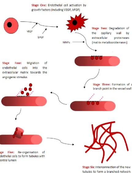
Figure 1.10 Stages involves in tumour angiogenesis 46
Figure 1.11 The cell cycle showing the two major events that takes place
within the cell
49
Figure 1.12 The four stages of mitosis 51
Figure 1.13 The cell cycle and its regulation by cyclins,
CDKs, and CDKIs
52
Figure 1.14 The processes involved in apoptosis 66
Figure 1.15 Stages of apoptosis 69
Figure 1.16 Morphology of apoptosis 71
Figure 1.17
Figure 1.18
Figure 1.19
Figure 3.1
Activation of apoptosis signaling pathway
Intrinsic and extrinsic pathway
Differences between apoptosis and necrosis
MTT cell viability assay for MDA468 cells
after treatment with cockle derived and
commercially sourced GAG chains
75
79
89
109
Figure 3.2 MTT cell viability assay for MDANQ01
cells after treatment with cockle derived
and commercially sourced GAG chains
111
Figure 3.3 MTT cell viability assay for MOLT-4
lymphoblastic leukaemia cells after treatment
xiii GAG chains
Figure 3.4 Concentration-dependent effect of Cockle GAG
mixtures on K562 erythroleukaemia cell viability
115
Figure 3.5 MTT cell viability assay for normal fibroblast (3T3)
cells after treatment with cockle GAGs
117
Figure 3.6 (i) Cell cycle analysis of MOLT-4 lymphoblastic
leukaemia cell line following treatment with
50µg/ml of cockle GAG extracts
119
Figure 3.6 (ii) Cell cycle analysis of MOLT-4 lymphoblastic
leukaemia cell line following treatment with low
concentration (20µg/ml) of cockle GAG extract.
123
Figure 3.7 Detection of Apoptotic MOLT-4 cells treated
with cockle GAG by Annexin V Staining.
126
Figure 3.8 MTT cell viability assay for MDANQ01 cells
after treatment with Whelk extract and
commercial GAGs
130
Figure 3.9 Concentration-dependent effect of whelk GAG
extracts on MDA468 breast cancer cell viability
132
Figure 3.10 MTT cell viability assay for MOLT-4 and K562
leukaemia cells after treatment with whelk GAG
extracts
134
Figure 3.11 MTT cell viability assay for HELA cervical cell
after treatment whelk GAG extracts
136
Figure 3.12 MTT cell viability assay of normal fibroblast 3T3
cells after treatment with whelk GAGs extracts
138
Figure 3.13 MTT cell viability assay for MDANQ01 and
MDA468 breast cancer cells after treatment with
xiv
heparinase depolymerised whelk GAG extracts
Figure 3.14 Cell cycle analysis of MDANQ01 breast cancer cell line following
treatment with crude whelk GAG extract
142
Figure 3.15 Cell cycle analysis of MDA468 breast cancer cell line following
treatment with crude whelk GAG extract
145
Figure 3.16 Detection of Apoptotic MDA468 breast cancer cells
treated with crude whelk GAG extracts by Annexin
V Staining
149
Figure 3.17 Detection of Apoptotic MDANQ01 breast cancer
cells treated with crude whelk GAG extracts by
Annexin V Staining
152
Figure 3.18 Detection of apoptotic MDA468 cells by DAPI
fluorescent microscopy following treatment
with crude whelk GAG extracts
155
Figure 3.19 Detection of apoptotic MDANQ01 cells
treated with crude whelk GAG mixtures
by DAPI fluorescent microscopy
157
Figure 3.20 Detection of Apoptosis in HELA cells by Annexin
V Staining following treatment with whelk GAG
extracts
160
Figure 3.21 Detection of apoptotic HELA cells by DAPI
fluorescent microscopy following treatment
with whelk GAG
162
Figure 3.22 Ion-exchange chromatography elution profile for
whelk GAG extracts
165
Figure 3.23 MTT cell viability assay for MDANQ01 breast
xv and purified fraction E
Figure 3.24 Concentration-dependent effect of purified whelk
GAG fraction E on MDA468 breast cancer cell viability
172
Figure 3.25 Cell cycle analysis of MDA468 breast cancer cell line following
treatment with purified whelk fraction E
174
Figure 3.26 Detection of Apoptotic MDANQ01 breast cancer cells
treated with purified whelk fraction E by Annexin V
Staining
176
Figure 3.27 Detection of Apoptotic MDANQ01 breast cancer
cells treated with purified whelk fraction E by Annexin V Staining
179
Figure 3.28 Detection of time-dependent Apoptotic MDA468
breast cancer cells treated with purified whelk
fraction E by Annexin V Staining
181
Figure 3.29 Kinectic of chondroitinase ABC enzyme degradation of
commercial CS
187
Figure 3.30 Analytical TSK2000PW gel-filtration profiles of
HS/Heparin and crude cockle GAGs after complete
digestion with Heparinase I, II and III enzymes
193
Figure 3.31 Superose 12 size exclusion chromatography and cellulose
acetate dot blotting result for crude cockle GAG mixture
194
Figure 3.32 PAGE analysis of enzymatic depolymerised GAGs. 196
Figure 3.33 Kinetic of chondroitinase ABC enzyme degradation
of GAGs
199
Figure 3.34 PAGE analysis of enzymatic depolymerised GAGs 203
Figure 3.35 Superose 12 size exclusion chromatography and cellulose
acetate dot blotting assay of crude whelk GAG extract
xvi
Figure 3.36 Analytical TSK2000PW gel-filtration profiles of HS/heparin
and purified whelk fraction E after complete digestion with
Heparinase I, II and III enzymes
215
Figure 3.37 PAGE analyses of enzymatic depolymerised whelk
derived and commercial GAGs.
217
Figure 3.38 Superose-12 size exclusion chromatography and cellulose acetate dot blotting assay for purified fraction E GAG mixtures
219
List of tables
Table 1
Table 2
Summary of biological effect of cockle GAGs
Ion-exchange chromatographic separation of
whelk GAG mixtures
127
158
Table 3
Table 4
Table 5
Summary of biological effect of whelk GAG
Summary of biological effect of fraction E
Disaccharides compositions of CS/DS-like
glycans found in cockle GAG mixtures
167
183
186
Table 6 HS Disaccharides compositions from
heparinase digest of cockle GAG extracts
190
Table 7 Disaccharides compositions of CS/DS-like
glycans found in whelk GAG mixtures
201
Table 8 Disaccharides compositions of CS/DS-like
GAGs present in ion-exchange purified whelk
peaks B and C
xvii
Table 9 Disaccharides compositions of HS-like GAGs
found in purified whelk fraction E
212
Table 10 Disaccharides compositions of HS-like GAGs 213
found in purified 204 whelk fraction E depolymerised
xviii Abbreviations
∆ delta
µg microgram
µl Microlitre
2-OST 2-O sulfotransferase
3-OST 3-O sulfotransferase
6-OST 6-O sulfotransferase
aFGF acidic fibroblast growth factor
AIF Apoptosis Inducing Factor
AKT Protein kinase B
Ala Alanine
ANOVA Analysis of variance
Apo2L/DR Apo2 ligand/ Death receptor
Apo3L Apo3 ligand
APS Ammonium Per Sulfate
AS Acharan sulfate
ATIII Antithrombin III
ATP Adenosine triphosphate
Bax BCL2 associated X protein
Bcl-2 B-cell lymphoma protein 2
bFGF basic fibroblast growth factor
C4ST Chondroitin 4- O –sulfotransferase
C6ST Chondroitin 6- O –sulfotransferase
Ca2+ Calcium ion
CaCl2 Calcium Chloride
CAD Caspase-Activated DNAse
Caspace Cysteinyl aspartic acid-protease
CD36 Cluster of differentiation 36
CD8+ Cluster of differentiation8+
CDK cyclin dependent kinase
CDKI cyclin dependent inhibitors
xix ChPF Chondroitin polymerizing factor.
ChSy Chondroitin synthase
COO- Carbonic acid
CPD cetylpyridinium chloride
CS Chondroitin sulfate
CS GlcAT-II Chondroitin sulfate GlcA transferase-II
CS/DS2ST uronyl 2- O –sulfotransferase
CTLs Cytotoxic T lymphocytes
CZE Capillary zone electrophoresis
D4ST Dermatan 4- O –sulfotransferase
DAPI 4',6-diamidino-2-phenylindole
DEAE Diethylaminoethyl
DIABLO Direct IAP binding protein with low PI
DISC Death-inducing signalling complex
DMSO Dimethyl sulfoxide
DNA Deoxyribonucleic acid
dp Degree of polymerization
dp10 decasaccharides
dp2 disaccharides
dp4 tetrasaccharides
dp6 hexasaccharides
dp8 octasaccharides
DR Death receptor
DS Dermatan sulfate
ECM Extracellular matrix
EDAC 1-ethyl-3-(3-dimethylaminopropyl) carbodiimide
EDTA Ethylenediaminetetraacetic acid.
ER Eostrogen receptor
ER-ve Eostrogen receptor negative
ER+ve Eostrogen receptor positive
EXT Exostosin glycosyltransferase
FADD Fas-associated death domain
xx FasL Fatty acid synthetase ligand
FasL/ FasR Fatty acid synthetase ligand /Fatty acid synthetase receptor
FasR Fatty acid synthetase receptor
FCS fetal calf serum
FGF Fibroblast growth factors
FGFR’s Fibroblast growth factors receptors
FITC Flourescein isothiocyanate
FPLC Fast Protein Liquid Chromatography
G1 Gap 1
G2 Gap 2
GAG-PG Glycosaminoglycans proteoglycans
GAGs Glycosaminoglycans/Galactosaminoglycans
Gal Galactose
GalNAc N-acetyl galactosamine
Gal-NAc4S-6ST GalNAc 4-sulfate 6- O –sulfotransferase
GalNAcT, GalNAc transferase
GalT β4-galactosyl transferase
G-CSF Follistatin Granulocyte colony-stimulating factors
GlcA Glucuronic acid
GlcAT β3-GlcA transferase
GlcNAc N-acetyl glucosamine
GlcNAc(6S) 6-O-sulfated N-acetylglucosamine
GlcNS N-sulfoglucosamine
GlcNS(6S) 6-O-sulfated N-sulphoglucosamine
GlcUA β-D-glucuronic acid
Gly Glycine
HA Hyaluronic acid
HCG Human chorionic gonadotropin
HCl Hydrochloride acid
HEPES 2-[4-(2-hydroxyethyl)piperazin-1-yl]ethanesulfonic acid
HGF Hepatocyte growth factor
xxi
HS-GAGs Heparan sulfate glycosaminoglycans
HSPG Heparan sulfate proteoglycans
IAP inhibitors of apoptosis proteins
IC50 half maximal inhibitory concentration
ICAD Inhibitor of Caspase Activated DNAse
IdoA Iduronic acid
IdoA (2S) 2-O-sulfated IdoA
IdoUA α-L-iduronic acid
Ig immunoglobulin-like
Kb kilo bytes
kDa Kilodalton
KS Keratan sulfate
LDL low density lipoprotein
L-IdoA L- iduronic acid
M Mitosis
Mg2+ Magnesium Ion
ML Millilitre
mM millimolar
MOPS 3(n-Morpholino) Propanesulfonic acid
MPT mitochondrial permeability transition
MTT 3-(4,5-Dimethylthiazol-2-yl)-2,5-Diphenyltetrazolium Bromide
NDST N-deacetylase, N-sulfotransferase
nm nanometre
NO Nitric oxide
NuMA Nuclear mitotic apparatus protein
O-Ser O Serine
PAGE polyacrylamide gel electrophoresis
PAP 3ˈPhosphoadenosine 5ˈphosphosulfate
PARP Poly (ADP-ribose) polymerase
PBS Phosphate Buffer Saline
xxii pRB retinoblastoma gene products
Procaspase-8 Pro cysteinyl aspartic acid-protease 8
Rb Retinoblastoma
RIP Receptor-interacting protein
ROS Reactive oxygen species
Rpm Rotation Per Minute
RPMI Roswell Park Memorial Institute
Rt retention time
RTKs receptor tyrosine kinases
S Synthesis
SDS-PAGE sodium dodecyl sulfate-polacrylamide gel electrophoresis
SEM Standard Error of Mean
Ser Serine
SGBS Simpson–Golabi–Behmel syndrome
SMAC Second mitochondrial activator of caspase
SNPs Single nucleotide polymorphisms
SO3 Sulfur trioxide
SRC Steroid receptor coactivator
TEMED N,N,N’,N’-Tetramethylethylendiamine
TGF-β tumour growth factor beta
Th2 Type 2 helper
TK Tyrosine kinase
TKR’s Tyrosine kinase receptors
TNF tumour necrosis factor
TNFR1 Tumour necrosis factor receptor
TNF-α tumour necrosis factor alpha
TRADD TNF receptor-associated death domain
Tris Trizma base
UCN-01 7-hydroxystaurosporine
UDP Uridine diphosphate
UV Ultraviolet
V0 Void volume
xxiii Vt Final volume
xxiv Acknowledgement
I dedicate this thesis to God almighty and the three special people in my life: my husband and
our twins, David and Esther. Thank you for your support and understanding. I would like to
thank my parents and my in-laws for their prayers and support over the past four years.
My sincere appreciation is also extended to Dr Jeremy Allen, Dr Lucy Smith, Helen
and Ross for their support and training in flourescent microscopy. Jeremy spent many hours
with me in the laboratory to ensure I achieved the best results. I also appreciate the immense
contribution of Ray (a former cell culture laboratory technician) during my cell culture assay
in his laboratory. I would also like to thank Eyad for providing all the necessary support and
help for my apoptosis detection assay. I am also grateful to the laboratory technicians in the
Cockroft Building for their unfliching technical and moral support throughout my laboratory
work.
I appreciate all the contributions of my colleagues, the masters and the placement
students in Dave’s laboratory. I would also like to thank Drs James Wilkinson, Elder
Rhoderik and Natalie Ferry for their help and support over the past four years. Jon Deakon is
also highly appreciated for his professional support and help each time I became stuck with
GAGs palava. ELS staff (Catriona and others) have been wonderful and supportive
throughout my study. I also appreciate my mum for coming all the way from Nigeria to help
care for my babies while I was busy writting this thesis. All my friends and family have been
very supportive over the past four years. My sincere thanks also goes to my able boss and
mentors back home, Professors E.O Agbedana, J.I Anetor, J.O Moody, J. Omotade, A.
xxv
Finally, my sincere appreciation goes to my supervisor, Dr David Pye, for giving me
unrestricted access to his office and providing invaluable advice and support. Without him,
xxvi Abstract
Glycosaminoglycans (GAGs) are a family of complex mixture of linear
polysaccharides that are present in both vertebrates and invertebrates. This polysaccharide
plays important roles in physiological and pathological conditions, including cancer. In this
study, GAGs were isolated from two different fish (whelks and cockles) belonging to mollusc
invertebrates. The crude GAGs isolated from each shellfish demonstrated variable selective
anti-cancer activities against many cancer cell lines including breast (MDANQ01 and
MDA468), leukemia (MOLT-4 and K562) and ovarian (HeLa) cancer. None of the
commercial GAGs exhibited any anti-cancer activities against all the cancer cells studied.
Previous studies conducted on the isolation of GAGs from molluscs reported mainly its
anti-coagulant and anti-inflammatory activities; thus neglecting its record of anti-cancer activity.
All purified whelk fractions (A – D & F) obtained failed to show any anti-cancer
activity; with the exception of fraction E, which showed equal levels of selective anti-cancer
activity against breast cancer cells. Mechanism of cell death caused by the three novel GAGs
on cancers cells were investigated via cell cycle analysis and apoptosis detection assay. Cell
cycle analysis revealed significant perturbations in the cancer cell cycle showing cell cycle
arrests at different stages. Similarly, there were significant apoptosis inductions induced by
the three novel GAGs on each of the cell lines investigated.
Structural elucidations of the two fish GAGs, using chemical, enzymatic,
Polyacrylamide, Superose 12 size exclusion chromatography , gel filtration and SAX-HPLC
methods of analysis, revealed the presence of both chondroitin sulfate (CS)/dermatan sulfate
(DS)-like and HS-like GAGs. Discrepancies in the structural elucidations of the novel GAG
mixtures and the commercial GAGs may be partly responsible for the anti-cancer activity of
the novel polysaccharides, as changes in exogenous GAGs structural composition, especially
sulfation levels or patterns, can alter its binding to growth factors, which is essential for cell
1
Section 1
2 1.0General introduction
Glycosaminoglycans (GAGs) are a family of linear, complex, polydisperse
polysaccharides extracted and purified from different animal tissues (Cassaro and Dietrich,
1977; Heinegard and Sommarin, 1987; Pejler et al, 1987a). They are very heterogeneous
polysaccharides in terms of relative molecular mass, charge density, physico-chemical
properties, biological and pharmacological activities (Jackson et al, 1991). GAGs are
ubiquitous in nature, occurring in a wide variety of organisms, including both vertebrates and
invertebrates. They are linked typically to a protein core in order to form proteoglycans.
These anionic polysacharides elicit many biological functions, largely through their
interaction with proteins (Cesaretti et al, 2004). Major GAGs in vertebrates and invertebrates
include heparin, heparan sulfate (HS), chondroitin sulfate/dermatan sulfate (CS/DS), acharan
sulfate (AS), and hyaluronic acid (HA) and keratin sulfate (KS).
All GAGs chains except KS comprise alternating units of an amino sugar, either N -acetyl-D-glucosamine (GlcNAc) or N-acetyl-D-galactosamine (GalNAc) and a hexuronic
acid (HexA), either glucuronic acid (GlcA) or iduronic acid (IdoA). Keratan sulfate contains
GAGs chains comprising of galactose attached to acetyl-D-glucosamine (Afratis et al., 2012:
Yamada et al, 2011). HS and heparin are the most widely studied of these polysaccharides
due to their involvement in many important biological processes. They have been shown to
play important roles in the control of stem cell growth and differentiation (Patel et al, 2014
and Desai 2013). Moreover, they are involved in a number of important biological activities,
such as developmental processes, angiogenesis, blood coagulation, cell adhesion and tumour
metastasis (Sasisekharan et al, 2006: Wei et al, 2005: Vives et al, 1999: Lindahl et al, 1998:
3
HS is also vital in regulation of embryonic development, as well as in
pathophysiological conditions, such as inflammation, wound healing, angiogenesis and
cancer (Tamm et al 2012: Lo et al, 2011: Sasisekharan et al, 2006). These activities are
facilitated by the ability of HS proteoglycans (HSPG) to interact and regulate proteins, such
as enzymes, cytokines, growth factors, and extracellular matrix (ECM) bimolecules (Wei et
al, 2005: Vives et al, 1999). The divergent structure of HS allows it to bind to and regulate
the activity of various growth factors, like fibroblast growth factors 1, 2, 4 etc (1,
FGF-2, FGF-4 etc), chemokines, morphogens and enzymes at the cell surface and in the ECM:
hence influencing cell adhesion, motility, growth and differentiation (Lo et al, 2011:
Sasisekharan et al, 2006: Vives et al, 1999). The ability of HS to modulate the activities of
many so-called heparin-binding growth factors, serve to co-ordinate the interaction of FGF
with their tyrosine kinase receptors (FGFRs); moreover, this appears to play a critical role in
receptor dimerization and activation (Iozzo and San Antonio 2001: Dickson et al 2000: Vives
et al, 1999: Ernst et al, 1995). The modulatory effect of HS is critical to angiogenesis that
occurs in pathological conditions, including tumour growth and progression; this has been
implicated in the vascularisation of all solid tumours (Iozzo and San Antonio, 2001).
Biological activities of HS depend broadly on the interaction between polysaccharides
and proteins, which appears to be mediated by distinct saccharides sequences within HS
chains. Moreover, the expression of different sequences within the HS chains demonstrates
various levels of specificity, selectivity and molecular organisation. Modulation of tumour
growth occurs through the interaction with angiogenic growth factors, like FGF-10, or with
negative regulators of angiogenesis like anti-angiogenic antithrombin III (Iozzo and San
Antonio, 2001: Ernst et al, 1995).
The modulatory effects of HS on the growth factors and chemokines involved in tumour
4
The voluntary binding of the growth factors to soluble ECM associated or cell-surface
HS results in the fine control of the bioavailability of growth factors; for example, binding of
tumour growth-factor beta (TGF-β) to betaglycan, a cell-associated PG, and decorin, which is
present in the ECM, and also for fibroblast growth-factor-2 (FGF-2) that binds basement
membrane perlecan in addition to cell-membrane syndecans (Mellor et al, 2007 and
Massague 1992).
The binding of HS to growth factors protects them from proteolytic degradation and
acid inactivation (Samuel et al, 2000: Szabo 1991 and Saksela et al, 1988).
HS modulates the access of FGF growth factors to specific signaling receptors by
different mechanisms that may involve independent interaction of HS with an aspect of the
transmembrane tyrosine kinase glycoprotein component of the FGFR complex (Kan et al,
1993).
HS may also trigger an intracellular transduction signal by interacting directly with
growth-factor receptors with no growth factors being present (Rusnati et al, 1993).
Indeed, Rusnati and others have reported that heparin/HS can activate FGFRs in the absence
of the growth-factor. This observation goes against the traditionally accepted proposal of a
ternary complex involving GAGs, growth factors, and tyrosine kinase (TK) receptors
(Rusnati et al, 1993). The introduction of this model led to an extensive study involving many
research groups and there is still much debate on actual mechanisms by which GAGs mediate
the activation of the FGF’s. Regardless of this debate some general properties of the
signalling complex can be surmised and these are outlined below:
Heparin/HS-related molecules can be useful tools to modulate the biological activity
5
Heparin/HS-related molecules can be designed with various structures that are able to
affect differently the biological activities of angiogenic growth (Mellor et al, 2007).
The ideas expressed above show some promise, therefore, there is need for the development
of inhibitors of the activation of growth factors by GAGs and the subsequent development of
new anti-cancer treatments aimed at the inhibition of tumour-cell proliferation.
The ability of low molecular weight heparin fragments administered systemically to
reduce the angiogenic activity of FGF2 and vascular endothelial growth-factor (VEGF)
validate this hypothesis (Ghandi and Mancera, 2010; Norrby 2000: Norrby and Ostergaard
1997 and 1996). Consequently, many HS/heparin fragments and mimics have been shown to
have potential as inhibitors of tumour angiogenesis. In addition to the role played by HS in
regulation of tumour angiogenesis, we shouldn’t forget the potential roles for HS in directly
regulating the growth of cancers through interactions at the tumour cell surface. The
structural complexity of tumour-cell HS (both at the cell surface and extruded into the ECM)
has been shown to play vital roles in tumour development. It enables them to modulate
directly several aspects of tumour-cell phenotype, including growth kinetics, invasiveness
and metastatic potential (Sasisekharan et al, 2002). Hence, a given tumour-derived HS-GAG
or HSPG can be protumorigenic or antitumorigenic, depending on whether it is anchored at
the cell surface or present in the ECM as soluble-free GAGs or/and depending on the
HS-GAG sequence ( Gomes et al., 2013: Liu et al, 2002 and Sasisekharan et al, 2002).
Targeting tumour angiogenesis by interfering with the interaction of HS with
angiogenic growth factors is a well-established model for the development of HS mimetic
therapeutics. However, we shouldn’t forget the ability of HS to modulate the growth and
development of tumours via other mechanisms. This could certainly lead to the determination
of, as yet unknown, HS structures and interactions that could pave way toward the
6
The study describe in this thesis employs GAGs derived from shellfish in a novel
approach that targets directly the growth of cancer cells, rather than the vasculature and
angiogenic processes within the tumour. The anti-cancer activity of these shellfish-derived
GAGs is a unique finding, with none of the commercially derived GAGs from mammalian
sources demonstrating any anticancer activity.
The GAGs derived from these shellfish have not been characterised in terms of their
7 1.1 Glycosaminoglycans
GAG’s are natural heteropolysaccharides, belonging to the family of polyanionic
polysaccharides. These highly negatively-charged polysaccharides are ubiquitous in nature,
occurring in a wide variety of organisms and tissues attached typically to a protein core to
form molecules known as proteoglycans (PGs). They are composed of repeating
disaccharides units comprising either sulfated or non-sulfated disaccharides. Their properties
have been shown to vary considerably depending on the tissue or cell type from which they
are extracted. (Andrade et al, 2013: Vijayabaskar and Somasundaram 2012: Afratis et al,
2011: Heinegard and Sommarin 1987; Pejler et al, 1987: Cassaro and Dietrich 1977).
GAGs elicit numerous biological functions, largely through their interaction with proteins,
including growth factors.
GAGs are found located within the cell mebrane and in the extra-cellular matrix
(ECM). They act by binding selectively to a variety of proteins and pathogens and are crucial
to many disease processes, such as inflammation, neurodegeneration, angiogenesis,
cardiovascular disorders, cancer and infectious diseases (Ghandi and Mancera et al, 2010:
Mark et al 2002).
The GAG family has several archetypal members including heparan sulfate (HS), heparin,
chondroitin sulfate/ dermatan sulfate CS/DS, keratan sulfate (KS) and acharan sulfate (AS).
There is also the non-sulfated GAGs consists of hyaluronic acid HA (Figure 1.1). HA lack a
covalently protein core and are generally synthesised at the intracellular surface of the plasma
membrane. HA GAGs has been reported to be a key player in tissue homeostasis, cancer
progression, inflammatory conditions, myocardial infarction and angiogenesis (Afratis et al.,
2012: Slevin et al., 2002). HS and heparin are the most widely studied members of the
8
angiogenesis, cell proliferation, wound healing, inflammation etc (Afratis et al., 2012: Vives
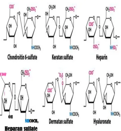
9 Fig. 1.1: Schematic structures of GAGs.
Heparin/HS and HA are glycosaminoglycans; chondroitin sulfate (CS) and dermatan sulfate (DS) are
galactosaminoglycans; KS is a sulfated polylactosamine. Abbreviation: R= H or SO3: R’= SO3 or COCH3
NH
COCH
3OH
COO
10 1.2 GAG’s structure and functions
GAGs comprise a linear chain of 10–200 disaccharide units of repeating N
-acetyl-D-glucosamine/galactosamine, which is linked to uronic acid (iduronic or glucuronic acid)
(Tumova et al, 2000: Lindahl et al, 1998). The disaccharide repeat unit can be modified to
include N- and O-sulfation and also epimerisation of β-D-glucuronic acid to α-L-iduronic
acid (Conrad, 1998). In addition, the five different modifications for disaccharides give rise to
25 = 32 combinations. Thus, this modification makes the complexity of the GAG much
greater than that of proteins, which are made up of 20 amino acids. With these 32 building
blocks, a GAG octasaccharide could have in excess of a million possible sequences; thereby
making HS-GAGs not only the most acidic, but also the most information-dense biopolymers
found in nature (Conrad, 1998).
The physical form of GAGs and the specific interactions between them and other
ECM components are controlled frequently by the sequence of disaccharide residues that they
are comprised of. Usually, GAGs interact with transient components of the ECM, either at
the cell surface, in solution or sequestered in the matrix. For instance, FGF-2 induced
endothelial-cell proliferation requires trimolecular interactions between FGF-2, FGF -TK
receptors (FGFR) and HS (Jaye et al., 1992: Kiefer et al, 1991: Klagsbrun and Baird, 1991:
Yayon et al, 1991). This interaction activates TK activity of the intracellular domain of the
11 1.3 Heparin/HS structures and functions
Heparin/HS, usually isolated from animal tissue including vertebrates and
invertebrates, is an important and chemically-distinct polysaccharide of considerable
biological relevance. As explained above they are polydisperse, highly sulfated, linear
polysaccharides, consisting of a repeat unit of uronic acid in either of its glucuronic or
iduronic form attached to D-glucosamine residues (Figure 1.1) ( Koo et al., 2008: Volpi et al,
2005: Islam and Linhardt, 2003). This group of GAGs have a very complex structure yet they
are the most widely studied members of the GAG family. The structural complexity of
Heparin/HS can be considered at both PG and GAG levels;
At the PG level, different numbers of polysaccharide chain (possibly with different
disaccharide sequences) can be attached to the various serine residues present in the protein
core (Koo et al, 2008).
At the GAG level, some of HS/Heparin structural complexity results from their
polydispersity.
GAG especially heparin has a molecular weight ranging from 5-40kDa (degree of
polymerisation (dp) 10-80) with an average molecular weight of 13 kDa. Heparin/ HS
structure has been partially characterised by studying their biosynthesis (Kjellen et al, 1992).
Twelve sulfation profiles for HS/heparin disaccharides have been reported and these have
equally been resolved and characterised by various separation techniques including chemical,
enzymatic, and spectroscopic techniques (Malavaki et al., 2011: Sasisekharan et al., 2006:
Linhardt, 1991).
Problems arise in the structural analysis of HS/Heparin GAGs since they are extensivley
12
the most structurally diverse members of the glycosaminoglycan family. The amino group
may be either sulfated or acetylated and each disaccharide unit may have up to three of the
hydroxyl groups sulfated (2-O-S, 3-O-S and 6-O-S). Glucuronic acid rich polymers are sometimes classed as HS while the iduronic acid rich polymers are also sometimes refers to
as heparin. Heparin contain a pentasaccharide region
(GlcNAc/NS(6S)-GlcA-GlcNS(3S,6S)-IdoA(2S)-GlcNS(6S) that bind to antithrombin III and this makes it the most widely used
carbohydrate based therapeutic because the binding allows it to inhibit blood coagulation
(Loganathan et al,1990: Marcum and Rosenberg,1989a: Linhardt et al,1988b: Atha et
al,1985: Lindahl et al, 1984). Relating structure to biological activities, such as that described
for heparin, is critical in our understanding of the role sulfation patterns and epimerisation
play in the natural function of heparin/HS chains.
Sulfation patterns of HS/heparin play a very crucial role in their interactions with growth
factors, proteins and cytokines, and therefore may affect their biological roles. HS/heparin
have been implicated in numerous biological processes at the cell–tissue–organ interface (
Malavaki et al., 2011: Conrad 1998), which are triggered mainly by their interactions with a
wide range of proteins, such as cofactors of the coagulation cascade, extracellular-matrix
components and a variety of cytokines and growth factors (Vives et al 1999: Permimom and
Belfield, 2000), angiogenesis (Sasisekharan and Venkataraman, 2000), viral invasion (Shukla
et al, 1999 and Chen et al, 1997), and anticoagulation (Petitou et al, 1999).
The HS/heparin also exerts its modulatory effects on key biological processes by binding
to growth factors, cytokines, morphogens, enzymes and other signaling molecules (Taipale
and Keski-Oja, 1997). They can regulate the activity of signaling molecules by modulating
13
the ECM, However, these processes become especially important at the cell–tissue–organ
interface.
Previous studies have added an extra dimension to this cell-surface event by indicating
that HS mediates dimerization, either of FGF-2 of FGF receptor or of both, which may be a
requirement for signal transduction (Ornitz et al, 1996: Pantoliano et al, 1994: Springer et al,
1994: Mascarelli et al, 1993: Ornitz et al,1992). The binding of HS chains to FGF-2 and
FGFR occurs through N- sulfated glucosamine (GlcNS) and 2-O-sulfated iduronic acid
(Ido2S) units or 6-O- sulfated glucosamine (Glc6S). Once the FGFs bind to their receptors, FGFR complexes dimerise, in conjuction with HS/heparin moiety, leading to the activation of
tyrosine kinase through autophosphorylation. Ultimately, these events facilitate the binding of
second messenger proteins which in turn trigger many other intracellular signalling
(Gallagher 2001: Dickson et al 2000).
The various roles played by these polysaccharides are becoming clearer as researchers
continue studying them.
1.4 CS structure and functions
Chondroitin and dermatan sulfate make up a second GAG family, also known as
galactosaminoglycans. These polysaccharide chains are, again as in HSPG’s, normally found
covalently bound to a protein core forming high molecular weight units, with a large number
of GAG chains attached (Ernst et al, 1995). The basic structural unit of this class of GAG is a
disaccharide in which a uronic acid (D-glucuronic and L-iduronic acid) is attached (β1→3) to
N-acetyl galactosamine (Figure 1.1). CS is the most abundant GAG in the body skeletal
14
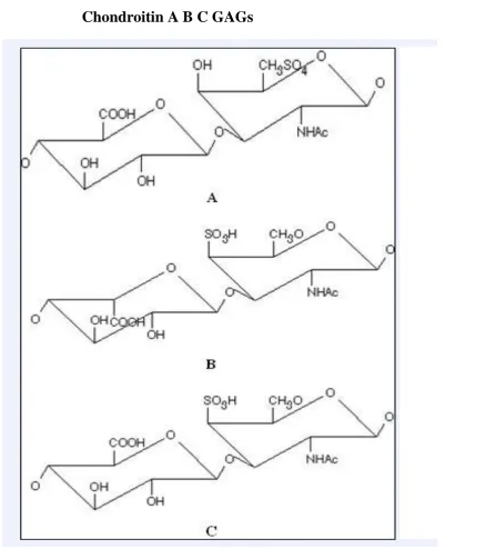
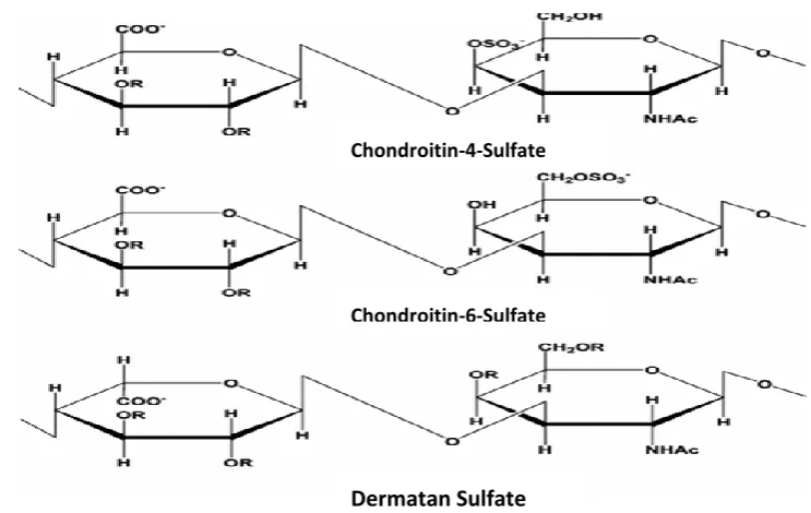
There are various sulfation patterns of CS and among the most studied sulfated CS
GAGs are the two common isomers CS-A (O-sulfo group attached at position 4 of galactosyl
residue) and CS-C (O-sulfo group attached at position 6 of galactosyl residue) see Figure 1.2. Similarly, disulfated CS are also classified as CS-D (O- sulfo group attached at position 2 of
glucuronic acid and position 6 of galactosyl residue) or CS-E (O- sulfo group attached at position 4 and 6 of galactosyl residue). The other CS isoform formerly designsted as CS-B is
called DS which is characterised by the presence of IdoA moiety in his disaccharide chains.
Similar to HS/heparin, the various sulfation patterns of CS GAGs enable specific interaction
with many molecules, including growth factors, cytokines, chemokines, adhesion molecules
and lipoproteins (Asimakopoulou et al., 2008).
The size of CS chains varies greatly, with an average of about 40 repeating
disaccharides units for the cartilage proteoglycans, corresponding to a molecular weight of
approximately 20,000 (Iozzo, 1985). Their sulfo group content also varies depending on the
number(s) attached to both or either of the galactosyl and uronic acid residues (Iozzo, 1985).
Once again they all show a considerable degree of microheterogeneity within the polymer
chain in a manner very similar to HS. The disaccharide unit present in CS/DS may be
non-sulfated, mono-sulfated or disulfated and both iduronic and glucuronic acids may be present
in a given polymer chain.
DS (CS-B) is a polydisperse; microheterogenous sulfated copolymer of D-Gal-NAc
and primarily L- iduronic acid (L-IdoA), with O-Sulfate groups most commonly attached to position 4 of the galactosyl residue and position 2 of the L- Iduronic acid residues (Linhardt
and Hileman, 1995). Many DS core proteins such as decorin and biglycan have been
identified (Kresse et al, 1993: Coster, 1991). Decorin and biglycans have been reported to
15
regulation of the extravascular activities of thrombin, act as anticoagulants by inhibiting
thrombin and lipid metabolism (Pangrazzi and Gianese, 1987). ). CS-B or DS is abundant in
skin and is also found in heart valves, tendons and arterial walls. It is made up of linear
repeating units containing D-galactosamine and either L-iduronic acid. Its molecular weight
ranges from 15,000 to 40,000 daltons.
Moreover, CS-A is commonly found in humans in cartilage, bone, cornea, skin and
the arterial wall. The molecular weight of CS-A ranges from 5,000 to 50,000 daltons and
contains about 15 to 150 basic units of D-galactosamine and D-glucuronic acid (Wastenson,
197). CS-C, is primarily found in fish and shark cartilage, but also in humans, is also made
16
Figure 1.2: Structure of different types of CS
17 1.5 Glycosaminoglycans isolated from shellfish
Many researchers, including Dietrich and co-workers, reported the isolation of GAGs
similar to heparin with comparable anti-thrombin activities from mollusc’s invertebrates.
Similarly, heparin-like substance with relative ability to bind antithrombin III (ATIII) has
been isolated from the marine clams, anomalocardia brasiliana (Pejler et al, 1987: Dietrich et
al, 1985). Another GAG similar to heparin GAG, with an AT-binding region comparable to
that of mammalian heparin has also been purified from another clam species, Mercenaria
mercenaria (Jordan and Marcum, 1986). Additionally, heparin, (a highly sulfated polysaccharide, commonly isolated from mast cell or mucosa and also used as an
anticoagulant in the clinic) (Bjork and Lindahl, 1982) has been reported to have some other
biological activities, such as; anti-viral activity, bind to a variety of growth factors, inhibit
complement activation, and regulate angiogenic activity (Weiler et al, 1992; Jackson et al,
1991; Casu 1985; Folkman et al, 1983).
1.6 Biosynthesis of GAGs
HS and CS are synthesised in the Golgi, whereby the individual GAG chains are
O-linked to a core protein, forming a large polysaccharide-protein conjugate known as
proteoglycan (PG) (Sasisekharan et al, 2006: Silbert and Sugumaran, 2002: Sugahara and
Kitagawa, 2002). However, KS can be either N-linked or O-linked to the core protein of the
PG (Funderburgh 2002). HA is not synthesised in the Golgi from the core protein but rather
by an integral plasma membrane synthase, which secretes the nascent chain immediately
(Itano and Kimata, 2002). All GAGs, with the exception of HA, are biosynthesised as
18
linkage region consists of the tetrasaccharide; glucuronic acid (GlcA), galactose (Gal), Gal,
and xylose (Xyl)-linked to the hydroxyl group of serine in the polypeptide core (Sugahara
and Kitagawa, 2000). Biosynthesis of all GAGs, except HA is initiated from a core protein,
rich in serine-glycine repeats, which create the platform for the attachment of one or more
GAG chains via the linkage region (Kimura et al, 1984). Contrarily, HA GAGs lack a
covalently bound protein core and is synthesised at the intracellular surface of the plasma
membrane (Afratis et al., 2012)
Biosynthesis of GAG is a complex non-template-driven process that involves many
enzymes that assemble the GAG polymer and then sulfate them at specific positions. GAGs
are synthesised as homopolymers, which are modified subsequently by N-deacetylation and
N-sulfation. This is followed by numerous modifications, including sulfation, epimerisation
and desulfation; all of which are performed in a spatiotemporal manner, producing mature
and functional GAG chains that exert biological functions dependent on their specific
structure. This is common to both types of GAG chain and is formed through the stepwise
addition of each monosaccharide residue by the respective specific glycosyltransferase
(Grimshaw,1997).
After the attachment of the linker, the first modification to the chain determines
whether the chain matures into a CS or HS. This modification involves the transfer of a
GlcNAc (HS) or a GalNAc (CS) monosaccharide. The enzyme GlcNAcT-I transfers GlcNAc
to the tetrasaccharide linker, initiating HS biosynthesis (Rohrmann et al, 1985). This enzyme
is different from the glycosyltransferase GlcNAcT-II that is involved in the elongation of the
HS GAG chains. In the case of CS, the initiating GalNAc residue is transferred to the linker
region by the enzyme GalNAcT. It is not very clear whether the activity of this enzyme
19
chain-elongation process. In all cases real chain-building ensues after the transfer of GlcNAc
(HS) or GalNAc (CS) to the linker.
In the case of HS chains, a multidomain glycosyl-transferase successively adds
GlcNAc and GlcA; thus, initiating the chain-elongation process. This multidomain enzyme is
encoded by EXT1 and EXT2 of the EXT family of genes. Although EXT1 and EXT2 possess
the ability to transfer both GlcNAc and GlcA, it has been established that these enzymes form
a single oligomeric unit, which is required for complete in vivo chain-elongation activity.
Similarly, in the case of CS, chondroitin synthase, also a known multidomain enzyme,
transfers GalNAc and GlcA successively for chain elongation (Sashisekhare et al, 2006).
Molecular cloning has been achieved for four of the glycosyl transferases responsible for the
biosynthesis of the linkage region tetrasaccharide (Uyama et al, 2007: Kitagawa et al, 1998).
Firstly, xylosyl transferase (XylT) transfers a Xyl residue from UDP-Xyl to specific
serine residues in core proteins in the endoplasmic reticulum and the cis-Golgi compartments
(Figure 1. 3) (Uyama et al, 2007). Secondly, Two XylTs, XylT-1 and XylT-2, were cloned,
and their amino acid sequences found to be significantly homologous. In contrast enzymatic
activities were only shown by XylT-1, not by XylT-2. Following the transfer of a Xyl
residue, two Gal residues are transferred to the Xyl residue by two kinds of galactosyl
20
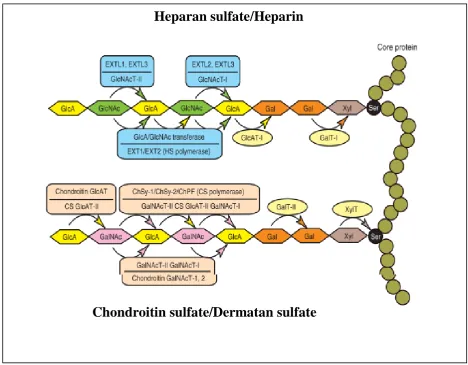
Figure 1.3; Schema of the biosynthetic assembly of the GAG backbones by various glycosyltransferases.
A number of glycosyltransferases are required for the synthesis of the backbones of GAG’s.e.g XylT, Xyl transferase; GalT-I, Gal transferase-I; GalT-II, Gal transferase-II, GlcAT-I, GlcA transferase-I; GalNAcT-I, GalNAc transferase-I; CS GlcAT-II, CS GlcA transferase-II;GalNAcT-II, GalNAc transferase-II; CS polymerase, GlcA/GalNAc transferase; GlcNAcT-I, GlcNAc transferase-I; GlcNAcT-II,GlcNAc transferase-II; HS polymerase, GlcA/GlcNAc transferase; ChSy-1, chondroitin synthase-1; ChSy-2, chondroitin synthase-2; and ChPF, chondroitin polymerising factor.
The biosynthesis of the linkage region is completed by the actions of GlcAT-I;
transferring a GlcA residue to the linkage trisaccharide, Galβ1→3Galβ1→4Xylβ. Moreover,
one study highlights that the expression level of GlcAT-I correlates well with the amount of
Heparan sulfate/Heparin
21
GAGs; thereby suggesting that GlcAT-I regulates the overall expression of GAGs (Uyama et
al, 2007). The resulting GAG chain backbone undergoes extensive sulfation modifications,
during chain elongation, thus making GAG chains some of the most negatively-charged
families of biopolymers in mammalian cells.
1.7 Biosynthetic modification of HS-GAG chain (HS chain elongation)
The nascent HS chain is first modified by N-deacetylase, N-sulfotransferase (NDST),
which is a multidomain enzyme that cleaves the N-acetyl group from the GlcNAc
monosaccharides and transfers a sulfate group to the N-position. This is followed by the
action of the C-5 uronyl epimerase, which converts GlcA to IdoA (Figure 1.4) in selected
positions. 2-O-sulfation of the uronic acid by the 2-O sulfotransferase (2-OST) can follow the
epimerisation process. The 2-OST acts on IdoA- as well as GlcA-containing HS chains; however, the IdoA containing chains are the catalytically preferred substrates (Pinhal et al,
2001: Rong et al, 2001). The final set of modification enzymes are the 6-O (6-OST) and 3-O (3-OST) sulfotransferases, which sulfate the 6-O and 3-O positions of the GlcNAc moiety, respectively. Many of the above biosynthetic enzymes have distinct isoforms that are
selectively expressed in different tissues (Kusche-Gullberg and Kjellen 2003). Four different
isoforms of NDST, three different 6-OSTs, and six different 3-OSTs have been characterised
in humans and mice (Kusche-Gullberg and Kjellen 2003), they all have different specificities
22
Figure 1.4 HS chain elongation
The chains are synthesised on the core protein by the sequential action of individual glycosyl transferases. A
common tetrasaccharide linkage region is formed, followed by the addition of alternating GlcA and GlcNAc
residues, producing in turn the precursor chain. This chain is then enzymatically modified by deacetylation and
N-sulfation, epimerisation and O-sulfation, yielding individual chains whose sequence is distinct from all the other chains.
GLCNAc/GlcA transferase (tout velu/Ext)
UDPglucose Dehydrogenase sugarless
N-deacetlase/ N-sulfotransferase (sulfteless)
GlcA C5-epimerase
2,6-and 3-O-sulfotransferase
UDP- GlcNac UDP-GlcA
GlcA
IdoA PAPS Sulfate
23
1.8 Biosynthetic modifications of CS/DS GAG chains (CS/DS chin elongation)
CS/DS sulfated GAGs [(CS] are covalently attached to their respective core proteins
through the GAG–protein linkage region, GlcAβ1→3Galβ1→3Galβ1→4Xylβ1→O-Ser
(Figure 1.3). The biosynthesis of CS can be broadly divided into that of chondroitin and DS.
CS is comprised of predominantly the GlcA epimer, whereas dermatan is predominantly
comprised of IdoA. Thus, the CS C-5 uronyl epimerase is more prominent in the biosynthesis
of DS. The first GalNAc transfer to the linkage region by the alleged GalNAc transferase-I
leads to the synthesis of the repeating disaccharide region [(→4GlcAβ1→3GalNAcβ1-)n] of
CS/DS through alternate additions of β-GalNAc and β-GlcA by the concerted actions of the
putative GalNAc transferase-II and GlcA transferase-II enzymes, which are specific to CS
backbone synthesis (Figure 1.5).
The three main sulfation events of the CS chains are 4-O and/or 6-O-sulfation of
GalNAc and 2-O-sulfation of uronic acid. There are four different isoforms of the CS GalNAc 4-OST (C4ST-1, C4ST-2, C4ST-3, and D4ST-1) and three different isoforms of the CS GalNAc 6-OST (C6ST, C6ST-2, and GalNAc4S-6ST) (Kusche-Gullberg and Kjellen
24 Figure 1.5; Pathways of CS/DS chain elongation.
25
1.9 Analysis of GAGs and their oligosaccharide fragments
There are various ways to study the biological function of GAGs structures in order to
understand the structure-activity relationships (SAR). Traditionally, the determinations of
GAG’s structures are usually derived from the following steps:
Direct extraction and purification of HS populations from tissues or culture cells arising
from different organs.
Partial chemical and enzymatic modifications of GAGs to produce oligosaccharides of
defined modifications and size (Yates et al, 2004). The structure of these fragments can
be confirmed using disaccharide compositional analysis as well as sequencing methods.
Chemically synthesised GAGs oligosaccharides (Grootenhuis et al, 1995) may shed light
on specific HS structure activity relationships. Synthesis of GAGs structures is a very
long and specialized process. However, solid-phase synthesis and combinatorial
chemistry has greatly advanced this field in recent years (Vohra et al, 2008: Seeberger
and Werz, 2005: Seeberger and Haaze, 2000)
1.10 Traditional methods for extraction and purification of GAGs from Tissue
Samples and Cultured Cells
In general, extraction of GAGs is initiated by the action of proteolytic enzyme
(e.g.Alcalase) on the core protein of PGs. This enzyme degrades the core protein into short
peptides, thereby releasing the corresponding GAGs chain. Dialysis with high concentrations
of harsh organic solvents such as 8 M urea and addition of trichloroacetic acid (Lyon and
Gallagher, 1991) or guandinium chloride are also employed (Yanagishita et al, 1987). These
26
structures of proteins, DNA and RNA. Urea and other chaotrophic agents usually disrupt the
3D structure of proteins by interfering with the stabilizing inter-molecular interactions which
mediate non-covalent forces such as hydrogen bonds, van der Waals forces and hydrophobic
effects (Puvirajesinghe and Turnbull, 2012). The disadvantages of these methods include:
they are lengthy; they can involve multiple sample transfers; they can contain harsh
chemicals; and they may alter pH and cause de-N-sulfation (Inoue and Nagasawa, 1976). Other methods include β-elimination, which involves alkaline borohydride treatment, which
also acts to disrupt the serine-xyloside linkage that attaches GAG chains to the core protein
backbone; all these methods result in the release of intact GAGs from protein cores (Stenstad
et al, 1993).
Another method that can be used for GAGs purification, from cultured cells, involves
the use of a non-ionic detergent solution, such as 1% Triton X-100 0.5 M KCl, which can
then be subjected to density centrifugation using caesium chloride gradients. This separates
the molecules according to their density (Jalkanen et al, 1988). Electrophoretic techniques
can be used for separation of proteoglycans. This technique can be used either separately or
in combination with SAX-chromatography, depending on the degree of purification required
or type of HSPG to be purified.
1.11 Chemical and enzymatic depolymerisation of GAGs used in structural analysis
The polydisperse nature and large size of GAG chains means that it impossible to
determine the structure of individual GAG chains. Structure activity relationships between
GAG chains and proteins must therefore be determined by reducing the size of the chains into
27
GAGs derived from a variety of sources is the analysis of disaccharides composition, which
is commonly achieved by depolymerisation of the GAGs chain by chemical or enzymatic
treatment. Site-specific enzymes are used to degrade the polysaccharides for structural and
analytical purposes (Murata and Yokoyama, 1987: Linhardt 1986). Depolymerisation of the
polysaccharides can results in the creation of smaller saccharides chains, known as
oligosaccharides by partial degradation enzymatic or chemical treatment.
They can also be degraded further by a combination of site-specific enzymes in order
to yield disaccharides, subsequent analysis by SAX-HPLC can give the total disaccharide
composition of the GAG chains. While similar disaccharides may be present in different
tissues, the proportion of each varies considerably thereby allowing the identification of
structural differences between tissues. Furthermore, the domain structures of GAGs can be
analysed to understand the relative distributions of nonsulfated and sulfated domains from
different cells and tissues (Esko and Lindhart, 2001). The structures found in different
domains can be analysed further using oligosaccharide mapping, which provides a
‘fingerprint’ of the domain structures (Turnbull and Gallagher, 1991: Turnbull 1990:
Turnbull and Gallagher, 1988). The development of GAG sequencing technology has been
used to provide more detailed information on the structure of individual oligosaccharides
involved in important biological interactions (Turnbull et al, 1999). GAG oligosaccharides
structures have been studied extensively by using one or more of the following methods and
have provided a wealth of information on the relationships between the fine structures of
GAG chains.
Originally, depolymerisation of HS/heparin was achieved using nitrous acid
hydrolysis. Nitrous acid cleaves in between the hexosamine residues and the hexuronate