D
E
V
E
LO
P
M
E
N
T
INTRODUCTION
Vertebrate neural crest cells originate at the border between the neural plate and the epidermis in the embryonic ectoderm. After their specification, neural crest cells migrate extensively along defined pathways, ultimately giving rise to diverse cell types that include the peripheral nervous system, melanocytes and the craniofacial skeleton (Hall, 1999; Le Douarin and Kalcheim, 1999). Several factors secreted from the neural plate, non-neural ectoderm or mesoderm show neural crest inducing activity in ectodermal explants, when applied alone or in combination. Yet, how the neural crest is specified at the junction between the neural plate and the epidermis remains unclear.
It has been proposed that bone morphogenetic protein (BMP) signaling plays an important role in specification of cell fates at the neural plate/ectodermal border in mouse, chick and frog embryos (Kanzler et al., 2000; Liem et al., 1995; Marchant et al., 1998). During neural development in Xenopus, BMPs are first expressed in the whole ectoderm, but they are then gradually downregulated in the presumptive neural plate, while BMP antagonists such as Noggin, Chordin and Follistatin are secreted from the dorsal axial mesendoderm (reviewed by Sasai and De Robertis, 1997). Several in vitro experiments suggest that a gradient of BMP activity in the
ectoderm is established by interactions between BMPs expressed in the ectoderm and BMP inhibitors secreted from the axial mesendoderm, resulting in the specification of the neural crest at a specific threshold of BMP signaling levels (LaBonne and Bronner-Fraser, 1998; Marchant et al., 1998; Nguyen et al., 1998). However, neural crest formation is specifically restricted to the posterior border of the neural plate, excluding the anterior neural plate (Hopwood et al., 1989; Mayor et al., 1995). This cannot be explained by the BMP gradient alone, and posteriorizing factors such as Wnts, fibroblast growth factors (FGF) and retinoic acid (RA), are required for this anteroposterior localization (Villanueva et al., 2002). Activation of Wnt signaling in the Xenopusembryo or in neuralized animal cap induces neural crest specification, while Wnt inhibition by either Wnt8 knockdown or overexpression of a dominant-negative Tcf3abrogates neural crest specification in zebrafish (LaBonne and Bronner-Fraser, 1998; Lewis et al., 2004; Mayor et al., 1995; Tan et al., 2001). These results led to the two-signal model, suggesting that the neural crest is specified in the posterior neural plate border by the intersection of canonical Wnt signaling with moderate levels of BMP signaling (LaBonne and Bronner-Fraser, 1998; Marchant et al., 1998; Villanueva et al., 2002). Recently, the Notch signaling pathway has also been shown to play an important role in neural crest specification, by regulating the expression of BMP4and the BMP target gene Msx1 at the lateral edge of the neural plate (Glavic et al., 2004).
Here, we show that the Xenopus Tsukushi gene (X-TSK) is strongly expressed at the border of the neural plate at the time of neural crest specification, and that its expression in the ectoderm is regulated by BMP signaling. By biochemical analysis and overexpression assays in Xenopus, we show that: (1) X-TSK works as a BMP antagonist by direct binding to BMPs; (2) X-TSK modulates activation of Notch signaling by directly binding to X-delta-1 extracellular region. Furthermore, X-TSK can regulate
Tsukushi
controls ectodermal patterning and neural crest
specification in
Xenopu
s by direct regulation of BMP4 and
X-delta-1 activity
Sei Kuriyama1,2, Giuseppe Lupo3, Kunimasa Ohta1,4,*, Shin-ichi Ohnuma5, William A. Harris3and Hideaki Tanaka1,2
In Xenopus, ectodermal patterning depends on a mediolateral gradient of BMP signaling, higher in the epidermis and lower in the neuroectoderm. Neural crest cells are specified at the border between the neural plate and the epidermis, at intermediate levels of BMP signaling. We recently described a novel secreted protein, Tsukushi (TSK), which works as a BMP antagonist during chick gastrulation. Here, we report on the Xenopus TSKgene (X-TSK), and show that it is involved in neural crest specification. X-TSK expression accumulates after gastrulation at the anterior-lateral edges of the neural plate, including the presumptive neural crest region. In gain-of-function experiments, X-TSKcan strongly enhance neural crest specification by the dorsolateral mesoderm or X-Wnt8in ectodermal explants, while the electroporation ofX-TSK mRNA in the lateral ectoderm of embryos after gastrulation can induce the expression of neural crest markers in vivo. By contrast, depletion of X-TSK in explants or embryos impairs neural crest specification. Similarly to its chick homolog, X-TSK works as a BMP antagonist by direct binding to BMP4. However, X-TSKcan also indirectly regulate BMP4mRNA expression at the neural plate border via modulation of the Delta-Notch signaling pathway. We show that X-TSK directly binds to the extracellular region of X-delta-1, and modulates Delta-dependent Notch activity. We propose that X-TSKplays a key role in neural crest formation by directly regulating BMP and Delta activities at the boundary between the neural and the non-neural ectoderm.
KEY WORDS: Ectoderm, Neural crest, Neural plate, Epidermis, X-TSK, BMP, Notch, Xenopus Development 133, 75-88 doi:10.1242/dev.02178
1Division of Developmental Neurobiology, Graduate School of Medical Sciences, Kumamoto University, Kumamoto 860-8556, Japan. 2The 21st Century COE program “Cell Fate Regulation Research and Education Unit”, Kumamoto University, Kumamoto 860-0811, Japan. 3Department of Anatomy, University of Cambridge, Downing Street, Cambridge CB2 3DY, UK 4PRESTO, JST, 4-1-8 Honcho, Kawaguchi, Saitama 332-0012, Japan. 5Department of Oncology, The Hutchison/MRC Research Centre, University of Cambridge, Hills Road, Cambridge CB2 2XZ, UK.
*Author for correspondence (e-mail: [email protected])
D
E
V
E
LO
P
M
E
N
T
BMP4 transcription indirectly via modulation of the Notch signaling pathway. Both gain- and loss-of-function assays demonstrate that X-TSK plays a crucial role in patterning of the ectoderm and especially in neural crest specification. We argue that X-TSK functions in the crosstalk of BMP and Notch signaling pathways at the boundary between the neural and non-neural ectoderm to determine the correct specification of the neural crest progenitors.
MATERIALS AND METHODS Screening and RT-PCR
A XenopuscDNA library at stage 24 (high density cDNA filter 725, RZPD)
was screened using C-TSK cDNA as a probe to isolate X-TSK cDNA
(GenBank Accession Number, AB176536).
For RT-PCR, total RNA was isolated by RNeasy Kit (QIAGEN) and TRIZOL purification system (Invitrogen). The obtained total RNA was treated with DNase (Invitrogen) for removing genome DNA contamination,
and first-strand cDNA was synthesized using oligo d(T)12-18primer and
Superscript III reverse transcriptase (Invitrogen). PCR was performed with the Expand Long template PCR system 3 (Roche), with the following
primers: Xslug-U 5⬘-CAATGCAAGAACTGTTCC-3⬘; Xslug-D 5⬘
-TC-TAGGCAAGAATTGCTC-3⬘; XAG-1(Sive and Bradley, 1996); XBF-1U,
5⬘-TCAACAGCCTAATGCCTGAAGC-3⬘; XBF-1D, 5⬘
-GCCGTCCAC-TTTCTTATCGTCG-3⬘;Xotx2 (Blitz and Cho, 1995); Sox2(De Robertis et
al., 1997); Msx1(Tríbulo et al., 2003); BMP4(Dale et al., 1992); XK81
(LaBonne and Bronner-Fraser, 1998);ODC(Agius et al., 2000); Cardiac
actin(Stutz et al., 1986); Sox9and Zic5(Monsoro-Burq et al., 2003);
XESR-1 (Wittenberger et al., 1999); X-notch-1U, 5⬘
-TCCTGATTTATATTG-CTTATCCGAGT-3⬘; and X-notch-1D, 5⬘
-TTACAGAAGTGTTAACA-GCAACAACA-3⬘.
Embryological methods, in situ hybridization and immunostaining
Xenopusembryos were obtained as previously described (Newport and Kirschner, 1982) and staged according to Nieuwkoop and Faber (Nieuwkoop and Faber, 1967). For animal cap assays, mRNA was injected into the animal pole of two- or four-cell stage embryos. Animal caps (ACs) were dissected
from stage 8-9 embryos in 1⫻MBS and cultured in 0.5⫻MBS until early
neurula (stage 14), mid neurula (stage 17) or early tailbud (stage 21/22) stages, depending on the experiment. For dissections of ventral marginal zone (VMZ) or dorsal marginal zone (DMZ) explants, embryos were marginally
injected into ventral or dorsal blastomeres at the four-cell stage with X-TSK,
BMP4or BMP4+X-TSKmRNAs. After culturing to the early gastrula stage
(stage 10-10+), VMZ or DMZ explants (comprising about 60° of the VMZ or
DMZ) were dissected in 1⫻MBS and cultured in 0.5⫻MBS until stage
24-26. Dorsal lateral marginal zone (DLMZ)-AC conjugates were prepared as described by Bonstein et al. (Bonstein et al., 1998).
Whole-mount in situ hybridization was performed as previously described (Harland, 1991; Shain and Zuber, 1996). Pigmented embryos and explants were bleached after the color reaction (Mayor et al., 1995). The
following probes were used: Xdlx3(Woda et al., 2003); ADAM13(Alfandari
et al., 1997); XAG-1(Bradley et al., 1996); XK81(Jonas et al., 1985); Sox2
(Mizuseki et al., 1998); Xrx1(Casarosa et al., 1997); N-tubulin(Richter et
al., 1988); cardiac actin (Mohun et al., 1984); Xotx2 (Pannese et al., 1995);
Xotx5b (Vignali et al., 2000); X-ESR-1 (Wettstein et al., 1997). X-delta-1
was cloned independently (Kiyota et al., 2001). Xbmp4 cDNA was
subcloned into pBSSK (–) from a BMP4expression vector. Xslug, Sox9,
Zic5, Msx-1, Hairy2AandTrp-2cDNAs were isolated by RT-PCR. We raised polyclonal antibody against X-TSK in rabbits (QIAGEN). The specificity of this antibody was checked by SDS-PAGE and western blot. Myc-His tagged X-TSK and X-TSK protein in the lysate of embryo can be detected in blot (data not shown). For double or triple staining, the hybridized embryo was sectioned by cryostat, and detected by anti-X-TSK antibody with Cy3-conjugated secondary antibody (Jackson Immuno Research).
Microinjection of mRNA, short inhibitory double strand RNA (siRNA) or morpholino oligonucleotides
Capped mRNAs were synthesized from linearized plasmid templates with the mMessage Machine kit (Ambion). Embryos were injected with
500-4000 pg mRNA/embryo at the two- or four-cell stage in 0.1⫻MMR with
4% Ficoll and kept in it for a few hours. They were then transferred into
0.1⫻MMR for subsequent culturing. The following RNAs were made as
previously described (Kuriyama and Kinoshita, 2001): X-TSK (pCS2+-
X-TSK), truncated BMP receptor (tBR) (Graff et al., 1994), Chordin(Sasai et
al., 1994), BMP4(Fainsod et al., 1994), Xnr1 (Jones et al., 1995), Notch
Intracellular domain, X-delta-1 and dominant-negative form of Delta
(Chitnis et al., 1995), Suppressor of hairlessDNA binding mutant, and
Su(H)/ Notch ankyrin repeats (Ank) fusion construct (Wettstein et al., 1997). We used the computer program provided on Dr Gregory Hannon’s website (http://www.cshl.edu/gradschool/hannon.html) to predict the
short-hairpin-siRNA sequence for gene silencing of X-TSK. Myc-His tagged
X-TSK was transfected into COS-7 cells with either a control or X-X-TSK siRNA-expressing vector and downregulation of X-TSK protein synthesis was checked by western blotting. After selecting the effective sequences, we
used a X-TSK siRNA designed for the following sequence: 5⬘
-GA-TACCTCGTATCTGGATCTC-3⬘. C-TSK siRNA, which degrades C-TSK
mRNA effectively, was used as control siRNA (Ohta et al., 2004). C-TSK mRNA was used for a rescue experiment of X-TSK siRNA. The applicable
region of the C-TSKsequence was 5⬘-GATACCTCCTACTTGGATTTG-3⬘.
In another loss-of-function experiment, we used a morpholino antisense
oligo (MO) against X-TSK, designed to target the following sequence: 5⬘
-TCTAACAATGGCTCTTTCTTCTTGG-3⬘(Gene Tools). To distinguish
the effect of MO on lateral expression of X-TSK from MO injection to deplete the organizer expression (Ohta et al., 2004), we injected it into ventral border of animal dorsal blastomere at the eight-cell stage corresponding to future D121 and D122 in the cell fate map of 32-cell stage embryo (Moody, 2000).
Western blotting and immunoprecipitation
Myc-His tagged X-TSK, Myc-His tagged C-TSK, a Flag tagged BMP4 plasmids or a Flag tagged X-delta-1 extracellular domain plasmids were transfected into COS-7 cells with LipofectAMINE 2000 (Invitrogen) and the supernatants were harvested after 96 hours culture in serum-free Opti-MEM (Invitrogen). The immunoprecipitation experiment was performed as previously described (Ohta et al., 2004). Myc-tagged and Flag-tagged proteins were detected after blotting using an anti-Myc antibody 9E10 or anti-Flag antibody M2 (Sigma), respectively.
XenopusmRNA electroporation
mRNA electroporation was performed as described by Sasagawa et al.
(Sasagawa et al., 2002). mRNA (200 nl) of X-TSK (2g/ l) and GFP (1
g/ l), or only GFP (3 g/ l) were injected into the space under the
vitelline membrane, and electroporated at stage 11.5. The position of electroporation was at the border between the lightly pigmented dorsal ectoderm and darkly pigmented lateral ectoderm. Electroporated embryos were selected under the fluorescent microscope for GFP expression at stage 14, fixed at the stage 15/16, then analyzed by whole-mount in situ hybridization.
Staining of the branchial arch cartilages
The same procedure was used as previously described (Berry et al., 1998). Samples were fixed overnight in 4% paraformaldehyde/PBS, dehydrated in 95% ethanol and stained with Alcian Blue solution [95% ethanol:acetic acid:0.3% Alcian Blue in 70% ethanol (8 ml:2 ml:1 ml)]. After the staining, samples were left in 95% ethanol for 2 days and then stained with Alizarin Red solution [0.1% Alizarin Red in 95% ethanol:1% potassium hydroxide (0.25 ml:10 ml)] for 2 days. After 6 hours incubation in 1% potassium hydroxide, samples were transferred into 100% glycerol solution for image acquisition.
RESULTS
D
E
V
E
LO
P
M
E
N
T
Xenopus TSKgene (X-TSK). We have previously reported about other Tsukushi homologues in zebrafish, mouse and human (Ohta et al., 2004), all of which have similar characteristics of structure. At the amino acid level, X-TSK shares 59% identity with C-TSK, and 48-49% identity with zebrafish, mouse and human TSK.
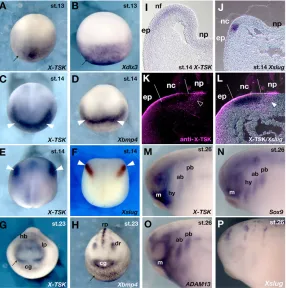
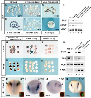
Maternal X-TSKexpression is observed in the animal hemisphere from unfertilized eggs to early gastrula embryos (Ohta et al., 2004). However, at the early neurula stage (stage 13), X-TSKexpression is hardly detectable in the presumptive neural plate region, and restricted to the non-neural ectoderm (Fig. 1A), where its levels increase by stage 14, especially in the presumptive anterior neural fold (Fig. 1C). At these stages, the spatiotemporal expression pattern of X-TSKclosely resembles that of Xbmp4(Fig. 1D) (Fainsod et al., 1994; Hemmati-Brivanlou and Thomsen, 1995; Schmidt et al., 1995) and BMP target genes, such as Xdlx3(Fig. 1B) (Woda et al., 2003).
In addition, X-TSKis expressed in the prospective cranial neural crest, in a similar domain to that of Xslug(Fig. 1E,F) (Mayor et al., 1995). Sections of hybridized embryos at these stages revealed that X-TSKis expressed in a broad area overlapping with the neural fold, but it is hardly observed inside the neural plate or in the underlying mesoderm (Fig. 1I), while Xslugexpression is restricted to the neural crest region (Fig. 1J). We immunostained Xslughybridized embryos with an anti-X-TSK antibody on sections (Fig. 1K,L). X-TSK protein is expressed in the superficial layer of the epidermis and the neural crest region, and also in the proximal edge of the neural crest region in the same layer of the Xslugexpression domain (Fig. 1L, arrowhead). At the early tailbud stage (stage 23), X-TSKexpression is observed in cranial neural crest cells, the dorsal retina and the lens placode (Fig. 1G). Comparison with Xbmp4showed that X-TSKhas a more distinct expression pattern at this stage (Fig. 1H). In the migrating cranial neural crest, X-TSK expression is observed strongly in the mandibular crest segment, weakly in the distal tip of the hyoid crest segment, and in the anterior and posterior branchial
crest segments (Fig. 1M). This expression is similar to that of Sox9 (Fig. 1N) (Spokony et al., 2002) and ADAM13(Fig. 1O) (Alfandari et al., 1997), while Xslugis downregulated in neural crest cells after they leave the neural tube (Fig. 1P).
X-TSKexpression is regulated by BMP4 signaling
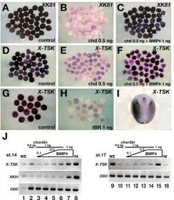
As the early expression domains of X-TSK and Xbmp4 in the ectoderm partially overlap, we speculated that X-TSKexpression might be under the control of BMP signaling. To test this hypothesis, we analyzed X-TSK expression in stage 14 animal caps from embryos injected at the two-cell stage with either 0.5 ng of the BMP antagonist ChordinmRNA or 1 ng of truncated, dominant-negative, BMP2/4receptor (tBR) mRNA. In accordance with its expression in the non-neural ectoderm (Fig. 2I), X-TSKwas clearly expressed in uninjected caps (Fig. 2D,G), which have epidermal character as shown by expression of the epidermal marker XK81(Fig. 2A). By contrast, inhibition of BMP signaling by injection of Chordinor tBR mRNAs downregulated X-TSKexpression, similar to XK81(Fig. 2B,E,H). In caps co-injected with 0.5 ng Chordinand 1 ng of BMP4 mRNAs, both XK81and X-TSKexpression were restored (Fig. 2C,F). Similar results were obtained by semi-quantitative RT-PCR (Fig. 2J). In particular, Chordin-injected caps showed dose-dependent upregulation of X-TSK expression when increasing amounts of BMP4mRNA were co-injected with ChordinmRNA, at both stages 14 and 17. These results indicate that BMP activity positively regulates X-TSKexpression in the ectoderm.
X-TSK antagonizes BMP4 activity and directly binds to BMP proteins in vitro
[image:3.612.54.340.450.738.2]In a previous study, we showed that C-TSKhas dorsalizing activity when overexpressed in Xenopusembryos, which is due to its ability to bind to BMPs directly and antagonize BMP signaling in the extracellular space (Ohta et al., 2004). In addition, C-TSK expression partially overlaps with BMP expression only at the
D
E
V
E
LO
P
M
E
N
T
posterior marginal zone and the posterior primitive streak during chick gastrulation. However, the expression of X-TSK largely overlaps with BMP4 in the ectoderm of Xenopusneurula embryos, and is controlled by BMP4. Thus, to confirm whether these dorsalizing and anti-BMP activities are conserved in X-TSK, we performed overexpression experiments of X-TSKmRNA in Xenopus embryos, and found that X-TSKcan dorsalize ventral mesoderm both in ventral marginal zone explants and in animal caps injected with low doses of Xnr1mRNA, as detected by explant elongation and induction of cardiac actin expression (see Fig. S1 in the supplementary material). In addition, in co-injection experiments, X-TSK can antagonize the ventralizing effects of BMP4 overexpression in dorsal marginal zone explants (see Fig. S1 in the supplementary material). To examine whether X-TSK can bind to
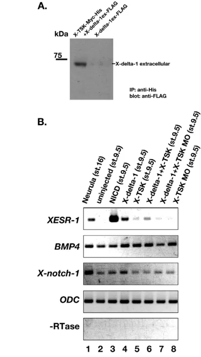
BMP4 directly, we carried out an immunoprecipitation assay between BMP4 and X-TSK or C-TSK proteins (Fig. 3A). When Myc-tagged X-TSK or C-TSK was reacted with FLAG-tagged BMP4, immunoprecipitation of both proteins with nickel chelating resins pulled down BMP4. These data indicate the direct binding of X-TSK to BMP4. Thus, the basic molecular characteristics of Tsukushiare also conserved in the Xenopushomologue.
X-TSKoverexpression induce the identities of
peripheral area of anterior neural plate in animal caps
[image:4.612.50.302.58.347.2]BMP signaling has a crucial role in mediolateral patterning of the ectoderm (Knecht and Harland, 1997; Wilson et al., 1997). To further test the ability of X-TSK as a BMP antagonist, we Fig. 2. X-TSK expression requires BMP signaling.Results of the
[image:4.612.326.538.60.459.2]animal cap (AC) assays. (A-H) Animal caps were prepared from stage 8-9 embryos injected with 0.5 ng Chordin(B,E), 0.5 ng Chordin+ 1 ng BMP4(C,F), 1 ng truncated BMP-receptor(tBR) (H), or none of the above (controls; A,D,G). ACs were harvested at the time equivalent to stage 14 and hybridized with XK81(A-C) or X-TSK(D-H) probes. Chordindownregulated XK81expression (B), which was rescued by BMP4(C). The expression of X-TSKwas repressed by Chordin(E), but rescued by BMP4(F). (H) Truncated BMP receptors repressed the expression of X-TSK. (I) X-TSKexpression in a normal embryo at stage 17. (J) RT-PCR analysis with ACs harvested at stages 14 (lanes 1-8) and 17 (lanes 9-16). ODCis used as an internal control. X-TSK expression in the ACs was recovered by increasing amounts of BMP4. BMP4 overexpression induced X-TSKexpression at both stages. WE, whole embryo; NI, non-injected ACs.
D
E
V
E
LO
P
M
E
N
T
performed animal cap assays and analyzed the induction of the dorsoanterior ectodermal markers. Using whole-mount in situ hybridization, we could detect strong induction of cement gland markers, but weak induction of neural markers, in X-TSK-injected caps (see Fig. S2 in the supplementary material). We then analyzed X-TSK-injected caps by semi-quantitative RT-PCR, which provides more sensitivity than in situ hybridization (Fig. 3B). In these experiments, X-TSKinduced the cement gland marker XAG-1, the telencephalic marker XBF-1, the forebrain and cement gland marker Xotx2, and the pan-neural marker Sox2in a dose-dependent manner (Fig. 3B, lanes 3-5). Interestingly, X-TSKoverexpression had only a weak, if any, effect on the expressions of BMP4 and its target geneMsx1 (Fig. 3B). Comparison with the effects of another BMP antagonist, Noggin, showed that, even at high doses of injected mRNA (4 ng), the effects of X-TSK mRNA on the induction of cement gland/neural markers and on the repression of BMP4and Msx1were clearly weaker than those of NogginmRNA. Co-overexpression of X-TSKand Nogginalso slightly reinforced the effects of Noggin(Fig. 3B, lanes 1,2). These effects were direct and not due to the induction of dorsal mesoderm, as no activation of cardiac actin was detected in the injected caps. Altogether, these data indicate that, while BMP antagonists such as Noggin can strongly inhibit BMP activity and strongly neuralize ectodermal explants, X-TSKoverexpression causes a moderate repression of BMP signaling. Thus, X-TSK may induce peripheral neural plate character in the ectoderm via direct induction of intermediate levels of BMP signaling.
Loss of X-TSK function inhibits neural crest formation in vivo
Several studies in Xenopushave suggested that intermediate levels of BMP signaling are necessary for neural crest specification at the neural plate border (LaBonne and Bronner-Fraser, 1998; Marchant et al., 1998). Thus, it was interesting to see, as described above, that X-TSKis a BMP antagonist strongly expressed in the presumptive cranial neural crest, which can promote intermediate levels of BMP signaling in ectodermal explants. To determine the role of X-TSK, we performed loss-of-function experiments using siRNA (Zhou et al., 2002). The inhibitory effect of X-TSK siRNA (X-TSK-si) sequence was confirmed by the inhibition of X-TSK-Myc protein production after co-transfection in COS cells (data not shown). X-TSK-si was also effective in reducing the levels of endogenous X-TSKmRNA in Xenopusembryos, as shown by RT-PCR (Fig. 4C). Although the endogenous expression of X-TSKincreased from stage 13 in uninjected embryos, this increase was completely prevented in embryos injected with X-TSK-si. Injection of X-TSK-si into one animal blastomere at the four-cell stage (0.3-0.5 pmol/cell) caused abnormal neural fold formation in the neural plate and increased pigmentation at the site where the neural fold is normally formed (Fig. 4A,B).
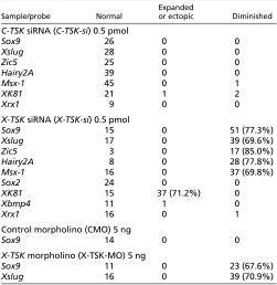
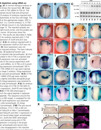
We then analyzed the effects on X-TSK depletion on ectodermal patterning using molecular markers after co-injection of X-TSK-si with -gal mRNA (see Table 1). The expression of the neural crest markers Sox9, Zic5 and Xslug was inhibited in the -gal positive area (Fig. 4E,G,L). No effects were detected after injection of C-TSK siRNA (C-C-TSK-si) (Fig. 4D,F). In addition, while the expression of the neural plate markers Sox2and Xrx1were not affected by X-TSK-si injection (Fig. 4H,J), the expression of the epidermal marker XK81was enhanced (Fig. 4I) to the same extent of the increased pigmentation as shown in Fig. 4B. In these embryos, no changes were detectable in the expression of Xbmp4 (Fig. 4K). These effects were rescued by co-injection of both 500
[image:5.612.312.563.81.339.2]pg C-TSK mRNA (Fig. 4L,M) or 250 pg X-TSKmRNA (Fig. 4N,O), indicating that these phenotypes are specifically caused by the depletion of the TSK protein (Table 2). Previously, we performed morpholino oligonucleotide (MO) injections into the prospective dorsal midline of the embryo to determine the effects of X-TSK depletion on the Spemann organizer and neural induction. In those experiments, we showed that X-TSK MO impairs anterior neural plate specification, as shown by a reduction in the expression domain of Sox2and Xrx1(Ohta et al., 2004). To confirm siRNA effects and to avoid indirect effects due to abnormal organizer formation, we performed MO injections into the prospective lateral ectoderm, as described in the Materials and methods section. In these conditions, both Sox9and Xslugwere diminished by X-TSK MO injection (Fig. 4P,Q). These effects were rescued by the injection of cad-X-TSK mRNA, where the N-terminal region of X-TSK, including the initiation codon and the signal peptides, were replaced by a myc-tagged N-cadherin signal peptide (Fig. 4R,S; see also Table 2). We also examined the effects of X-TSK depletion on the expression of Hairy2Aand Msx1, which identify a pre-neural crest region before neural crest specification (Glavic et al., 2004; Tríbulo et al., 2003). Both Hairy2A and Msx1 were downregulated by X-TSK-si (Fig. 4U,W), but not C-TSK-si (Fig. 4T,V). To confirm the relationship between X-TSK protein reduction and the observed phenotypes, we sectioned the embryos hybridized with neural crest markers and immunostained them with an anti-TSK antibody (Fig. 4X). In the embryos showing downregulation of Sox9and upregulation of XK81after X-TSK-si injection, X-TSK protein expression in the neural crest was reduced on the injected side, while X-TSK endogenous expression was detectable in the proximal edge of the neural crest region on the control side (Fig. 4X, arrowheads). Morphological and molecular analysis at later stages of development confirmed that, in agreement Table 1. Effects of X-TSKdepletion on the expression of various genes
Expanded
Sample/probe Normal or ectopic Diminished
C-TSKsiRNA (C-TSK-si) 0.5 pmol
Sox9 26 0 0
Xslug 28 0 0
Zic5 25 0 0
Hairy2A 39 0 0
Msx-1 45 0 1
XK81 21 1 2
Xrx1 9 0 0
X-TSKsiRNA (X-TSK-si) 0.5 pmol
Sox9 15 0 51 (77.3%)
Xslug 17 0 39 (69.6%)
Zic5 3 0 17 (85.0%)
Hairy2A 8 0 28 (77.8%)
Msx-1 16 0 37 (69.8%)
Sox2 24 0 0
XK81 15 37 (71.2%) 0
Xbmp4 11 1 0
Xrx1 16 0 1
Control morpholino (CMO) 5 ng
Sox9 14 0 0
X-TSKmorpholino (X-TSK-MO) 5 ng
Sox9 11 0 23 (67.6%)
Xslug 16 0 39 (70.9%)
D
E
V
E
LO
P
M
E
N
T
with the reduction of early neural crest markers at neurula stages, inhibition of X-TSK function strongly repressed formation of neural crest derivatives such as melanocytes and the branchial arch cartilages (see Fig. S3 in the supplementary material). Altogether, these results suggest that X-TSKis required at an early step of neural crest specification upstream of Hairy2Aand Msx1, and that in the absence of TSK function in the ectoderm the presumptive neural crest region is at least partially specified as epidermis.
X-TSKis required in the ectoderm for neural crest formation, and can induce neural crest
specification in cooperation with the dorsolateral mesoderm or XWnt-8
[image:6.612.133.548.172.705.2]As previously described, dorsolateral marginal zone (DLMZ) explants dissected from early gastrula embryos at stage 10.25-10.5 induced expression of the neural crest marker Sox9in conjugated animal caps grown to the late neurula stage (Fig. 5A; 78.9%, n=38)
Fig. 4. Effects of X-TSK depletion using siRNA on ectodermal patterning.(A) A normal Xenopusembryo at stage 17; the arrowhead shows the neural fold. (B) Stage 17; an embryo injected with X-TSK siRNA (X-TSK-si). The arrowhead shows a flattened neural fold and enhanced pigmentation. (C) RT-PCR analysis of embryos injected with siRNA radially into all blastomeres at the four-cell stage. The embryos were harvested at the appropriate stages. ODCis used as an internal control. Co, Control embryo; Si, X-TSK-si-injected embryo. (D-S) Whole-mount in situ hybridization of injected samples at stage 15. The injected samples are indicated in the upper right-hand corner, and the probes are in the lower right-hand corner. All pictures show the injected side on the right. The results are described in Table 1, except M-O,R,S. (D,F) An embryo injected with C-TSK siRNA (C-TSK-si). The expression ofSox9and Zic5was unchanged (arrowheads). (E,G) An embryo injected with X-TSK siRNA (X-X-TSK-si). The expressionSox9 or Zic5was not observed (arrowheads). (H) Sox2expression was not disturbed in the X-TSK-si-injected embryo. The bars indicate the width of neural plates. (I) Epidermal keratin was activated in the X-TSK-si-injected side (arrowheads). (J) Xrx1 expression was not disturbed in the X-TSK-si-injected side (arrowhead). (K) Xbmp4expression was not activated (arrowhead). (L,M) Results of the rescue experiment with co-injection of X-TSK-si and mRNAs. (L) X-TSK-si (0.5 pmol) injection diminished Xslugexpression (arrowhead). (M) X-TSK-si (0.5 pmol) and C-TSKmRNA (0.5 ng) were injected into one blastomere of a stage 2 embryo. Xslugexpression was weak, but regionally rescued (arrowhead). (N,O) X-TSK-si (0.5 pmol) and X-TSKmRNA (250 pg) were injected. (N) Sox9 expression was slightly extended alongside -gal staining (arrowhead). (O)Xslugexpression was restored to the same level as control side (arrowhead). (P,Q) An embryo injected with a morpholino oligo of X-TSK (X-TSK MO) (5 ng). Neural crest marker expression, Sox9(P) and Xslug (Q) levels were decreased. (R,S) X-TSK MO phenotype is restored by X-TSK in which the signal peptide is replaced with N-cadherin signal peptide. The sequence around the initiation codon, which is a MO target, is swapped for the N-cadherinsequence. (R) Sox9 expression was observed alongside -gal-positive cells (arrowhead). (S) Xslug expression was restored (arrowhead). (T-W) The pre-neural crest genes in injected embryos. (T,V) C-TSK-si did not change the expression of Hairy2Aor Msx-1 (arrowheads). (U,W) X-TSK-si diminished Hairy2A and Msx-1expression in the cranial to trunk lateral neural plate (open arrowheads). (X) Triple staining sections. Sox9and XK81phenotypic embryos are sectioned, and stained using anti-X-TSK antibody. (Top) Sox9expression is missing in the  -gal-positive region (left, open arrowhead) and neural crest expression of X-TSK has also disappeared in the injected side (left). Normal expression of X-TSK protein was observed
D
E
V
E
LO
P
M
E
N
T
(Bonstein et al., 1998). Sox9induction was much reduced in X-TSK-si-injected caps conjugates with DLMZ explants (Fig. 5B; 15.9%, n=44), but not in uninjected caps conjugated with X-TSK-si-injected DLMZ (Fig. 5C; 75%, n=28). Expressions of the neural crest marker, Zic5 and Xslug were also decreased in a dose-dependent manner in X-TSK-si-injected caps after conjugation with DLMZ, as detected by RT-PCR analysis (Fig. 5G). By contrast, the expression of Sox9 was strongly increased in conjugates of DLMZ with animal caps overexpressing X-TSK mRNA (Fig. 5A,D; 100%, n=15), while it was decreased in uninjected caps conjugated with X-TSK-overexpressing DLMZ (Fig. 5A,E; 41.6%, n=24). These results indicate that X-TSK expression in the ectoderm enhances the response to neural crest
inducing signals derived from DLMZ, while X-TSK
overexpression in the dorsolateral mesoderm has the opposite effect. As X-TSK overexpression in the mesoderm has a dorsalizing effect (see Fig. S1 in the supplementary material), this opposite effect may be explained by dorsalization of the DLMZ.
In Xenopusembryos, neural crest specification requires the action of posteriorizing signals such as Wnts, FGFs and retinoic acid acting on cells with intermediate levels of BMP signaling at the border of the neural plate (Villanueva et al., 2002). These signals may be at least partially produced by the DLMZ (Bang et al., 1999; Monsoro-Burq et al., 2003; Monsoro-Monsoro-Burq et al., 2005; Wu et al., 2003). XWnt-8 overexpression induces ectopic neural crest markers in vivo
(LaBonne and Bronner-Fraser, 1998), and it can induce neural crest specification in animal caps in cooperation with BMP antagonists (Chordinor Noggin) (Christian et al., 1991; LaBonne and Bronner-Fraser, 1998). Therefore, we tested whether X-TSKcould induce neural crest specification in cooperation with XWnt-8. X-TSK -injected caps induced the cement gland marker XAG-1(Fig. 3B, Fig. 5I), but did not show any significant expression of the neural crest marker Xslug(Fig. 5L), while XWnt-8-injected caps showed weak expression of both XAG-1and Xslug(Fig. 5J,M). By contrast, when X-TSKand XWnt-8 were co-overexpressed, Xslugwas strongly induced, while XAG-1expression was reduced compared with X-TSK-injected caps (Fig. 5H,K). RT-PCR analysis confirmed that co-injection of X-TSKand XWnt-8significantly enhanced expression of the neural crest markers Sox9and Zic5, compared with single X-TSK or XWnt-8injection (Fig. 5N). Therefore, the BMP antagonistic activity of X-TSK is sufficient for neural crest specification in cooperation with XWnt-8.
[image:7.612.248.559.365.700.2]The misexpression of X-TSKafter gastrulation can induce neural crest markers in the epidermal area In Xenopus, mRNA injections often show only earlier functions of the corresponding gene, preventing the analysis of later roles for the same gene. For example, while X-TSKor Chordinoverexpression by mRNA injections into early embryos represses neural crest formation (see below), overexpression of a hormone-inducible
Fig. 5. X-TSK induces the neural crest when in combination with Wnt. (A-E) Conjugate assay of ACs (stage 8-9) and the dorsolateral marginal zone (DLMZ) (stage10.5) analyzed by whole-mount in situ hybridization with a Sox9probe. The numbers of phenotypic explants are described in the lower right-hand corners. (A) Sox9expression in the conjugate of a non-injected animal cap and non-injected DLMZ. (B) The conjugate of X-TSK-si-injected AC and DLMZ; Sox9expression is decreased. (C) The conjugate of AC and X-TSK-si-injected DLMZ. (D) The conjugate of X-TSK overexpressing AC and DLMZ; Sox9is expressed in all explants. (E) The conjugate of AC and X-TSK expressing DLMZ. (F) The expression of Sox9at the same stage as ACs. (G) RT-PCR analysis of the AC/DLMZ conjugate assay. X-TSK-si (0-4 pmol) was injected into the animal blastomeres evenly. (H-M) Whole-mount in situ hybridization analysis of the animal cap assay. ACs were prepared from stage 8-9 embryos injected with 0.5 ng X-TSK+ 0.5 ng XWnt-8(H,K), 0.5 ng X-TSK(I,L), or 0.5 ng XWnt-8(J,M). ACs were harvested at the time equivalent to stage 21/22 and hybridized with XAG-1(H-J) and Xslug(K-M) probes. (N) RT-PCR analysis of the animal cap assay. ODC was used as an the internal marker. The amount of injected X-TSKmRNA was 1 ng/embryo, which is enough to induce anterior markers. XWnt-8mRNA (0.5 ng) was co-injected with X-TSK. X-TSKinduced neural crest marker expression in combination with XWnt-8. (O-R) The embryos are transfected by the mRNA electroporation at stage 11.5 after gastrulation. The distribution of GFP fluorescence is indicated in the inset of each figure. (O) Sox9
D
E
V
E
LO
P
M
E
N
T
version of the BMP antagonist Smad6 causes ectopic neural crest induction when its activity is induced after stage 9 (Wawersik et al., 2005). Thus, BMP antagonistic activity is required for neural crest formation after gastrulation. To study X-TSKfunction in vivo at these later stages, we performed mRNA electroporation in stage 11.5 Xenopusembryos (Fig. 5O-R). In these conditions, overexpression of X-TSKin the anterior neural fold extended the expression domain of Sox9(Fig. 5P, arrowhead), and induced ectopic expression in the epidermal region (Fig. 5Q, arrowhead), while GFP mRNA electroporation had no effect (Fig. 5O, arrowhead). Xslugexpression was also enhanced in the electroporated side (Fig. 5R). The electroporation of X-TSKmRNA after early neurula stage 13 caused a slight enhancement of the cranial-facial neural crest at stage 21 (data not shown). These data strongly suggest that X-TSK upregulation in the lateral ectoderm after gastrulation during normal development (Fig. 1C,M) is involved in cranial neural crest specification.
Overexpression experiments suggest that X-TSK can modulate BMP4 transcription and regulate activation of Notch signaling
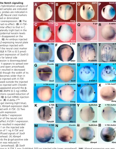
In this study, gain-of-function and loss-of-function analyses clearly suggest the importance of X-TSK-mediated BMP antagonism in ectodermal patterning and neural crest specification. To shed light on X-TSK activity during ectodermal development, we performed additional overexpression experiments in Xenopusembryos. When X-TSKmRNA was injected into one dorsoanimal blastomere of four-cell stage embryos (1 ng/cell; co-injected with 200 pg of  -gal mRNA as a lineage tracer), the expression of the neural crest marker Xslugwas suppressed on the injected side, while -gal alone did not cause any specific phenotype (Fig. 6A,B). Sox9 expression was also suppressed by X-TSK, and even more strongly by a membrane-bound form of X-TSK (X-TSK-CD2),obtained after fusion with the CD2 transmembrane domain (Chang et al., 2001) (Fig. 6C,D). The fact that the effects of both the secreted and membrane-bound forms of X-TSK were very similar, and that the secreted form of X-TSK affected only the area immediately around the -gal-stained region, suggest that X-TSK acts as a short-range factor and does not disperse far away. Unilateral injection of X-TSKalso caused the expansion of the neural plate marker Sox2 (Fig. 6E), the inhibition of the epidermal marker XK81(Fig. 6F), and a downregulation of Hairy2Aand Msx-1in the presumptive
neural crest territory (Fig. 6K) (Table 3). -Gal mRNA did not change the expression of Hairy2Aand Msx-1(data not shown) (Table 3). These effects were dose dependent. Similar to X-TSK overexpression, injection of Chordin mRNA, another BMP antagonist, also caused an expansion of the neural plate (Fig. 6G), and the inhibition of neural crest formation (Fig. 6H). These data confirm that X-TSK acts as a BMP antagonist in vivo, although less efficiently than Chordin. However, Chordinwas not effective in restoring neural crest gene expression in X-TSK-depleted embryos (Fig. 6I,J), suggesting that the function of X-TSK in neural crest formation may not be simply explained by its anti-BMP activity.
In support of the above interpretation, we found that Chordin inhibited Xbmp4 expression in the lateral ectoderm (Fig. 6N), while X-TSKoverexpression enhanced it (Fig. 6M), and that BMP4 overexpression also caused a repression of neural crest formation (Fig. 6O). It has been shown that Notch signaling and its target gene Hairy2Aare involved in neural crest specification in Xenopus, by regulating the expression of BMP4and Msx-1at the lateral border of the neural plate (Glavic et al., 2004). In fact, similar to the results described by Glavic et al. (Glavic et al., 2004), overexpression of Notch-ICDmRNA into early embryos repressed Xslugexpression (Fig. 6P), while overexpression of X-delta-1Stu,
a dominant-negative ligand of Notch, caused upregulation of Xbmp4expression (Fig. 6R). As these effects are similar to those of X-TSK overexpression, we then examined in more detail whether the effects of TSK overexpression are mediated by modulation of the Notch signaling pathway. We first checked the effects of TSKoverexpression on primary neurogenesis, which is known to be controlled by Notch signaling (Chitnis et al., 1995). Analogous with what has previously been shown for Notch overexpression (Chitnis et al., 1995; Chitnis and Kintner, 1996), TSK overexpression repressed the expression of N-tubulin, a marker of differentiated neurons, and that of X-delta-1(Fig. 6S,T). Moreover, X-TSKoverexpression upregulated the Notch-target gene, X-ESR-1(Fig. 6V). Finally, we found that the repression of neural crest specification by TSK overexpression could be overcome by co-injection with Su(H)DBMmRNA, which acts as an antagonist of Notch signaling (Wettstein et al., 1997) (Fig. 6W) (Table 3), while Su(H)DBMmRNA alone very slightly enhanced Sox9 expression (data not shown) (Table 3). By contrast, co-overexpression of X-TSKwith Su(H)/Ank, which works as an agonist of Notch signaling as described by Wettstein et al. (Wettstein et al., 1997), strongly inhibited Xslug and Sox9(Fig. 6Z, data not shown) (Table 3). Su(H)/Ankalone also inhibited the expression of neural crest markers, though more weakly than Notch ICD(Fig. 6Y) (Table 3). However, Xbmp4 upregulation by X-TSK overexpression in the lateral ectoderm could not be rescued by co-injection of Su(H)DBMmRNA (Fig. 6X) (Table 3), consistent
with the fact that the same effect is obtained after overexpression of X-delta-1Stu(Fig. 6R). By contrast, the upregulation ofXbmp4
[image:8.612.49.299.83.223.2]expression by X-TSKoverexpression could be partially prevented by co-injection of Su(H)/Ank mRNA, though some patches of ectopic Xbmp4expression in the lateral area of -gal-positive cells were still detectable (Fig. 6BB, arrows). Su(H)/Ank mRNA alone inhibited Xbmp4expression very weakly (Fig. 6AA, arrowhead). Altogether, these results suggest that TSK overexpression has complex, bimodal effects on the regulation of Notch signaling. Specifically, the repression of neural crest markers and primary neurogenesis appear to be consistent with enhanced Notch signaling, while the activation of Xbmp4 transcription seems to be related to diminished Notch signaling.
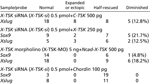
Table 2. Rescue experiments on the X-TSK-si/X-TSKMO phenotype
Expanded
Sample/probe Normal or ectopic Half-rescued Diminished
X-TSKsiRNA (X-TSK-si) 0.5 pmol+C-TSK500 pg
Xslug 18 8 8 5 (12.8%)
X-TSKsiRNA (X-TSK-si) 0.5 pmol+X-TSK250 pg
Sox9 9 2 7 5 (21.7%)
Xslug 5 3 6 2 (12.5%)
X-TSKmorpholino (X-TSK-MO) 5 ng+Ncad-X-TSK500 pg
Sox9 14 0 6 1 (4.8%)
Xslug 18 0 9 6 (18.2%)
X-TSKsiRNA (X-TSK-si) 0.5 pmol+Chordin100 pg
Sox9 3 0 19 0
Xslug 8 0 11 0
D
E
V
E
LO
P
M
E
N
T
X-TSK modulates Notch signaling activation by direct binding to X-delta-1
Our results suggest that X-TSK overexpression can modulate Notch signaling in the neural crest region. In order to determine whether there is a direct interaction between X-TSK and the Notch signaling pathway, we stained Notch-transfected cells and Delta-transfected cells with TSK-myc tagged protein and anti-myc antibody. Binding of TSK protein to Delta-transfected cells, but not to Notch-transfected cells, was detectable (data not shown). To confirm this binding, we performed immunoprecipitation experiments, and found that the X-TSK protein precipitates the X-delta-1 extracellular domain (Fig. 7A). This result suggests that X-TSK modulates Notch signaling by binding to Delta in the extracellular space.
[image:9.612.162.544.54.545.2]To clarify whether TSK works as an activator or an inhibitor of Notch signaling, we performed animal cap experiments by using the Notch-target gene XESR-1expression as a readout for the activation of the Notch signaling pathway (Fig. 7B). As the expression of X-delta-1in early gastrula stages is localized to the marginal zone, uninjected animal caps express Notch, but not X-delta-1 (Wittenberger et al., 1999). Thus, Notch activation does not normally occur in isolated animal caps (Kiyota and Kinoshita, 2002). We then activated Notch signaling in animal caps by injection of X-delta-1 mRNA, and assessed the effects of X-TSK gain- or loss-of-function on this activation. Synthetic mRNAs and/or X-TSK MO were injected into four-cell stage embryos, animal caps were excised from stage 8.5 embryos, and analyzed by RT-PCR at stage 9.5. As Fig. 6. X-TSKinteracts with the Notch signaling
pathway. Whole-mount in situ hybridization analysis of mRNA-injected embryos. Injected samples are indicated in the upper right-hand corners; probes are indicated in the lower right-hand corner. (A,C) Neural crest markers Xslug(A) or Sox9 (C) disappeared or diminished (arrowheads) with X-TSK(1 ng) overexpression. (B) The -gal mRNA (0.5 ng) injection had no effect. (D) X-TSK-CD2mRNA (1 ng) showed a similar effect to that in C. (E) Sox2expression expanded laterally (right bar) in the X-TSK(1 ng)-injected side. (F) Epidermal keratin levels diminished. The anterior border disappeared on the injected side (open arrowhead). (G) An embryo injected with Chordin(100 pg). The Sox2-expressing neural plate is expanded (right bar). (H) An embryo injected with Chordin(100 pg). Expression of the neural crest marker Sox9diminished (arrowhead). (I,J) X-TSK-si (0.5 pmol) and ChordinmRNA (100 pg). Faint expression of Sox9(I) or Xslug(J) was observed in the far lateral side
(arrowheads). (K) Hairy2Aexpression is downregulated on X-TSK-injected side, though it appears to spread over a broader domain (arrowhead and open arrowhead). (L)X-TSK mRNA (1 ng) injection resulted in decreased expression of Msx-1(arrowhead) though the width of its expression area (right bracket) becomes wider than in the control side. (M) An embryo injected with X-TSK (1 ng). Xbmp4expression increased outside the injected side (arrowhead). (N) An embryo injected with Chordin (100 pg). Xbmp4expression disappeared around the  -gal-positive cells (arrowhead). (O) BMP4(0.5 ng) mRNA and (P) Notch ICD (0.5 ng) injections caused reduction of Xslugexpression (arrowheads). (Q)-Gal mRNA injection did not affect Xbmp4expression. (R) X-delta-1Stu injected areas are marked by -gal staining (light blue), the arrowhead indicates ectopic Xbmp4expression (dark purple). (S,T) An embryo injected with X-TSK. (S) Two out of the three strips of N-tubulinexpression disappeared (arrowheads). (T) X-delta-1expression disappeared on the outer border of the neural crest (arrowhead). (U) -gal did not affect X-ESR-1expression. (V) X-TSKmRNA (1 ng) injection resulted in expanded peripheral expression of X-ESR-1on the injected side (arrowheads). (W,X) Co-injection of 1 ng X-TSKand 0.5 ng Su(H)DBMmRNAs. (W) Diffused signals of Sox9
were observed around the arrowhead. (X) Xbmp4 expression was extended towards the -gal-positive side (arrowhead). (Y) Sox9expression was reduced in Su(H)/Ank500 pg injected side (arrowhead). (Z) Sox9
D
E
V
E
LO
P
M
E
N
T
expected, both Notch ICDmRNA and X-delta-1 mRNA caused induction of XESR-1(Fig. 7B, lanes 3, 4). By contrast, co-injection of X-TSKwith X-delta-1caused a much lower induction of XESR-1 compared with X-delta-1 alone, while X-TSKalone could not induce X-ESR1(Fig. 7B, lanes 4-6). Remarkably, co-injection of X-TSK-MO together with X-delta-1 also strongly blocked X-ESR1
activation by X-delta-1 (Fig. 7B, lanes 7, 8). Taken together, these data suggest that the endogenous levels of X-TSK present in uninjected animal caps (Fig. 2D,J) are required for activation of Notch signaling by X-delta-1. Conversely, higher levels of TSK, as those resulting from X-TSK overexpression, can cause inhibition of X-delta-1 activity, suggesting that X-TSK may differentially modulate X-delta-1 activity in a dose-dependent manner in vivo.
DISCUSSION
In this paper, we describe the Xenopushomolog of the Tsukushi gene. The present results suggest that X-TSK is required for proper ectodermal patterning and neural crest specification, acting both as a BMP antagonist and as a modulator of Notch signaling.
Signaling activities of X-TSK
[image:10.612.51.304.157.711.2]X-TSKencodes for a secreted protein belonging to the SLRP family. Although some members of this family, such as decorin, have previously been shown to bind TGF-and modulate its activity Table 3. Gene expression in X-TSKoverexpressing embryos
Expanded
Sample /probe Normal or ectopic Diminished
X-TSK250 pg
Sox9 8 0 5
Sox2 3 10 1
XK81 5 0 6
X-TSK500 pg
Sox9 1 0 6
Sox2 4 1 0
Xslug 5 0 14 (73.7%)
Xbmp4 6 22 (78.6%) 0
Xrx1 9 17 (60.7%) 2
Xotx2 14 35 (71.4%) 0
Six3 3 22 (88.0%) 0
X-TSK1 ng
Sox9 11 0 41 (78.8%)
Xslug 3 0 22 (88.0%)
Sox2 11 44 (80.0%) 0
Xbmp4 16 22 (57.9%) 0
XK81 7 0 26 (78.8%)
N-tubulin 16 1 49 (74.2%)
X-ESR-1 10 22 (57.9%) 6
X-delta-1 18 3 60 (74.1%)
MyoD 26 5 9
Hairy2A 16 0 39 (70.9%)
Msx-1 12 0 40 (76.2%)
-gal500 pg
Sox9 14 0 0
Xslug 16 1 1
Sox2 19 0 0
Xbmp4 62 2 4
XK81 15 0 2
Xrx1 37 0 3
Xotx2 14 0 0
Six3 37 1 2
N-tubulin 28 0 0
X-ESR-1 35 5 2
X-delta-1 44 0 1
Hairy2A 38 0 0
Msx-1 39 0 0
X-TSK1 ng+Su(H)DBM500 pg
Sox9 0 16 (84.9%) 3
Xbmp4 7 16 (69.6%) 0
Su(H)DBM500 pg Weakly
enhanced
Sox9 15 12 0
Su(H)/Ank500 pg Weakly Weakly
enhanced diminished
Sox9 11 3 9
Xslug 10 4 12
Xbmp4 14 2 7
X-TSK1 ng+Su(H)/Ank500 pg
Sox9 13 0 28 (68.3%)
Xslug 4 0 32 (88.9%)
Xbmp4 18 19 (51.3%) 0
X-TSKmRNA (250 pg-1 ng), -galmRNA (500 pg), other RNAs or their mixtures were injected into unilateral blastomeres at the four-cell stage. Embryos showing identical effects on the injected side were counted at stage 15.
[image:10.612.317.523.285.651.2]D
E
V
E
LO
P
M
E
N
T
(Yamaguchi et al., 1990), their role during embryonic development was not clear. We recently described the chick TSK homolog and showed that it works as a BMP antagonist during chick gastrulation (Ohta et al., 2004), while another SLRP member, Biglycan, has also been shown to modulate BMP activity during Xenopus early development (Moreno et al., 2005). Similar to its chick counterpart, X-TSK works as a BMP antagonist, as indicated by overexpression experiments in Xenopusembryos and in vitro assays. In fact, X-TSK overexpression can dorsalize ventral mesoderm (see Fig. S1 in the supplementary material), and it can induce cement gland and neural tissue, but not the dorsal mesodermal marker cardiac actin in animal caps (Fig. 3, see Fig. S2 in the supplementary material), thus mimicking the effects of other known BMP inhibitors, such as Chordin, Nogginor a dominant-negative BMP receptor (Sasai et al., 1995; Sive and Bradley, 1996). In whole embryos, X-TSK overexpression can enhance dorsoanterior fates and repress ventrolateral fates, and these effects are similar to those produced by Chordinoverexpression (Fig. 6G). The opposite effect, namely a reduction of dorsal and an expansion of ventral structures, is observed after X-TSK depletion (Fig. 4) (Ohta et al., 2004). Finally, X-TSK can antagonize the ventralizing activity of BMP4 in mesodermal explants, and it can bind BMP proteins in vitro. Altogether, these data indicate that X-TSK is endowed with a clear BMP antagonistic activity.
However, not all the effects of TSKgain or loss of function can be simply ascribed to its anti-BMP function. In particular, although chick TSKcould efficiently rescue the reduction in neural crest formation caused by X-TSK depletion, Chordincould not (Fig. 4M-O,R,S, Fig. 6I,J). Moreover, although Chordin overexpression repressed BMP4expression in the ectoderm, X-TSKoverexpression had the opposite effect (Fig. 3B, Fig. 6M,N). These data suggest that X-TSK has some signaling activities that are different from BMP antagonism. Thus, it is interesting to see (Figs 6 and 7) that modulation of the Notch signaling pathway is also involved in TSK function, as evidenced by the direct binding of TSK to X-delta-1 in vitro (Fig. 7A). Moreover, both Notch ICDand X-TSKhad similar effects on neurogenesis (Chitinis et al., 1995), neural crest formation (Glavic et al., 2004) and the expression of the Notch-target gene X-ESR-1(Wettstein et al., 1997). Finally, suppression of neural crest formation by X-TSKoverexpression was efficiently rescued by a dominant-negative Su(H), which inhibits Notch signaling, but not by an active form of Su(H)(Fig. 6). Together, these data clearly suggest that X-TSK may also work, at least in part, via activation of Notch signaling. However, the effect of X-TSK on BMP4expression in the anterior peripheral region of the neural plate is similar to that of a dominant-negative Notch ligand, X-delta-1Stu (Glavic et al., 2004), and this X-TSK effect can be prevented, at least in part, by an active form of Su(H), but not by a dominant-negative Su(H)(Fig. 6). This suggests that X-TSKoverexpression may also cause inhibition of Notch signaling at the neural/epidermal border. To gain more insight into the interaction between TSK and delta-1, we performed gain- and loss-of-function experiments of X-TSK in isolated animal caps where Notch signaling was simultaneously activated by X-delta-1overexpression (Fig. 7B). Surprisingly, we found that while the endogenous levels of X-TSK in isolated animal caps are necessary for X-delta-1 activity, higher X-TSK levels resulting from X-TSKoverexpression inhibits X-delta-1 function. Although further work will be needed to dissect the biochemical mechanism by which X-TSK exerts these dual effects on X-delta-1 activity, these results identify X-TSK as a new modulator of the Notch signaling pathway, which works by directly binding X-delta-1 in the extracellular space.
X-TSK is required for ectodermal patterning and neural crest specification
The work described in this paper uncovers a novel function for TSK family members, i.e. the control of neural crest specification. During gastrulation and neurulation, X-TSKexpression is downregulated in the middle of the neural plate, while it remains in the non-neural ectoderm and it accumulates to strong levels at the neural plate border (Fig. 1). This is the region where the neural crest is specified, and therefore it was reasonable to see that both X-TSKgain and loss of function affected neural crest formation. In particular, neural crest specification was inhibited in X-TSK-depleted embryos, which also showed an expansion of the epidermal ectoderm and, depending on the approach, a reduction of the neural plate (Fig. 4) (Ohta et al., 2004). In these experiments, the observation that the neural plate was reduced in embryos injected with a X-TSK-targeted morpholino (Ohta et al., 2004), but not siRNA, may be explained with the fact that siRNA did not apparently affect X-TSK levels during gastrulation, when X-TSKis expressed in the dorsal ectoderm and mesoderm, while the morpholino may be already effective at these stages (Ohta et al., 2004). By contrast, X-TSKoverexpression after gastrulation caused an expansion of the neural crest (Fig. 6). In addition, though X-TSKon its own was not able to induce neural crest markers in animal caps, it could strongly cooperate in this process with the dorsolateral mesoderm or XWnt-8, both of which provide a posteriorizing signal required for neural crest specification (Fig. 5) (Villanueva et al., 2002). These experiments also showed that, consistent with it expression pattern at the neural plate border (Fig. 1), X-TSK function is required in the ectoderm for neural crest specification.
How does X-TSK control neural crest specification? Clearly, its BMP antagonistic activity is likely to play a role. In fact, other BMP inhibitors, such as Chordinor Noggin,can mimic X-TSK ability to induce neural crest markers in cooperation with XWnt-8(LaBonne and Bronner-Fraser, 1998; Mayor et al., 1995). In addition, both gain- and loss-of-function analysis suggest that X-TSK has a more general role in the mediolateral patterning of the ectoderm, which is best explained with its anti-BMP function. A specific gradient of BMP signaling is well-known to be required to specify the neural plate, neural crest and epidermal domains in the Xenopus ectoderm (Marchant et al., 1998). Therefore, one possibility is that the BMP-inhibitory activity of X-TSK, localized at the neural plate border and in the non-neural ectoderm, is essential for the proper shaping of the BMP gradient required for ectodermal patterning, and that the action of BMP antagonists secreted from the dorsal midline, such as Chordin and Noggin, is not sufficient in this respect. This would be in agreement with previous observations that removal of the dorsal marginal zone does not prevent neural crest specification, though it strongly affects neural plate induction (Marchant et al., 1998).
D
E
V
E
LO
P
M
E
N
T
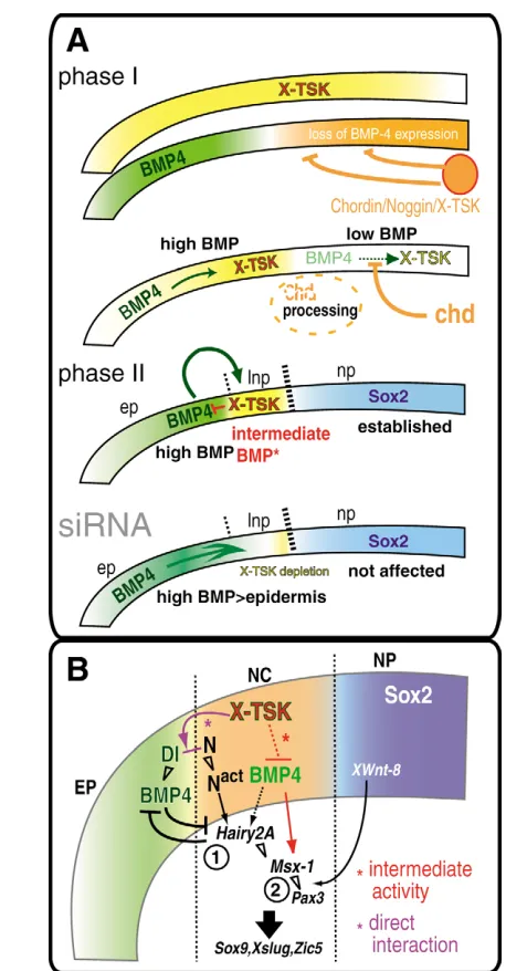
Moreover, duringXenopusdevelopment, it has been described that Notch and its target gene Hairy2A are expressed in the neural crest territory, while the Notch ligands, Delta and Serrate, are expressed in the cells surrounding the prospective crest cells (Glavic et al., 2004). An attractive hypothesis is that, by directly interacting with both BMP4 and X-delta-1 in the extracellular space at the neural/epidermal border, and by indirectly regulating BMP4 transcription in this region, X-TSK may work as a crucial molecular intersection between BMP and Notch signaling in the territory where the neural crest is specified.
Our results also suggest that X-TSK may differentially modulate X-delta-1 activity in a dose-dependent manner, by activating it at lower X-TSK levels and repressing it at higher X-TSK levels. On this respect, it is interesting to note that the distribution of TSK expression levels shows some correlation with the localization of the neural crest. For example, the neural crest is specified at the lateral neural border, where X-TSK expression is lower, and excluded from the anterior neural border, where X-TSK expression is higher (Fig. 1A,C). Moreover, the proximal edge of the neural crest in the lateral neural border overlaps with a region of higher X-TSK expression, while X-TSK expression inside the neural crest domain is lower (Fig. 1K,L; Fig. 4X). As Notch is expressed in the whole neural plate, while X-delta-1 is expressed both at the anterior neural boundary and at both the proximal and the distal edges of the neural crest in the lateral neural boundary (Glavic et al., 2004), the expression pattern of Notch and Delta can only partially account for the specific localization of the neural crest region. Furthermore, although caudalizing signals such as Wnt, FGF and RA are thought to be responsible for the localized induction of the neural crest at the lateral, but not the anterior, neural border, some FGFs and RA-producing enzymes, such as FGF8 and Raldh2, are expressed in the anterior neural border from early neurula stages (Lupo et al., 2005). Therefore, it is tempting to speculate that the presence of higher levels of X-TSK in the anterior neural border and in the proximal edge of the neural crest in the lateral neural border, compared with the neural crest itself, might be an additional mechanism to restrict the boundaries of the neural crest domain
A model of consecutive steps neural crest specification
Previously, two-signal models of neural crest specification were proposed (LaBonne and Bronner-Fraser, 1998; Villanueva et al., 2002), which suggested the requirement of intermediate levels of BMP signaling and posteriorization factors. Based on the results presented in this paper, we propose a model of consecutive steps in neural crest specification that contemplates the dynamic expression pattern of X-TSK and sequential molecular interaction during ectodermal development.
First, during gastrulation, X-TSK is expressed in the whole animal ectoderm and in the dorsal mesoderm (Ohta et al., 2004). Dorsal midline signals, such as Chordin, Nogginand X-TSK, bind to BMPs and directly inhibit their activity in the dorsal ectoderm (Sasai et al., 1994) (Fig. 8A, phase I), leading to the specification of the neural plate at these stages (reviewed by Sasai and De Robertis, 1997).
[image:12.612.51.280.56.495.2]Subsequently, X-TSK expression is downregulated in the presumptive neural plate, while it is maintained and enhanced in the ectoderm flanking the neural plate (phase II). At these stages, in the presumptive neural crest region, X-TSK controls BMP activity through direct binding to BMPs and it also modulates BMP transcription, possibly via modulation of the Notch signaling Fig. 8. A model of consecutive step of the neural crest