IJPSR (2020), Volume 11, Issue 1 (Research Article)
Received on 04 April 2019; received in revised form, 24 July 2019; accepted, 12 August 2019; published 01 January 2020
CYANOBACTERIA ASSISTED BIO-REDUCTION OF SILVER NANOPARTICLE CONJUGATES AND STUDY ON THEIR CHARACTERIZATION AND ANTIBACTERIAL ACTIVITY AGAINST PATHOGENIC BACTERIA
Gunaswetha Kuraganti, Sujatha Edla * and Thrimothi Dasari
Department of Microbiology, Kakatiya University, Warangal - 506009, Telangana, India.
ABSTRACT: Green nanotechnology has recently emerged as an area of
research involving more eco-friendly and energy-efficient approaches for the synthesis of inorganic nanoparticles. The point of the present investigation is to evaluate the capacity of selected strains of freshwater Cyanobacteria (Microalgae) for their capability to biosynthesize silver nanoparticles by utilizing both live biomass of microalgae and cell-free extract by suspending in AgNO3 solution for 72 h of incubation period in both in presence of light and
dark conditions. In most of the cases, silver nanoparticles were formed both in the presence of biomass as well as in the cell-free extract under continues light emission, which indicates that formation of silver nanoparticles involves an extracellular compounds. Synthesized silver nanoparticles were characterized by Ultraviolet-Visible (UV-Vis) Spectroscopy, Scanning Electron Microscopy (SEM), and Transmission Electron Microscopy (TEM). Biosynthesis of silver nanoparticles was indicated by observing gradual colour change of the extract pale yellow to visible brownish-yellow. UV-Vis spectrophotometry study showed absorption maxima at 420-470 nm. The embedment and size of the synthesized silver nanoparticles were characterized by SEM and TEM. The bio-reducted silver nanoparticles size ranged between 20-22 nm depending on organism used in TEM analysis. The presence of bio-component in Cyanobacteria was confirmed by Fourier Transmittance Infrared Spectrum (FTIR) analysis, which is a source of nanoparticle synthesis. The antibacterial activity of synthesized silver nanoparticles was studied on clinically important gram+ve and gram –ve pathogenic bacteria.
INTRODUCTION: Green technology has recently emerged as an area of research involving more environ safe and energy-efficient approach for the synthesis of inorganic nanoparticles 1, 2, 3. Production of nanoparticles using minimal or negligible toxic substances grasps cynosure while ensuring that it poses no damage to the environment and risk on human health 4.
QUICK RESPONSE CODE
DOI:
10.13040/IJPSR.0975-8232.11(1).163-72
The article can be accessed online on
www.ijpsr.com
DOI link: http://dx.doi.org/10.13040/IJPSR.0975-8232.11(1).163-72
Nanoparticles are gaining enormous scientific interest as multifaceted materials exhibiting unique, novel and advanced like electronic, optical, mechanical, magnetic characteristics and chemical properties that are significantly different from those of bulk materials 5. Smaller sized nanoparticles display higher surface to volume ratio; a feature vital to catalytic reactivity, thermal conductivity, antimicrobial activity, chemical steadiness, and non- linear optical performance 6.
Nanoparticles with such type of characteristics playing crucial roles in medical diagnostics, drug delivery systems, antisense and gene therapy applications, and tissue engineering 7. The size, shape and intercalation properties are the special
Keywords:
Cyanobacteria, Silver nanoparticles, Conjugate linked silver nanoparticles,
FTIR, Antibacterial activity
Correspondence to Author: Dr. Sujatha Edla
Assistant Professor,
Department of Microbiology, Kakatiya University, Warangal- 506009, Telangana, India.
attributes of nanomaterials. Nanoparticles are clusters of atoms in the size range of 1-100 nm. Biologically synthesized nanoparticles show slow kinetics which offers better control over crystal growth, reduced capital involved in the production than the chemical synthesis which is not enviro safe
8
whereas physical methods are very expensive and time consuming 9. Biological routes for the synthesis of nanoparticles include use of plant extracts 10, enzymes 11, fungi 12 and algae 13, 14, 5.
Amongst biological systems utilized, microalgae (Cyanobacteria) draw in uncommon consideration since they can bio-remediate toxic metals, in this manner changing over them to more kinds of amiable forms 15. Green syntheses of metallic nanoparticles at high rate have been extensively explored by cyanophycean members, probably due to their capacity of metal accumulation 4. They provide great potential as a source of fine chemicals, pharmaceuticals, and biofuels and are a rich source of pigments/proteins 16. Synthesis and stabilization of the nanoparticles may be facilitated by cell extract of Cyanobacteria containing a vast array of active bio-molecules 16.
Silver ions and silver-based compounds are known bactericides and have great research interest towards nanoparticles and antibacterial agents 17, 18. The efficient antibacterial activity of silver nanoparticle that comes in contact with the microbial cells is due to the large surface area, therefore, the larger particles have a higher percentage of interaction than of the same parent material 19, 20. The bactericidal component includes the arrangement of free radicals that instigate membrane harm as clarified by 21. The different strains of Cyanobacteria produce intracellular and extracellular metabolites have diverse biological activities such as antialgal, antibacterial, antifungal and antiviral activity 22.
The antimicrobial compound production influenced by these metabolites depends on various important factors such as the temperature of incubation, pH of the culture medium, incubation period, medium constituents and light intensity 23. Due to the high growth rate and high biomass productivity, Cyanobacteria are considered a better biological template for nanoscale particle synthesis 24. Cyanobacteria have stress tolerance during adverse
conditions because they possess highly heterogeneous polymers containing a number of distinct polysaccharides and non-carbohydrate constituents including proteins, phospholipids and nucleic acids.
Studies have shown that polysaccharides play a crucial role in bio absorption and binding of toxic heavy metals (silver particles) 25. These properties of bio nanoparticles have significant applications in bioremediation, food & pharmaceutical industries. Mahdieh et al., 26, Chug, Mathur 27, Roychoudhury, Pal 28 have used Cyanobacteria for the production of Ag, Au, pd, Pt nanoparticles. Since alginate and its composites have been used in many biomedical applications including drug delivery and wound dressings. It can also be effectively used for the immobilization of silver nanoparticles 29.
The aim of the present study is to screen the ability of freshwater Cyanobacteria for synthesizing silver nanoparticles (biomass and cell-free extract) in the presence and absence of light. The present work additionally incorporates the utilization of Cyanobacteria as a steady transporter of silver nanoparticles and calcium alginate as a crosslinker between silver nanoparticles and Cyanobacteria. Immobilized Cyanobacterial silver nanoparticles were bio-evaluated against five clinically important bacteria.
MATERIALS AND METHODS:
Isolation and Cultivation of Cyanobacterial Cultures: Silver nanoparticles were synthesized from two Cyanobacterial strains, isolated from freshwater habitats. The cultures were maintained through the common subculturing method in BG11 medium in laboratory conditions at 25 °C under continuous cool white fluorescent light. The isolates were identified based on their morphological characters 30.
Biosynthesis of Ag-NPs by Cyanobacterial Cultures: Detection of Ag NPs formation was performed by a modified method of Mahdieh et al. 26
each culture was then suspended in 20 ml of 1mM aqueous AgNO3 (Sigma Aldrich) solution pH 7.0.
The cell-free algal extract was used for the synthesis of silver nanoparticles by adding 1ml of 100mM AgNO3 to 19 ml of cell-free algal extract
(Patel et al., 2015).
Both sets of experiments (with and without biomass) were incubated at 25 ± 1 °C under cool white fluorescent light (50 μ mol photons m-2s-1) and in dark condition for 72 h of incubation. Fresh BG11 medium with addition of AgNO3 was used as
control. During the incubation period change in color from pale yellowish-green to brown indicates nanoparticles synthesis. The darkening of the brownish color was time-dependent and it was quantified by recording the absorbance spectra during the 72 h of the incubation period. One ml aliquot samples were taken every 12 h and the absorbance of the UV-Vis spectra at a resolution of 1nm at in the range of 300 and 800 nm were recorded by using a spectrophotometer (Shimadzu, Model: UV- 2450).
Electron Microscopic Analysis: Transmission Electron Microscopy (TEM) images were obtained from an FEI TECHNI G₂ TEM operating at an accelerating voltage of 200 KV. Samples were prepared by placing a drop of aqueous biomass solution on the carbon-coated copper grid and dried under infrared lamp prior to examination. Crystalline of structures was confirmed by the selected area electron diffraction (SAED) pattern31.
Scanning Electron Microscopy (SEM): SEM analysis (Hitachi, model: S-3400N) was used to observe microalgal cell with entrapped silver nanoparticles. Thin films of the sample were prepared on a carbon-coated copper grid and drying it by putting it under a mercury lamp for 5 min 32.
FTIR Spectroscopy Analysis: For Fourier Transform Infrared Spectroscopy (FTIR), freeze-dried biomass was used. A small amount of freeze-dried biomass was ground with potassium bromide (KBr). FTIR spectrum of the sample was recorded on a PerkinElmer FT-IR system Spectrum GX model. All measurements were carried out in the range of 400-4000 cm-1 at a resolution of 4 cm-1.
Preparation of Immobilized Cyanobacterial Cells for Antibacterial Activity: The 15-day old
cyanobacterial cells were separated from the medium by centrifugation (4000 rpm / 15 min) followed by filtration with Whatman filter paper. Finally, the Cyanobacterial pellets were lyophilized and stored at -20 °C. Later the cultures were dissolved in PBS (Phosphate buffer saline). 0.2 g of Cyanobacterial cultures Chroococcus sp. MBKG 08 and Microcystis sp. MBKG 16 was dissolved in 10 ml of PBS buffer separately 25.
Preparation of Linker with Cyanobacterial Extracts (Cyanobacteria Linker Solution): 3mM concentration of sodium alginate was prepared by dissolving 70 mg of sodium alginate in 20 ml of distilled water and calcium chloride solution (0.03 M) was added dropwise on continuous stirring to sodium alginate solution. After 10 min, PBS dissolved Cyanobacterial samples were added dropwise to the above mixture. These solutions were used in 5:1:4 ratios 25.
Conjugation of Silver Nanoparticles with Cyanobacterial - Linker Solution: Silver nanoparticles were immobilized by Cyanobacterial linker solution by mixing 5 ml of Cyanobacterial- linker sample to 5 ml of silver nanoparticle solution on continuous stirring for 3 to 4 h.
Test Organisms: Five clinically important bacteria cultures including Staphylococus aureus (MTCC- 3381), Escherichia coli (MTCC-1541), Klebsiella pneumonia (MTCC- 3384), Salmonella paratyphi
(MTCC- 3220), Micrococcus luteus (MTCC- 1541) were obtained from Microbial Type Culture Collection Center, Chandigarh, India and used in this study. The bacterial strains were maintained on nutrient agar slants.
Antibacterial Assay: The antibacterial
susceptibility of silver nanoparticles (AgNPs) was assessed by well diffusion method. Bacterial broth cultures were prepared prior to the experimental setup and used for the antimicrobial assay.
incubation period diameter of the zone of inhibition were measured manually by using a millimeter scale 32.
RESULTS AND DISCUSSION: The two
Cyanobacterial strains Chroococcus sp. MBKG08
Microcystis sp. MBKG16 silver nanoparticles synthesis was confirmed by a color change from pale yellow to reddish-brown. The intensity of color increased with the time of incubation till 72 h, further there was no color change. Synthesis of AgNPs by Cyanobacterial cells was determined by
studying UV absorbance peak in the range of 300- 800 nm (Shimazu, Model: UV- 2450), because of the strong absorption of visible light due to excitation of the nanoparticles associated with Surface Plasmon Resonance, a significant property of silver nanoparticles 33, 34, 35.
The color change of AgNO3 solution was observed
only under light conditions by both the strains. Both the biomass and cell-free extract were able to synthesize silver nanoparticles Fig. 1.
FIG. 1: REDUCTION OF SILVER NITRATE TO NANO SILVER BY CHROOCOCCUS SP. (MBKG08) AND
MICROCYSTIS SP (MBKG16) SUPERNATANT AND BIOMASS
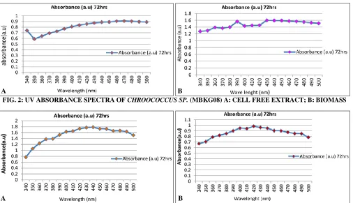
[image:4.612.53.565.226.339.2]FIG. 2: UV ABSORBANCE SPECTRA OF CHROOCOCCUS SP. (MBKG08) A: CELL FREE EXTRACT; B: BIOMASS
FIG. 3: UV ABSORBANCE SPECTRA OF MICROCYSTIS SP. (MBKG16) A: CELL FREE EXTRACT, B: BIOMASS
The UV absorbance of the biomass of Microcystis
(MBKG16) showed a wide peak with 0.984 at 420nm, where for cell-free extract with 1.785 at 440nm Fig. 2, whereas UV absorbance of the peak of 1.598 at 430nm for the biomass of Chroococcus
sp. (MBKG 08) and for cell-free extract the peak was 0.908 at 470 nm Fig. 3. Silver nanoparticle synthesis by Cyanobacterial cells at 420 and 470 nm, corresponding to the Plasmon absorbance suggested that Cyanobacterial extract is efficient in
A B
[image:4.612.56.562.372.663.2]nanoparticle synthesis. According to Sastry et al.,
36
; Pal et al., 19 narrow peak at 425 nm and wide peak at 490 nm correspond to 29 and 89 nm silver nanoparticle respectively. Reduction of silver ions to silver nanoparticles may be due to the capping of microalgal protein metabolites 13. It is reported that the absorption spectrum of silver nanoparticle present maximum peak height between 420 and 450 nm with a blue or red shift with an increase in particle size 37. Though photoreduction of silver ions has been reported by 38 so far the mechanism of action of Ag nanoparticles synthesis was not known, but later it was hypothesized that the silver ions required the NADH dependent nitrate reductase enzyme for their reduction 39, 40. It is reported that proteins, enzymes, sugars, and lipids act as reducing agents in the cell extract in the biological synthesis of nanoparticles. Active functional groups such as hydroxyl groups in tyrosine residues and carbonyl group in aspartic acid and glutamic acid residues were reported for
silver reduction and silver nanoparticles synthesis
41
.
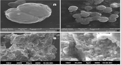
Characterization of SNPs: Scanning Electron Microscopy (SEM) analysis was carried out to understand the topology and size of the cyanobacterial silver nanoparticles.
Light microscopy of Chroococcus sp. (MBKG08),
Microcystis sp. (MBKG16) showed that clusters of synthesized nanoparticles were attached to the surface of the Cyanobacterial cells Fig. 4. This was confirmed by scanning electron microscopy (SEM) which showed that silver nanoparticles were present and evenly distributed throughout the biomass. It has been already demonstrated that the bacterial cell walls may serve as nucleation sites at which Ag+ ions get deposited and get transformed into AgNP’s 20. Particles were not seen in the control culture.
FIG. 4: SCANNING ELECTRON MICROSCOPIC ANALYSIS A) CHROOCOCCUS SP. (MBKG08) AND B)
MICROCYSTIS SP. (MBKG16) AFTER INCUBATION WITH AGNO3
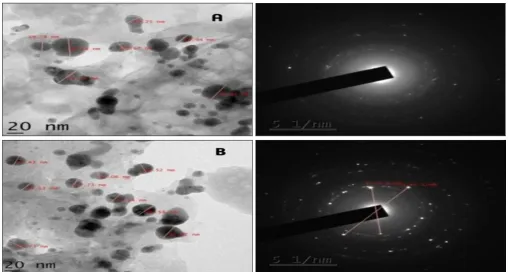
Transmission Electron Microscopy: (TEM) analysis provided information on morphology and size of the silver nanoparticles. TEM images showed that the shape and size of AgNPs varied considerably among the species used Fig. 4. The spherical shape was predominant in the case of
Chroococcus sp. MBKG 08 and ovoid in
Microcystis sp MBKG16. TEM analysis showed
that the particles have a tendency to aggregate within the organic matrix, presumably polysaccharide. It appears that a significant number of nanoparticles are formed or trapped within this matrix Fig. 5, a phenomenon already reported by
42
[image:5.612.53.561.354.628.2]AgNPs synthesized in the range of 20-23 nm with an average size of 22.73 nm in Chroococcus sp.
MBKG08 and 20.67 nm in Microcystis sp.
MBKG16. The crystalline structure of the particles was confirmed by the selected area electron diffraction pattern (SAED).
FIG. 5: TEM ANALYSIS AND SELECTED AREA OF ELECTRON DIFFRACTION PATTERN (SAED) A)
MICROCYSTIS SP. (MBKG16) AND B) CHROOCOCCUS SP. (MBKG08)
FTIR Analysis: FTIR analysis of cyanobacterial
cell biomass and AgNp’s synthesized Cyanobacterial
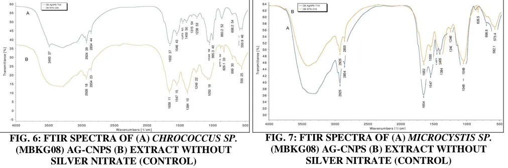
cell biomass showed a different number of peaks representing different functional groups of biological origin. The major stretching and bending frequencies of Chrococcus sp. MBKG08 AgNp’s
Fig. 6 showed a peak at 3493 cm-1 might be assigned to O-H stretching to alcohol and phenol compound. The peaks at 2924 cm-1, 2854 cm-1 may be assigned to C-N, C-H alkanes.
Similarly, stretching was also seen at 1315 cm-1. The stretching’s at 1651 cm-1
and 1546 cm-1 may be assigned to primary and secondary amides i.e.
amide I and amide II bands involving carbonyl and N-H stretching of the proteins. Biomass after bioreduction showed a shift in few peak positions and appearance of new peaks at 3493 cm-1, 1400 cm-1, and 995 cm-1 could be responsible for the adsorption of biomolecules on their surface. Relative investigation of the spectra demonstrated that the cell concentration and Ag-CNPs share certain basic functional groups. However we observed a significant reduction in the frequency curve from 2926 cm-1 to 2924 cm-1, 1661 cm-1 to
1651 cm-1 and 1547 cm-1 to 1546 cm-1 in the cell extract during the formation of Ag-CNPs, this reduction might be due to the utilization of –OH groups in the reduction of Ag+ to Agº.
The stretching and binding frequencies of
Microcystis sp. MBKG16 Fig. 7 showed major peaks. The FTIR peaks of frequencies of 1658 cm-1 and 1547 cm-1 of Ag-CNPs corresponding to the bending vibrations of the amide I and amide II respectively. The peaks reduction from 2926 cm-1 to 2924 cm-1, 1658 cm-1 to 1654 cm-1 may be caused by the reduction of Ag+ to Agº. And the formation of new peaks at 1452 cm-1, 1405 cm-1 is maybe the adsorption of biomolecules on their surface.
[image:6.612.52.560.103.375.2]reduced in the presence of nitrate reductase, in the cell extraction during biosynthesis leading to the formation of Ag-CNPs and stabilization by the capping agent in the medium 43. The amino acid residues bound through amide linkage offer ascent to understand peaks in the infrared region of the electromagnetic spectrum. The stabilization of the silver nanoparticles by proteins may occur by protein nanoparticle interactions either through free amino groups or cysteine residues in the protein
and via the electrostatic attraction of negatively charged carboxylate groups in enzyme present in the cell wall 32, 44. FTIR analysis suggests the presence of phycobiliprotein as the major fraction in Ag-CNPs. In consent to this examination 45, affirmed the existence of protein shell which is in charge of the nanoparticle biosynthesis. Apart from providing harbor ability to nanoparticles on bacterial membranes enabling them to attain antibacterial property 46.
Antibacterial Assay: The antibacterial activity of biosynthesized silver nanoparticles was performed against both gram-negative bacteria (Escherichia. coli, Klebsiella pneumonia, Salmonella typhi, Salmonella paratyphi) and gram-positive bacteria (Staphylococcus aureus, Micrococcus luteus) by well diffusion method. The antibacterial activity is
[image:7.612.56.562.203.371.2]studied by measuring the zone of inhibition 32. The silver nanoparticles synthesized from the isolates showed remarkable antibacterial activity against all tested bacterial strains at a volume of 80 μl, whereas at the volume of 40 μl and 60 μl showed moderate zone size Table 1.
TABLE 1: ANTIBACTERIAL ASSAY AGAINST E. COLI, S. AUREUS, S. PARATYPHI, K. PNEUMONIA, M. LUTEUS
Samples Zone of Inhibition ( diameter in mm)
Conc. E. coli S. aureus S. paratyphi K. pneumonia M. luteus
Silver nitrate (Positive control)
40 μl 1 1 2 1 1
60 μl 1 1 4 1 1
80 μl 3 3 4 2 2
PBS Buffer (Negative control)
40 μl 0 0 0 0 0
60 μl 0 0 0 0 0
80 μl 1 1 1 1 1
Chroococcus sp. MBKG 08 conjugated linked silver
nanoparticles
40 μl 0 0 0 0 0
60 μl 1 1 3 1 1
80 μl 0 1 7 3 3
Mictocystis sp. MBKG 16 conjugated linked silver
nanoparticles
40 μl 1 1 2 1 1
60 μl 2 1 4 2 1
80 μl 3 3 7 3 3
Where, AgNO3 as positive control and PBS buffer as negative control, MBKG: Microbiology Kuraganti Guna
Silver nanoparticles serve as positive control and PBS buffer as the negative control. After 24 h of incubation the conjugated silver nanoparticles of
Chroococcus sp. linker solution of 40 μl showed no inhibition against pathogenic bacteria and 60 μl
showed zone diameter of 3 mm against S. paratyphi and 1mm for four other bacteria whereas for 80 μl of concentration showed considerable zone of inhibition (7 mm) on S. paratyphi and 3 mm, 1 mm of zone diameter against K. pneumonia
FIG. 6: FTIR SPECTRA OF (A) CHROCOCCUS SP.
(MBKG08) AG-CNPS (B) EXTRACT WITHOUT SILVER NITRATE (CONTROL)
FIG. 7: FTIR SPECTRA OF (A) MICROCYSTIS SP.
[image:7.612.46.570.489.657.2]and M. luteus respectively. However, there was no inhibition against E. coli. The conjugated silver nanoparticles with Microcystis sp. MBKG16 linker solution with 40 μl concentration inhibited the growth of S. paratyphi with 2 mm of zone diameter and 1 mm of zone diameter against other four pathogenic bacteria and with 60 μl of concentration, 4 mm of inhibition zone was
recorded against S. paratyphi and 2 mm of diameter against E. coli and K. pneumonia, 1 mm of inhibition zone against S. aureus and M. luteus
whereas for 80 μl of concentration exhibited maximum zone of inhibition (7 mm) against S. paratyphi and 4 mm of zone against E. coli
whereas on S. aureus, K. pneumonai and M. luteus
3 mm of zone of inhibition was observed Fig. 8.
FIG. 8: PETRI PLATES EXHIBITING THE ZONE OF INHIBITION AGAINST PATHOGENIC BACTERIA
In spite of the fact that silver nanoparticles are widely utilized as an antimicrobial specialist, their correct antimicrobial mechanism is yet not clear. It is well known that silver nanoparticles are toxic to microorganisms because they have bacteriostatic and bactericidal effects. The difference in the composition of the cell wall of gram-positive and gram-negative induce different antimicrobial activity. As per prior reports, silver nanoparticles may attach to the surface of cell film disturbing penetrability and respiratory activity of the cell. It is also possible that the silver nanoparticles can also penetrate inside the bacteria 47. In our study, we observed that silver nanoparticles have
comparatively higher anti-bacterial activity against gram-negative organisms than gram-positive, probably due to the thinner peptidoglycon layer and presence of porins 43.
cyanobacteria did have agglomerate and were stabilized for a long period of time due to the secretion of protein from Cyanobacteria which acted as a capping agent to help these conjugates to not lose their bioactivity, whereas non- conjugated silver nanoparticles showed a decreased size of zone of inhibition and bioactivity.
CONCLUSION: In our study, silver nanoparticles with a mean diameter of 20 nm were synthesized using sodium alginate linker solution. The nanoparticles were characterized by UV/Vis spectroscopy. UV/Vis spectra showed the characteristic Plasmon absorption peak for the silver nanoparticles ranging from 420 and 470 nm. FTIR analysis confirmed the reduction of Ag+ to Agº. The results of this study clearly reveals that the Cyanobacterial conjugated silver nanoparticles inhibited the growth and multiplication of the tested bacteria, including drug-resistant strains of S. aureus, S. typhi and S. paratyphi. The conjugated silver nanoparticles of Cyanobacteria improve the stability of silver nanoparticles by the secretion of proteins (capping agent).
The bio-nano-formulation of Cyanobacteria and silver nanoparticles conjugate were effective against pathogenic bacteria than the silver nanoparticles alone. Thus these conjugates may have applications in the fields of biomedical, pharmaceutical, health, food and agriculture.
ACKNOWLEDGEMENT: The authors are
thankful to the University Grant Commission- RGNF (Rajiv Gandhi National Fellowship) F1-17.1/2014-15/ RGNF-2014-15-C-AND -57992, New Delhi India for providing financial assistance to carry out the research work.
CONFLICTS OF INTEREST: There is no
conflict of interest among the authors.
REFERENCES:
1. Kharissova OV, Dias HR, Kharisov BI, Pérez BO and Pérez VM: The greener synthesis of nanoparticles. Trends in Biotechnology 2013; 31(4): 240-8.
2. Mittal AK, Chisti Y and Banerjee UC: Synthesis of metallic nanoparticles using plant extracts. Biotechnology Advances 2013; 31(2): 346-56.
3. Jeffryes C, Agathos SN and Rorrer G. Biogenic nanomaterials from photosynthetic microorganisms. Current Opinion in Biotechnology 2015; 33: 23-31. 4. Sharma A, Sharma S, Sharma K, Chetri SP, Vashishtha A,
Singh P, Kumar R, Rathi B and Agrawal V: Algae as
crucial organisms in advancing nanotechnology: a systematic review. Journal of Applied Phycology 2016; 28(3): 1759-74.
5. Rajeshkumar S, Malarkodi C, Paulkumar K, Vanaja M, Gnanajobitha G and Annadurai G: Algae mediated green fabrication of silver nanoparticles and examination of its antifungal activity against clinical pathogens. International Journal of Metals 2014; 1-8.
6. Kamal SS, Sahoo PK, Vimala J, Premkumar M, Ram S and Durai L: A novel green chemical route for synthesis of silver nanoparticles using Camellia sinensis. Acta Chimica Slovenica 2010; 57: 808-12.
7. Kubik T, Bogunia-Kubik K and Sugisaka M: Nanotechnology on duty in medical applications. Current Pharmaceutical Biotechnology 2005; 6(1): 17-33.
8. Umer A, Naveed S, Ramzan N and Rafique MS: Selection of a suitable method for the synthesis of copper nanoparticles. Nano 2012; 7(05): 1230005.
9. Tan S, Erol M, Attygalle A, Du H and Sukhishvili S: Synthesis of positively charged silver nanoparticles via
photoreduction of AgNO3 in branched poly-ethyleneimine / HEPES solutions. Langmuir 2007; 23(19): 9836-43. 10. Gilaki M: Biosynthesis of silver nanoparticles using plant
extracts. Journal of Biological Sciences 2010; 10(5): 465-7.
11. Schneidewind H, Schüler T, Strelau KK, Weber K, Cialla D, Diegel M, Mattheis R, Berger A, Möller R and Popp J: The morphology of silver nanoparticles prepared by enzyme-induced reduction. Beilstein Journal of Nanotechnology 2012; 3(1): 404-14.
12. Balaji DS, Basavaraja S, Bedre MD, Prabhakar BK and Venkataraman A: Biosynthesis of silver nanoparticles by fungus Trichoderma Reesei Insci J 2011; 1: 65-79. 13. Sudha SS, Rajamanickam K and Rengaramanujam J:
Microalgae mediated synthesis of silver nanoparticles and their antibacterial activity against pathogenic bacteria. Indian Journal of Experimental Biology 2013; 52: 393-399.
14. Jena J, Pradhan N, Nayak RR, Dash BP, Sukla LB, Panda PK and Mishra BK: Microalga Scenedesmus sp.: a potential low-cost green machine for silver nanoparticle synthesis. Journal of Microbiology and Biotechnology 2014; 24(4): 522-33.
15. Parial D, Patra HK, Dasgupta AK and Pal R: Screening of different algae for green synthesis of gold nanoparticles. European Journal of Phycology 2012; 47(1): 22-9. 16. MubarakAli D, Gopinath V, Rameshbabu N and
Thajuddin N: Synthesis and characterization of CdS nanoparticles using C-phycoerythrin from the marine cyanobacteria. Materials Letters 2012; 74: 8-11.
17. Crabtree JH, Burchette RJ, Siddiqi RA, Huen IT, Hadnott LL and Fishman A: The efficacy of silver-ion implanted catheters in reducing peritoneal dialysis-related infections. Peritoneal Dialysis International 2003; 23(4): 368-74. 18. Furno F, Morley KS, Wong B, Sharp BL, Arnold PL,
Howdle SM, Bayston R, Brown PD, Winship PD and Reid HJ: Silver nanoparticles and polymeric medical devices: a new approach to prevention of infection? Journal of Antimicrobial Chemotherapy 2004; 54(6): 1019-24. 19. Pal S, Tak YK and Song JM: Does the antibacterial
activity of silver nanoparticles depend on the shape of the nanoparticle? A study of the gram-negative bacterium
Escherichia coli. Applied and Environmental Microbiology 2007; 73(6): 1712-20.
synthesize silver nanoparticles with antibacterial activity. Biotechnology Reports 2015; 5: 112-9.
21. Kim JS, Kuk E, Yu KN, Kim JH, Park SJ, Lee HJ, Kim SH, Park YK, Park YH, Hwang CY and Kim YK: Antimicrobial effects of silver nanoparticles. Nanomedicine: Nanotechnology, Biology and Medicine 2007; 3(1): 95-101.
22. Soltani N, Khavari-Nejad RA, Tabatabaei Yazdi M, Shokravi S and Fernandez-Valiente E: Screening of soil Cyanobacteria for antifungal and antibacterial activity. Pharmaceutical Biology 2005; 43(5): 455-9.
23. Noaman NH, Fattah A, Khaleafa M and Zaky SH: Factors affecting antimicrobial activity of S. leopoliensis. Microbiological Research 2004; 159(4): 395-402.
24. Shaheen H, Meryam S and Tesneem F: Screening of Cyanoabaterial extracts for synthesis of silver nanoparticles. World Journal of Microbiology and Biotechnology 2015; 31: 1279-83.
25. Ritika C, Minakshi L and Naveen Namita S: Synthesis characterization and antimicrobial efficacy of Cyanoabacterial (polymer) silver nanoparticle conjugates. International Journal of Scientific Engineering and Technology 2015; 3: 1061-68.
26. Mahdieh M, Zolanvari A and Azimee AS: Green biosynthesis of silver nanoparticles by Spirulina platensis. Sci Iran 2012; 19(3): 926-9.
27. Chug R and Mathur S: Extracellular polymeric substances from Cyanobacteria: Characteristics, isolation and biotechnological applications-A. International Journal of Engineering, Science and Technology 2013; 3: 49-53. 28. Roychoudhury P and Pal R: Synthesis and characterization
of nanosilver-a blue-green approach. Indian Journal of Applied Research 2014; 4(1): 54-56.
29. Panáček A , Kvitek L , Prucek R , Kolář M , Večeřová R , Pizúrová N, Sharma VK, Nevěčná TJ and Zbořil R: Silver colloid nanoparticles: synthesis, characterization, and their antibacterial activity. The Journal of Physical Chemistry B 2006; 110(33): 16248-53.
30. Anagnostidis K and Komárek J: Modern approach to the classification system of cyanophytes. 3-Oscillatoriales. Archivfur Hydrobiologie Supplement 1988; 50:: 327-72. 31. Jena J, Pradhan N, Dash BP, Sukla LB and Panda PK:
Biosynthesis and characterization of silver nanoparticles using microalga Chlorococcum humicola and its antibacterial activity. International Journal of Nanomaterials and Biostructures 2013; 3(1): 1-8.
32. Issa AA: Antibiotic production by the Cyanobacteria
Oscillatoria angustissima and Calothrix parietina. Environmental Toxicology and Pharmacology 1999; 8(1): 33-7.
33. Hartland GV: Optical studies of dynamics in noble metal nanostructures. Chemical Reviews 2011; 111(6): 3858-87. 34. Lee J, Kwon MJ, Wark AW and Lee HJ: Nanoparticle-enhanced surface plasmon resonance detection of proteins at attomolar concentrations: comparing different nanoparticle shapes and sizes. Analytical Chemistry 2012; 84: 1702-07.
35. Banerjee P, Satapathy M, Mukhopahayay A and Das P: Leaf extract mediated green synthesis of silver nanoparticles from widely available Indian plants: synthesis, characterization, antimicrobial property and toxicity analysis. Bioresources and Bio-processing 2014; 1(1): 3.
36. Sastry M, Patil V and Sainkar SR: Electrostatically controlled diffusion of carboxylic acid derivatized silver colloidal particles in thermally evaporated fatty amine films. Journal of Physical Chemistry B 1998; 102(8): 1404-10.
37. Sönnichsen C, Franzl T, Wilk T, Von Plessen G and Feldmann J: Plasmon resonances in large noble-metal clusters. New Journal of Physics 2002; 4(1): 93.
38. Nam KT, Lee YJ, Krauland EM, Kottmann ST and Belcher AM: Peptide-mediated reduction of silver ions on engineered biological scaffolds. Acs Nano 2008; 2(7): 1480-6.
39. Roh Y, Lauf RJ, McMillan AD, Zhang C, Rawn CJ, Bai J and Phelps TJ: Microbial synthesis and the characterization of metal-substituted magnetites. Solid State Communications 2001; 118(10): 529-34.
40. Shankar S, Rai A, Ahmad A and Sastry M: Transverse plasmon vibration in silver nanoparticles. Journal of Colloid and Interface Science 2004; 275: 496.
41. Xie J, Lee JY, Wang DI and Ting YP: Silver nanoplates: from biological to biomimetic synthesis. ACS Nano 2007; 1(5): 429-39.
42. Morones JR, Elechiguerra JL, Camacho A, Holt K, Kouri JB, Ramírez JT and Yacaman MJ: The bactericidal effect of silver nanoparticles. Nanotechnology 2005; 16(10): 2346.
43. Metuku RP, Pabba S, Burra S, Gudikandula K and Charya MS: Biosynthesis of silver nanoparticles from
Schizophyllum radiatum HE 863742.1: their characterization and antimicrobial activity. 3 Biotech 2014; 4(3): 227-34.
44. Keskin S, Oya N, Koçberber Kılıç N, Dönmez G and Tekinay T: Green synthesis of silver nanoparticles using Cyanobacteria and evaluation of their photocatalytic and antimicrobial activity. Journal of Nanoparticle Research 2015; 40: 120-127.
45. Ali DM, Sasikala M, Gunasekaran M and Thajuddin N: Biosynthesis and characterization of silver nanoparticles using marine Cyanobacterium, Oscillatoria willei
NTDM01. Digest Journal of Nanomaterials and Biostructures 2011; 6(2): 385-90.
46. Singh G, Babele PK, Shahi SK, Sinha RP, Tyagi MB and Kumar A: Green synthesis of silver nanoparticles using cell extracts of Anabaena doliolum and screening of its antibacterial and antitumor activity. Journal of Microbiology and Biotechnology 2014; 24(10): 1354-67. 47. Prabhu S and Poulose EK: Silver nanoparticles:
mechanism of antimicrobial action, synthesis, medical applications, and toxicity effects. International Nano Letters 2012; 2(1): 32.
All © 2013 are reserved by International Journal of Pharmaceutical Sciences and Research. This Journal licensed under a Creative Commons Attribution-NonCommercial-ShareAlike 3.0 Unported License.
This article can be downloaded to Android OS based mobile. Scan QR Code using Code/Bar Scanner from your mobile. (Scanners are available on Google Playstore)
How to cite this article: