British Journal of Dermatology (ig^^) 120, 1-7.
Gamma interferon induces different keratinocyte
cellular patterns of expression of HLA-DR and
DQ and intercellular adhesion molecule-1
(ICAM-i) antigens
C.E.M.GRIFFITHS, JJ.VOORHEES AND B.J.NICKOLOFF*
Departments of Dermatology and *Pathology, University of Michigan Medical Center, Ann Arbor, Michigan, U.S.A.
Accepted for publication 30 June 1988
SUMMARY
With indirect immunofluorescetice techniques we demonstrated that recombinant gamma-interferon induced the expression of the class II antigens HLA-DR and HI.A-DQ as well as intercellular adhesion molecule-i (ICAM-i) on normal, cultured human keratinocytes grown in low-calcium, serum-free medium. Each antigen displayed a distinctive cellular staining pattern. HLA-DR was strongly localized to perinuclear zones with intense cell surface expression; HLA-DQ displayed a perinuclear accentuation, but with minimal cell surface staining, and ICAM-i was strongly expressed in a diffuse cytoplasmic pattern with intense cell surface expression. Keratinocytes grown in medium supplemented with 10",, fetal calf serum underwent differentiation, with a diminished expression of all three antigens as compared to those grown in low-calcium, serum-free medium.
These results confirm that gamma interferon can differentially regulate DR and HLA-DQ expression; that there are probably different biochemical metabolic pathways by which these three molecules are expressed on keratinocytes, and that the expression is also a function of the degree of keratinocyte differentiation. The strong cell surface expression of ICAM-i is suggested to be of major importance as the recognition molecule, by which T cells bind to gamma interferon exposed keratinocytes, and suggests an integral role for this molecule in epidermal lymphocyte trafficking.
The human class II major histocompatibility complex (MHC) antigens are coded for by three distinct loci on chromosome six: HLA-DR, HLA-DQ and HLA-DP.' Unlike class I MHC
Correspondence: Brian J.Nickoloff, M.D., Ph.D., Department of Pathology, University of Michigan Medical Center, M4232 Medical Science I, 1301 Catherine Street, Ann Arbor, MI 48109-0602, U.S.A.
antigens, which are ubiquitously expressed, class 11 antigens have a limited cellular distribution on human nucleated cells.""^ HLA-DR expression on macrophages is necessary for antigen to be presented to T lymphocytes/ and for induction of cytolytic T lymphocyte activity,*^ and is involved in the autologous mixed lymphocyte reaction (AMLR).^ Much less is known about the functional significance of HLA-DQ. However, it has similar effects to HLA-DR and Gonwa et
alJ have shown it to be required for antigen presentation in the AMLR. In normal skin, both
HLA-DR and HLA-DQ are found on the same cells, namely vascular endothelium,** Langerhans cells'* and the acrosyringial epithelium.'^ Under normal circumstances neither antigen is expressed by keratinocytes (KC).^^ In diseased skin, particularly the inflammatory dermatoses, HLA-DR is commonly expressed by KC' ^ and has been related to the presence of intra-epidermal lymphocytes.'' HLA-DQ expression by KC is infrequent although it has been observed in cutaneous T cell lymphoma, lichen planus,'** Sorr^/ia-induced skin disease,'^ and allergic and irritant contact dermatitis.'*'
Gamma interferon IFN-y, induces HLA-DR expression on normal cultured human KC'^ and direct intradermal injection of IFN-7 will induce the appearance of HLA-DR on the overlying epidermal KC.' ^ However, IFN-v has not been demonstrated to induce HLA-DQ on normal cultured human KC'"* or on KCs in a short-term organ culture system.' '-^^
Intercellular adhesion molecule-i (ICAM-i) is a cell surface glycoprotein of molecular weight 90-114 kd expressed by both haemopoeitic and non-haemopoeitic cells.^''^^ It is involved in the regulation of lymphocyte-cell adherence mediated by lymphocyte function-associated antigen-i (LFA-i) which is a ligand for ICAM-i.^^ The adherence of resting and activated autologous and allogeneic T lymphocytes to IFN-y treated KC is blocked by anti-LFA-i antibodies^*^'^^ or anti-ICAM-i antibody,^*" thus implying a role for ICAM-i in lymphocyte-KC interactions in the inflammatory dermatoses.
We have investigated the cellular distribution patterns of HLA-DR, HLA-DQ and ICAM-i in normal human cultured KC in serum-free and serum-supplemented medium and the effect of IFN-y on the expression of these antigens.
METHODS
Preparation of keratmocyte cultures
Samples of normal human skin were obtained from patients undergoing cosmetic surgery and single cell suspensions prepared according to the method of Liu and Karasek.^^ Small, round, viable cells were seeded onto and grown in either 10 cm diameter Petri dishes (Lux Flow Laboratories, Inc) or Lab-Tek chamber slides (Miles Scientific, Inverhill, IL, U.S.A.) in either a serum-free KC growth medium containing low calcium^^ (KGM, Clonetics, CO., San Diego, CA, U.S.A.) or Dulbecco's modified Eagle's medium (DMEM, Gibco Laboratories, Grand Island, NY) supplemented with io'\, heat inactivated fetal calf serum (FCS, Gibco). Cells were maintained in a humidified incubator in 5",, Co2/95"() air at 37 C and used after passage 3.
Rccombinant IFN-7 (specific activity^ i 7 x 10^ U/mg; kindly provided by Dr M.Shepard, Genentech Inc., South Francisco, CA, U.S.A.) was used at a final concentration of 1200 U/ml and added to dishes or slides containing sub-confluent KC, after which incubation was continued for 3 days at 37 C.
Staining of keratinocyte for HLA-DR, HLA-DQ and ICAM-i
Gamma interferon induction of keratinocyte antigens
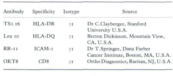
TABLE I. Monoclonal antibodies used in the study
Antibody Specificity Isotype Source
TS1.16 Leu 10 RR-ii OKT8 HLA-DR HLA-DQ ICAM-I CDS
yi Dr C.Clayberger, Stanford University U.S.A.
)'i Becton Dickinson, Mounlain View, CA, U.S.A.
71 Dr T.Springer, Dana Farber Cancer Institute, Boston, MA, U.S.A. ji Ortho Diagnostics, Raritan,NJ, U.S.A.
min on ice with lo/^g/ml of the panel of monoclonal antibodies listed in Table i. OKT8 was used as an isotype irrelevant control antibody. The cells were washed and then stained with i /ig of fluorescein isothiocyanate (FITC)-conjugated anti-mouse IgG (Tago Inc., Burlingame, CA, U.S.A.). In order to investigate the possibility of anti-HLA-DQ antibody binding to HLA-DR antigen, which may have been altered after acetone fixation,^^ KC were stained directly with FITC-conjugated Leu 10 (Becton Dickinson, Mountain View, CA, U.S.A.) for 25 min on ice and compared with KCs pre-incubated with TS i • 16 for i h on ice prior to staining with FITC-conjugated Leu 10. The cells were examined using an Olympus BH-2 fluorescence microscope.
Cultured KC were trypsinized using 003"o trypsin/ooi",, EDTA, an aliquot of 10'' KC was stained as described above, and staining intensity and numbers of cells stained determined using fluorescence activated cell sorter (Epics 541: Coulter Corp), as previously described.'^
RESULTS
Induction of HLA-DR, HLA-DQ and ICAM-i antigen expression by IFN-y
Normal human KC grown in KGM in the absence of IFN-y did not express DR, HLA-DQ or ICAM-I. However, the addition of IFN-y (1200 U/ml) induced distinct patterns of cellular expression for all three antigens. The staining patterns described combine the cell surface staining seen in the absence of fixation or permeabilization, and the intracellular staining seen after acetone treatment.
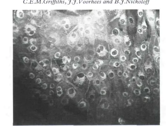
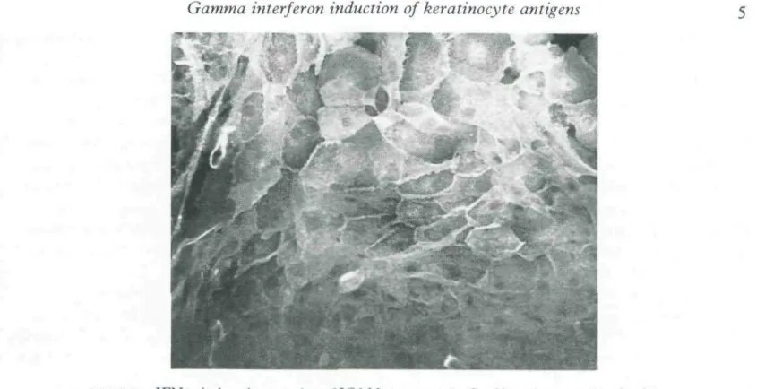
HLA-DR was localized to the perinuclear zone with intense cell suface expression (Fig. i), as has been observed previously.~^" HLA-DQ showed perinuclear localization, but with minimal cell surface staining (Fig. 2). ICAM-i was strongly expressed in a diffuse cytoplasmic pattern with intense cell surface expression (Fig. 3). Using FITC-conjugated Leu 10 (anti-HLA-DQ) there was no observable inhibition of staining when the KC were pre-incubated with TS 116, suggesting that the Leu 10 antibody was not binding to HLA-DR antigen (data not shown). This was also confirmed by the difference in the intensity of cell-surface staining between HLA-DR and HLA-DQ.
Effect of serum-supplemented medium on the IFN-y-induced expression of HLA-DR, HLA-DQ and ICAM-I
FIGURE I. IFN-y-itiduced expression of HLA-DR on acetone-fixed keratinocytes showing perinudear and intense cell surface staining (original x 150).
FIGURE 2. IFN-)'-induced expression of HLA-DQ on acetone-fixed keratinocytes showing perinudear and minimal ceil surface staining (origitial x 150).
not shown). There was no cell surface expression of HLA-DQ on KC cultured in DMEM + io"i, FCS and the intracellular staining was also markedly reduced.
Intensity of cell surface expression of HLA-DR, HLA-DQ and ICAM-i antigens using FACS analysis
[image:4.520.117.472.306.494.2]Gamma inierferon induction of keratinocyte antigens
FIGURE 3. IFN-j'-induced expression {i! it,AM-i an accKmc-tixcd kcrannocyiL-s showing intense cell surface and difFuse cytoplasmic staining (original x 150).
These values are for KC grown in KGM supplemented with IFN-;' (i 200 U/ml) for 3 days. In the absence of IFN-y, the mean channel readings were 0 for all three antigens, as well as for the 0KT8 stained KC. It is important to use isotype controls because we have previously observed that IFN-y induces expression of Fc receptors in cultured KC.' "^ It should also be noted that the mean channel readings presented are on a log scale; thus the values represent a considerable degree of fluorescence intensity.
DISCUSSION
This study has demonstrated the ability of IFN-y to induce contrasting and distinctive cell surface and intracellular expression of HLA-DR, HLA-DQ and ICAM-i antigens in keratinocytes. The expression of these antigens was diminished by growing the KC in medium supplemented with serum. The induction of HLA-DQ by IFN-y is of interest as Basham et al.' ^ in a previous study, were unable to demonstrate this antigen on normal human KC. This disparity can be explained by the fact that they cultured KC in DMEM + io'\, FCS which we have shown to abrogate the cell surface expression of HLA-DQ. The observed reduction in IFN-y-induced antigen expression in serum-supplemented medium was most probably a result of the greater degree of KC differentiation observed under these culture conditions.
higher concentrations of IFN-y produced by dermal or intra-epidermal activated T lympho-cytes in these particular dermatoses. An analogous situation has been observed in auto-immune thyroid disease where in class II antigen expression by thyrocytes HLA-DR is more commonly and intensely expressed than HLA-DQ.^'^ The same pattern is seen in cultured thyrocytes treated with IFN-"/.^^ Measurement of steady-state mRNA levels transcribed from different HLA-DQ loci may allow further identification of the genes upon which IFN-y acts in human KC. Further study of the regulation of HLA-DQ is required since one allele (HLA-DQ/0 has recently been found to be associated with pemphigus vulgaris.^^ Furthermore, HLA-DQ expression by lymphoid cells has been associated with antigen-specific suppression of the immune response. ^^^ Since IFN-y treated KCs have also been observed to induce antigen-specific unresponsiveness,^^ it will be of interest to determine whether such reactions are mediated via HLA-DQ.
The ability of IFN-7 to induce ICAM-1 expression on the surface of KC and the intense cell surface expression of this antigen is of major importance in the lymphocyte-KC adherence reaction in vitro~'*'^^ and may also be relevant in many inflammatory dermatoses in vivo}'' It has been demonstrated that anti-LFA-i and anti-ICAM-i antibodies, but not anti-HLA-DR antibodies, inhibit the binding of T lymphocytes to IFN-y-treated KC"'*"^'' thus producing additional evidence that ICAM-i is involved in this process. The prominent cell-surface expression of this antigen by KC after IFN-y exposure supports this idea.
Thus, it is postulated that activated T lymphocytes in the dermis induce ICAM-i expression on KC by means of IFN-7 production. The present work extends our previous suggestion that IFN-"; is the common molecule responsible for the expression of HLA-DR on KC in a variety of inflammatory dermatoses.**" ICAM-i expression of KC could promote lymphocyte-KC adherence reactions and the subsequent events leading to class II antigen induction on KC, which may contibutc to the pathophysiological changes observed in a variety of inflammatory dermatoses.'^
ACKNOWLEDGMENTS
This work was partially supported by NIH Grant AR38957 (B.J.N.) and the Babcock Dermatologieal Endowment Fund. C.E.M.G. was supported by the British Association of Dermatologists Dowling Travelling Fellowship.
REFERENCES
1 Giles RC, Capra JC. Structure, function and genetics of human class II molecules. In: Advances in Immunology, (Dixon FJ, ed.) London; Academic Press, 1985, 37: 1-71.
2 Hammerling GJ. Tissue distribution of la antigens and their expression on lymphocyte sub-populations. Transplam
Revig-j6, 30: 64-82.
3 Natali PG, Segatio O, Ferrone S ei al. Differential tissue distribution and ontogeny of DC-1 and HLA-DR antigens.
itniHunogenetics 1984; 19: IO9-16.
4 Rosenthal AS, Shevach EM. Function of macrophages in antigen recognition by guinea-pig T lymphocytes. I. Requirement for histocompatible macrophages and lymphocytes. J Exp Med 1973; 138: 1194-212.
5 Weinberger O, Herrman SH, Mescher MF et al. Cellular interactions in the generation of cytotoxic T-lymphocyte responses: roleof la positive splenic adherent cells in presentation of H-2 antigen. Proc Nail Acad Sci USA 1980; 77: 6091-95.
6 Raff HV, Picker LJ, Stobo J. Macrophage heterogeneity in man: a sub-population of HLA-DR bearing macrophages required for antigen induced T cell activation also contains stimulators for autologous-reactive T cells,
Gamma interferon induction of keratinocyte antigens 7
7 Gonwa TA, Picker LJ, Raff HV er al. Antigen-presenting capabilites of human monocytes correlates with their expression of HLA-DS, and la determinant distinct from HLA-DR. J Immunol 1983; 130: 706-11.
8 Natalj PG, de Martino C, Quaranta V er al. Expression of la-like antigens in normal non-lymphoid tissues. Transplantation 1981; 31: 75-1^.
9 Stingl G, Tamaki K, Katz SI. Origin and function of epidermal Langerhans cells. Immunol Rev. 1980; 53; 149-74. 10 Murphy GF, Shepard RS, Harrist TJ ei al. Ultrastructural documentation of HLA-DR antigen reactivity in normal
human acrosyringial epithelium. J Invesl Dermatol 1983; 81: 181-3.
11 Messadi DV, Pober JS, Murphy GF. Effects of recombinant-interferon-)' on HLA-DR and HLA-DQ expression by skin cells in short-term organ culture. Lab Invest 1988; 58: 61-7.
12 Aubock H, Romani N, Grubauer G, Friisch PI. HLA-DR expression on keratinocytes is a common feature of diseased skin. Br J Dermatol 1986; 114: 465-72.
13 Shiohara T, Moriya N, Tanaka Y et al. Immunopaihologic study of lichenoid skin diseases: Correlation between HLA-DR positive keratinocytes or Langerhans cells and. e^\dexn\oirop\cT csW^.J Am Acad Dermaiol 1987; 18167-
74-14 Volc-PlatzerB,GrohV, Wolff K. Differential expression of class 11 alloantigens by keratinocytes in disease. _7^"^^sf Dermatol 1987; 89: 64-8.
15 Tjernlund U, Scheynius A, Asbrink E, Hovmark A. Expression of HLA-DQ antigens on keratinocytes in Borrelia spirochete-induced skin lesions. J Immunol 1986; 23: 383-8.
16 Gawkrodger DJ, Carr MM, McVittie E et al. Keratinocyte expression of MHC class II antigens in allergic sensitization and challenge reactions and in irritant contact dermatitis. J Invest Dermalol 1987; 88; 11-16, 17 Basham TY, Nickoloff BJ, Merigan TC, Morhenn VB. Recombinant gamma interferon induces HLA-DR
expression on cultured human keratinocytes. _7 Invest Dermatol 1984; 83: 88-90.
18 Nathan CF, Kaplan G, Lewis WR el al. Local and systemic effects of iniradermal recombinam interferon-gamma in patients with systemic leprosy. N EngI J Med 1986; 31s: 6-11.
19 Basham TY, Nickoloff BJ, Merigan TC, Morhenn VB. Recombinant gamma interferon differentially regulates class II antigen expression and biosynthesis on cultured normal human keratinocytes. _7 Interferon Res 1985; 5: 23-7. 20 Griffiths CEM, Voorhees JJ, Nickoloff BJ. Characterization of intercellular adhesion molecuie-i and HLA-DR expression in normal and inflamed skin: modulation by recombinant interferon-7 and tumor necrosis factor. J Am Acad Dermatol (in press).
21 Dougherty GJ, Murdoch S, Hogg N. The function of human intercellular adhesion molecule-1 (ICAM-i) in the generation of an immune response. Hur J Immunol 1988; 18: 35-9.
22 Duscin ML, Rothlein R, Bhan AK ei al. Induction by IL-1 and interferon-y: Tissue distribution, biochemistry, and function of a natural adherence molecule (ICAM-i). J Immunol 1986; 137: 245-54.
23 Marlin SD, Springer TA. Purified intercellular adhesion molecule-1 (ICAM-i) is a ligand for lymphocyte function-associated antigen-i (LFA-i). Cell 1987; 51: 813-19.
24 Nickoloff BJ, Lewinsohn D, Butcher EC et al. Recombinant gamma interferon increases the binding of peripheral blood mononuclear leukocytes and a leu-3' T lymphocyte clone to cultured keratinocytes and to a malignant cutaneous squamous cell carcinoma line that is blocked by antibody against the LFA-i molecuie. J Invest Dermatol 1988; 90: 17-22.
25 Nickoloff BJ, Mitra RS. Phorbol treatment enhances binding of mononuclear leukocytes to autologous and allogencic gamma interferon treated keratinocytes which is blocked by anii-LFA-1 monoclonal antibody. J Invest Dermatol 1988; 90: 684-9.
26 Dustin ML, Singer KH, Tuck DT, Springer TA. Adhesion of T lymphoblasts to epidermal keratinocytes is regulated by interferon-y and is mediated by intercellular adhesion molecule-i (ICAM-i). J Exp Med 1988; 167:
1323-40-27 Liu SC, Karasek M. Isolation and growth of aduh human epidermal keratinocytes in cell culture. _7/«^"f Dermatol 1978;71:157-62.
28 Boyce ST, Ham RG. Calcium regulated differentiation of normal human epidermal keratinocytes in chemically defined clonal culture and serum-free serial culture. J Invest Dermalol 1983; 81 (Suppi.): 33-40.
29 Chen Y-X, Evans RL, Pollack MS et al. Characterization and expression of the HLA-DC antigens defined by anti-Leu 10. Hum Immunol 1984; 10: 221-35.
30 Wikner NE, Huff JC, Norris DA era/. Study of HLA-DR in cultured human keratinocytes. J Invest Dermatol 1986; 87: 559-64.
32 Todd I, Pujol-Borrell R, Abdui-Karim AS et al. HLA-D Subregion expression by thyroid epithelium in autoimmune thyroid diseases and induced in vitro. Clin Exp Immurtal 1987; 69: 532-42.
33 Sinha AA, Brautbar C, Szafer F er al. A newly characterized HLA-DQ allele associated with pemphigus vulgaris. Science 1988; 239: 1026-9.
34 Hirayama K, Matsusuita S, Kikuschi I et al. HLA-DQ is epistatic to HLA DR in controlling the immune response 10 schistosomal antigen in humans. Nature 1987; 327: 426-30.
35 Gaspari A, Jenkins M, Katz SI. Induction of antigen-specific T cell unresponsiveness by Ia"*^ keratinocytes. J Invest Dermatol 1988; 90: 562.
36 NickoloffBJ,BashamTY, MeriganTC, Morhenn VB. Keratinocyte class II histocompatibility antigen expression. Br J Dermatol 1985; 112: 373-4.