
Nephropathy in human immunodeficiency
virus-1 transgenic mice is due to renal transgene
expression.
L A Bruggeman, … , T M Coffman, P E Klotman
J Clin Invest.
1997;
100(1)
:84-92.
https://doi.org/10.1172/JCI119525
.
HIV-associated nephropathy (HIVAN) is a progressive glomerular and tubular disease that
is increasingly common in AIDS patients and one of the leading causes of end stage renal
disease in African Americans. A major unresolved issue in the pathogenesis of HIVAN is
whether the kidney disease is due to renal cell infection or a "bystander" phenomenon
mediated by systemically dysregulated cytokines. To address this issue, we have used two
different experimental approaches and an HIV-1 transgenic mouse line that develops a
progressive renal disease histologically similar to HIVAN in humans. In the murine model,
kidney tissue expresses the transgene and in heterozygous adults, renal disease develops
shortly thereafter. We demonstrate by terminal deoxynucleotide transferase-mediated
dUTP-biotin nick-end labeling assay that similar to the disease in humans, apoptosis of
renal tubular epithelial cells is a component of the molecular pathogenesis. To determine
whether apoptosis is due to transgene expression or environmental factors, we treated fetal
kidney explants (normal and transgenic) with UV light to induce transgene expression.
Apoptosis occurred in transgenic but not normal littermates after stimulation of transgene
expression. To confirm a direct effect of HIV expression on the production of HIVAN, we
transplanted kidneys between normal and transgenic mice. HIVAN developed in transgenic
kidneys transplanted into nontransgenic littermates. Normal kidneys remained disease free
when transplanted into transgenic littermates. Thus, the renal […]
Research Article
Find the latest version:
The Journal of Clinical Investigation Volume 100, Number 1, July 1997, 84–92
Nephropathy in Human Immunodeficiency Virus-1 Transgenic Mice Is Due to Renal
Transgene Expression
Leslie A. Bruggeman,* Steven Dikman,‡ Charis Meng,* Susan E. Quaggin,§ Thomas M. Coffman,i and Paul E. Klotman*
*Divison of Nephrology and ‡Department of Pathology, Mount Sinai Medical Center, New York 10029; §Division of Nephrology, Yale
University, New Haven, Connecticut 06510; and iDivision of Nephrology, Duke University Medical Center and Durham VAMC, Durham,
North Carolina 27710
Abstract
HIV-associated nephropathy (HIVAN) is a progressive glomerular and tubular disease that is increasingly common in AIDS patients and one of the leading causes of end stage renal disease in African Americans. A major unresolved is-sue in the pathogenesis of HIVAN is whether the kidney disease is due to renal cell infection or a “bystander” phe-nomenon mediated by systemically dysregulated cytokines. To address this issue, we have used two different experi-mental approaches and an HIV-1 transgenic mouse line that develops a progressive renal disease histologically simi-lar to HIVAN in humans. In the murine model, kidney tis-sue expresses the transgene and in heterozygous adults, re-nal disease develops shortly thereafter. We demonstrate by terminal deoxynucleotide transferase–mediated dUTP-biotin nick-end labeling assay that similar to the disease in hu-mans, apoptosis of renal tubular epithelial cells is a compo-nent of the molecular pathogenesis. To determine whether apoptosis is due to transgene expression or environmental factors, we treated fetal kidney explants (normal and trans-genic) with UV light to induce transgene expression. Apop-tosis occurred in transgenic but not normal littermates after stimulation of transgene expression. To confirm a direct ef-fect of HIV expression on the production of HIVAN, we transplanted kidneys between normal and transgenic mice. HIVAN developed in transgenic kidneys transplanted into nontransgenic littermates. Normal kidneys remained dis-ease free when transplanted into transgenic littermates. Thus, the renal disease in the murine model is intrinsic to the kidney. Using two different experimental approaches, we demonstrate a direct effect of transgene expression on the development of HIVAN in the mouse. These studies suggest that in humans, a direct effect of HIV-1 expression is likely the essential cause of HIVAN, rather than an
indi-rect effect of cytokine dysregulation. (J. Clin. Invest. 1997.
100:84–92.)Key words: HIV-associated nephropathy •
apop-tosis • chronic renal failure • glomerulosclerosis • tubular
in-terstitium
Introduction
Human immunodeficiency virus–associated nephropathy (HIVAN,1 reviewed in references 1–3) is a complication in
z 10% of African American AIDS patients (4, 5). HIVAN has been reported to occur at any point in AIDS progression with no apparent correlation with viral burden or CD41 T cell number (1, 6, 7). Clinically, HIVAN is characterized by heavy proteinuria and rapid progression to end stage disease. Renal biopsy material generally reveals both glomerulosclerosis and tubulo-interstitial lesions, with extensive epithelial cell injury and microcystic tubular dilation. Hyperplasia of both tubular and glomerular visceral epithelium can be observed in addi-tion to increased deposiaddi-tion of extracellular matrix in the mes-angium and tubulointerstitium.
One of the fundamental questions that remains to be an-swered is whether HIVAN is caused by infection of renal cells or is a secondary phenomenon due to HIV-1 infection at an-other site. Although initial studies were inconclusive as to whether renal cells were infected (8–11), recent studies have provided stronger evidence that renal cell infection may occur (12, 13). Likewise, several cytokines are released by HIV-1– infected lymphocytes and are present at high systemic levels including, IL-1, IL-6, interferon g, basic fibroblast growth fac-tor, transforming growth factor b, tumor necrosis factors a and
b (14). Many of these cytokines have been implicated as patho-genic in both in vitro and in vivo models of renal disease (15, 16). Not surprisingly, the pathological basis for HIVAN has centered on these two issues: (a) a direct effect of HIV-1 ver-sus (b) an indirect effect of the dysregulated cytokine milieu.
In the present manuscript, we describe studies addressing this issue using two different experimental approaches based on observations in humans and in the murine model. In hu-mans, the tubulointerstitial lesion and the formation of micro-cysts is the prominent pathogenic feature of HIVAN. Re-cently, apoptosis has been found to be a common mechanism of tubule damage in many different renal diseases including congenital polycystic disease, obstructive uropathy, hyperten-sion, crescentic glomerulonephritis, and ischemic renal disease (17–20). Apoptosis or programmed cell death is also an impor-tant feature in kidney development and regeneration (21, 22). Previous studies with knock-out mice for bcl-2, a gene which protects from apoptosis, reveal normal development of almost all organs except the kidney (23). The bcl-2 null mice develop polycystic kidneys and renal failure similar to autosomal domi-nant polycystic kidney disease, and this study suggests that the
Address correspondence to Leslie A. Bruggeman, Division of Ne-phrology, Box 1243, Mount Sinai School of Medicine, One Gustave L. Levy Place, New York, NY 10029. Phone: 241-8002; FAX: 212-987-0389; E-mail: [email protected]
Received for publication 16 January 1997 and accepted in revised
loss of this protective mechanism for apoptosis may contribute to renal tubule pathogenesis (24).
Apoptosis is an important feature of cell death associated with HIV infection. After exposure to HIV-1 envelope pro-tein, T cells undergo apoptosis (25). In a recent study using bi-opsy material from patients with HIVAN, apoptotic nuclei could be detected in both the interstitium and the tubules (26). Glomeruli in contrast, had little evidence of apoptotic change. Similarly, glomeruli from patients with different forms of nephropathy had very few apoptotic cells. This suggests that abnormal apoptosis either during development or in disease may be a critical mechanism for renal tubule damage and the development of micro or macrocysts.
In this study, we used two approaches to the HIV-1 trans-genic mouse model to demonstrate that expression of the viral transgene in renal cells and not exposure to the circulating mi-lieu is sufficient to induce the features of HIVAN. Using ex vivo fetal transgenic kidneys we demonstrate that transgene expression induces apoptosis in renal tubular structures. Using reciprocal transplantation of transgenic and normal kidneys into normal and transgenic mice, we demonstrate that the his-tological features of HIVAN develop only when the kidneys are transgenic. These studies support the hypothesis that a di-rect effect of HIV-1 gene products or even possibly HIV-1 in-fection of the kidney causes HIVAN.
Methods
Transgenic mice. The development of the transgenic mouse line TgN(pNL43d14)26Lom (“Tg26”) has been reported previously (27, 28). These mice contain 10 copies of the HIV-1 proviral DNA pNL4-3d1443 under the transcriptional control of the native long terminal repeat (LTR). The transgene was generated by deletion of a 3-kb SphI/BalI fragment within pNL4-3 spanning the gag and pol genes, rendering this construct nonreplicating and noninfectious (27, 28). We have previously shown the homozygous and heterozygous mice develop multiple phenotypes including a progressive renal disease in heterozygotes (28), a wasting syndrome (29), a myopathy and myosi-tis in homozygotes (30), and skin lesions and cataracts (31). We have also previously documented the pattern of transgene expression in the kidney and other tissues (28, 32). The transgenic animals used for these studies were from an inbred colony (over seven generations) es-tablished at Mount Sinai using animals obtained from the original National Institutes of Health (NIH) colony. In the Mount Sinai col-ony, all phenotypes and genotypes (as determined by restriction frag-ment length polymorphisms) have not changed when compared with the original colony. Inbreeding in our colony has, however, increased the penetrance of the phenotype of renal disease to 100% of adult an-imals when compared with our previous report of 60% at the NIH (28) when the animals were not strictly inbred. All studies using mice were approved by the Institutional Animal Care and Use Commit-tees of Duke University and Mount Sinai Medical Centers.
Light microscopy. Kidneys from adult normal and heterozygous transgenic mice with and without disease were prepared for light mi-croscopy. Kidneys were fixed in 4% paraformaldehyde (PFA)/PBS overnight and 4-mm paraffin sections were stained with hematoxylin and eosin. Apoptotic nuclei were counted in three sections per speci-men, five high power fields (HPF) per each section from normal mice (n5 7), transgenic mice with 11 proteinuria (n5 7), and transgenic mice with edema (n5 7). The total number of glomeruli observed in each group were 105 in the normals, 99 in the transgenics with 11
proteinuria, and 60 in the transgenics with edema. Analysis of vari-ance was performed and all numbers with a P value , 0.05 were sub-jected to Fisher’s post hoc test to determine critical difference (Stat-view 4.0.1, Abacus Concepts Inc., Berkeley, CA).
Embryo culture. Day 12 fetuses were dissected from pregnant Tg26 heterozygous mice and both kidneys were removed. Pups from three pregnant Tg26 heterozygotes were obtained and two litters (15 pups total) were used for in situ hybridization and one litter (eight pups) was used for electron microscopy. DNA was extracted from the remainder of each fetus and used for Southern blot analysis to identify transgenic and nontransgenic fetuses as previously described (33). Each kidney was placed in a separate 35-mm tissue culture dish with ME/ Ham F12 plus 10% FBS and cultured for 7 d (34). At days 3 and 5, one of each pair was exposed to 12.5J/m2 UV light (254 nm) (35). At the end
of the culture period, kidneys were either fixed for electron microscopy in 3% glutaraldehyde/PBS or processed for in situ hybridization.
Terminal deoxynucleotide transferase-mediated dUTP-biotin nick-end labeling assay. Adult mouse kidneys were fixed for 4 h in 4% PFA/PBS followed by freezing in OCT compound (Tissue-Tex; Miles Inc., Elkart, IN). Frozen sections (10 mm) were incubated with termi-nal deoxynucleotide transferase and FITC-dUTP as described (36).
In situ hybridization. The RNA expression plasmids pGM92 and pGM93 were obtained from the NIH AIDS Research and Reference Reagent Program (contributed by John Rossi). These plasmids con-tain (in opposite orientations) 1.1 kb of the 39 end of the HIV-1–cod-ing region that is common to all HIV-1 mRNAs. Both plasmids were digested with EcoRI and sense and antisense transcripts were synthe-sized using T7 and digoxigenin-UTP using a kit from Boehringer Mannheim (Genius; Indianapolis, IN). The probes were cleaved with alkali to z 500-bp fragments as recommended by the manufacturer.
Fetal kidney tissue was fixed for 30 min in 4% PFA/PBS and then transferred to 30% sucrose/PBS overnight. Adult kidneys were bi-sected and fixed for 2 h in 4% PFA/PBS. Fixed tissue was blocked in paraffin and cut into 4-mm sections and mounted onto poly L-lysine coated, RNase-free slides. In situ hybridization was carried out using a digoxygenin–antidigoxygenin technique as described previously (Wilson, P.D., O. Deruyst, L. Gatti, D. Falkenstein, S. Robinson, D. Fambrough, and C.R. Burrow, manuscript submitted for publication) and with the following modifications. After hybridization and wash-ing, slides were treated with 20 mg/ml RNase for 30 min at 378C, fol-lowed by two washes in 0.23 SCC at 378C. A 4-h incubation with 1 mM levamisole was also included before blocking with sheep serum. Color development was with 5-bromo-4-chloro-3-indolyl phosphate (BCIP) and nitroblue tetrazolium salt (NBT; Boehringer Mannheim). Sections were quantitated by counting the number of positively stained cells in at least three high power fields per each specimen.
Kidney transplantation. To further examine the relative contribu-tions of renal versus circulating factors in the pathogenesis of HIVAN, we performed kidney cross-transplantation experiments. To deter-mine the role of renal expression of HIV-1 genes in isolation, kidneys from heterozygous HIV-1 transgenic mice were transplanted into nontransgenic littermates (n5 2). Conversely, to examine the role of systemic HIV-1 expression in the pathogenesis of HIVAN, kidneys from nontransgenic donor mice were transplanted into heterozygous HIV-1 transgenic littermates (n5 2). Transplantation of fully vascu-larized mouse kidney grafts, followed by bilateral native nephrec-tomy was performed as described previously (37). Proteinuria was as-sessed in donor and recipient mice before transplantation using Chemsticks (trace to 31 scale), and monitoring was continued for up to 24 wk after transplantation. After 24 wk, animals were killed and the kidney grafts were harvested and fixed in PFA/PBS. Hematoxylin and eosin–stained thin sections were prepared and examined for the presence of histomorphological abnormalities.
Results
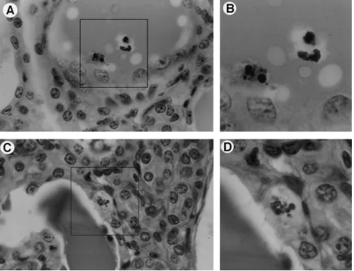
con-densed and fragmented chromatin typical of apoptotic cell death (Fig. 1). Normal adult kidneys had 0.17960.042 (P,
0.02) apoptotic cells/HPF as compared with 0.76460.090 (P,
0.001) in adult transgenic kidneys with early stages of disease (11 proteinuria). Edematous mice with end stage disease had 2.73060.187 (P, 0.001) apoptotic cells/HPF. Apoptosis was almost exclusively observed in tubules with only rare apoptotic cells observed in glomeruli from transgenic kidneys.
To confirm these renal epithelial cells were apoptotic, kid-neys from adult animals with end stage disease were evaluated for the presence of apoptotic cells using the terminal deoxynu-cleotide transferase–mediated dUTP-biotin nick-end labeling (TUNEL) assay. The TUNEL assay detects DNA fragmenta-tion by fluoresceinating the free ends of the DNA such that cells undergoing apoptosis can be detected by fluorescence mi-croscopy. Light microscopy of an unstained frozen section of adult, diseased kidney shows both a glomerulus and tubules (Fig. 2 A). The TUNEL assay from this region (Fig. 2 C) had a significant number of apoptotic cells, predominantly in the ar-eas of microcysts with little apoptosis observed in glomeruli. Normal adult kidneys typically had only rare apoptotic cells
(Fig. 2 B). The results of the TUNEL assay suggested that transgene expression in the kidney may induce apoptosis in the tubular epithelial cells.
Transgene expression in adult kidney. Previous studies evalu-ating transgene expression using Northern analysis and reverse transcriptase polymerase chain reaction indicated that the kid-ney expresses the transgene with peak expression near wean-ing age (28). Since these studies used mRNA prepared from intact kidneys, the specific cell types contributing to the trans-gene mRNA pool was unknown. To determine which cell types within the kidney express the transgene, in situ hybrid-ization on adult kidneys was performed (Fig. 3). Transgene ex-pression was detected in both cortex and medulla. Sporadic cells in the glomerulus and Bowman’s capsule were positive. However, the majority of staining was in the tubular epithe-lium with some staining in the tubular interstitium. Thus, cell types with higher levels of transgene expression correlated with the cell types undergoing apoptosis.
[image:4.612.57.558.60.443.2]Transgene expression induces apoptosis.To determine whether apoptosis was caused by alterations in cytokines that are known to affect apoptosis or by a direct effect of HIV-1 gene
Figure 1. Identification of apoptotic epithelial cells in adult transgenic mouse kidneys with HIVAN. (A) Epithelial cell being sloughed into a di-lated tubule lumen containing a proteinacious cast. (B) Magnification of boxed region in A showing chromatin condensation into apoptotic bod-ies. (C) Cell contiguous with the epithelial cells lining a dilated tubule containing a proteinacious cast. (D) Magnification of boxed region in C
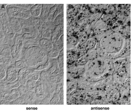
expression, we cultured kidneys ex vivo. Whole organ culture can only be accomplished in fetal tissue and we have previ-ously shown transgene expression during fetal development (33). Day 12 fetal kidneys from transgenic and normal fetuses were grown in culture for 7 d and developed normally. To in-crease transgene expression, fetal kidneys were irradiated with UV light to directly upregulate transcription of the HIV-1 LTR. Fetal kidneys treated with UV light had elevated steady-state levels of viral mRNA as confirmed by in situ hybridiza-tion (Fig. 4). Expression of the transgene was evident at a low level in the unirradiated kidneys (Fig. 4 B, average of five cells/ HPF, but was enhanced sixfold in the UV stimulated tissue (Fig. 4 C, average of 30 cells/HPF).
[image:5.612.57.299.71.339.2]Apoptosis in the UV irradiated kidneys was increased as detected by light and electron microscopy. Light microscopic comparisons of the irradiated and unirradiated kidneys showed
Figure 2. Identification of apoptotic cells in transgenic mouse kid-neys by TUNEL assay. TUNEL assay was performed on adult, dis-eased transgenic, and nontransgenic mice. (A) Unstained frozen sec-tion; “G,” glomerulus. (B) TUNEL fluorescence of a nontransgenic control in region of tubules. (C) TUNEL fluorescence in transgenic kidney corresponding to the boxed region of panel A exhibiting tubu-lar dilatation.
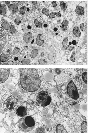
[image:5.612.57.518.350.739.2]dark staining aggregates in the transgenic kidneys irradiated with UV light (Fig. 5). The number of apoptotic nuclei in the irradiated transgenic kidney averaged 26 cells/HPF, a mini-mum twofold increase over unirradiated transgenic or irradi-ated normal kidneys. Because large clusters of apoptotic nuclei were detected in the luminal space of the developing comma bodies after irradiation, an exact apoptotic cell number was impossible to determine (see Fig. 5 C). Electron micrographs of the dark staining material showed condensed and frag-mented nuclei with a centrifugal pattern of condensation on the nuclear membrane, and many dense chromatin masses which form due to the disintegration of the nucleus (Fig. 6). These typical apoptotic changes were most evident in comma and S-shaped bodies, areas of tubule formation. These changes were not observed in nontransgenic or unirradiated transgenic kidneys. Since fetal kidney culture was performed in the ab-sence of potential circulating factors, these results suggest
transgene expression in renal cells alone is sufficient to induce apoptosis in the tubular epithelium.
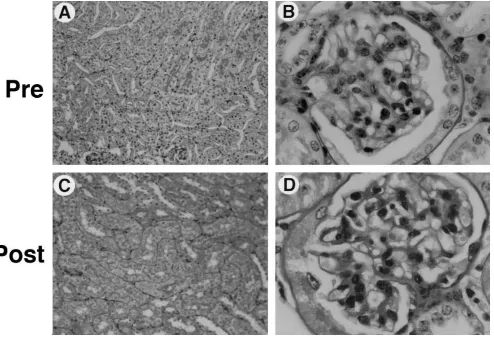
[image:6.612.58.556.59.255.2]Renal cross-transplantation. To establish the role of renal transgene expression in the development of renal disease in vivo, we performed reciprocal kidney transplantation between normal and transgenic mice. To examine the role of renal fac-tors in the pathogenesis of HIVAN, we transplanted kidneys from heterozygous HIV-1 transgenic mice into nontransgenic littermates. Histological examination of the contralateral kid-ney, harvested from the transgenic mouse donors at the time of surgery (Fig. 7, A and B), showed modest abnormalities that suggested early focal glomerulosclerosis; no microcystic changes were observed. One of the nontransgenic mice that received a transplant kidney from a transgenic donor became edematous and moribund with 31 proteinuria and was killed. At the time of death, the blood urea nitrogen was 302 mg/dl and the trans-plant kidney manifested marked histological changes that
Figure 4. In situ hybridization of HIV-1 transgenic and UV-irradiated cultured kidneys (fetal day 12). (A) Unirradiated HIV-1 transgenic kid-ney with control probe (sense). (B) Unirradiated HIV-1 transgenic kidney with antisense probes (serial section with panel A). (C) UV irradiated HIV-1 transgenic kidney with antisense probes showing elevation in steady-state mRNA levels from unirradiated. Original magnification of 250.
[image:6.612.58.556.528.703.2]were typical for HIVAN, including severe microcystic tubular dilatation, proteinacious casts and sclerotic glomeruli (Fig. 7, C and D). The other animal in the group survived to study termi-nation at 24 wk and was killed with 21 proteinuria. Similarly, the transplanted kidney also showed progressive changes as compared with the residual kidney from the transgenic donor, although not to the extent as seen in the animal that was in re-nal failure.
Kidneys from normal, nontransgenic donors were then transplanted into HIV-1 transgenic mice. The HIV-1 trans-genic mice that received transplants from nontranstrans-genic litter-mates remained healthy for 24 wk after transplant. These ani-mals did not manifest abnormal proteinuria. At the time of death, the histology of the nontransgenic kidney transplant was normal without histological features of HIVAN (Fig. 8).
These results suggest: (a) renal transgene expression is a necessary component in the development of kidney disease, and (b) the milieu associated with systemic transgene expres-sion, in itself, does not appear to cause HIVAN. This does not
exclude the possibility, however, that circulating factors may contribute to the rate of progression of this disease in the transgenic animal model or in humans.
Discussion
[image:7.612.58.359.57.512.2]Many of the complications of AIDS are thought to be conse-quences of immunodeficiency or a dysregulation of the im-mune response. In contrast, Kaposi’s sarcoma and AIDS demen-tia complex are complications that are secondary to cofactors (human herpesvirus 8) or a consequence of a more direct viral effect (tropism for the central nervous system [CNS]) (38–40). The pathological basis for the development of renal disease has been enigmatic. That HIVAN develops at any time during the course of HIV-1 infection, however, suggests that immuno-deficiency per se is not the mechanism responsible. As a result, HIVAN has generally been viewed as a disease that occurs as a direct effect of renal cellular HIV-1 infection, a direct effect
of HIV-1 gene products on the kidney, or as an indirect effect of the dysregulated cytokine milieu. The results from the cur-rent study, however, suggest that the milieu is less important than the direct expression of the HIV-1 transgene.
While the kidney is not the primary target for HIV-1 infec-tion, it may provide a suitable reservoir for the virus. Recent work by Ray and coworkers has provided evidence that not only do renal cells become infected, but HIVAN patients ap-pear to have viral isolates that have adapted to replication in renal cells (12). The evolution of HIV-1 strains with altered tropism has precedence in AIDS dementia (39, 40). AIDS pa-tients with dementia complex have macrophage tropic strains of HIV-1 that have adapted to replication in microglial cells through mutations in envelope as well as the LTR, allowing for viral gene expression in the CNS. Our previous studies with the Tg26 line of transgenic mice have shown that renal cells are able to express and appropriately process all viral mRNAs (32). Thus, should HIV-1 infect renal cells, there does not ap-pear to be an intracellular block to viral replication.
A potential route of viral entry into kidney cells is currently unknown. The renal tubular epithelial cells are known to be antigen presenting cells and one report suggests some cell types in the kidney may express CD4 (41, 42). Non-CD4 routes of viral entry into kidney cells may be possible through the recently identified secondary receptors for HIV-1
infec-tion, CCR-5 (chemokine receptor) and CXCR-4 (LESTR/ Fusin) (43–45). These receptors are important for viral entry into T cells and macrophages and homozygous mutations in the CCR-5 gene are associated with long term survival (46). This recent finding suggests a genetic basis for host resistance to HIV-1 infection (46, 47). Based on the racial prevalence of HIVAN in humans, a genetic basis for susceptibility to this HIV complication also appears likely. It remains to be determined, however, whether differences in receptors for viral entry into kidney or differences in the renal sensitivity to the effects of the virus will explain the molecular basis of HIVAN in African Americans.
Thus, in the present manuscript, we demonstrate that renal disease in the murine model of HIVAN is related to renal ex-pression of HIV-1 and is intrinsic to the kidney rather than a bystander phenomenon in response to the systemic milieu or to HIV-1 expression at a distant site. These studies support the hypothesis that the pathogenesis of HIVAN in humans in-volves a direct effect of HIV-1 expression in renal cells or di-rect exposure of renal cells to HIV-1 gene products.
Acknowledgments
[image:8.612.74.560.59.387.2]We are grateful to Dr. Patricia Wilson for assistance with the in situ hybridizations. We would like to thank Daphne Semet for facilitating
the renal transplant studies and the technical assistance of Bob Grif-fiths. We thank Drs. Wilson and Ruth Abramson for critical review of the manuscript.
This work was funded in part by Public Health Service grant DK50795.
References
1. Rao, T.K. 1991. Human immunodeficiency virus (HIV) associated ne-phropathy. Annu. Rev. Med. 42:391–401.
2. Bourgoignie, J.J. 1990. Renal complications of human immunodeficiency virus type 1. Kidney Int. 37:1571–1584.
3. Humphreys, M.H. 1995. Human immunodeficiency virus-associated glomerulosclerosis. Kidney Int. 48:311–320.
4. Simon, D.B., A. Farhi, R. Mahnensmith, and R.P. Lifton. 1996. Inherited
susceptibility to HIV nephropathy in African Americans. J. Am. Soc. Nephrol.
7:1343a. (Abstr.)
5. Bourgoignie, J.J., C. Ortiz-Interian, D.F. Green, and D. Roth. 1989. Race, a co-factor in HIV-1 associated nephropathy. Transplant. Proc. 21:3899– 3901.
6. Kimmel, P.L., S.S. VedBrat, P.F. Pierce, W.O. Umana, L. Shepherd, D.A. Verme, R.P. Hirsch, and K.B. Hellman. 1995. Prevalence of viremia in hu-man immunodeficiency virus-infected patients with renal disease. Arch. Intern.
Med. 155:1578–1584.
7. Winston, J., P.E. Klotman, and M.E. Klotman. 1996. Plasma HIV-1 RNA in seropositive patients with renal disease. J. Am. Soc. Nephrol. 7:1345a. (Abstr.)
8. Cohen, A.H., N.C.J. Sun, P. Shapshak, and D.T. Imagawa. 1989. Demon-stration of human immunodeficiency virus in renal epithelium in HIV-associ-ated nephropathy. Mod. Pathol. 2:125–128.
9. Alpers, C.E., J. McClure, and S.L. Bursten. 1992. Human mesangial cells are resistant to productive infection by multiple strains of human immunodefi-ciency virus types 1 and 2. Am. J. Kid. Dis. 19:126–130.
10. Green, D.F., L. Resnick, and J.J. Bourgoignie. 1992. HIV infects glom-erular endothelial and mesangial but not epithelial cells in vitro. Kidney Int. 41: 956–960.
11. di Belgiojoso, G.B., A. Genderini, L. Vago, C. Parravicini, S. Bertoli, and N. Landriani. 1990. Absence of HIV antigens in renal tissue from patients with HIV-associated nephropathy. Nephrol. Dial. Transplant. 5:489–492.
12. Ray, P.E., D. Henry, L. Xu, L. Dye, J.M. Orenstein, and T. Schuztbank. 1996. Infection and injury of human renal tubular epithelial cells (RETC) by
HIV-1 derived from children with HIVAN. J. Am. Soc. Nephrol. 7:13419a
(Abstr.)
13. Kimmel, P.L., A. Ferreira-Centeno, T. Farkas-Szallasi, A.A. Abraham, and C.T. Garrett. 1993. Viral DNA in microdissected renal biopsy tissue from HIV infected patients with nephrotic syndrome. Kidney Int. 43:1347–1352.
14. Fauci, A.S., G. Pantaleo, S. Stanley, and D. Weissman. 1996. Immuno-pathogenic mechanisms of HIV infection. Ann. Intern. Med. 124:654–663.
15. Kopp, J.B., and P.E. Klotman. 1995. Transgenic animal models of renal
development and pathogenesis. Am J. Physiol. 269:F601–F620.
16. Ketteler, M., W.A. Border, and N.A. Noble. 1994. Cytokines and L-argi-nine in renal injury and repair. Am. J. Physiol. 267:F197–F207.
17. Woo, D.D. 1995. Apoptosis and loss of renal tissue in polycystic kidney diseases. N. Engl. J. Med. 333:18–25.
18. Bonventre, J.V. 1993. Mechanisms of ischemic acute renal failure. Kid-ney Int. 43:1160–1178.
19. Shimizu, A., Y. Masuda, H. Kitamura, M. Ishizaki, Y. Sugisaki, and N.
Yamanaka. 1996. Apoptosis in progressive crescentic glomerulonephritis. Lab.
Invest. 74:941–951.
[image:9.612.65.559.59.396.2]20. Gobe, G.C., R. Buttyan, K.R.L. Wyburn, M.R. Etheridge, and P.J. Smith. 1995. Clusterin expression and apoptosis in tissue remodeling associated with renal regeneration. Kidney Int. 47:411–420.
21. Coles, H.S., Burne, J.F., and M.C. Raff. 1993. Large-scale normal cell death in the developing rat kidney and its reduction by epidermal growth fac-tor. Development (Camb.). 118:777–784.
22. Savill, J. 1994. Apoptosis and the kidney. J. Am. Soc. Nephrol. 5:12–21. 23. Sorenson, C.M., S.A. Rogers, S.J. Korsmeyer, and M.R. Hammerman. 1995. Fulminant metanephric apoptosis and abnormal kidney development in
bcl-2-deficient mice. Am. J. Physiol. 268:F73–F81.
24. Veis, D.J., C.M. Sorenson, J.R. Shutter, and S.J. Korsmeyer. 1993. Bcl-2-deficient mice demonstrate fulminant lymphoid apoptosis, polycystic kidneys, and hypopigmented hair. Cell. 75:229–240.
25. Laurnet-Crawford, A.G., K. Bernard, S. Muller, Y. Riviere, A.-M. Rey-Cuille, J.-M. Bechet, L. Montagnier, and A.G. Hovanessian. 1991. The cyto-pathic effect of HIV is associated with apoptosis. Virology. 185:829–839.
26. Bodi, I.S., A.A. Abraham, and P.L. Kimmel. 1995. Apoptosis in human immunodeficiency virus-associated nephropathy. Am. J. Kid. Dis. 26:286–291.
27. Dickie, P., J. Felser, M. Eckhaus, J. Bryant, J. Silver, N. Marinos, and A.L. Notkins. 1991. HIV-associated nephropathy in transgenic mice expressing HIV-1 genes. Virology. 185:109–119.
28. Kopp, J.B., M.E. Klotman, S.H. Adler, L.A. Bruggeman, P. Dickie, N.J. Marinos, M. Eckhaus, J.L. Bryant, A.L. Notkins, and P.E. Klotman. 1992. Pro-gressive glomerulosclerosis and enhanced renal accumulation of basement
membrane components in mice transgenic for HIV-1 genes. Proc. Natl. Acad.
Sci. USA. 89:1577–1582.
29. Santoro, T., J.A. Bryant, J. Pellicoro, M.E. Klotman, J.B. Kopp, L.A. Bruggeman, R.R. Franks, A.L. Notkins, and P.E. Klotman. 1994. Growth
fail-ure and AIDS-like cachexia syndrome in HIV-transgenic mice. Virology. 201:
147–151.
30. Adler, S.H., L.A. Bruggeman, J.B. Kopp, N. Dorfman, J. Bryant, M. Eckhaus, A.L. Notkins, and P.E. Klotman. 1992. Transcription of HIV-1 genes
in transgenic mice is associated with myopathy and myositis. AIDS (Phila.).
8:48a (Abstr.)
31. Kopp, J.B., J.F. Rooney, C. Wohlenberg, N. Dorfman, N.J. Marinos, J.L. Bryant, S.I. Katz, A.L. Notkins, and P. E. Klotman. 1993. Cutaneous
disor-ders and viral gene expression in HIV-1 transgenic mice. AIDS Res. Hum.
Ret-roviruses. 9:267–275.
32. Bruggeman, L.A., M.M. Thomson, P.J. Nelson, J.B. Kopp, J. Rappa-port, P.E. Klotman, and M.E. Klotman. 1994. Evidence for tissue-specific regu-lation of viral expression and mRNA splicing in HIV-1 transgenic mice. Virol-ogy. 202:940–948.
33. Franks, R.R., P.E. Ray, C.C. Babbott, J.L. Bryant, A.L. Notkins, T.J. Santoro, and P.E. Klotman. 1995. Maternal-fetal interactions affect growth of human immunodeficiency virus type 1 transgenic mice. Pediatr. Res. 37:56–63.
34. Robert, B., D. Pinson Hyink, P.L. St. John and D.R. Abrahamson.
Iden-tification of endothelial precursors in embryonic mouse kidneys. J. Am. Soc.
Nephrol. 6:708a. (Abstr.)
35. Schreck, S., J. Panozzo, J. Milton, C.R. Libertin, and G.E. Woloschak.
1995. The effects of multiple UV exposures on HIV-LTR expression.
Photo-chem. Photobiol. 61:378–382.
36. Quaggin, S.E., H. Yeger, and P. Igarashi. 1997. Antisense oligonucle-otides to cux-1, a cut-related homeobox gene, cause increased apoptosis in mouse embryonic kidney cultures. J. Clin. Invest. 99:718–724.
37. Coffman, T.M., S. Geier, S. Ibrahim, R. Griffiths, R. Spurney, O. Smith-ies, B. Koller, and F. Sanfilippo. 1993. Improved renal function in mouse kidney allografts lacking MHC class I antigens. J. Immunol. 151:425–435.
38. Chang, Y., E. Cesarman, M.S. Pessin, F. Lee, J. Culpepper, D.M. Knowles, and P.S. Moore. 1994. Identification of new human herpesvirus-like
DNA sequences in AIDS-associated Kaposi’s sarcoma. Science (Wash. DC). 266:
1865–1869.
39. Cauley, K., and I.M. Verma. 1994. Kappa B enhancer binding com-plexes that do not contain NF-kappa B are developmentally regulated in mam-malian brain. Proc. Natl. Acad. Sci. USA. 91:390–394.
40. Buzy, J.M., L.M. Lindstrom, M.C. Zink, and J.E. Clements. 1995. HIV-1
in the developing CNS: Developmental differences in gene expression.
Virol-ogy. 210:361–371.
41. Karlsson-Parra, A., E. Dimeny, B. Fellstrom, and L. Klareskog. 1989.
HIV receptors (CD4 antigen) in normal human glomerular cells. N. Engl. J.
Med. 320:741.
42. Rubin-Kelley, V.E., and A.M. Jevnikar. 1991. Antigen presentation by renal tubular epithelial cells. J. Am. Soc. Nephrol. 2:13–26.
43. Dragic, T., V. Litwin, G.P. Allaway, S.R. Martin, Y. Huang, K.A. Na-gashima, C. Cayanan, P.J. Maddon, R.A Koup, J.P. Moore, and W.A. Paxton.
1996. HIV-1 entry into CD41 cells is mediated by the chemokine receptor
CC-CKR-5. Nature (Lond.). 381:667–673.
44. Deng, H., R. Liu, W. Ellmeier, S. Choe, D. Unutmaz, M. Burkhart, P. DiMarzio, S. Marmon, R.E. Sutton, C.M. Hill, et al. 1996. Identification of a major co-receptor for primary isolates of HIV-1. Nature (Lond.). 381:661–666.
45. Feng, Y., C.C. Border, P.E. Kennedy, and E.A. Berger. 1996. HIV-1 en-try cofactor: function cDNA cloning of a seven-transmembrane, G protein-cou-pled receptor. Science (Wash. DC). 272:872–877.
46. Liu, R, W.A. Paxton, S. Choe, D. Ceradini, S.R. Martin, R. Horuk, M.E. MacDonald, H. Stuhlmann, R.A. Koup, and N.R. Landau. 1996. Homozygous defect in HIV-1 coreceptor accounts for resistance of some multiply-exposed individuals to HIV-1 infection. Cell. 86:367–377.


![(rac 2SR,3RS,5SR) Spiro[3 methoxycarbonyl 1 azabicyclo[3 3 0]octane 2,1′ acenaphthylen] 2′ one](data:image/gif;base64,R0lGODlhAQABAIAAAP///wAAACH5BAEAAAAALAAAAAABAAEAAAICRAEAOw==)