Copyright
q
1997, American Society for Microbiology
Single-Tube, Nested, Reverse Transcriptase PCR for Detection
of Viable Mycobacterium tuberculosis
NAINN-TSYR JOU,
1ROBERT B. YOSHIMORI,
2GREGORY R. MASON,
1JAMES S. LOUIE,
1AND
MICHAEL R. LIEBLING
1*
Department of Medicine
1and Department of Pathology,
2Harbor-UCLA Medical Center, Torrance, California 90509
Received 21 August 1996/Returned for modification 25 November 1996/Accepted 11 February 1997
Several problems remain before molecular biology-based techniques, such as PCR, are widely accepted for
the detection of infectious agents. Among the most formidable of these problems are the inability of the tests
to distinguish between viable and nonviable organisms. We approached this problem by using the fact that
bacterial mRNA has an extremely short half-life, averaging only a few minutes. We reasoned that by targeting
bacterial mRNA by a reverse transcriptase PCR (RT-PCR), a positive signal would indicate the presence of a
recently viable organism. To test our hypothesis, we chose to target the mRNA coding for the ubiquitous 85B
antigen of mycobacteria. After partially sequencing the gene coding for 85B, we developed primers that were
specific for Mycobacterium tuberculosis. In a single-tube, nested, RT-PCR (STN RT-PCR), these primers
detected fewer than 40 CFU in spiked sputum samples and as few as 12 CFU in clinical sputum specimens. The
sensitivity of STN RT-PCR with smear-negative samples was as good as that of culture. The specificity was
100%. More importantly, when M. tuberculosis was cultured with and without 1
m
g of isoniazid per ml, this
assay could distinguish between those cultures which contained the antibiotic and those which did not.
Subcultures on Lowenstein-Jensen agar confirmed the viability assessments of the STN RT-PCR. Control
experiments demonstrated that isoniazid did not inhibit the RT-PCR. In addition, when an IS6110-targeted,
DNA PCR was used to examine the same samples, all samples through 13 days (the last sample) continued to
be positive, irrespective of whether isoniazid was present, thereby demonstrating the superiority of an mRNA
target in the detection of mycobacterial viability.
The resurgence of tuberculosis has created a global public
health emergency, propelled in large measure by the AIDS
epidemic (10). Standard methods of diagnosis have proven to
be inadequate for the task of swift detection and therapy of
these infections, particularly in AIDS patients (2, 10).
More-over, this situation is worsened by an increasing number of
patients with drug-resistant cases of infection (2). Because of
these difficulties, PCR has become a popular method of
de-tecting Mycobacterium tuberculosis. However, several issues
must be resolved before PCR can be widely accepted for
clin-ical use with specimens from patients with tuberculosis. These
have been summarized by Bates (3): “The problems with any
PCR method for the diagnosis of tuberculosis will include the
risk of obtaining false-positive results due to contamination of
clinical specimens with M. tuberculosis DNA product from the
PCR laboratory, the inability of the PCR method to detect a
difference between viable and nonviable organisms, and the
inability of the PCR method to determine drug susceptibility.”
Considerable work has already gone into resolving the problem
of contamination (5, 13, 14). Therefore, we have focused on
the issues of assessing microbial viability and drug
susceptibil-ity using PCR.
To address these questions, we took advantage of the fact
that the average half-life of bacterial mRNA is 3 min (1).
Because mRNA is more rapidly destroyed in cells than rRNA
or genomic DNA, we reasoned that an assay targeting bacterial
mRNA would provide a better guide to mycobacterial viability
than amplification tests directed at DNA or rRNA targets.
Similarly, if it could distinguish viable from nonviable
organ-isms, the same test should be able to detect the effects of
antibiotics on these organisms and, thus, rapidly determine
antibiotic susceptibility or resistance.
We chose as a target for amplification the mRNA coding for
85B, one of three homologous proteins that are part of the 85
antigen complex of mycobacteria. This protein is known to be
secreted in large quantities from growing mycobacteria (19)
and thus should represent a relatively abundant target. The 85
antigen complex is present in all mycobacteria tested (7, 21),
and there is considerable evidence that the complex contains
both species-specific and shared epitopes (18). Thus, this antigen
would provide a target which is universally present but which still
permits species differentiation by primer manipulation.
MATERIALS AND METHODS
Mycobacterial cultures.M. tuberculosis H37Rv (ATCC 27294) was cultured in 7H9 broth for 7 to 14 days. Aliquots were shaken with five 3-mm-diameter Kimax glass beads (VWR Scientific, Cerritos, Calif.) for 5 to 10 min to facilitate dis-persion, and the aliquots were diluted so that their turbidities were equivalent to that of a McFarland no. 1 standard in 7H9 broth. Further dilutions were made as specified in individual experiments.
Various strains of mycobacteria including M. bovis, M. avium, M. kansasii, M. scrofulaceum, M. microti, and M. marinum, as well as photochromes and scoto-chromes, were obtained from the Mycobacteriology Laboratory at Harbor-UCLA Medical Center.
After processing as described below, all sputa were cultured on Lowenstein-Jensen slants, a Mycobactosel slant, and a Wallenstein slant.
Sputum processing.For those experiments which involved human sputum, residual, purulent sputum samples were obtained from the Harbor-UCLA Med-ical Center Microbiology Laboratory and were treated with the mucolytic agent Sputolysin (dithiothreitol) for 15 min. The samples were then incubated with equal volumes of 4% NaOH containing phenolphthalein for exactly 20 min and were back-titrated to neutral with 5% oxalic acid. The treated samples were centrifuged at 3,0003g for 15 min, and the pellets were examined by fluores-cence microscopy (8). The residual pellet was immediately sent for nucleic acid extraction or was pooled and refrigerated at 48C for no more than 3 days prior to use in experiments directed at determining the limits of detection of the single-tube, nested, reverse transcriptase PCR (STN RT-PCR) (see below).
* Corresponding author. Mailing address: Division of
Rheumatol-ogy, Box 470, Harbor-UCLA Medical Center, 1000 West Carson St.,
Torrance, CA 90509. Phone: (310) 222-3697. Fax: (310) 782-0595.
E-mail: [email protected].
1161
on May 15, 2020 by guest
http://jcm.asm.org/
RNA and DNA extraction.Lysis of the mycobacteria required a two-step process. Bacterial or sputum pellets were first incubated with 250ml of lysozyme buffer (lysozyme at 10 mg/ml, 50 mM glucose, 25 mM Tris-HCl [pH 8.0], and 10 mM EDTA [pH 8.0]) for 30 min on ice. This was followed by incubation with 250 ml of proteinase K buffer containing proteinase K (CalBiochem, La Jolla, Calif.) at 2 mg/ml, 2% sodium dodecyl sulfate, and 3% diethyl pyrocarbonate for 1 h at 508C. All reagents were prepared as RNase-free solutions with diethyl pyrocar-bonate-treated water.
RNA was purified from the aqueous phase by acid phenol-chloroform (5:1) extraction (pH 4.7) and ethanol precipitation in the presence of tRNA (1 mg/ml) as a carrier (4). Twenty units of DNase I and MgCl2at a final concentration of
0.01 M were added to the extracted RNA (15). The material was incubated at 378C for 30 min to digest the DNA. The RNA was purified again by acid phenol-chloroform extraction and ethanol precipitation.
DNA was purified from the organic phase generated during RNA extraction. Briefly, 1 volume of STE buffer (0.1 M NaCl, 10 mM Tris-HCl [pH 8.0], 1 mM EDTA [pH 8.0]) containing 1% sodium dodecyl sulfate and adjusted to pH 12.0 with 5 N NaOH was added to the organic phase (4). After vortexing for 30 s and centrifuging at 12,0003g for 20 min, the DNA was isolated from the aqueous phase by ethanol precipitation in the presence of 10mg of salmon sperm DNA. When not used immediately, the purified DNA and RNA were aliquoted and stored at2708C.
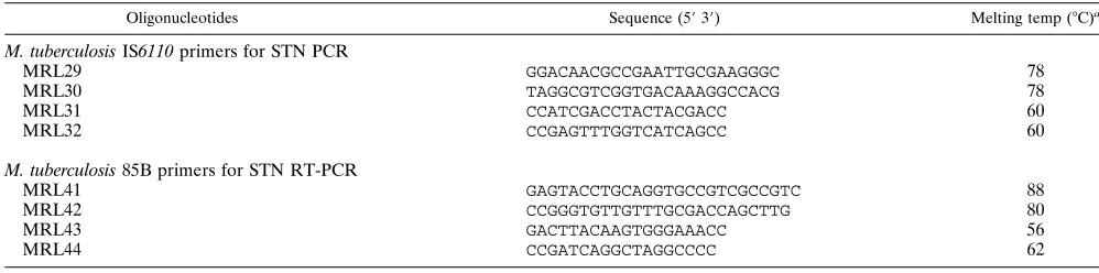
Partial sequencing the 85B antigen-coding region for M. tuberculosis.The 85B antigen-coding sequence has previously been determined for M. bovis and M. kansasii (16, 17). We used conserved segments of the 85B gene to develop primers which would amplify DNA from M. tuberculosis H37Rv. These same oligonucleotides were used as sequencing primers with the dsDNA Cycle Se-quencing System as described by the manufacturer (Gibco BRL, Grand Island, N.Y.). The primers used for sequencing (MRL41 and MRL42) as well as their reported positions in the M. bovis sequence are indicated in Table 1.
STN PCR.Amplification was performed in 0.5-ml thin-walled PCR tubes with a total reaction volume of 50ml by using a model 480 thermal cycler (Perkin-Elmer, Foster City, Calif.). The oligonucleotide primers used for amplification of the M. tuberculosis DNA target sequences in the IS6110 insertion element are indicated in Table 1. The outer and inner primers for M. tuberculosis were designated primers MRL29 and MRL30 and primers MRL31 and MRL32, respectively. They have been modified from those used by Wilson et al. (20) to permit two-cycle rather than three-cycle amplification in the first phase of the STN PCR (20).
The amplification reactions contained the individual outer and inner primers at final concentrations of 0.01 and 1mM, respectively, 2.5 U of Taq polymerase (Perkin-Elmer) in amplification buffer, 200mM (each) dATP, dCTP, and dGTP and 600mM dUTP (Perkin-Elmer), and 0.5 U of thermolabile uracil N-glycosy-lase (HK-UNG; Epicenter Technologies, Madison, Wis.) to control cross con-tamination (5). After a 10-min incubation at 508C to allow the HK-UNG to work, the temperature was raised to 948C for 2 min to inactivate the enzyme and break up any contaminating DNA.
The thermal cycler was programmed to carry out the DNA PCR in two stages. The first stage, for the outer primers, involved 30 cycles of denaturation at 948C for 45 s, with primer annealing and extension carried out in one step at 708C for 1.5 min. The second stage, for the inner primers, included 30 cycles of denatur-ation at 948C for 45 s, primer annealing at 608C for 45 s, and extension at 728C for 45 s, after which the reaction mixture was held at 728C for 30 min. Ten microliters of the amplification product was electrophoresed on 3% NuSieve GTG–1% agarose gels, stained with 1mg of ethidium bromide per ml, and visualized by UV transillumination. The presence of a 197-bp band indicated successful amplification of the IS6110 target.
STN RT-PCR.Recombinant Thermus thermophilus (rTth) DNA polymerase was used (GeneAmp EZ rTth RNA PCR Kit; Perkin-Elmer). The oligonucleo-tide primers used for amplification of the M. tuberculosis 85B mRNA target sequences are indicated in Table 1. The outer and inner primers for M.
tuber-culosis are designated primers MRL41 and MRL42 and primers MRL43 and MRL44, respectively. Their positions in the 85B sequence are indicated in Fig. 1. The amplification reactions contained the individual outer and inner primers at final concentrations of 0.01 and 0.8mM, respectively, 5 U of rTth DNA polymerase, 200mM (each) dATP, dCTP, and dGTP, 600mM dUTP, 0.5 U of HK-UNG, 10ml of 53EZ buffer, 5.0ml of 25 mM manganese diacetate, and a quantity of water sufficient to bring the volume to 50ml. After a 10-min incu-bation at 508C to allow the HK-UNG to work, the temperature was raised to 608C for 30 min to initiate synthesis of the first cDNA strand and then to 948C for 2 min to inactivate the HK-UNG. The first and second stages of the ampli-fication were then conducted as described above. The presence of a 216-bp signal on agarose gel electrophoresis indicated successful amplification of the 85B target.
RESULTS
Primer selection and specificity.
The partial coding
se-quence of 85B determined from M. tuberculosis DNA
demon-strated a few differences from that published for M. bovis (Fig.
1). One such difference consisted of an insertion of six bases
(GCCTAG) between positions 689 and 690 of the published
sequence (19). We exploited this difference to produce primers
which distinguished between M. tuberculosis and other
myco-bacteria, even closely related species such as M. bovis. The set
of nested primers that were selected successfully produced a
216-bp amplification product from M. tuberculosis DNA. These
primers were specific and proved to be capable of
distinguish-ing M. tuberculosis from 18 other species and strains of
myco-bacteria including M. bovis, M. avium, M. kansasii, M.
scrofu-laceum, M. microti, and M. marinum, as well as photochromes
and scotochromes (data not shown). In addition, the sequences
of the primers were compared to sequences in the GenBank
and EMBL DNA libraries. No two primers corresponded to or
complemented DNA registered in these libraries in a manner
that would permit amplification.
Limits of detection of the STN RT-PCR.
Seven-day, pure
cultures of M. tuberculosis H37Rv underwent serial 10-fold
dilutions with 7H9 medium. Aliquots of the dilutions were
cultured on Lowenstein-Jensen slants to assess the number of
bacilli present in the dilutions and their viabilities. Using the
four primers described above, the STN RT-PCR detected 85B
mRNA in dilutions of pure M. tuberculosis cultures that
pro-duced as few as 38 CFU from 0.1-ml culture aliquots on agar
slants. With clinical sputum samples, the technique detected as
few as one to nine bacilli per high-power field on fluorochrome
staining and 12 CFU on culture (Fig. 2).
To further define the limits of detection of this assay,
resid-ual, purulent sputum samples from individuals not suspected
of having tuberculosis were obtained by the hospital
microbi-ology laboratory, pooled, decontaminated, treated with a
mu-colytic agent, and concentrated as described above.
Fluoro-TABLE 1. PCR primers used in the study
Oligonucleotides Sequence (5939) Melting temp (8C)a
M. tuberculosis IS6110 primers for STN PCR
MRL29
GGACAACGCCGAATTGCGAAGGGC
78
MRL30
TAGGCGTCGGTGACAAAGGCCACG
78
MRL31
CCATCGACCTACTACGACC
60
MRL32
CCGAGTTTGGTCATCAGCC
60
M. tuberculosis 85B primers for STN RT-PCR
MRL41
GAGTACCTGCAGGTGCCGTCGCCGTC
88
MRL42
CCGGGTGTTGTTTGCGACCAGCTTG
80
MRL43
GACTTACAAGTGGGAAACC
56
MRL44
CCGATCAGGCTAGGCCCC
62
a[4(G1C)12(A1T)].
on May 15, 2020 by guest
http://jcm.asm.org/
[image:2.612.56.555.81.205.2]chrome staining and subsequent culture on Lowenstein-Jensen
slants confirmed the lack of mycobacteria in the pooled
sam-ples.
Aliquots of the treated, pooled sputa were spiked with
dilu-tions of a culture containing a known quantity of M.
tubercu-losis H37Rv. Samples were mixed by vortexing for 30 s. The
RNA was extracted and subjected to STN RT-PCR with
prim-ers to detect the 85B mRNA targets. Simultaneous
fluoro-chrome smears and cultures of the dilutions of M. tuberculosis
on Lowenstein-Jensen agar served as additional controls for
the number and viability of the mycobacteria detected by the
PCR.
The results of one such experiment displayed in Table 2
demonstrate that the STN RT-PCR could detect between 1
and 10 bacilli. For smear-negative samples (1 to 1,000 bacilli/
ml), this PCR assay was at least as sensitive as the single
agar-based culture method (sensitivity of PCR, 83%; sensitivity
of culture, 75%). The specificity of PCR was 100% (Table 2).
Detection of mycobacterial viability by the STN RT-PCR.
To
determine if the STN RT-PCR would detect living but not
dead M. tuberculosis bacilli, isoniazid at 0.1
m
g/ml was added to
cultures of M. tuberculosis H37Rv in 7H9 medium but not to
control cultures.
The STN RT-PCR was able to detect the difference between
a culture which contained antibiotic and one which did not
after 7 days of incubation (Fig. 3A). In an important control
experiment, neither isoniazid nor rifampin inhibited the STN
RT-PCR when they were added directly to the reaction
mix-ture (data not shown).
When an IS6110-targeted, DNA PCR was used to examine
the same samples, all samples through 13 days (the last
sam-ple) continued to be positive, irrespective of whether isoniazid
was present (Fig. 3B). The viability (or lack thereof) of the
organisms contained in these cultures was confirmed by
simul-taneous standard culture on Lowenstein-Jensen agar.
DISCUSSION
[image:3.612.73.557.68.250.2]The STN RT-PCR procedure described in this report
ad-dresses all of the problems outlined by Bates (3) and noted
above. One objective was to avoid contamination. The STN
PCR was initially designed to avoid the inherent
contamina-tion potential of standard nested PCR without relinquishing
the extremely powerful amplification potential of the nested
format (20). To our knowledge, this is the first attempt to use
this strategy with an RT-PCR. The addition of UNG provided
FIG. 1. Comparison of the DNA sequences of the 85B antigen in M. bovis BCG and M. tuberculosis H37Rv. The sequence for M. tuberculosis was compared and aligned with the published sequence for M. bovis BCG (12). Dashes, identical sequences; dots, sequences that have not been determined; N, unresolved residues; S, G or C; Y, C or T; R, A or G. Primers MRL41 and MRL42 are shown in enclosed boxes, while the underlined sequences represent primers MRL43 and MRL44. MRL44 contains the 6-base insertion not present in the M. bovis 85B sequence. [image:3.612.102.254.541.651.2]FIG. 2. STN RT-PCR can detect M. tuberculosis in spear-positive sputum. Lane 1, 100-bp DNA ladder; lane 2, positive control (216-bp amplification product of STN RT-PCR performed with RNA extracted from M. tuberculosis H37Rv); lane 3, no-target negative control; lanes 4 to 6, 216-bp amplification products from sputa containing 10 to 90, 10 to 90, and 1 to 9 M. tuberculosis isolates per high-power field, respectively, on fluorochrome staining; lanes 7 and 8, negative amplification of sputa containing no bacteria on fluorochrome stain-ing. All spear-positive sputa were later culture positive; smear-negative sputa were culture negative.
TABLE 2. Results of simultaneous smear, culture, and PCR for
spiked sputum samples
No. of bacilli/ml of sputum Smear resulta Culture resultb PCR result
1
3
10
30
m
1
5
3
10
20
m
1
1
3
10
20
m
1
5
3
10
10
3
1
1
3
10
10
0
1
1
3
10
00
0
1
1
3
10
210
0
2
0
0
0
2
aNumber of bacteria in 10 high-power fields on smear. bNumber of CFU; m,.50 CFU.
on May 15, 2020 by guest
http://jcm.asm.org/
[image:3.612.316.557.616.709.2]further protection against contamination (5). The use of UNG
would normally be impossible with a nested format because its
use in the second stage of the nested PCR would eliminate the
amplified target generated in the first stage. Since our STN
RT-PCR procedure is performed in a single tube and no
trans-fers are necessary, the UNG can be inactivated prior to the
initiation of amplification without sacrificing effective
decon-tamination. Although UNG rapidly removes uracil from DNA,
the target in this assay, mRNA, remains unaffected.
Our primary objective was to develop a PCR technique
which could distinguish viable from nonviable tubercle bacilli.
The STN RT-PCR accomplishes this (Fig. 3A) while still
re-taining sufficient sensitivity to detect the equivalent of
smear-negative samples. It is equally clear that DNA-targeted PCR
was incapable of determining mycobacterial viability over the
time period tested. This is not a trivial distinction. Yuen and
colleagues (22), who used an IS6110 DNA target, reported that
the sputa of 70% of 41 patients with proven tuberculosis had a
positive PCR signal 4 weeks after the onset of therapy, whereas
cultures were positive for only 32% of the cohort at the same
point in time. Moreover, Eisenach (9) has described the
de-tection of the same PCR target more than 500 days after the
initiation of therapy and long after cultures were negative. Is
this the detection of dead organisms, inadequate therapy, or
merely cross contamination? These are extremely important
questions that have considerable bearing on clinical decision
making and the natural course of treated disease. The STN
RT-PCR should be able to resolve these questions.
Because the STN RT-PCR can rapidly detect the effect of
antibiotics on mycobacterial viability (Fig. 3A), it may be
pos-sible to design a PCR-based system that can be used to quickly
perform antibiotic susceptibility testing. Our experiments
doc-ument that STN RT-PCR can detect mycobacterial
suscepti-bility to isoniazid in 4 to 7 days when an initial inoculum of
20,000 to 50,000 organisms/ml is used. Recently, a survey of
state microbiology laboratories sponsored by the Centers for
Disease Control and Prevention noted that even those which
use rapid radiometric techniques for both culture and
suscep-tibility testing still require, on average, 31 days from the time of
specimen processing to the time that the mycobacterial drug
susceptibility result is reported (11). Even if a PCR-based
system took 10 days to determine antibiotic susceptibilities, it
would be a significant improvement. This would be particularly
true in the case of smear-negative, STN RT-PCR-positive
sam-ples, for which there would be an even greater delay by
stan-dard techniques.
What these experiments do not address is whether the STN
RT-PCR system can deal with the more difficult problem of
assessing antibiotic resistance. A method would have to be
developed to detect samples that contained more resistant
bacilli than the current cutoff for resistance, i.e., more than 1%
of the total inoculum.
Although the STN RT-PCR appears to demonstrate
detec-tion limits that are clinically acceptable, its sensitivity and
spec-ificity with a large number of clinical sputum samples
contain-ing a variety of potential PCR inhibitors are unknown.
Moreover, the RNA extraction procedures used in the present
studies are cumbersome and would probably require
modifi-cation to be clinically practical. Whether this can be
accom-plished without compromising detection limits remains to be
determined.
Finally, although we used STN RT-PCR, other RNA-based
amplification systems such as transcription-mediated
amplifi-cation or nucleic acid sequence-based amplifiamplifi-cation should be
capable of producing similar results with an mRNA target (6,
12). Indeed, it is the utility of detecting an mRNA target to
establish bacterial viability that we wish to emphasize here,
rather than the particular method used to do so.
ACKNOWLEDGMENTS
This work was supported by ACRC Seed Grant B394-90-0004 from
the UCLA AIDS Clinical Research Center, grant R95-REI-169 from
the University of California Universitywide AIDS Research Program,
and a grant from the Pfizer Pharmaceutical Company, Inc.
REFERENCES
1. Alberts, B., D. Bray, J. Lewis, M. Raff, K. Roberts, and J. D. Watson. 1989. Control of gene expression, p. 595. Molecular biology of the cell, 2nd ed. Garland Publishing, Inc., New York, N.Y.
2. Barnes, P. F., and S. A. Barrows. 1993. Tuberculosis in the 1990s. Ann. Intern. Med. 119:400–410.
3. Bates, J. H. 1994. New diagnostic methods, p. 81–92. In L. N. Friedman (ed.), Tuberculosis. Current concepts and treatment. CRC Press, Inc., Boca Raton, Fla.
4. Chevillard, S. 1993. A method for sequential extraction of RNA and DNA from the same sample, specially designed for a limited supply of biological material. BioTechniques 15:22–24.
5. Cimino, G. D., K. C. Metchette, J. W. Tessman, J. E. Hearst, and S. T. Issacs. 1991. Post-PCR sterilization: a method to control carryover contamination FIG. 3. (A) RT-PCR can distinguish cultures of M. tuberculosis containing
isoniazid from those without isoniazid. Lane 1, a 100-bp ladder; lane 2, positive control (216-bp amplification product of STN RT-PCR performed with RNA extracted from M. tuberculosis H37Rv); lane 3, no-target negative control; lane 4, 216-bp amplification product in day 0 culture of H37R immediately after the addition of isoniazid; lane 5, day 0 medium-only control; lane 6, 216-bp ampli-fication product from day 3 control culture without isoniazid; lane 7, 216-bp amplification product from day 3 control culture without isoniazid; lane 8, 216-bp amplification product from day 7 culture with isoniazid; lane 9, absence of 216 bp amplification product from day 7 culture containing isoniazid; lane 10, 216-bp amplification product from day 11 control culture without isoniazid; lane 11, absence of 216-bp amplification from day 11 culture containing isoniazid; lane 12, 216-bp amplification product from day 13 control culture without isoniazid; lane 13, absence of 216-bp amplification product from day 13 containing isoni-azid. (B) IS6110-targeted, DNA PCR cannot distinguish cultures of M. tubercu-losis containing isoniazid from those without isoniazid. Lane 1, a 100-bp ladder; lane 2, no-target negative control; lane 3, positive control (197-bp amplification product of STN PCR performed with DNA extracted from M. tuberculosis); lane 4, 197-bp amplification product in day 0 culture of H37Rv immediately after the addition of isoniazid; lane 5, day 0 medium-only control; lanes 6 and 7, 197-bp amplification product from day 3 cultures with isoniazid (lane 6) and without isoniazid (lane 7); lanes 8 and 9, 197-bp amplification product from day 7 cultures with isoniazid (lane 8) and without isoniazid (lane 9); lanes 10 and 11, 197-bp amplification product from day 11 cultures with isoniazid (lane 10) and without isoniazid (lane 11); lanes 12 and 13, 197-bp amplification product from day 13 cultures with isoniazid (lane 12) and without isoniazid (lane 13).
on May 15, 2020 by guest
http://jcm.asm.org/
[image:4.612.62.297.67.279.2]for the polymerase chain reaction. Nucleic Acids Res. 19:99–107. 6. Compton, J. 1991. Nucleic acid sequence-based amplification. Nature 350:
91–92.
7. Daniel, T. M., and J. J. Ellner. 1993. Immunology of tuberculosis, p. 75–91. In L. B. Reichman and E. S. Hershfield (ed.), Tuberculosis. A comprehen-sive international approach. Marcel Dekker, Inc., New York, N.Y. 8. Ebersole, L. L. 1992. Acid fast stain procedures, p. 3.5.1–3.5.11. In H. D.
Isenberg (editor in chief), Clinical microbiology procedures handbook. American Society for Microbiology, Washington, D.C.
9. Eisenach, K. D. 1995. Clinical monitoring of patients with tuberculosis using SDA and PCR, abstr. 20, p. 135. In Proceedings of the Conference on Advances in Genetic Diagnostics for Infectious Diseases. Cambridge Health-tech Institute, Washington, D.C.
10. Guide for Grants and Contracts. 1993. Vol. 22, no. 34, p. 7, September 24. National Institutes of Health, Bethesda, Md.
11. Huebner, R. E., R. C. Good, and J. I. Tokars. 1993. Current practices in mycobacteriology: results of a survey of state public health laboratories. J. Clin. Microbiol. 31:771–775.
12. Jonas, V., M. J. Alden, J. I. Curry, K. Kamisango, C. A. Knott, R. Lankford,
J. M. Wolf, and D. F. Moore.1993. Detection and identification of Myco-bacterium tuberculosis directly from sputum sediments by amplification of rRNA. J. Clin. Microbiol. 32:2410–2416.
13. Kitchin, P. A., Z. Szotyori, C. Fromholc, and N. Almond. 1990. Avoidance of false positives. Nature 344:201.
14. Kwok, S., and R. Higuchi. 1989. Avoiding false positives with PCR. Nature
339:237–238.
15. Liebling, M. R., and E. V. Barnett. 1977. Substrate competition between DNase I and anti-DNA antibody. Clin. Immunol. Immunopathol. 8:80–89. 16. Matsuo, K., R. Yamaguchi, A. Yamazaki, H. Tasaka, K. Terasaka, and T.
Yamada.1990. Cloning and expression of the gene for the cross-reactivea antigen of Mycobacterium kansasii. Infect. Immun. 58:550–556.
17. Matsuo, K., R. Yamaguchi, A. Yamazaki, H. Tasaka, and T. Yamada. 1988. Cloning and expression of the Mycobacterium bovis BCG gene for extracel-lularaantigen. J. Bacteriol. 170:3847–3854.
18. Salata, R. A., A. J. Sanson, I. J. Malhotra, H. G. Wiker, M. Harboe, N. B.
Phillips, and T. M. Daniel.1991. Purification and characterization of the 30,000 dalton native antigen of Mycobacterium tuberculosis and characteriza-tion of six monoclonal antibodies reactive with a major epitope of this antigen. J. Lab. Clin. Med. 118:589–598.
19. Wiker, H. G., M. Harboe, and T. E. Lea. 1986. Purification and character-ization of two protein antigens from the heterogeneous BCG85 complex in Mycobacterium bovis BCG. Int. Arch. Allergy Appl. Immunol. 81:298–306. 20. Wilson, S. M., R. McNerney, P. M. Nye, P. D. Godfrey-Faussett, N. G.
Stoker, and A. Voller.1993. Progress toward a simplified polymerase chain reaction and its application to diagnosis of tuberculosis. J. Clin. Microbiol.
31:776–782.
21. Yoneda, M., Y. Fukui, and T. Yamanouchi. 1965. Extracellular proteins of tubercle bacilli. V. Distribution ofaandbantigens in various mycobacteria. Biken J. 8:201–223.
22. Yuen, K. Y., K. S. Chan, C. M. Chan, B. Ho, L. Dai, P. Chau, and M. Ng. 1993. Use of PCR in routine diagnosis of treated and untreated pulmonary tuberculosis. J. Clin. Pathol. 46:318–322.